the practice building bulletin volume iv issue xx · pdf filethe issue xxpractice building...
TRANSCRIPT

The Practice Building BULLETIN VOLUME IVISSUE XX
incisors. Although it is impossible to predict precisely the size of the permanent dentition during this period of development, one can confi dently predict that the arches will be too small and the permanent incisors will be crowded and irregular, if the primary incisors contact each other proximally.2
• Skeletal Cross-bites - In the deciduous and mixed dentition, the creation of a posterior cross-bite is often a consequence of prolonged abnormal habits such as fi nger sucking, mouth breathing, and nasal blockage. These chil dren will often develop a high narrow palatal vault as well. When diagnosed early, it is possible to widen the maxilla itself by opening the midpalatal suture.3,4
• Anterior and Posterior Crowding - Patients with arches that are underdeveloped will usually have inadequate space for proper tooth alignment. When this occurs, either of two condi tions may develop. One possibility is that the teeth may remain upright and well positioned over the basal bone of the maxilla or mandible but will rotate or tip labially or lingually. In this case, the crowding is diffi cult to miss. How ever, in other cases, the crowded teeth may stay completely aligned and mani fest their crowding by moving off the basal bone, displacing the lips forward.3
» PRACTICE POTENTIAL
A healthy Temporomandibular joint, excellent facial esthetics, and a solid functional occlusion are the goals that every clinician should strive toward when performing orthodontic care.1
To accomplish these goals we usually need to look at both the orthopedic and orthodontic components of a patient’s occlusion.
Functional appliances that retrain muscles, active plates that have suffi cient power to reshape bone, and fi xed appliances that level, align and rotate the teeth, are often needed to ensure that a patient will have proper lip seal, normal tongue position, and ade quately developed arches, when treat ment is complete.1
One of the most important concepts regarding the solving of orthodontic treatment problems is the notion of proper arch size. Collapse of the dental arches in either an anteroposterior direction, a lateral direction, or a combination of both, is one of the major components of most types of malocclusion. Some malocclusions which are directly related to improper arch shape and size are:
• Arch Size Discrepancy in the Primary Dentition - In the primary denti-tion, spacing between the incisors is normal and in fact is necessary to allow for alignment of the adult
• Anterior Open Bites - Often because of a lower tongue position and unopposed muscle forces of the buccinator mechanism on the maxillary buccal segment, a child’s abnormal sucking habit will cause an open bite, and an excessive overjet.4
• Skeletal Class II Relationships - A narrow upper arch caused by improper function or habits such as thumb sucking, a deviant swallowing pattern or tongue thrusting, is one of the common causes of a Class II Division1 malocclusion. The functional occlusion during this process usually drags the lower teeth lingually as the inclined plane action of the cusps of the upper teeth act to narrow the lower teeth into an arch of approximately the same proportions. If this inclined plane action fails to move the lower posteri ors lingually, posterior cross-bites develop or the entire mandible is locked back by the occlusion, keeping it where it may intercuspate itself more completely with the upper arch.1
• Improper Tongue Volume - A properly developed arch is essential to give the tongue enough volume in which to operate. If the tongue does not have enough space and is impinged upon by the teeth and bone, the latter will even-tually give way. Therefore, proper arch form is also critical for stability and long term retention of your orthodontic result.1,3
THE SCHWARZ MODEL ANALYSIS (with Korkhaus (K) Measurement)
TheThe Practice BuildingPractice Building BULLETINBULLETIN
The Practice Building BULLETINTheThe Practice BuildingPractice Building BULLETINBULLETIN
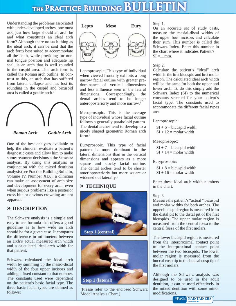
Leptoprosopic. This type of individual when viewed frontally exhibits a long narrow facial outline with greater pre-dominance of vertical dimensions and less infl uence seen in the lateral dimensions. Correspondingly, the dental arches tend to be longer anteropos teriorly and more narrow.1
Mesoprosopic. This is the average type of individual whose facial outline follows a generally paraboloid pattern. The dental arches tend to develop to a nicely shaped geometric Roman arch form.1
Euryprosopic. This type of facial pattern is more dominant in the lateral dimensions than in the vertical dimensions and appears as a more square and stocky facial outline. The dental arches tend to be shorter anterioposteriorly but more square or widened out laterally.1
» TECHNIQUE
Step 1.On an accurate set of study casts, measure the mesial-distal widths of the upper four incisors and calcu late their sum. This number is called the Schwarz Index. Enter this number in the chart where it indicates Patient’s SI =__mm.
Step 2.Calculate the patient’s “ideal” arch width in the fi rst bicuspid and fi rst molar region. The calculated ideal arch width will be the same for both the upper and lower arch. To do this simply add the Schwarz Index (SI) to the numerical constants selected for your patient’s facial type. The constants used to accommodate the different facial types are:
Leptoprosopic:SI + 6 = bicuspid width SI + 12 = molar width
Mesoprosopic:SI + 7 = bicuspid width SI + 14 = molar width
Euryprosopic:SI + 8 = bicuspid width SI + 16 = molar width
Enter these ideal arch width numbers in the chart.
Step 3.Measure the patient’s “actual “ bicuspid and molar widths for both arches. The upper bicuspid region is measured from the distal pit to the distal pit of the fi rst bicuspids. The upper molar region is measured from the central fossa to the central fossa of the fi rst molars.
The lower bicuspid region is mea sured from the interproximal con tact point to the interproximal con tact point between the two bicus pids. The lower molar region is measured from the buccal cusp tip to the buccal cusp tip of the fi rst molars.
Although the Schwarz analysis was designed to be used in the adult dentition, it can be used effectively in the mixed dentition with some minor modifi cations.
Understanding the problems associated with under-developed arches, one must ask, just how large should an arch be and what constitutes an ideal arch form? Although there no such thing as the ideal arch, it can be said that the arch form best suited to accommodate all the teeth, while providing for nor-mal tongue position and adequate lip seal, is an arch that is well rounded in the cuspid area. This arch form is called the Roman arch outline. In con-trast to this, an arch that has suffered from lateral collapse and has lost its rounding in the cuspid and bicuspid area is called a gothic arch.1
One of the best analyses available to help the clinician evaluate a patient’s diagnostic casts and allow him to make some treatment decisions is the Schwarz analysis. By using this analysis in conjunction with the mixed dentition analysis (see Practice Building Bulletin, Volume IV, Number XIX), a clinician can make an assessment of arch size and development for every arch, even when serious problems like a pos terior cross-bite or obvious crowding are not apparent.
» DESCRIPTION
The Schwarz analysis is a simple and easy-to-use formula that offers a good guideline as to how wide an arch should be for a given case. It compares the difference in millimeters between an arch’s actual measured arch width and a calculated ideal arch width for that patient.
Schwarz calculated the ideal arch width by summing up the mesio-distal width of the four upper incisors and adding a fi xed constant to that number. The constants used were dependent on the patient’s basic facial type. The three basic facial types are defi ned as follows:
Roman Arch Gothic Arch
Lepto Meso Eury
Step 1 (central)
Step 1 (lateral)
(Please refer to the enclosed Schwarz Model Analysis Chart.)

The Practice Building BULLETINTheThe Practice BuildingPractice Building BULLETINBULLETINWhen measur ing the patients “actual” arch width, on the upper arch, measure across the arch from the distal pits of the D’s. On the lower arch, measure across the arch from the buccal interproximal contact points between the D’s and E’s. Then add 2mm to these measurements. This will give you the approximate position of the unerupted fi rst bicuspids.
Step 4.
» TREATMENT
When the Schwarz analysis indicates that the arch is collapsed laterally, we will usually see the four anteriors jumbled up, exhibiting crowding and rota tions. However, it is possible even if the arches are extremely narrow, that these teeth may align themselves completely or partially at the expense of the lip, displacing it forward. When this is the case, the anteriors bow out labially causing a very pointed appearance to the outline of the arch.3
If by some miracle, the four anteriors retain their lateral spread across the front of the arch and lateral collapse is still present, it will be very obvious to the eye that the arch form is distorted with extremely lingually positioned bicuspids and molars. Treatment in all three of these examples is a matter of lateral development.
Common sense indicates that the larger the difference between the ideal and actual arch width, the less chance that arch development alone will be enough to properly align the teeth and arches. Although it is impossible to say with certainty where the borderline is between arch development and the need for extraction, initial treatment should consist of a relatively conserva tive attempt to expand the arches, followed by an evaluation of the results. If the patient tolerates the arch expansion well and does not develop excessive incisor protrusion, expansion treat ment may ultimately be successful even in spite of a large difference between the actual and ideal arch mea surements. On the other hand, if the expansion proceeds with diffi culty with the signs of excessive arch expansion - incisor protrusion and labial tipping of the teeth - arch expansion is probably inappropriate.3,4
As a general guideline, one can say that space discrepancies up to 4mm usually can be resolved through arch develop-ment without extraction. In cases where the discrepancy is in the 5 to 9 mm range, arch development can be tried, but these cases frequently require extraction of some teeth. Discrepancies of 10 mm or greater almost always require premolar or second molar extraction to resolve the crowding.1
» THE APPLIANCES
Transverse appliances are a group of appliances used to gain arch width by moving the posterior quadrants away from each other laterally across the arch. This may in fact provide a gener-ally wider arch but not necessarily a longer arch.
These appliances range from the deli cate Crozat, all the way up to the suture splitting, rapid palatal expanders and may utilize orthodontic move ment, orthopedic movement, or a com bination of both. They also may be either fi xed or removable.1 For an excellent overview of these appliances, please see Chapter 8 in the Principles of Appliance Therapy for Adults and Chil dren textbook
The Basic Lower Crozat
The appliance seen here is the basic lower Crozat. This appliance is usually used simultaneously with the upper Crozat for lateral development of the arches. In this basic design, the fi rst permanent molars are clasped and dis-tal extensions are carried back to the second molars to upright them during development. Lingual lap springs have also been added to align the lower ante riors. From the patient’s point of view, there are several advantages to the Crozat. Esthetically, the appliance is very inconspicuous and therefore is widely accepted by both children and adults. Being acrylic free and easy to take in and out, patients fi nd it easier to maintain their oral hygiene. There also seems to be less interference with normal speech.5
Rapid palatal expansion is most commonly used in the mixed or early adult dentition that exhibits a bilateral max illary posterior crossbite. This crossbite is usually due to a defi ciency of the maxillary apical base. Rapid
Step 3 (bicuspid measurement)
Step 3 (molar measurement)
Fill in the chart and calculate the difference between the actual arch measurements and the ideal arch measurements.
Step 3 (molar measurement)
Step 3 (bicuspid measurement)

The Practice Building BULLETINTheThe Practice BuildingPractice Building BULLETINBULLETINexpansion is obtained over a two to three week period by means of turning the expan sion screw twice a day. This creates enough pressure to separate the mid palatine suture. The appliance is then worn for another three to four months to allow time for osseous healing. In this design, the expansion screw is positioned high in the palate for greater patient comfort.4,5
Rapid Palatal Expander
» CONTRAINDICATIONS AND CONCERNS
Dental crowding is one of the major aspects of any type of malocclusion and is usually a result of the collapse of the dental arch in both a lateral direction and an anteroposterior direction. Therefore, in order to properly determine the course future treatment will take, the clinician should perform a Schwarz analysis and a mixed dentition analysis.
When using the Schwarz analysis to determine the ideal arch width, remember the numbers are just that - numbers. They are only to act as a guide in helping the doctor decide what is best for the patient. The doc tor is the one who assumes responsi bility for determining when the case is correct, not some chart or mathemati cal formula!
The dimensions that are necessary for ideal alignment and those clinically achievable are not necessarily the same. The Schwarz analysis simply suggests ideal maxillary premolar and fi rst molar cross arch measurements according to the mesiodistal width of the maxillary incisors. Clinically however, it is often the patient’s mandibular intercanine width that is the limiting factor to treatment, as it only increases slightly throughout growth. In fact, most of the
mandibular correction in arch width that is seen during treat ment is the uprighting of lingually tipped teeth back over the basal bone.4
This is not to say that an increase in mandibular arch width cannot be accomplished. It can. Small amounts of widening of the basal bony width of the mandible can occur by orthodontic stimulation of bony deposition on the lateral borders of the corpus mandibularis.4
When using this analysis always evaluate the patient’s facial type carefully. It would be remiss to try to force the development laterally of an extremely narrow set of arches in a leptoprosopic type individual trying to make him accommodate the larger arch width reserved for the naturally wider-based euryprosopic individuals. This could potentially result in over expansion of the arches leading to an undesirable penchant for instability.
When measuring the sum of the four anteriors for a patient who has peg laterals, it is best to use a value 2mm less than that of the central as a substitute for the laterals. This will give you a better idea of the ideal arch for that patient. Even though expanding the arches towards this ideal may create gaps between the laterals and the other anterior teeth, it is still better to expand and close the gaps with bonding or veneers than to leave the arch underdeveloped.1
When performing a Schwarz analysis in the mixed dentition remember to take the bicuspid measurements from the deciduous D’s and E’s and add 2mm as a substitute for the fi rst bicus pids which are developing right beneath them.1
If the deciduous cuspids are lost prematurely either unilaterally or bilaterally, the D’s, E’s, and fi rst permanent molars behind the space created by the lost C will tend to drift mesially en masse. In drifting forward, the D’s will end up in the narrower front portion of the young growing dental arch. When this is the case, the results of a Schwarz analysis could easily fool you into surmising the case is narrower than it actually is.1
Having this smaller arch measurement may lead you into thinking the treat ment of choice is to regain the arch width by lateral development with appliances like a Schwarz or Jackson when, in fact, the true treatment of choice is to distalize the posterior quadrants and regain the lost space given up by the premature loss of the C’s.1
When you have moved the teeth back to a wider point in the arch, the arch width may be reassessed again at that position and any major gross arch width defi ciencies still present can be corrected by lateral development techniques if need be.1
» SUPPLY LIST
The supply list for this procedure is minimal. All you will need is:
Diagnostic Template*Diagnostic Calipers*Arch Symmetry Grid*Range of Motion Guide*Intra Oral Photo Mirrors*Digital Camera*Per-Fect Bites*Base Plate Wax*Hot Water Bath*
* Available through Success Essentials, call 800-423-3270
» CUSTOMARY FEESAND INCOME POTENTIAL
Taking the time to do a proper diagnosis is the key to prevention and treat ment of orthopedic and orthodontic problems.” For whatever unknown rea son, we are often hesitant to collect a fee for the time we spend analyzing and diagnosing our patient’s dental prob lems. I feel this is a mistake.
Consider the time involved in just collecting records like impressions for diagnostic casts, intra-oral and extra-oral photographs, and all the neces sary x-rays (FMX, Lateral Ceph, Panorex). Even if you send the patient out to a service to get these records, you will still need to take the time to analyze them and devise a treatment strategy.

The Practice Building BULLETINTheThe Practice BuildingPractice Building BULLETINBULLETINThe Practice Building Bulletin is a special service of Space Maintainers Laboratory produced solely for the private use of our clients. It is designed to help expand and enhance your ability to provide comprehensive patient care. Information included is the opinion of the author and may not be reproduced in any form without written consent.Appliance Therapy Group Headquarters:Space Maintainers LaboratoryP.O. Box 4184, Van Nuys, California 91409-4184Copyright © 2006www.ApplianceTherapy.com
Regional Labs:Southwest 800-423-3270Northwest: 800-423-6509Northeast 866-310-5800Southeast 800-289-0118Midwest: 800-325-8921CANADA 800-661-1169AUSTRALIA 03-9521-0299MALAYSIA 03-6251-8599TAIWAN 886-7-235-5612
Therefore, I feel it is reason able to charge your patient for your time. The range of fees for this service, of course, will be dependent upon the area that you live in and the economic make up of your community. While lecturing across the country, I have noticed that it is not unusual to receive a fee in the range of $150 to $350 for this diagnosis.
by Rob Veis D.D.S.Director of Practice Development
» REFERENCES:
1. Witzig, Spahl: The Clinical Management of Basic Orthopedic Appliances Vol 1, Chicago, Year Book Medical Publishers Inc., 1987 Chapter 5, pp279-414.
2. Pinkham,J: Pediatric Dentistry, Philadel phia W.B. Saunders, 1988 Chapter 25, 311-315.
3. Profi t,W: Contemporary Orthodontics, St. Louis, C.V. Mosby, 1986,Chapter 7, pp 168-197.
4. Moyers, Robert: Handbook of Orthodon tics, Year Book Medical Publishers Inc., 1980 Chapter 7, pp192-207.
5. Veis, Salzer, Christian: The Manual of Appliance Therapy for Adults and Chil dren. L.A., Space Maintainers Laborato ry, 1995, Chapter 7.
6. Caldwell, Stallard: A Textbook of Preven tive Dentistry, Philadelphia W.B. Saun ders, 1977 Chapter 20, pp 351-389.

*To photocopy for individual patient records, place your patient’s study casts here, occlusal side down, and cover area with a towel.
STEP 1: Calculate the sum of the mesial-distal widths of the four upper incisors: SI= ___mm
STEP 2: Calculate the patient’s “ideal” arch width in the first bicuspid and first molar region. These numbers are the same for both the upper and lower arch. The ideal arch width measurements are based on facial type:
For Leptoprosopic: 4|4 = SI + 6m 6|6 = SI + 12mmFor Mesoprosopic: 4|4 = SI + 7mm 6|6 = SI + 14mmFor Euryprosopic: 4|4 = SI + 8mm 6|6 = SI + 16mm
STEP 3: Measure the patient’s “actual bicuspid” and molar arch widths at the points indicated on the diagram.
For the upper measure from the distal pit to the distal pit*For the upper measure from the central fossa to the central fossa
For the lower measure from the interproximal contact point to the interproximal contact point at 4&5*For the lower measure from the buccal cusp tip to the buccal cusp tip
* In mixed dentition cases, when measuring the patient’s “actual” arch width, on the upper measure across the arch from the distal pits of the D’s. On the lower, measure across the arch from the buccal interproximal contact points between the D’s and E’s. Then add 2mm to each measurement.
STEP 4: Fill in the chart and calculate the difference between the actual and the “ideal” (+ or -).
K=1/2 of the measurementIf patient’s actual measurement is less than K value: needs premaxilliary developmentIf patient’s actual measurement is greater than K value: needs lingual retraction or torquing
Patient’s S.I. = ______mmPatient’s Ideal Actual Difference
Schwarz Analysis(with Korkhaus (K) Measurement)
SAME AS
SAME AS
Patient Name _____________________________________________________________________________________________________________________________Date ____________________________
Last First Middle Initial