the paraffin lamp - a r esurrection - microscopic … · assembly by soldering was not fea- ......
TRANSCRIPT

1Journal of the Microscopical Society of Southern California Sep/Oct 2003
Volume 8 Number 5 September/October 2003THE MICROSCOPICAL SOCIETY OF SOUTHERN CALIFORNIA
Journal of
THE PARAFFIN LAMP - A RESURRECTION
byDave Hirsch
We have among us MSSC members who are eBayafflicted. Assiduously, and with great frequency,we peruse the Antiques and Collectibles sectionscovering scientific instruments. Juicy scientificmorsels are contemplated and bids placed. Af-ter an eternity the auction ends, and all the whilewe hope that some sniper didn’t grab on to theprize at the very last second. This time the ham-mer drops and we are the holder of the winningbid. You know the winner when he or she comesto the meeting grinning from ear to ear, shleppingtheir prize to the envy and admiration of the groupassembled. Regrettably, such is not always thecase.
The auction advertisement mentioned a “mysteryinstrument,” denoting that the seller didn’t havethe foggiest idea of what was being offered. Bot-tom line: I was the one and only bidder, acquir-ing the item for six bucks plus another fiver tohave the brass fragments shipped to me. What Ireceived is shown in fig. 1. It was a well-patinatedbrassthingy consisting of two main assemblies;a ring base with an attached post and an assem-bly with a two-inch diameter compound lens fit-ted to an arm which rode on a rack and pinionarrangement. The item had no signature or otheridentification. Astute MSSC mavens steeped inthe lore of historical technology might have sug-gested some sort of Victorian period scientificlaboratory device, a lamp, perhaps.
Gerard I’E. Turner’s, “The Great Age of the Micro-scope” has a section on paraffin (kerosene) lamps.
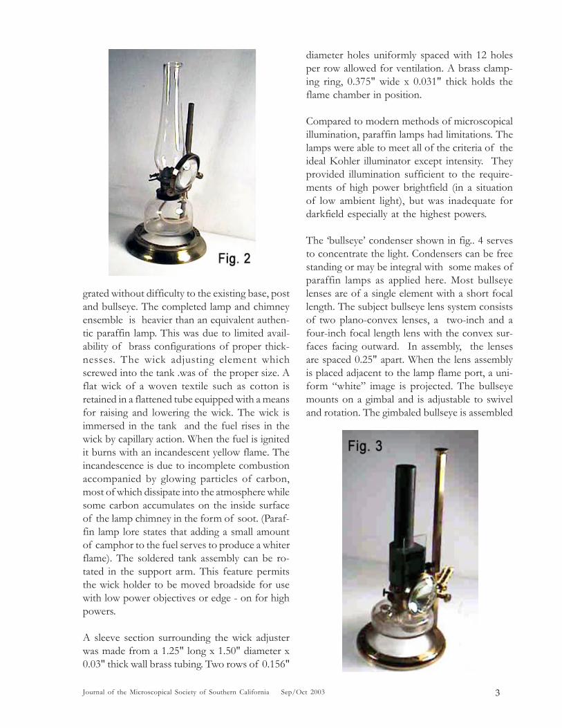
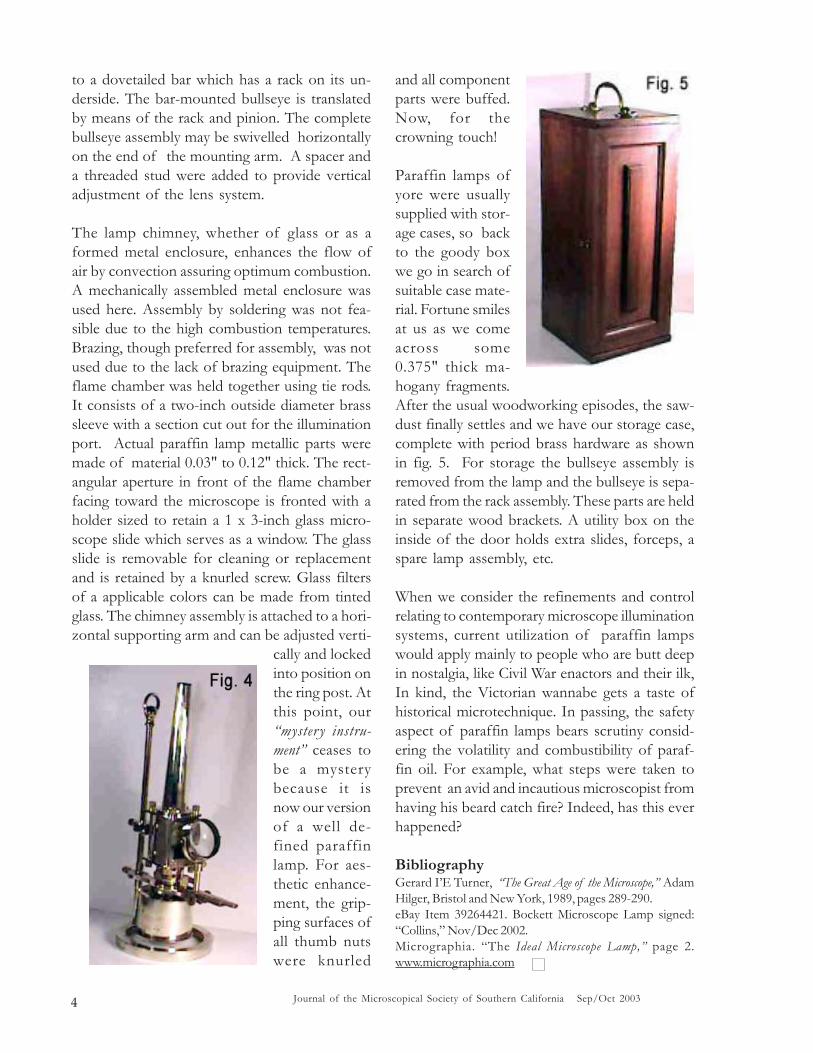
Paraffin lamp illumination was used by Britishand other microscopists well into the 1930s. Brit-ish microscopical societies retained a number ofthese lamps for general use during meetings. Twosuch lamps are shown here. The lamp shown infig. 2 has a glass chimney and is signed: “CollinsLondon. Bockett Microscope Lamp.” The lamp shownin fig. 3 has a metal chimney and is signed: “Swift& Son. London.W.” The assembled fragment ofthe “mystery lamp” bore no resemblance to any ofthe lamps pictured. What then, would the miss-ing parts, namely the lamp assembly and a chim-ney look like? It is no wonder the “mystery object”went for six bucks! In contrast, a piéce de resis-tance, the “Bockett Microscope Lamp, 19th Century,

Journal of the Microscopical Society of Southern California Sep/Oct 20032
MSSC Journal Volume 8 Number 5 Sep/Oct 2003
CONTENTSMICROSCOPICAL SOCIETY OF
SOUTHERN CALIFORNIA
President: James D. Solliday(714) 775-1575 [email protected]
Vice President: Stuart Warter714-847-0529 [email protected]
Treasurer: Herb Gold *2065 Balmer Drive, LA, CA 90039-3047(323) 665-8391 [email protected]
Education Chair: Alan deHaas(310) 475-2873 [email protected]
Facilities Chair: Pete Teti(323) 660-9259 [email protected]
Webmaster and Leonie FedelJournal Editor: 3273 Provon Lane, LA, CA 90034 -2714
(310) 839-9881 [email protected]
Corresponding George VittSecretary: [email protected]
Program Chair: Dr. Ken Gregory(562) 596-1762 [email protected]
Program Committee: Ed Jones (805) 654-8548 [email protected] Ken Miller (818) 906-1032 [email protected]
* Prospective new members, please contact Herb Gold formembership application. Dues are $50 yearly for regular membersand $40 yearly for corresponding members who are geographicallytoo distant to attend regular meetings. Please make checkspayable to “Herb Gold - MSSC”.
The Paraffin Lamp - A Resurrection 1by Dave Hirsch
MSSC September 2003 Workshop 5recorded by Herb Gold and written by Jim Solliday
MSSC September 2003 Meeting 24by Leonie Fedel
MSSC October 2003 Workshop 26recorded by Herb Gold and written by Jim Solliday
MSSC October 2003 Meeting 34by Leonie Fedel
Announcements:
MSSC Sat Workshop Announcements 35First Saturday of every month
MSSC Meeting Announcements 367:00pm November 19th, 2003Holiday Banquet 4:00pm December 14th, 2003
Editor’s Note 26
signed Collins” appeared on eBay. The first bidwas $100.00, reaching $911.00 without meet-ing the reserve. The pristine condition and con-figuration of the lamp would put any collectorof microscopes and accessories into a state offrenzy. Being thus challenged, the augmentationof the eBay find got underway.
Beyond that which already existed, the originalin-toto form of my “mystery lamp” was indeed, amystery. Back we go then to our old scientificinstrument books and related references. Hope-fully, the gleanings will offer clues as to how the“mystery instrument” cum lamp might have looked
when it illuminated a specimen for some longdeparted bearded and swallowtail-coated Vic-torian gentleman peering into his Powell & Lelandbinocular microscope.
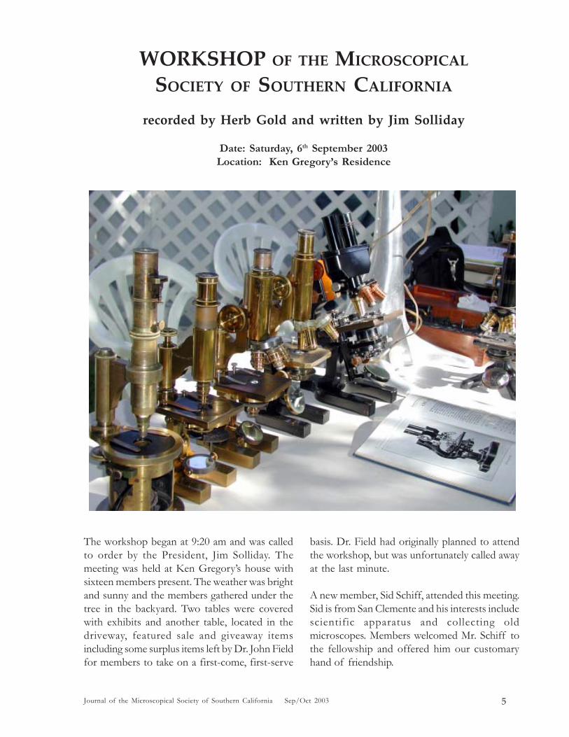
After perusing several photos and sketches ofold microscope lamps, I dug into my treasurechest loaded with dribs and drabs of sundry castoff materials. I recovered a conglomeration ofscrap brass in the form of rod, tubing and sheetswhich came together as the lamp assemblyshown in fig. 4. This end product was pompouslyalluded to as: “An extrapolated reconstruction of a19th century paraffin lamp.” The assembly inte-
3Journal of the Microscopical Society of Southern California Sep/Oct 2003
grated without difficulty to the existing base, postand bullseye. The completed lamp and chimneyensemble is heavier than an equivalent authen-tic paraffin lamp. This was due to limited avail-ability of brass configurations of proper thick-nesses. The wick adjusting element whichscrewed into the tank .was of the proper size. Aflat wick of a woven textile such as cotton isretained in a flattened tube equipped with a meansfor raising and lowering the wick. The wick isimmersed in the tank and the fuel rises in thewick by capillary action. When the fuel is ignitedit burns with an incandescent yellow flame. Theincandescence is due to incomplete combustionaccompanied by glowing particles of carbon,most of which dissipate into the atmosphere whilesome carbon accumulates on the inside surfaceof the lamp chimney in the form of soot. (Paraf-fin lamp lore states that adding a small amountof camphor to the fuel serves to produce a whiterflame). The soldered tank assembly can be ro-tated in the support arm. This feature permitsthe wick holder to be moved broadside for usewith low power objectives or edge - on for highpowers.
A sleeve section surrounding the wick adjusterwas made from a 1.25" long x 1.50" diameter x0.03" thick wall brass tubing. Two rows of 0.156"
diameter holes uniformly spaced with 12 holesper row allowed for ventilation. A brass clamp-ing ring, 0.375" wide x 0.031" thick holds theflame chamber in position.
Compared to modern methods of microscopicalillumination, paraffin lamps had limitations. Thelamps were able to meet all of the criteria of theideal Kohler illuminator except intensity. Theyprovided illumination sufficient to the require-ments of high power brightfield (in a situationof low ambient light), but was inadequate fordarkfield especially at the highest powers.
The ‘bullseye’ condenser shown in fig.. 4 servesto concentrate the light. Condensers can be freestanding or may be integral with some makes ofparaffin lamps as applied here. Most bullseyelenses are of a single element with a short focallength. The subject bullseye lens system consistsof two plano-convex lenses, a two-inch and afour-inch focal length lens with the convex sur-faces facing outward. In assembly, the lensesare spaced 0.25" apart. When the lens assemblyis placed adjacent to the lamp flame port, a uni-form “white” image is projected. The bullseyemounts on a gimbal and is adjustable to swiveland rotation. The gimbaled bullseye is assembled
Journal of the Microscopical Society of Southern California Sep/Oct 20034
to a dovetailed bar which has a rack on its un-derside. The bar-mounted bullseye is translatedby means of the rack and pinion. The completebullseye assembly may be swivelled horizontallyon the end of the mounting arm. A spacer anda threaded stud were added to provide verticaladjustment of the lens system.
The lamp chimney, whether of glass or as aformed metal enclosure, enhances the flow ofair by convection assuring optimum combustion.A mechanically assembled metal enclosure wasused here. Assembly by soldering was not fea-sible due to the high combustion temperatures.Brazing, though preferred for assembly, was notused due to the lack of brazing equipment. Theflame chamber was held together using tie rods.It consists of a two-inch outside diameter brasssleeve with a section cut out for the illuminationport. Actual paraffin lamp metallic parts weremade of material 0.03" to 0.12" thick. The rect-angular aperture in front of the flame chamberfacing toward the microscope is fronted with aholder sized to retain a 1 x 3-inch glass micro-scope slide which serves as a window. The glassslide is removable for cleaning or replacementand is retained by a knurled screw. Glass filtersof a applicable colors can be made from tintedglass. The chimney assembly is attached to a hori-zontal supporting arm and can be adjusted verti-
cally and lockedinto position onthe ring post. Atthis point, our“myster y instru-ment” ceases tobe a mysterybecause it isnow our versionof a well de-fined paraffinlamp. For aes-thetic enhance-ment, the grip-ping surfaces ofall thumb nutswere knurled
and all componentparts were buffed.Now, for thecrowning touch!
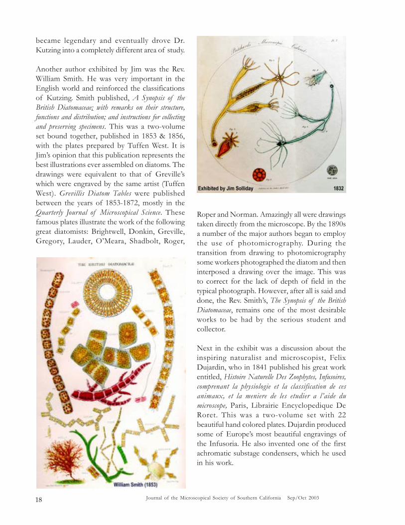
Paraffin lamps ofyore were usuallysupplied with stor-age cases, so backto the goody boxwe go in search ofsuitable case mate-rial. Fortune smilesat us as we comeacross some0.375" thick ma-hogany fragments.After the usual woodworking episodes, the saw-dust finally settles and we have our storage case,complete with period brass hardware as shownin fig. 5. For storage the bullseye assembly isremoved from the lamp and the bullseye is sepa-rated from the rack assembly. These parts are heldin separate wood brackets. A utility box on theinside of the door holds extra slides, forceps, aspare lamp assembly, etc.
When we consider the refinements and controlrelating to contemporary microscope illuminationsystems, current utilization of paraffin lampswould apply mainly to people who are butt deepin nostalgia, like Civil War enactors and their ilk,In kind, the Victorian wannabe gets a taste ofhistorical microtechnique. In passing, the safetyaspect of paraffin lamps bears scrutiny consid-ering the volatility and combustibility of paraf-fin oil. For example, what steps were taken toprevent an avid and incautious microscopist fromhaving his beard catch fire? Indeed, has this everhappened?
BibliographyGerard I’E Turner, “The Great Age of the Microscope,” AdamHilger, Bristol and New York, 1989, pages 289-290.eBay Item 39264421. Bockett Microscope Lamp signed:“Collins,” Nov/Dec 2002.Micrographia. “The Ideal Microscope Lamp,” page 2.www.micrographia.com
5Journal of the Microscopical Society of Southern California Sep/Oct 2003
WORKSHOP OF THE MICROSCOPICAL
SOCIETY OF SOUTHERN CALIFORNIA
recorded by Herb Gold and written by Jim Solliday
Date: Saturday, 6th September 2003Location: Ken Gregory’s Residence
The workshop began at 9:20 am and was calledto order by the President, Jim Solliday. Themeeting was held at Ken Gregory’s house withsixteen members present. The weather was brightand sunny and the members gathered under thetree in the backyard. Two tables were coveredwith exhibits and another table, located in thedriveway, featured sale and giveaway itemsincluding some surplus items left by Dr. John Fieldfor members to take on a first-come, first-serve
basis. Dr. Field had originally planned to attendthe workshop, but was unfortunately called awayat the last minute.
A new member, Sid Schiff, attended this meeting.Sid is from San Clemente and his interests includescientific apparatus and collecting oldmicroscopes. Members welcomed Mr. Schiff tothe fellowship and offered him our customaryhand of friendship.

Journal of the Microscopical Society of Southern California Sep/Oct 20036
Ken Gregory then mentioned that he hadsuccessfully obtained two excellent speakers forthe next lectureship meetings (third Wednesdayof the Month). Dr. Clifton Franklund, from theDepartment of Biological Sciences, CSULB, willgive a presentation on the microscopic analysisof oral biofilms at the September 2003 meeting.Ken described biofilms as populations ofmicroorganisms that adhere to any surface withsufficient moisture and adequate nutrients. Thesubject of the October 2003 meeting will behuman parasitology.
Jim Solliday displayed an example of the latestedition of The Journal of the MicroscopeHistorical Society (edited by Dan Kile,[email protected].) The Journal is aprofessional quality publication, boasting a newand colorful format, is well illustrated and packedwith researched articles. The President (JimSolliday) gave his highest recommendation forthis publication, especially for anyone interestedin the history of the microscope and collectingthe instrument. There was also a discussion ledby Stuart Warter on the value of maintaining ahard copy of our own fine Journal. The memberswere then reminded that the next workshop(October 4th) would be held at Izzy Lieberman’sresidence.
Exhibits and Discussions:
Alan deHaas described for the group a collectionof rare books he had set up on the desk in thehouse. All the books brought to the meeting werefor sale at very reasonable prices. Alan hasdecided to keep all his books on optics, butliquidate books dealing with subjects such as thedescriptions of microscopical discoveries. Thetwo books that he brought outside to the meetingarea were by Joseph Leidy and Saville Kent, bothwere authors of superbly illustrated works on theProtozoa.
Joseph Leidy’s book was entitled Fresh-WaterRhizopods of North America (1879), published inWashington by the Government Printing Office
(U.S.G.S.) for the Department of the Interior. Thiswas part of the U.S.G.S. report of the Territories.Volume XII, with beautiful color plates. Alan wasasking a mere $80.00 for this near perfect copy.
The second book, by Saville Kent, was entitledA Manual of the Infusoria: Including a Description ofAll Known FLAGELLATE, CILIATE, ANDTENTACULEFEROUS PROTOZOA, Britishand Foreign, and an account of the Organization andAffinities of the Sponges, three volumes, (1880-1882). This is indeed one of the best-illustratedbooks ever produced on the subject. If onecarefully studies the plates, he cannot help butrealize that it should have taken a lifetime tocreate all the spectacular drawings. Volume Three,representing the plates, has literally thousandsof illustrations all of the finest detail. DavidBogue was the publisher of this three-volumeset in London. At the time it was considered bythe author to be a complete account of thesubject.

7Journal of the Microscopical Society of Southern California Sep/Oct 2003
Alan also talked a bitabout the works ofLedermüller (Pub. 1760-1763) and exhibited twocopies of this magnificentbook. The hand-paintedillustrations throughoutthis collection can only bedescribed as among thebest ever published. Ofinterest was the fact thatthe engraving of themosquito found inLedermuller’s work is verymuch the same as thatdone over a century earlierby Swammerdam (see theillustrations for acomparison).

Journal of the Microscopical Society of Southern California Sep/Oct 20038

9Journal of the Microscopical Society of Southern California Sep/Oct 2003
Alan then set out for sale a copy of Amimalclesdes Infusions Vegetales by P. Laurent (1854). Thisalso had splendid plates illustrating primarily theProtozoa (see illustrations).
Inside the house, Alan exhibited a rather largecollection of valuable books that representedcopies from the 16th through the 19th Centuries.It would not be practical to list all the books thatwere on exhibit, but you are directed to theillustrations for a representation of a few of thebetter known examples.

Journal of the Microscopical Society of Southern California Sep/Oct 200310
Ken Gregory exhibited a large early Reichertstand with a five-objective nosepiece. The yearof manufacture was estimated to be circa 1884.The body along with the circular stage can beswiveled around the axis of the scope much likethat of the early Oberhaeuser drum microscopes.Also on exhibit was a Carl Zeiss stand that offeredthe same feature. The two provided a goodexample for comparison and illustrated theuniversal appeal of this feature. This option wasvery useful for dissection work and essential fortechniques in arranging diatoms by hand. Thespecimen could be rotated along with the stageand body of the microscope preventing anydisplacement from the axis of the instrument.The object would remain in perfect alignmentwith the objective and eyepiece but could berotated to match any position for application ofthe dissection instrument. The hand of theworker could remain steady on an armrest next

11Journal of the Microscopical Society of Southern California Sep/Oct 2003
to the stage. It was mentioned that this featurebecame less popular with the introduction ofmore mechanically accurate rotating stages in thelate 19th Century.
Ken also brought out for the group a very niceBausch & Lomb portable microscope (Portable
Microscope APS). This was to illustrate thesimilarity to a portable stereo Bausch & Lombexhibited by our new member, Sid Schiff. Bothscopes were from the early Twentieth Century(circa 1912-1918).
Ken also presented a very unique microscope thatwas designed to be used in the hair care industry.It featured a large view screen and included botha polarizer and analyzer. The image of anindividual hair could be projected on to the screenfor both the worker and the customer to see. It isthought that McBain Instruments sold thisparticular design. Ken said the scope wasavailable for sale for a mere $40.00.

Journal of the Microscopical Society of Southern California Sep/Oct 200312
Stuart War ter exhibited two interestingEuropean microscopes, one an early Drum typeand the other a later Continental model. TheDrum was signed by Nachet and manufacturedsome time in the 1850s; it was sometimes referredto as a compound-dissecting microscope. TheContinental was by Hartnack who succeededOberhaeuser in 1860. Both the Drum and theContinental featured the same rotation capability,as did the two stands exhibited by Ken Gregory.The stage, arm and bodytube could be rotatedaround the axis of the microscope with the mirrorand foot remaining stationary.
Edmund Hartnack (1826-1891) was located atthe time at Place Dauphine 21, Paris. By 1870,he had moved to 39 Waisenstrasse, Potsdam(Berlin), Germany (1870-c.1927). Hartnackbecame one of Europe’s most influential makers,adding much to the improvements of themicroscope. According to Mayall, he was first tosubstitute the horseshoe foot in place of the
Drum-base introduced earlier by Oberhaeuser.This allowed the use of oblique illumination andbetter access to the substage apparatus. He alsowas said to have placed the fine adjustment screwat the top of the limb instead of at the underside.This improved form became what was to beknown as the “Continental type.” Zeiss andNachet added the heel that extended the basebehind the pillar making it more appropriate forthe inclination joint.
The above remarks by Mayall must be consideredin the light of the fact that earlier instrumentssigned by Oberhaeuser have been found havingan inclination joint; also in a few early stands thefine adjustment screw was placed to the top ofthe limb (Oberhaeuser, c.1853). It is likely thatHartnack was working with Oberhaeuser in theearly 1850s, long before the partnership of 1857began. (See Oberhaeuser/McAllister stand,c.1853, Warter collection.) In contrast to whatMayall said, Nuttall states that Oberhaeuser

13Journal of the Microscopical Society of Southern California Sep/Oct 2003
developed the horseshoe foot in about 1848 andthat Chevallier advertised an intermediate formof horseshoe in 1841. However, there can be verylittle doubt that it was Hartnack who popularizedthe horseshoe continental microscope.
Larry McDavid gave us a report on the NorthAmerican Sundial Society, which recently met inBanff, Canada. He also had the chance to visitboth Lake Louise and the glacier where he wasable to walk out over the ice. He indicated thatthe scenery was not nearly as nice as it couldhave been due to the extensive forest fires burningat the time of the meeting. Larry did pass arounda ring-dial that he won during his visit. For moreinformation on the North American SundialSociety please see http://www.nass.org.
Allen Bishop brought in a new book dedicatedto the work of Amici. It was packed with rareillustrations of a large number of Amici’smicroscopes. The book was by Alberto Meschiariand was entitled The Microscopes of Giovanni BattistaAmici (only the introducton is in English). Formore information you can contact; FondazioneGiorgio Ronchi, Redazione e Amministrazione:Via S. Felice A EMA, 20-S0125, Firenze, Italy.Or contact the author at: [email protected].
Allen then exhibited a beautiful large Winkel-Zeiss polarizing stand, Model IV-M that wasmanufactured in 1936. This outfit came completewith a case and all the appropriate accessories.The Winkel (R. Winkel GMBH, Gottingen)Catalogue refers to this microscope as thePetrological Working Stand IV-M. This is a largestand with a similar design to the Winkel StandV-M, differing only in that the compoundmechanical revolving stage is replaced by a fixedrevolving stage. The stage on this microscope isdivided into 360 degrees and has verniers reading0.1 degree. The point was made by Alan deHaasthat the Fedorow universal rotating stage couldbe used with this stand. This instrument wasintended for original scientific investigationsrelating to mineralogy and petrography as well
as for the technical study of natural and artificialminerals.
The stand has a large, heavy foot with a hingedjoint for inclination and a long projecting heel,which ensures an absolutely steady position whenthe stand is inclined. The coarse adjustment iscontrolled by rack and pinion, while the slowmotion is produced by a side micrometer screw,which can be operated from both sides. The fineadjustment screw carries on one side a divideddrum with fifty intervals, each of whichcorresponds to a tube displacement of 0.002mm.The eyepiece drawtube is secured against turning,has a millimeter scale and is displaced by a rackand pinion attachment. After the removal of anintermediate sleeve the drawtube will receiveeyepieces with an enlarged field of view. Thestand is also furnished with a revolving nosepiecewith centering collars having the standard RMSthreads. A slot just above the nosepiece servesfor the introduction of a quartz wedge andgypsum plate. The mirror can be raised andlowered as well as inclined in all directions. The

Journal of the Microscopical Society of Southern California Sep/Oct 200314
tube has a Telan lens incorporatedas well as the analyzer beingprotected from the accumulation ofdust by a cover-glass and can beturned through an angle of 90degrees. The amount of rotation isshown by a graduated arc withintervals of two degrees.
An important note is that the Amici-Bertrand lens slides into the tubeabove the analyzer and is computedfor use with the achromaticobjective 42M (also the a42M andthe Fluorite objective 41M inconjunction with the Huygens 6xeyepiece). The Catalogue re-commends that the following opticsshould be used with the IV-M.Achromatic objectives include:2.5M, 10M, 23M, 42M and the 69Mhomogeneous immersion lens. The eyepiecesavailable were the Huygens 6x with cross-linesand movable eye-lens, Huygens 9x withmicrometer scale and the 12x with cross-hair andadjustable eye-lens. Also recommended were thefollowing auxiliary items: quartz wedge withcolors of the I.-III. Orders, in metal mount,gypsum red one order, in metal mount and a micaplate with ¼ W.L. phase-difference, in metalmount.
Reino Mascarino told the group about someunusual rotifers that he discovered in a nearbylake. He talked about his ongoing efforts to locateand study his favorite microscopic subject. Hementioned a book that he was looking for on thesubject of rotifers. He also talked a bit about hiswife’s work with frogs. He emphasized the valueof the microscope in their efforts to raise exoticfrogs. One facet that he described was his delightin watching frog’s eggs develop with his LeitzGreenough stereomicroscope. Alan deHaas then
described the benefits and effects ofthe high numerical aperture versusthe long working distancestereoscope. We also talked aboutthe differences between the newerphoto-stereos and the effect of theolder Greenough type.
Jim Solliday, after previously talkingwith Alan deHaas decided to bringin a collection of antique books thatwere published during the first halfof the 19th Century. This was to helpcomplement the contribution ofbooks brought in by Alan and focusadditional attention on the subject

15Journal of the Microscopical Society of Southern California Sep/Oct 2003
of books. The books that were chosen were someof the finest produced on the microscope of thattime and were indeed very popular amongmicroscopists.
The most important item that was exhibited wasa collection of works by Christian GottfriedEhrenberg (1795-1876). This was a collectionof eight papers bound together into a two-volumeset (Berlin, 1830-1849). This series of papersrepresented Ehrenberg’s fundamentalcontributions to the fields of micro-zoology, inwhich he studies the muscles, nervous andvascular systems, digestive and sexual organs ofa range of microscopic animals. Ehrenberg didnot yet separate the multicelled animals from thesingle-celled forms, a concept that becamepopular in systematic zoology only after 1850;rather, he believed that he could demonstrate thepresence of complete organ systems in single-celled animals. He made a rather interesting casesupporting this position in the work he did withrotifers, and used this to argue againstspontaneous generation and the “chain of being.”
The second volume concentrated on what wasconsidered the study of the infusoria. This is Jim’sfavorite work as it features some of the mostspectacular illustrations of marine deposits(diatoms, forams, et al) in Ehrenburg’s collection.
Ehrenberg is considered the father of thestudy of diatoms and was the first topublish a description of diatomaceousearth. The revelation that large rockformations were actually the result of themassive accumulation of living organismswas both revolutionary and inspired a surgein the study of geology. He remains solidlyassociated with the study of diatoms, asmore than one-third of all the knownspecies were named and described by him.This volume included the following works:Verbreitung und Einflufs des mikroskopischenLebens in Sud-und Nord-Amerika; DerLandesschule PFORTA widmet beim300jahrigen Stiftungsfeste (1843).Mikroskopische Analyse des curlandischenMeteor papiers von 1686 und Erlauterungdesselben als ein Produkt jetzt lebender Confervenund Infusorien (1839). Die Bildung derenropaischen, libyschen und arabischenKreidefelsen und des Kr eidemer gels ausmikroskopischen Organismen, dargestellt und

Journal of the Microscopical Society of Southern California Sep/Oct 200316

17Journal of the Microscopical Society of Southern California Sep/Oct 2003
physiologisch crlautert (1839). Passet-Staub und Blut-Regen ein grofses organisches unsichtbares Wirken andLeben in der Atmosphare (1849). All were publishedin Berlin and accompanied by magnificent plates.The above account has been included in thesenotes as a reference and helps to illustrate themagnitude of work assembled in these twovolumes. The contents of the first volume arenot included as they would be too exhaustive.
Next, Jim discussed a small book put together byL. Mandl (1839) and entitled, Traite Pratique duMICROSCOPE, et de son emploi Dans L’etude descor ps Organises. Including, Recherches surL’Organisation des Animaux Infusoires , byEhrenberg, Paris. This book was exhibited toillustrate just one more author who extensivelyused Ehrenberg as a source of authority and tofill major subject matters.
The next book Jim talked about was by AndrewPritchard (1832), The Microscopic Cabinet of SelectAnimated Objects; with a description of the Jewel anddoublet microscope, test objects, &c, London. It isillustrated with thirteen hand-colored engravedplates of original drawings by Dr. Goring.Pritchard included in his text most of the beliefs
of Ehrenberg. He continued to hold ontoEhrenberg’s interpretations through just about allof his publications up to at least the 1860s. Thiswould include Pritchard’s History of Infusoria(1861), a very well known work and referencesource for the time. Pritchard did finally mentionthe views of other contemporary workers in hisfinal editions.
One of the most controversial of Ehrenberg’spositions was that he was convinced that diatomsshould be catalogued in the animal kingdom. Hehypothesized that creatures that could moveabout using their own power were animals andthose that were fixed to a substrate were plants.Most of the other well-known authors of the timesuch as Carpenter and Hogg held to the positionsof Dr. Friedrich Kutzing who classified thediatoms as the Bacillarien oder Diatomeen (goldenalgae, or plants). The animosity and competitionfor authority between Ehrenberg and Kutzing
Journal of the Microscopical Society of Southern California Sep/Oct 200318
became legendary and eventually drove Dr.Kutzing into a completely different area of study.
Another author exhibited by Jim was the Rev.William Smith. He was very important in theEnglish world and reinforced the classificationsof Kutzing. Smith published, A Synopsis of theBritish Diatomaceae: with remarks on their structure,functions and distribution; and instructions for collectingand preserving specimens. This was a two-volumeset bound together, published in 1853 & 1856,with the plates prepared by Tuffen West. It isJim’s opinion that this publication represents thebest illustrations ever assembled on diatoms. Thedrawings were equivalent to that of Greville’swhich were engraved by the same artist (TuffenWest). Grevillis Diatom Tables were publishedbetween the years of 1853-1872, mostly in theQuarterly Journal of Microscopical Science. Thesefamous plates illustrate the work of the followinggreat diatomists: Brightwell, Donkin, Greville,Gregory, Lauder, O’Meara, Shadbolt, Roger,
Roper and Norman. Amazingly all were drawingstaken directly from the microscope. By the 1890sa number of the major authors began to employthe use of photomicrography. During thetransition from drawing to photomicrographysome workers photographed the diatom and theninterposed a drawing over the image. This wasto correct for the lack of depth of field in thetypical photograph. However, after all is said anddone, the Rev. Smith’s, The Synopsis of the BritishDiatomaceae, remains one of the most desirableworks to be had by the serious student andcollector.
Next in the exhibit was a discussion about theinspiring naturalist and microscopist, FelixDujardin, who in 1841 published his great workentitled, Histoire Naturelle Des Zoophytes, Infusoires,comprenant la physiologie et la classification de cesanimaux, et la meniere de les etudier a l’aide dumicroscope, Paris, Librairie Encyclopedique DeRoret. This was a two-volume set with 22beautiful hand colored plates. Dujardin producedsome of Europe’s most beautiful engravings ofthe Infusoria. He also invented one of the firstachromatic substage condensers, which he usedin his work.

19Journal of the Microscopical Society of Southern California Sep/Oct 2003
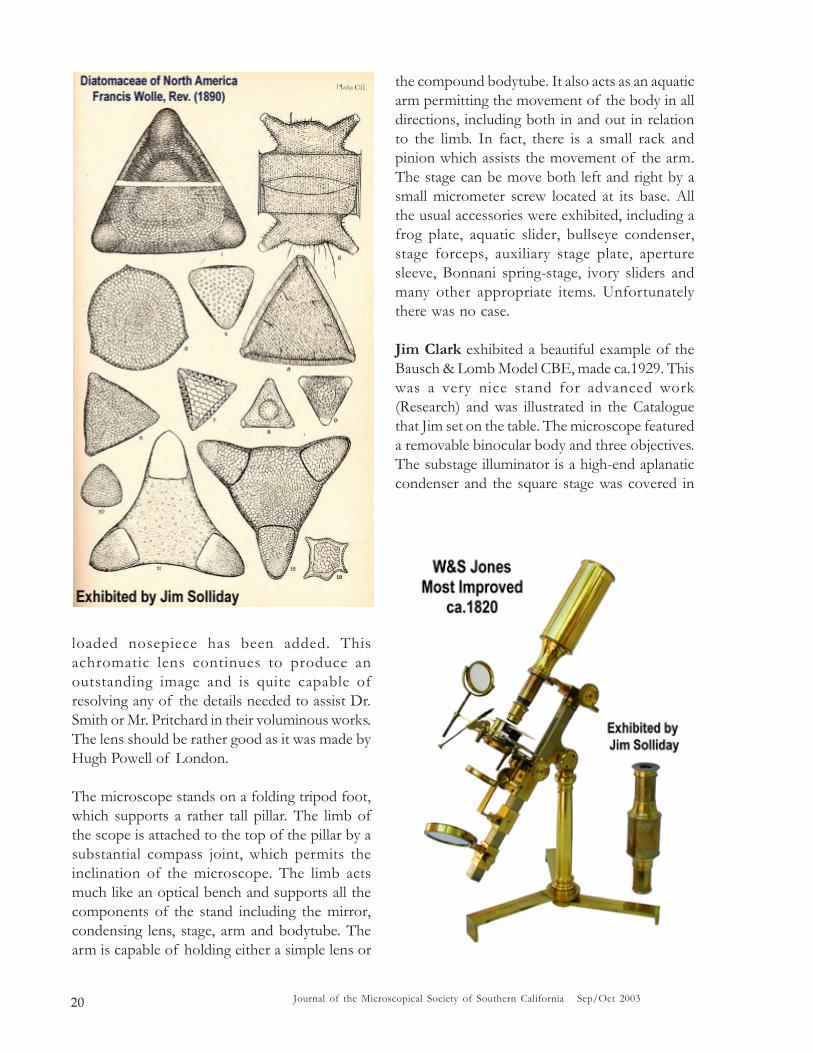
Here in the United States, the Rev. Francis Wollein 1890, published his encyclopedic, Diatomaceaeof North America. Illustrated with 2300 figures,all drawings by the author and published inBethleham, PA. If you leaf through thismagnificent work you would be convinced thatthis was the accumulated work of a lifetime.Each plate is filled with sometimes dozens ofbeautifully detailed drawings of the diatoms. Thisis also one of the rarest of books on thediatomaceae and “worth its weight in gold.”
Just about all the books exhibited above wereproduced during the first half of the 19th Centuryand relied on microscopes that were either pre-achromatic or made in the very early years of theachromatic microscope.
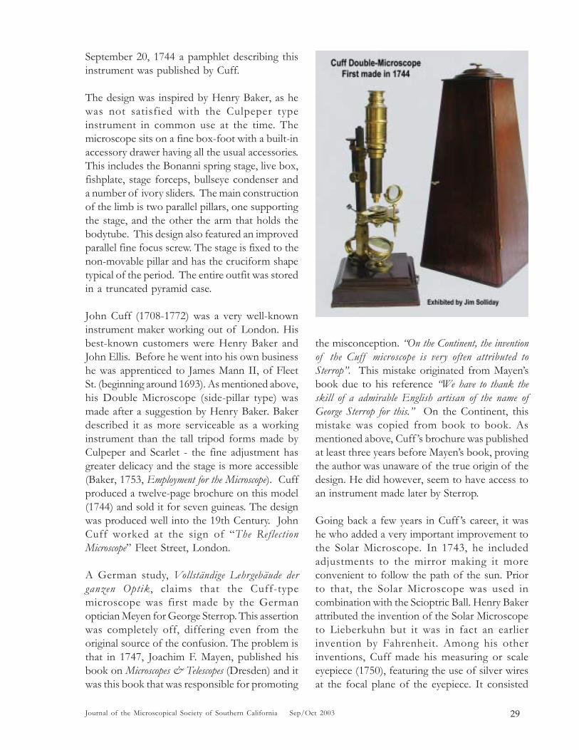
In order to illustrate the types of microscopesused at the time, Jim exhibited a very popular
type of microscope that potentially could havebeen used by one of the above authors. Thismicroscope was described by John Mayall as thebest instrument one could buy during the firstquarter of the 19th Century. The microscopestanding before the group was a large Englishinstrument signed by W & S, Jones, London. Itwas known as the Jones “Most ImprovedCompound Microscope,” made after the originaldesign of George Adams. Billings states that thisis the best microscope made prior to theachromatic period.
The lacquered finish remains in almost perfectcondition and all mechanical functions are in goodworking order. Included are two eyepiece tubesand a rotating objective turret with six lenses,representing six different magnifications. Also, adevisable achromatic objective with a spring-
Journal of the Microscopical Society of Southern California Sep/Oct 200320
loaded nosepiece has been added. Thisachromatic lens continues to produce anoutstanding image and is quite capable ofresolving any of the details needed to assist Dr.Smith or Mr. Pritchard in their voluminous works.The lens should be rather good as it was made byHugh Powell of London.
The microscope stands on a folding tripod foot,which supports a rather tall pillar. The limb ofthe scope is attached to the top of the pillar by asubstantial compass joint, which permits theinclination of the microscope. The limb actsmuch like an optical bench and supports all thecomponents of the stand including the mirror,condensing lens, stage, arm and bodytube. Thearm is capable of holding either a simple lens or
the compound bodytube. It also acts as an aquaticarm permitting the movement of the body in alldirections, including both in and out in relationto the limb. In fact, there is a small rack andpinion which assists the movement of the arm.The stage can be move both left and right by asmall micrometer screw located at its base. Allthe usual accessories were exhibited, including afrog plate, aquatic slider, bullseye condenser,stage forceps, auxiliary stage plate, aperturesleeve, Bonnani spring-stage, ivory sliders andmany other appropriate items. Unfortunatelythere was no case.
Jim Clark exhibited a beautiful example of theBausch & Lomb Model CBE, made ca.1929. Thiswas a very nice stand for advanced work(Research) and was illustrated in the Cataloguethat Jim set on the table. The microscope featureda removable binocular body and three objectives.The substage illuminator is a high-end aplanaticcondenser and the square stage was covered in

21Journal of the Microscopical Society of Southern California Sep/Oct 2003
vulcanite. It also features a built in mechanicalslide holder with X-Y movement.
The Catalogue describes this microscope asfollows: “CBE-A Binocular Instrument for Research.”After pointing out the disadvantages of themonocular tube the catalogue touts theconvenience and comfort of the binocular,especially when used for long uninterruptedperiods of observation. “To overcome these conditionswe offer in the Microscope CBE a research microscopewhich not only permits the simultaneous use of botheyes, but which accommodates the entire series of singleobjectives ordinarily used on compound microscopes ofthe monocular type. The compound slide, upon which theeyepiece tubes are mounted, is adjustable so as to permitany observer to accommodate the eyepieces to his owninterpupillary distance. A scale on this slide enables thetubes to be set instantly at the correct distance, once it isdetermined.”

Journal of the Microscopical Society of Southern California Sep/Oct 200322
The catalogue continues to describe the binocularas follows: “The binocular feature affords relief fromeyestrain and fatigue. The eyepieces are parallel and theoptical construction of the body tube is such that there isno necessity for converging the eyes. The effect is as thoughthe observer was looking into the distance and is, ofcourse, very restful to the eyes. This means that lessconcentration is required on the part of the observer,enabling him to give all of his attention to studying thespecimen.” This led to a discussion of theadvantages versus the disadvantages of theconverging binocular body tube. Some of themembers stated that it was impossible for themto use the parallel type binocular while othercould not use the convergent type, which werepopularly made by Spencer. It all depended onwhether the user was near or far-sighted. Itseemed that the near-sighted user preferred theconvergent type body tubes.
Izzy Lieberman shared with the group anumber of problems he experienced with theairlines. This of course brought out a variety of
similar experiences that other members haddealing with the same service.
Sid Schiff, our new member exhibited a very niceexample of a traveling Bausch & LombGreenough stereoscope. This instrument camefolded inside a very nice hardwood case and wasin almost new condition. Sid introduced thisinstrument with the hope that some of ourmembers could tell him more about it and if theprice he had paid was reasonable. Afterrecognizing some of the features of this scope,Ken Gregory went into the house and obtainedan example of a Bausch & Lomb compoundtraveling scope that was manufactured at aboutthe same time period. Ken’s portable scope waslisted in the B&L Catalogue as the PortableMicroscope APS and was made in about the year1915. The consensus of the B&L experts wasthat Sid’s traveling stereo scope was indeed quiterare and was something that none of us had seenbefore. Our new member was assured that theprice he paid was without question very fairindeed.

23Journal of the Microscopical Society of Southern California Sep/Oct 2003
Herb Gold exhibited a wonderful LeitzIntegrating stage. The vintage of the instrumentand the look of the case caused the members tobelieve it to be prewar. Also, its condition wasseen to be quite good. Alan deHaas gave thegroup a description of the primary purpose ofthis sophisticated stage and how it was used. Itwas explained that it was needed in determiningthe total area or percentage of a certain mineralwithin the matrix of a rock section (this is butone example of its use). Other more convenientmethods for this function have been recently
introduced in association with computer imageanalytical programs. A page from a German Leitzcatalogue illustrating the stage could be seen onthe table.
Finally, it was announced that the members wereinvited to go to lunch after the meeting at a localrestaurant. The President brought the meetingto a close at 11:50 a.m. leaving plenty of timefor photographing the exhibits. A hearty thankswere expressed to the members for theirparticipation and continued support.

Journal of the Microscopical Society of Southern California Sep/Oct 200324
MSSC MONTHLY MEETINGWednesday 17th September 2003
at New Roads Schoolreported by Leonie Fedel
At this meeting, Dr. Clifton Franklund,Department of Biological Sciences, CaliforniaState University, Long Beach gave a lecture onthe microscopic analysis of oral biofilms.Biofilms are populations of microorganisms thatadhere to any environmental surface withsufficient moisture and adequate nutrients. Dentalplaque is an example of the development of abiofilm.
Dr. Franklund started his presentation by givingan overview of periodontal disease which affectsover 60 million people in the US and is the maincause of tooth loss in adults. Periodontal diseaseis the result of a chronic host inflammatoryresponse to oral bacteria - over 400 differentspecies of bacteria are involved includingFusobacterium, Por phyromonas, Pr evotella,Actinobacillus, and Bacteroides. Gram-positive coccicolonize the tooth and gums, followed by gram-negative anaerobes, leading to mineralization,inflammation (gingivitis), and finally tissuedestruction of the gums and hence tooth loss.
The progression of the disease is affected by thepresence of a number of virulence factors. Ofthese Lipopolysaccharide (LPS) has been
implicated in processes as diverse as attachmentto host tissues, evasion of phagocytosis,molecular mimicry, and (most importantly)immune stimulation. From the literature, it is clearthat LPS, proteinaceous virulence factors,metabolic end products, and host factors allcontribute to the tissue damage seen inperiodontal disease. Furthermore, in many casesthese factors may perform the same or similarfunctions. The complexity of this clinicalsituation makes it very difficult to determinecausal relationships between bacterial species,their products, and tissue damage.
However, one of the most commonly isolatedmicrobes from the gingiva is the Gram-negativeanaerobe, Fusobacterium nucleatum. Given thepredominance of F. nucleatum in oral infections,Dr. Franklund is focusing on researching the roleof LPS in promoting the inflammatory responseobserved in advanced periodontal disease. Aclear understanding of this important moleculewill enable him to determine the role(s) that itplays in forming biofilms, evading or subvertingthe immune response, and promoting of hosttissue damage.

25Journal of the Microscopical Society of Southern California Sep/Oct 2003
Dr. Franklund continued by discussing thetechniques he uses to the study oral biofilms:
• BacLight staining;• SYTO9 staining ;• Immunofluorescence analysis;• Fluorescence in situ hybridization (FISH)
analysis;• Confocal scanning laser microscopy;• Microscopic flow cell analysis.
For further information and updates on the re-search please visit Dr. Franklund’s research pageat www.csulb.edu/~cfranklu/research.html.

Journal of the Microscopical Society of Southern California Sep/Oct 200326
WORKSHOP OF THE MICROSCOPICAL
SOCIETY OF SOUTHERN CALIFORNIA
recorded by Herb Gold, written by Jim Solliday
Date: Saturday, 4th October 2003Location: Izzy Lieberman’s Residence
The workshop was called to order by ourPresident, Jim Solliday at 9:10am at IzzyLieberman’s residence with 17 members present.The weather was unusually pleasant with the sunshining and the temperature being mild. As usual,there were plenty of refreshments availableincluding doughnuts and coffee. A number oftables were set up with one featuring the exhibitsand the other holding the sale and giveaway items.The President noted the quality of microscopesand accessories set out on the exhibition table.
Dr. Ken Gregory, announced that Dr. Larry Ashwould be the guest speaker at the next WednesdayLectureship meeting on October 15, 2003. Dr.Ash works at UCLA and is a specialist inParasitology. He will speak about parasitescommon to Southern California which can becontracted by children. Examples of transmissionfrom animals (often pets) to humans will bedescribed. This talk continues the current seriesof topics dealing with microscopic organismsthat can affect humans.

27Journal of the Microscopical Society of Southern California Sep/Oct 2003
The members were reminded that the popularNovember Exhibition meeting was approaching.Individuals should begin preparing their exhibitsfor this annual event. Each year a wealth ofwonderful displays and projects are presentedduring this meeting. This is an appropriatemeeting to which to bring the kids or a guest.The President reiterated that all types of exhibitsare welcome and anything from an elaborateposter display to a simple microscope with yourfavorite slide is appropriate. Participation is themain thing and the more members that add tothe effort the better the event will be.
Finally, we were informed that the March/Apriledition of the Journal was now ready and shouldappear at the next lectureship meeting.
Exhibits and Discussions:
Ken Gregory set up a number of opticalinstruments to share with the group. First he toldus about a Bausch & Lomb microscope hepurchased from eBay that came in an elaboratehardwood case. The case obviously did notbelong to this rather common microscope but wasof 19th Century European origin. Having seen anumber of very similar cases exhibited here atour workshops by Stuart Warter and Jim Solliday
it was determined that it belonged to a Seibert(German) Microscope. The question was justwhat should he done with this wonderful case asthe B&L just did not fit properly.
A short time later Ken noticed a single Seibertmicroscope offered on eBay without its propercase. On top of that, it was up for sale by thesame dealer that had sold Ken the case. Withouthesitation Ken bid appropriately in order to winthis rare and important Seibert stand. Lo andbehold after the microscope arrived it was aperfect match for this beautiful mahogany case.Thus Ken acting as the conservationist he hasbecome, brought these two valuable items backtogether as they should be. It is to his credit andvigilance that he continues to make the world abetter place for all those lost microscopes. Theillustrations show this Seibert microscopestanding in front of its proper case, also exhibitedis an example of a similar Seibert brought in byStuart Warter to illustrate the same style ofstorage case. Both instruments feature the “C”-shaped pillar and horseshoe-type foot. Accordingto John deHaas, Ken’s Siebert was madesometime around 1882 or 1883.
Ken Gregory also offered for sale a largeophthalmic device, which was intended tomeasure the curvature of the cornea. Thisinstrument was dominated by a very large

Journal of the Microscopical Society of Southern California Sep/Oct 200328
graduated sector, which can be seen in theaccompanying illustration. The condition of thisinstrument seemed quite good. Also offered byKen was a large retinal microscope, which wasmanufactured by Bausch & Lomb (ca.1929). Themembers first noticed the horizontally mountedbinocular observation body. This item wasdescribed as a binocular with a large “auto-collimator like” telescope. One of the fellowsreferred to it as an “Opthalmometer”. These twoitems were prominently displayed and were bothfor sale.
Stuart Warter exhibited a beautiful Seibertmicroscope with mahogany case, very similar tothe stand brought in by Ken and described above.It was pointed out that the storage cases werequite similar and of the same pattern. Theinstrument was identified as a Siebert Model IIand was the same type of microscope used byGram of the Gram Stain fame. Stuart kindlybrought this particular instrument in to help Kenin the description of his outfit. It should be saidthat it is rather rare and unusual to see more thenone early Seibert in the same place, especiallyout here in California. Seibert did not sell thatwell in the United States when compared tomakers like Leitz and Zeiss. In fact at the turnof the Century, the most common imports weresmall French microscopes. However, among theserious instruments, the most ubiquitous werethose imported by Zeiss, then Leitz, Reichert and
finally, R&J Beck. Seibert hardly even registeredand could be counted with stands imported byPaul Waechter and R. Wasserlein of Berlin.
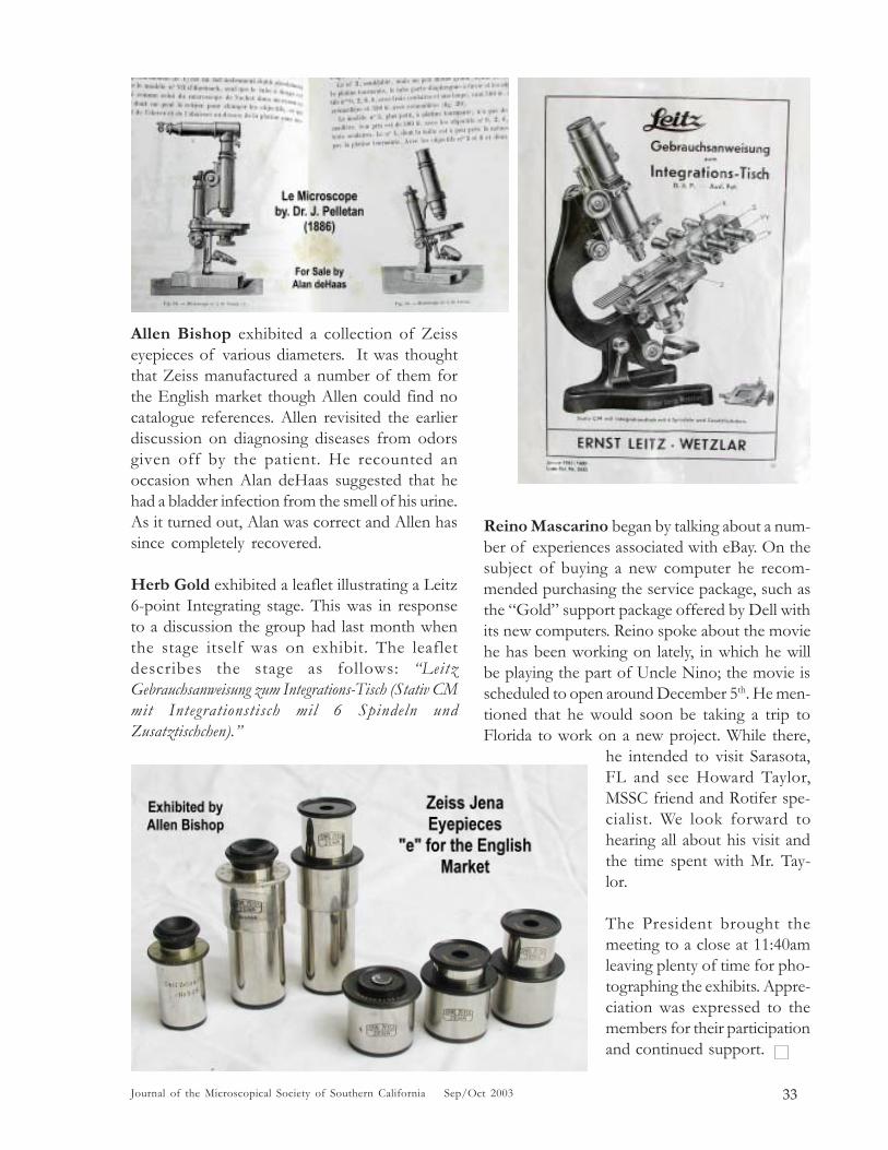
Jim Solliday exhibited a very early 18th Centurymicroscope, the type of which was invented byJohn Cuff. This example is in excellent conditionand good working order. The microscope iscomplete, and accompanied by all knownaccessories provided. The instrument is normallyreferred to as Cuff ’s Double Microscope and is agood example of his 1744 design. However, thisparticular instrument may have been made a bitlater, perhaps in the 1750s or 1760s. On
29Journal of the Microscopical Society of Southern California Sep/Oct 2003
September 20, 1744 a pamphlet describing thisinstrument was published by Cuff.
The design was inspired by Henry Baker, as hewas not satisfied with the Culpeper typeinstrument in common use at the time. Themicroscope sits on a fine box-foot with a built-inaccessory drawer having all the usual accessories.This includes the Bonanni spring stage, live box,fishplate, stage forceps, bullseye condenser anda number of ivory sliders. The main constructionof the limb is two parallel pillars, one supportingthe stage, and the other the arm that holds thebodytube. This design also featured an improvedparallel fine focus screw. The stage is fixed to thenon-movable pillar and has the cruciform shapetypical of the period. The entire outfit was storedin a truncated pyramid case.
John Cuff (1708-1772) was a very well-knowninstrument maker working out of London. Hisbest-known customers were Henry Baker andJohn Ellis. Before he went into his own businesshe was apprenticed to James Mann II, of FleetSt. (beginning around 1693). As mentioned above,his Double Microscope (side-pillar type) wasmade after a suggestion by Henry Baker. Bakerdescribed it as more serviceable as a workinginstrument than the tall tripod forms made byCulpeper and Scarlet - the fine adjustment hasgreater delicacy and the stage is more accessible(Baker, 1753, Employment for the Microscope). Cuffproduced a twelve-page brochure on this model(1744) and sold it for seven guineas. The designwas produced well into the 19th Century. JohnCuff worked at the sign of “The ReflectionMicroscope” Fleet Street, London.
A German study, Vollständige Lehrgebäude derganzen Optik, claims that the Cuff-typemicroscope was first made by the Germanoptician Meyen for George Sterrop. This assertionwas completely off, differing even from theoriginal source of the confusion. The problem isthat in 1747, Joachim F. Mayen, published hisbook on Microscopes & Telescopes (Dresden) and itwas this book that was responsible for promoting
the misconception. “On the Continent, the inventionof the Cuff microscope is very often attributed toSterrop”. This mistake originated from Mayen’sbook due to his reference “We have to thank theskill of a admirable English artisan of the name ofGeorge Sterrop for this.” On the Continent, thismistake was copied from book to book. Asmentioned above, Cuff ’s brochure was publishedat least three years before Mayen’s book, provingthe author was unaware of the true origin of thedesign. He did however, seem to have access toan instrument made later by Sterrop.
Going back a few years in Cuff ’s career, it washe who added a very important improvement tothe Solar Microscope. In 1743, he includedadjustments to the mirror making it moreconvenient to follow the path of the sun. Priorto that, the Solar Microscope was used incombination with the Scioptric Ball. Henry Bakerattributed the invention of the Solar Microscopeto Lieberkuhn but it was in fact an earlierinvention by Fahrenheit. Among his otherinventions, Cuff made his measuring or scaleeyepiece (1750), featuring the use of silver wiresat the focal plane of the eyepiece. It consisted

Journal of the Microscopical Society of Southern California Sep/Oct 200330
of 1/50-inch squares. In 1752, Ellis had hisaquatic microscope made by J. Cuff and in 1758,Cuff issued a pamphlet entitled “The Descriptionof a Double and Single Microscope Very Convenient toView All Sorts of Objects,” illustrating an aquaticmicroscope on to which a compound body couldbe added. The above is only a few of thecontributions Cuff made towards theimprovement of the microscope. In fact his newDouble Microscope was perhaps the single mostimportant improvement in the microscope for theentire 18th Century.
Dave Hirsch exhibited a Seibert lamp, whichwas coupled to a Seibert projection microscopehaving a heating stage. To enhance usability Daveconstructed the large support stand. Alan deHaasmentioned that this sort of set up was often usedfor melting point experiments. This outfit wassaid to be from the mid 1920s (see illustration).
Pete Teti has been for some time the caretakerof our Society collections. This includes not onlythe slides but also a number of books as well asa few compound and stereomicroscopes. Peteinformed the group that we have approximately800 prepared slides in the collection. At this time
they are stored in boxes of about 100 each withsome in cardboard trays of about 20. We are inthe process of cataloguing this collection withmany of them already identified. This work needsto be completed and a proper list published inthe Journal. Members may borrow slides from thecollection for a maximum of one month at a time.Contact Pete if you are interested, specifyingwhat slide box he should bring to the meeting.The member must return the slide box to Pete atthe next meeting (i.e.: 4 weeks later). Pete willmaintain a checkout list and see that all slidesand boxes are accounted for. This is just one morearea in which our good friend, Pete Teti continuesto serve our Society.
Dr. Fred Kahn talked to us about a physicianwhose name was Jacob da Silva Solis-Cohen(1838-1927). Dr. Cohen was active in the CivilWar as both a general and a physician. He wasprobably the first surgeon to adopt laryngologyas a specialty. He was to have cured cancer ofthe larynx and diagnosed patients by smelling theirbreath. Dr. Kahn confirmed that the odor froma patient’s breath could often be of great help inidentifying the problem. Fred also exhibited hisfather’s “Wappler” otoscope, which was said tobe from the 1920’s. He also exhibited a collectionof otoscopes representing examples from the1920’s to ones currently in use.

31Journal of the Microscopical Society of Southern California Sep/Oct 2003
Ellen Cohen talked about becoming a bit morecomputer-savvy and stimulated a discussion onthe MAC versus the PC. It was to be expectedthat the group represented advocates from bothsides of the fence. Alan deHaas pointed out thatthe MAC continues to offer great conveniencefor users working with graphics. However, thePC continues to enjoy the greatest variety ofsoftware applications. There were a number offellows in the group that offered Ellen anyassistance appropriate that would help her makethe best choice for her needs.
Ken Miller offered for sale an early example ofan Olympus PM-6 photomicrographic camera.It included a Luna Pro light meter that was invery good working order. There are two importantfeatures of this camera. First, it has a very nicefocusing telescope that utilizes a proper beam-splitter; second, it has a floating shutter that helpsprevent vibration during exposure. The shuttersits on a foam bed and is operated manually witha cable shutter-release. Please contact Ken if youhave any questions about this offer.
John deHaas proudly exhibited two of his oilpaintings. The first featured a large tree nestledwithin a small European village with a vegetablegarden in the foreground. The second was of asnowbound village with an alpine flavor. Bothwere nice representations of John’s artistic talents.For sale, John offered a Nikon camera body ingood working order.
John Fedel brought up the continued need foradvice on proper maintenance of the microscope.About a year and a half ago we had a veryinformative workshop given by Alan deHaas onthis subject. All agreed that it would be helpfulif Alan were available to continue hisinstructions on this subject. Alan said that hewould indeed be available and would be happyto share his expertise. A few problems associatedwith microscope maintenance were discussedwith suggestions coming from all sides of thegroup.
Izzy Lieberman talked about printers and thehistory of color cartridges. He compared the inksproduced by a number of makers including HP

Journal of the Microscopical Society of Southern California Sep/Oct 200332
versus Epson. He provided examples of printsto illustrate his points. He again recommendedthe use of cartridges with a built-in injection-head, rather than ones that have to be filled witha hypodermic needle.
Larry McDavid talked about an Archimedesspecial on television featuring a “Palimpset”manuscript for calculating the volume of solidfigures. This was a NOVA program that suggestedArchimedes was the original inventor of integralcalculus. A discussion followed led by Larry.
Alan deHaas placed on the table and offeredfor sale a large collection of microscopeobjectives. This was an exceptional offer forthose in the group who needed contemporarylenses. The makers represented were primarilyLeitz and Zeiss, with some others includingOlympus. Alan’s prices were reasonable andcreated excitement amongst the members. Hetold the group that a number of importantobjectives obtained in this lot were decementedand no longer usable. Those lenses had been setaside and were designated as possible projectsfor repair. Alan described some of the methodshe intended to employ in his attempt to fix them.He talked about decementing, cleaning andreassembly. A discussion about optical cementsensued and how best to apply them.
Alan also offered for sale a rarebook written by J. Pelletan,entitled Le Microscope (1886).See the illustration of thebeautiful binding. This bookwas at one time in thelaboratory of Dr. Philip B.Ayres as can be seen in theillustration of the letter thataccompanied the book as itchanged hands in 1888.
33Journal of the Microscopical Society of Southern California Sep/Oct 2003
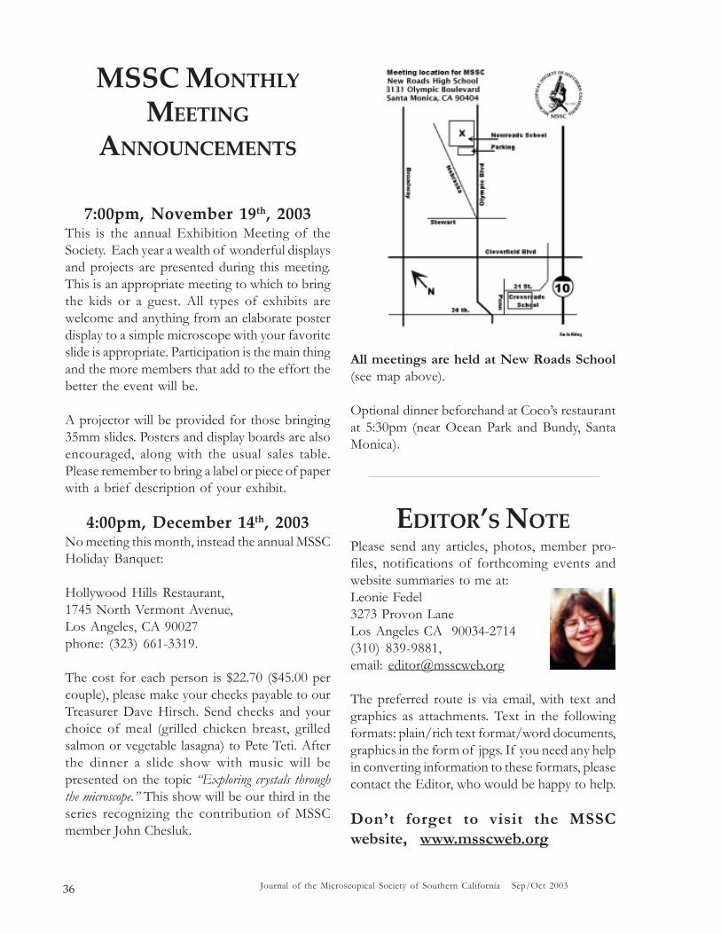
Allen Bishop exhibited a collection of Zeisseyepieces of various diameters. It was thoughtthat Zeiss manufactured a number of them forthe English market though Allen could find nocatalogue references. Allen revisited the earlierdiscussion on diagnosing diseases from odorsgiven off by the patient. He recounted anoccasion when Alan deHaas suggested that hehad a bladder infection from the smell of his urine.As it turned out, Alan was correct and Allen hassince completely recovered.
Herb Gold exhibited a leaflet illustrating a Leitz6-point Integrating stage. This was in responseto a discussion the group had last month whenthe stage itself was on exhibit. The leafletdescribes the stage as follows: “LeitzGebrauchsanweisung zum Integrations-Tisch (Stativ CMmit Integrationstisch mil 6 Spindeln undZusatztischchen).”
Reino Mascarino began by talking about a num-ber of experiences associated with eBay. On thesubject of buying a new computer he recom-mended purchasing the service package, such asthe “Gold” support package offered by Dell withits new computers. Reino spoke about the moviehe has been working on lately, in which he willbe playing the part of Uncle Nino; the movie isscheduled to open around December 5th. He men-tioned that he would soon be taking a trip toFlorida to work on a new project. While there,
he intended to visit Sarasota,FL and see Howard Taylor,MSSC friend and Rotifer spe-cialist. We look forward tohearing all about his visit andthe time spent with Mr. Tay-lor.
The President brought themeeting to a close at 11:40amleaving plenty of time for pho-tographing the exhibits. Appre-ciation was expressed to themembers for their participationand continued support.

Journal of the Microscopical Society of Southern California Sep/Oct 200334
MSSC MONTHLY MEETINGWednesday 15th October 2003
at New Roads Schoolreported by Leonie Fedel
At this meeting, Dr. Larry Ash, Department ofBiological Sciences, UCLA, and a specialist inparasitology, gave a lecture on parasites commonto Southern California titled “Zoonotic LarvalNematode Infections and Childhood Diseases.”
Dr. Ash explained how children, in particulartoddler-age children, are especially vulnerable toinfection by parasites, both those occurring inhousehold pets (i.e., dogs and cats) and parasitesof wild animals living in urban and peri-urbanenvironments. Such infections, deriving fromanimals, are referred to as zoonoses or zoonoticinfections.
He continued by discussing the syndrome, viscerallarval migrans, first described in the early 1950s,in which young children acquired infectionprincipally from ingestion of Toxocara canis, thecommon roundworm of dogs, but also fromingestion of Toxocara cati, a similar parasiteinfecting cats. With these infections, larvalnematodes invaded a variety of tissues,particularly the liver, the central nervous systemand the eye. Severity of infection was generallydependent upon the numbers of infective eggsingested but fatalities were uncommon.
Beginning in the 1980s, a related ascarid parasiteof raccoons, Baylisascaris procyonis, was found tocause severe, often fatal, disease in children. It isnow recognized that this raccoon parasite is wellestablished on the west coast of the U.S. and anumber of cases have been reported in California.
Dr. Ash continued by discussing the interactionof animals and their parasitic infections, with theexternal environment such as where fecal materialis deposited, the influence of weather, and thebehavior of adults and children - all of whichcontribute to the public health problemassociated with B. procyonis and other similarnematode infections.

35Journal of the Microscopical Society of Southern California Sep/Oct 2003
MSSC MONTHLY
SATURDAY WORKSHOP
ANNOUNCEMENTS
The MSSC holds a workshop from:
9:00am to 12:00pm on the firstSaturday of every month
Locations alternate between two members’houses, Izzy Leiberman’s and Ken Gregory’s.
The workshops provide a chance for fellow mi-croscopists to talk about our favorite subject. Youare invited to bring any manner of items relatedto microscopy to share it with the fellowship. Ifyou have something you would like to sell, pleasefeel free to bring it and set it up at the sales table.All are encouraged to participate and join in thefun.
An optional lunch after each workshop will beheld at the local Coco’s.
Remaining schedule for 2003 is as follows:Nov. 1, 2003 at Izzy Lieberman’sDec. 6, 2003 at Ken Gregory’s
The schedule for 2004 is as follows:January 3, 2004, Izzy Lieberman’sFebruary 7, 2004, Izzy Lieberman’sMarch 6, 2004, Izzy Lieberman’sApril 3, 2004, Ken Gregory’sMay 1, 2004, Ken Gregory’sJune 5, 2004, Ken Gregory’sJuly 3, 2004, Izzy Lieberman’sAugust 7, 2004, Izzy Lieberman’sSeptember 4, 2004, Ken Gregory’sOctober 2, 2004, Izzy Lieberman’sNovember 6, 2004, Izzy Lieberman’sDecember 4, 2004, Ken Gregory’s
Izzy Leiberman’s Residence:3300 Corinth Avenue
Los Angeles CA 90066310-391-6076
Ken Gregory’s Residence:2124 Ocana Avenue
Long Beach, CA 90815562-596-1762
Journal of the Microscopical Society of Southern California Sep/Oct 200336
EDITOR’S NOTEPlease send any articles, photos, member pro-files, notifications of forthcoming events andwebsite summaries to me at:Leonie Fedel3273 Provon LaneLos Angeles CA 90034-2714(310) 839-9881,email: [email protected]
The preferred route is via email, with text andgraphics as attachments. Text in the followingformats: plain/rich text format/word documents,graphics in the form of jpgs. If you need any helpin converting information to these formats, pleasecontact the Editor, who would be happy to help.
Don’t forget to visit the MSSCwebsite, www.msscweb.org
MSSC MONTHLY
MEETING
ANNOUNCEMENTS
7:00pm, November 19th, 2003This is the annual Exhibition Meeting of theSociety. Each year a wealth of wonderful displaysand projects are presented during this meeting.This is an appropriate meeting to which to bringthe kids or a guest. All types of exhibits arewelcome and anything from an elaborate posterdisplay to a simple microscope with your favoriteslide is appropriate. Participation is the main thingand the more members that add to the effort thebetter the event will be.
A projector will be provided for those bringing35mm slides. Posters and display boards are alsoencouraged, along with the usual sales table.Please remember to bring a label or piece of paperwith a brief description of your exhibit.
4:00pm, December 14th, 2003No meeting this month, instead the annual MSSCHoliday Banquet:
Hollywood Hills Restaurant,1745 North Vermont Avenue,Los Angeles, CA 90027phone: (323) 661-3319.
The cost for each person is $22.70 ($45.00 percouple), please make your checks payable to ourTreasurer Dave Hirsch. Send checks and yourchoice of meal (grilled chicken breast, grilledsalmon or vegetable lasagna) to Pete Teti. Afterthe dinner a slide show with music will bepresented on the topic “Exploring crystals throughthe microscope.” This show will be our third in theseries recognizing the contribution of MSSCmember John Chesluk.
All meetings are held at New Roads School(see map above).
Optional dinner beforehand at Coco’s restaurantat 5:30pm (near Ocean Park and Bundy, SantaMonica).