the osteology and relationships of the anglerfish … · 2012-05-08 · the osteology and...
TRANSCRIPT

THE OSTEOLOGY AND RELATIONSHIPS OF THE ANGLERFISH GENUSTETRABRACHIUM WITH COMMENTS ON LOPHIIFORM CLASSIFICATION 1
THEODORE W. PIETSCH'
ABSTRACf
The shallow-water anglerfish, Tetrabrachium ocellatum, now represented by 36 specimens fromAustralian, New Guinean, and Indonesian waters, is redescribed and compared osteologicaIly with itsallies within the Antennarioidei. Phylogenetic analysis based on a search for shared, derivedcharacters shows that Tetrabrachium is most closely related to Antennarius, and is classified on thisbasis as a sister-family of the Antennariidae. That the Tetrabrachiidae has entered a "new adaptivezone" relative to the Antennariidae is evidenced morphologicaIly by a number of unique derivedfeatures. The most conspicuous of these include small, dose-set eyes protruding from the dorsal surfaceof the head, and a peculiar webbing between the pectoral fin and the body, and between the pectoral andpelvic fins, characters that reflect a benthic existence in soft substrata (mud or fine sand!.
It is further shown that a group including the Antennariidae and Tetrabrachiidae forms theprimitive sister group of the Lophichthyidae and that these two groups together form the primitivesister group of the Brachionichthyidae. Although evidence is provided to establish a sister-grouprelationship between the Chaunacidae and Ogcocephalidae, no convincing synapomorphy is known atthe present time that will establish monophyly for a group containing all six families. An analyticalkey to the major subgroups of the Antennarioidei is provided and a revised classification of the orderLophiiformes is proposed.
One of the more curious forms described byGunther (1880) in his report on the shore fishesprocured by the Challenger Expedition of1873-76,was a single specimen of an antennarioid anglerfish from off the southern coast of New Guinea. Inreference to a peculiar, double pectoral fin andnumerous ocellilike markings on the dorsal halfofthe body, the species was named Tetrabrachiumocellatum. Since the original description perhapsa dozen authors have cited Gunther (1880), butnone have been able to offer any new informationon this species other than a report of the discoveryof two additional specimens (Whitley 1935). Forthe purposes of this study, 36 specimens of T.ocellatum have been located, all collected fromAustralian, New Guinean, and Indonesian watersat depths of between approximately 5 and 55 m.Although a close phylogenetic relationship withthe genus Antennarius has been implied (byrecognition of a subfamily Tetrabrachiinae of theAntennariidae; Regan 1912, Berg 1940, Norman
lContribution No. 561, College of Fisheries, University ofWashington, Seattle, Wash.
'CoIlege of Fisheries, University of Washington, Seattle,WA 98195.
Manuscript accepted February 1981.FISHERY BULLETIN: VOL. 79, NO.3, 1981.
1966), no evidence for this alignment has beenprovided.
The objectives of this paper are to describe thestructure of T. ocellatum, to compare it morphologically with its nearest allies, and to speculate on the phylogenetic relationships of this andother members of the suborder Antennarioidei.Representatives of the six major antennarioidsubgroups (here recognized as families) are considered in detail. In addition to Tetrabrachium,these are Antennarius Daudin, recognized hereas the least derived genus (see PhylogeneticRelationships below) of some eight genera ofthe Antennariidae (a modification of Schultz1957; Pietsch in prep.); Lophichthys, a monotypicgenus recently described by Boeseman (1964)and heretofore not adequately placed within anyhigher taxonomic category (see Boeseman 1964and Le Danois 1979); Brachionichthys Bleeker(= Sympterichthys Gill), containing approximately four southern Australian species, andrecognized as constitutingan antennarioid familyby nearly all authors since Regan (1912); ChaunaxLowe, the only genus of the family Chaunacidae;and Dibranchus Peters, an underived genus ofthe Ogcocephalidae (see Bradbury 1967).
387

METHODS AND MATERIALS WAM:
FISHERY BULLETIN: VOL. 7~. NO. ~
Western Australian Museum, Perth
Standard lengths (SL) are used throughout. Allmeasurements were taken on the left side androunded to the nearest 0.5 mm. Head length is thedistance from the anterior tip of the upper jaw tothe posteriormost margin of the preopercle. Theillicial bone is the first spinous dorsal ray (Bradbury 1967). Sockets indicating missing teeth inthe jaws and on the vomer were included in totaltooth counts. The analysis ofrelationships follows.in a general way. the phylogenetic approachsuggested by Hennig (1966) with the exceptionthat not all branching points in the cladogramare formally named. The loss of convenience indiscussing individual sister groups by a singleepithet is outweighed by the avoidance of adding amultiplicity of new taxonomic categories andnames. as well as the necessity of altering namesthat are well established in the scientific literature. The relative primitiveness of characterstates is identified by the procedure of outgroupcomparison as discussed by Eldredge and Cracraft(1980:63).
The osteology of Tetrabrachiu11l ocellatu11l isbased on two specimens (AMS IB.7177. 7178, 56and 61 mm SL) cleared and stained with alizarinred S following the trypsin digestion techniqueofTaylor (1967>. All additional material examinedfor comparative purposes is listed in the Appendix. Bone terminology follows Nybelin (1963).Bradbury (1967). and Pietsch (1972). In osteological drawings cartilage is stippled, and wherenecessary for clarity, open spaces are renderedin solid black.
Material is deposited in the following institutions:
AMS:BMNH:
KFRS:
MCZ:
NMV:
RMNH:
lTSNM:
UW:
388
Australian Museum. SydneyBritish Museum (Natural History),LondonKanudi Fisheries Research Station.Konedobu, Papua, New GuineaMuseum of Comparative Zoology, Harvard University, CambridgeNational Museum of Victoria. MelbourneRijksmuseum van Natuurlijke Historie. Leiden. The NetherlandsNational Museum of Natural History,Washington, D.C.College of Fisheries, University ofWashington, Seattle
SYSTEMATICS
Te/rabracbill1l1 ocella/1I111 GuntherFigures 1, 1
Tetrabrarhiu11l orellatu11l Gunther 1880:44-45,78. pI. 19, fig. C (original description. single specimen, 51 mm SL, holotype BMNH 1879.5.14.618.Challenger Station 188, south of New Guinea,9°59' S. 139°42' E, 51 m>. Gill 1883:551 (afterGunther 1880; Pedicalidae of Gunther 1880:78 amisprint for Pediculati>. Regan 1912:283 lafterGunther 1880; Tetrabrachiinae). Fowler 1928:476(after Gunther 1880; Pedicalidae after Gunther1880:78>. Gregory 1933:394 (after Gunther 1880).Whitley 1934:xxx (second known specimenL Whitley 1935:249 (second and third known specimens;Tetrabrachiidae). Berg 1940:499 (subfamily Tetrabrachiini of Antennariidael. Gregory 1951:224.fig. 9.154C (obliteration of postopercular cleft bybranchiostegal membrane>. Beaufort and Briggs1962:222. fig. 50 (description. holotype reexamined). Le Danois 1964:141 lafter Gunther 1880,Whitley 1935). Norman 1966:590 (in key; Tetrabrachiinae of Antennariidael. Kailola and Wilson1978:26, 58-59 (additional material, Papua NewGuineal.
Material.-Thirty-six specimens. 17-67 mm SL.Holotype of T. orella.tu11l: BMNH 1879.5.14.618.
51 mm SL, Challenger Station 188, south of NewGuinea. 9°59' S, 139°42' E, 51 m.
Additional nontype material: AMS IB.5836, 64mm SL, Townsville District. Queensland. 19°16'S, 146°49' E, trawled. AMS IA.6003, 17 mmSL,offHayman Island. Queensland, 20°03' S, 148°53' E.9 m. AMS IA.6136, 27 mm SL, Lindeman Island,Queensland, 1934. AMS IA.6759, 2(20 and 26.5mm SU, Lindeman Island. Queensland. 20°27' S,149°02' E, trawled. AMS IB.7173-7178, 6(42.5-61mm SL). Gulf of Carpentaria, Queensland (56 and61 mm SL specimens cleared and stainedl. AMS1.15557-281, 7142-61.5 mm SL), Gulf of Carpentaria, Queensland, 17°29' S, 140°24' E, trawled,5.5 m. 24 November 1963. AMS 1.19289-003, 31.5mm SL. Alpha Helix, Arafura Sea, 10°27.5' S,136°47.0' E, trawled on bottom ofmud, gravel, andshells. 55 m, 17 March 1975. AMS 1.20907-041,41.5 mm, south of Cooktown, Queensland, 16°01'S, 145°29' E, trawled on bottom ofmud and shells,20 m, 6 February 1979.
PIETSCH: OSTEOLOGY AND RELATIONSHIPS OF TETRABRACHIUM
KFRS 871, 46 mm SL, northwest of Yule Island,Gulf of Papua, New Guinea, March 1963. KFRS1483, 52 mm SL, Kerema Bay, Gulf of Papua, 5May 1969. KFRS 2953, 62 mm SL, 6.5-8 km offKerema Point, GulfofPapua, prawn trawl, 14.6 m,9 May 1973. KFRS 3017, 58 mm SL, Kerema Bay,Gulf of Papua, 9-13 m, September-October 1970.KFRS 3023, 61 mm SL, Kerema Bay, Gulf ofPapua, 9-13 m, September-October 1970. KFRS3082, 50 mm SL, FRV Rossel, Yule Island, Gulf ofPapua, trawl, 18-24 m, January-February 1971.
USNM 177873, 52 mm SL, between Haymanand Magnetic Islands, Queensland, trawl, 18-46m, May-June 1957.
WAM P.21473-00l, 67 mm SL, Vansittart Bay,West Australia, 14°04/ S, 126°17/ E, 26 May 1968.WAM P.26130-00l, 2(58 and 67 mm SL), BroomeBay, Napier, West Australia, 14°00' S, 126°36' E,26 November 1968. WAM P.26832-00l, 34 mm SL,Wokam and Uru Islands, Indonesia, 5°30' S,134°12/ E, 15 June 1970. WAM P.26833-001, 47mm SL, Aru Island, Indonesia, 5°30' S, 134°12/ E,16 June 1971. WAM P.26540-001, 53 mm SL,Mermaid Passage, Dampier Arch, West Australia,16°25/ S, 123°20' E, prawn trawl, 8 September1977. WAM P.26834-001, 35 mm SL, west ofDongara, West Australia, 29°15' S, 114°01/ E, 20March 1972.
Diagnosis. -Mouth small, opening dorsally,bones of jaws nearly vertical, nearly completelyhidden by folds of skin; lower lip lined with small,cutaneous papillae; eyes small, close-set, protruding from dorsal surface of head; anterior halfof frontals separate, posterior half meeting onmidline; pterosphenoid present; parietals separated by supraoccipital; mesopterygoid absent;ectopterygoid triradiate, T-shaped; dorsal headof quadrate narrow, less than width ofmetapterygoid; interhyal with a medial, posterolaterallydirected process; interopercle flat, broad; pharyngobranchial I present; epibranchial teeth absent; ceratobranchials toothless; toothed portionof ceratobranchial V expanded; hypohyals II andIII bifurcated; ossified basibranchials absent;small basihyal present; neural spines of preuralcentra 14-22 short, spatulate, not interdigitatingwith proximal radials of soft dorsal fin; epuralsabsent; three dorsal fin spines without interconnecting membrane; illicial cavity absent (Bradbury 1967); illicium reduced, without esca,emerging anterior to eyes; second dorsal fin spinecovered with cutaneous filaments, emerging from
between eyes; third dorsal fin spine nearly completely covered with skin of head, distal tipemerging on posterior margin of cranium; illicialpterygiophore and pterygiophore of third dorsalfin spine with highly compressed, bladelike dorsalexpansions, each expansion with a foramen withinwhich lie medially directed prongs ofproximal endof respective dorsal fin spine; soft dorsal fin rays16-17; anal fin rays 11-12; pectoral fin rays9, divided into dorsal portion of 4 rays interconnected by membrane, ventral portion of 5 interconnected rays, dorsalmost ray attached to lateralsurface of body by membrane; pectoral lobe attached to post.eriormost ray of pelvic fin by membrane; three pectoral radials; skin naked exceptfor very few microscopic spinules associated withpores of acoustico-Iateralis system.
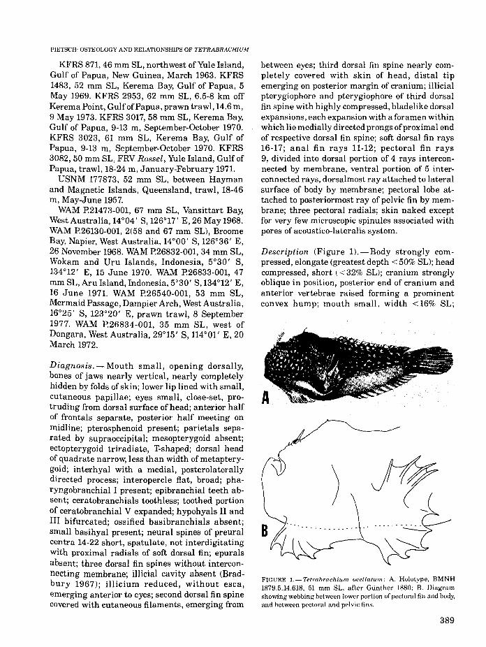
Description (Figure l).-Body strongly compressed, elongate (greatest depth < 50% SL); headcompressed, short (< 32% SL); cranium stronglyoblique in position, posterior end of cranium andanterior vertebrae raised forming a prominentconvex hump; mouth small, width <16% SL;
FIGURE l.-Tetrabrachium ocellatum: A. Holotype. BMNH1879.5.14.618, 51 mm SL. after Gunther 1880; B. Diagramshowing webbing between lower portion of pectoral fin and body,and between pectoral and pelvic fins.
389

anterior nostril opening on edge of upper lip,posterior nostril opening approximately half-waybetween edge of lip and eye; oral valve presentlining both upper and lower jaw; gill openingsmall, situated just below and behind base ofpectoral fin lobe; no opening behind fourth gillarch; holobranchs present on ventral half ofceratobranchial I, full length of ceratobranchialsII and III, ventral half of epibranchial II, andventral tip of epibranchial III; hemibranchs present on dorsal half of ceratobranchial IV andventral tip of epibranchial IV; pseudobranchabsent; swim bladder absent; ovaries paired.
Pterygiophore of illicium completely coveredwith skin ofhead; illicial bone short (< 8% SL) andthin, tapering to a point; bases of soft dorsal andanal fins long (>48% and 42% SL, respectively),rays short; dorsal and anal fin rays enveloped inmembrane; in some specimens (7 of 16 specimensexamined) distal tips of first 9 rays of soft dorsalfin free, each terminating in a tight ball of tissue,remaining dorsal rays enveloped in membrane;caudal fin long (>30% SL), rounded.
Teeth small, slender, recurved, and depressible;each premaxilla with a single row of 22-25 teeth,each dentary with approximately 35 teeth arranged in two rows; vomerine teeth in two patches,about 25 teeth in each patch; palatine teethabsent; pharyngobranchials II and III and ceratobranchial V toothed.
Color in preservative white on lower halfofbodyto brown on upper half of body, with numerous,small, white spots continuing onto soft dorsal fin,remaining fins white; oral cavity and visceraunpigmented.
Length to 67 mm SL.Complete counts and measurements of repre
sentative material are given in Table 1.
FISHERY BULLETIN: VOL. 79. NO.3
Habitat. -Specific information on the habitatfrequented by T. ocellatum is available for onlytwo captures: a 31.5 mm SL specimen (AMS1.19289.003) and a 41.5 mm SL specimen (AMS1.20907-041) were trawled off a bottom of mud,gravel, and shell. A number of other specimenswere collected in prawn trawls most likely fishedover similar, soft-bottom substrates of mud orsand.
Distribution (Figure 2).-Tetrabrachium ocellatum is known from 36 specimens collected inshallow water (55 m or less) off the western (as farsouth as lat. 290 S) and northern coasts of Australia, the southern coast of Papua, New Guinea, andthe south Molucca Islands, Indonesia.
Osteology of Tetrabrachium ocellatumFigures 3-13
The osteology oflophiiform fishes has been dealtwith by numerous authors (Garman 1899; Regan
HC::4~~-+--+,-"';,---+--iO·
\--.J ~f.:.-i";1III~·····~··-.f·-··- ....
: ._...~.--~_ .
-·"~T .._·"~t ..: .} j
'101'-~90~·.-'--'---:IZO~·"""-..;.,c~==::-=--...i..-~~"""c
FIGURE 2.-Known distribution of Tetrabrachium ocellatum.One symbol may indicate more than one capture.
TABLE I.-Counts and measurements (in percentage of standard length) of representative specimens of Tetrabrachium ocellatum.
KFRS UW AMS KFRS KFRS USNM AMS AMS KFRS KFRS3067 20771 IB.7173 671 3062 177673 IB.7174 IB.7175 3023 2953
Standard length, mm 39 39.5 42.5 46 50 50.5 54 56 61 62Length:
Head (snout 10 posteriormosl margin of preopercfe) 27.7 30.4 25.9 26.1 26.0 23.6 20.4 25.0 23.0 22.6Snout to emergence of dorsal spine III 22.6 .25.3 25.9 25.4 23.2 23.6 25.0 23.0 25.0lIIicial bone 7.2 4.6 2.6 4.1 4.0 4.0 3.3 4.8Dorsal spine II 7.9 6.3 4.7 6.7 6.6 5.2 4.1 4.6 5.4 4.6Base of soft dorsal fin 67.9 64.6 49.4 56.5 56.0 63.4 63.0 54.9 53.2Base of anal fin 51.3 51.9 42.3 46.9 51.0 53.5 49.1 48.4 48.4Caudal fin 43.1 3B.2 39.3 34.6 32.0 35.6 36.1 36.7 37.7 33.9
Width:Between eyes (from center of lens) 11.0 12.4 10.6 11.1 10.4 9.9 9.1 10.8 10.3Least between frontal bones 6.7 B.6 4.9 6.1 5.6 5.5 5.4 4.9 5.6Greatest between sphenotic bones 20.2 19.7 19.8 20.6 19.4 19.2 17.2 17.9 20.5 20.2
Greatest body depth 36.5 43.0 35.3 46.7 46.0 45.5 42.6 37.5 42.6 41.9Dorsal fin rays 1B 17 16 16 16 17 16 16 17 16Anal fin rays 11 11 11 11 11 12 12 11 12 12
390

PIETSCH: OSTEOLOGY AND RELATIONSHIPS OF TETRABRACHIUM
1903,1912; Gregory 1933; Eaton et al.1954; Monod1960; Le Danois 1964, 1974, 1979; Field 1966;Bradbury 1967; Rosen and Patterson 1969; andadditional references cited by Pietsch 1972, 1974,1978,1979), yet no published osteological information on the genus Tetrabrachium is available. Inthe following account only those comparativeaspects that differ from those previously describedin other anglerfishes are discussed.
Cranium (Figures 3-6).-The ethmoid cartilageof T. ocellatum broadly covers the posterior half ofthe vomer meeting with the lateral ethmoidslaterally and the supraethmoid medially. Thesupraethmoid forms a narrow, vertical interorbital septum lying between, but well separatedfrom the orbital· portions of the frontals. Thelaterally compressed, ventral end of the supraethmoid meets with the ethmoid cartilage anteriorly and lies within a groove on the dorsalsurface of the parasphenoid posteriorly. The dor-
Sphenotic
Supraethmoid
Vomer __
lateral ethmoij
sal end of the supraethmoid is overlapped on eachside by central extensions of the frontals. Eachlateral ethmoid has a narrow, cylindrical posterior portion that lies ventral to an anterior extension of the respective frontal, and a larger, ventrally directed, anterior portion that meets withthe ethmoid cartilage.
The head of the vomer lies ventral to theethmoid cartilage. Its anterior margin is indentedmedially. The ventral surface of the vomer isstrongly concave (as seen in anterior view, Figure6). A laterally compressed, keellike posteromedialprocess emerges from the ventral surface of thisbone and fits within a deep groove on the anteroventral surface of the parasphenoid; the ventralmargins of the posteromedial process ofthe vomerand the anterior end of the parasphenoid arestrongly convex (as seen in lateral view, Figure 4).Vomerine teeth are present in two lateral patches,each patch containing approximately 25 teetharranged in perhaps three irregular rows.
/ Exoc:cipilJI
[pkltic
FIGURE 3.-Dorsal view of cranium of Tetrabrachium oce//atum, AMS IB.7178, 61 mm SL.
lateral ethmoid
Vomer
Suprmcipilal Plerotic
Basioccipital FIGURE 4.-Lateral view of cranium of Tetrabrachium ocel/atum, AMS IB.7178, 61 mm SL.
391

FISHERY BULLETIN: VOL. 79. NO.3
Pterosphenoid
Parasphenoid
Vomer
,,\
\,
\SPhenotic
Plerotic
".- 22nd pre·ural. centrum
Basioccipital
FIGURE 5. - Ventral view ofcranium of Tetrabrachiumocellatum, AMS IB.7178, 61 mm SL.
\,\, Posllemporal
FIGURE 6.-Anterior view of cranium of Tetrabrachium oce/latum, AMS IB.7178, 61 mm SL.
The frontals are relatively large and irregularin shape. Each has a laterally compressed, anterior half, well separated from its counterpart ofthe other side, and a dorsoventrally depressedposterior half that meets its counterpart on themidline. In dorsal view (Figure 3), the frontalsform a relatively narrow orbital region to accommodate the closely set, dorsally directed eyes. Inlateral view (Figure 4), the depressed posteriorhalf of the frontals form a concavity between theelevated, laterally compressed anterior half ofthese bones and the posterior half of the cranium.
The parietals are irregularly shaped elementswith deeply pitted and grooved external surfaces.They are well separated from each other by thesupraoccipital. Each parietal overlaps the respective frontal anteriorly, the sphenotic and pterotic
Lateralethmoid
" Sphenotic
laterally, the supraoccipital medially, and theepiotic posteriorly.
A small pterosphenoid lies on the ventromedialsurface of the frontal in contact with the prootic.
The orbitosphenoid and basisphenoid are absentin alliophiiforms.
The parasphenoid is a stout, well-ossified element with a convex ventral margin (Figure 4). Itsanterior end is overlapped by the ethmoid cartilage dorsally and by the narrow shaft of the vomerventrally. Medially, the dorsal surface ofthis boneforms a deep groove within which lies the laterallycompressed, posteroventral part of the supraethmoid. Posteriorly, the parasphenoid is broadlyconnected with the prootics laterally and thebasioccipital medially. At no point does the parasphenoid make contact with the frontals.
Each sphenotic forms a dorsoventrally depressed flange that extends outward in an anterolateral direction, considerably beyond the widthof the ethmovomerine region of the cranium(Figure 3).
The remaining elements of the cranium (pterotics, epiotics, prootics, supraoccipital, and exoccipitals) do not differ substantially from those described for other lophiiforms (Regan 1912, fig. 5;Gregory 1933, fig. 265, 267-271; Pietsch 1972,1974).
Otoliths (Figure 7), - The sagitta of T. ocellatumis roughly oval in shape with a length to heightratio of about 1.4:1. The sulcus is only slightly
392

PIETSCH: OSTEOLOGY AND RELATIONSHIPS OF TETRABRACHIUM
Sulcus
Premaxilla
FIGURE 7.-Medial view of right sagitta of Tctrabrachiumoce/latum, AMS IB.7178, 61 mm SL.
similar to those described for other lophiiforms(Gregory 1933, fig. 265, 266, 269-271; Pietsch 1972,1974). Each dentary bears approximately 35 depressible teeth arranged in two rows.
Palatine arch (Figure 9). - Each metapterygoid isin contact with four other bones; dorsally andposterodorsally with the hyomandibular, posteroventrally with the upper half of the symplectic,and ventrally with the quadrate and ectopterygoid. The ectopterygoid is large and T-shaped,overlapping the medial surface of the metapterygoid dorsally, the quadrate ventrally, and thepalatine anteriorly. The mesopterygoid (cartilaginous or ossified) is absent. The palatine is unusually large, approximately twice the length ofthe ectopterygoid. Palatine teeth are absent.
Hyoid arch (Figures 9, lO).-Dorsally, each hyomandibular is forked forming two heads, both ofwhich articulate with the cranium: an anteriorhead fits into a concavity formed by the sphenoticand prootic, and a posterior head articulates onthe ventrolateral face of the pterotic (Figures 4,5,9). The symplectic is separated from the hyomandibular by cartilage dorsally, and lies within ashallow groove on the medial surface of thequadrate ventrally. The dorsal head of the quadrate is narrow, considerably less than the width ofthe metapterygoid. The interhyal bears a prominent medial, posterolaterally directed processthat wraps around the posterior margin of therespective preopercle when the interhyal rotatesupward (e.g., during a feeding event). This contactbetween the interhyal and the preopercle limitsthe dorsal rotation of the interhyal and, in turn,limits the extent of abduction of the lower jawvia ligamentous connections to the respectiveinteropercle.
The epihyal and ceratohyal do not differ substantially from those described for other lophiiforms (Pietsch 1974, 1979). There are two hypohyals on each side (Figure 10), both of whichare connected to the ceratohyal by a posteriorlydirected strut. The dorsal hypohyal is furtherconnected to an anterodorsal extension of theceratohyal by a cylindrical piece of cartilage.
There are six branchiostegal rays all borne onthe ceratohyal (Figure 10); the two anteriormostrays articulate on the medial surface, the fourposterior rays on the lateral surface. Branchiostegal rays 3 and 4 are curved in an anteroventraldirection, in contrast to the posterodorsal direc-
ASP
Rostrum
grooved. The rostrum is poorly developed, and anantirostrum is absent.
Mandibular arch (Figures 8, 9).-The premaxillae (Figure 8) are each characterized by having anarrow ascending process, nearly as long as thetapering toothed portion of the bone; a roundedarticular process; and an elongate, spatulate post·maxillary process (pmpmx of Rosen and Patterson1969, fig. 56A). The ascending and articular processes together form an oblique angle with thepostmaxillary and toothed processes. The toothedportion of each premaxilla bears a single rowof 22 to 25 depressible teeth, the largest atthe symphysis, becoming progressively smallerposteriorly.
Each maxilla consists of a broad posterior portion (completely hidden from behind by a thickfold of skin when the mouth is closed), and anexpanded anterior head that, in turn, consists ofan anterior process that overlaps the respectivepremaxilla and a medially directed process that isattached by a short ligament to the articularprocess of the respective premaxilla. The dentaries, articulars, and angulars (Figure 9) are
FIGURE 8.-Upper jaw bones of Tctrabrachiurn Deellaturn, AMS1B.7178, 61 mm 8L. AP = anterior process of maxilla; ARP =articular process of premaxilla; ASP = ascending processof premaxilla; MP = medial process of maxilla; PMP = post·maxillary process of premaxilla; PP = posterior process ofmaxilla.
393

FISHERY BULLETIN: VOL. 79, NO.3
Hyomandibular
Metapterygoid
Ectopterygoid
Palatine
Quadrate
\
\ Dentm
~Angular
/Opercle
Subopercle
-- Symplectic
Preopercle
Interopercle
FIGURE 9.-Medial view oflower jaw, suspensorium, interhyal, and opercular apparatus of Tetrabrachium ocellatum, AM8 IB.7178,61 mm 8L, right side.
tion of the remaining rays. On the left ceratohyalof the 61 mm cleared and stained specimen of T.ocellatum (Figure 10), the fifth branchiostegal rayis bifurcated at midlength, giving the impressionof having seven total rays.
A small, triangular basihyal is present (Figure10). The urohyal is absent in alllophiiforms.
Opercular apparatus (Figure 9).-The opercle istriangular in shape with a slightly concave posterior margin. An elongate, crescent-shaped sub-
394
opercle lies medial to the ventral tip ofthe opercle.A subopercular spine is absent. The interopercle islarge, flat, and broad. The crescent-shaped preopercle is also large, strengthening the entirelength of the sllspensorium,
Branchial arches (Figure ll).-There are threepharyngobranchials. That of the first arch is asmall, toothless, suspensory pharyngobranchial;those of the second and third arches are considerably larger, tooth-bearing elements closely at-

PIETSCH: OSTEOLOGY AND RELATIONSHIPS OF TETRABRACHIUM
element. Hypobranchials II and III are bifurcatedproximally. Ossified basibranchials are absent.
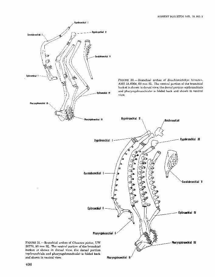
FIGURE H.-Branchial arches of Tetrabrachium ocel/atum,AMS 1B.7178, 61 mm SL. The ventral portion of the branchialbasket is shown in dorsal view, the dorsal portion (epibranchialsand pharyngobranchialsl is folded back and shown in ventralview.
Epibrancbial IV
Ceratobranchial V
Hypobrancbial III
PharynCobranchial III
Pharyncobranchial II
Epibrancbi.1 I
Hyp.brancbial 1_---1/
Hyp.branchial 1\
Ceratobranchial I
Pbary,cobranchiai 1-_=.,,(/
Venlralhypohyal
FIGURE 1O.-Hyoid apparatus of Tetrabrachium ocel/atum:A. AMS IB.7178, 61 mm SL, left lateral view; B. Basihyal,AMS IB.7177, 56 mm SL, ventral view, anterior to the left.
tached to each other and to the dorsal end ofepibranchials II through IV. Epibranchial I istriradiate in shape, articulating with ceratobranchial I proximally, bearing pharyngobranchial I distally, and attached by a short ligament tothe proximal end of epibranchial II medially.Ceratobranchials I through IV are toothless. Theexpanded, proximal end of each ceratobranchial Vbears about 19 to 21 depressible teeth arranged intwo rows. Hypobranchial I is a simple, rod-shaped
Dorsalhypohyal
14 th pre -uralcentrum
FIGURE 12.-Vertebrae, caudal skeleton, and median fins of Tetrabrachium ocel/atum, AMS IB.7178, 61 mm SL.
395
FISHERY BULLETIN: VOL. 79, NO.3
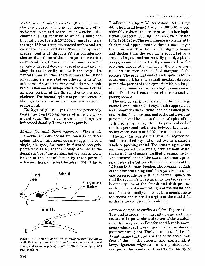
Median fins and illicial apparatus (Figures 12,13).-The spinous dorsal fin consists of threespines. The anteriormost two are supported by asingle, elongate, horizontally situated pterygiophore (Figure 13) that is loosely attached to thedorsal surface ofthe cranium between the anteriorhalves of the frontal bones by three pairs ofextrinsic illicial muscles (Bertelsen 1951:18, fig. 4;
Vertebrae and caudal skeleton (Figure 12).-Inthe two cleared and stained specimens of T.ocellatum examined, there are 22 vertebrae (including the last centrum to which is fused thehypural plate; Pietsch 1972:38). Preural centra 2through 18 bear complete haemal arches and areconsidered caudal vertebrae. The neural spines ofpreural centra 14 through 22 are considerablyshorter than those of the more posterior centra;correspondingly, the seven anteriormost proximalradials of the soft dorsal fin are also short so thatthey do not interdigitate with the respectiveneural spines. Further, there appears to be little ifany connective tissue between the elements ofthesoft dorsal fin and the vertebral column in thisregion allowing for independent movement of theanterior portion of the fin relative to the axialskeleton. The haemal spines of preural centra 14through 17 are unusually broad and laterallycompressed.
The hypural plate, slightly notched posteriorly,bears the overlapping bases of nine principlecaudal rays. The central seven caudal rays arebifurcated distally. There are no epurals.
Bradbury 1967, fig. 2; Winterbottom 1974:284, fig.44). The illicial bone (Bradbury 1967:401) is considerably reduced in size relative to other lophiiforms (Gregory 1933, fig. 265, 266, 267; Pietsch1972,1974,1979). The second spine is considerablythicker and approximately three times longerthan the first. The third spine, slightly longerand thicker than the second, is supported by asecond, elongate, and horizontally placed, cephalicpterygiophore that is tightly connected to theposterior, dorsomedial surface of the supraoccipital and anterior, dorsomedial margins of theepiotics. The proximal end of each spine is bifurcated, each fork bearing a small, medially directedprong; the prongs of each spine fit within a large,rounded foramen located on a highly compressed,bladelike dorsal expansion of the respectivepterygiophore.
The soft dorsal fin consists of 16 biserial, segmented, and unbranched rays, each supported bya cartilaginous distal radial and an ossified proximal radial. The proximal end ofthe anteriormostproximal radial lies above the neural spine of the19th preural centrum, while the proximal end ofthe last proximal radial lies between the neuralspines of the fourth and fifth preural centra.
The anal fin consists of 11 biserial, segmented,and unbranched rays. The first two rays share asingle supporting radial. The remaining rays areeach supported by a small, cartilaginous distalradial and an elongate, ossified proximal radial.The proximal ends of the two anteriormost proximal radials lie between the haemal spines of the12th and 13th preural centra. The proximal radialsof the nine remaining anal fin rays have a one-toone correspondence with the haemal spines, sothat the radial ofthe last anal ray lies between thehaemal spines of the fourth and fifth preuralcentra. The posteriormost rays of the dorsal andanal fins are broadly connected by a membrane tothe dorsal and ventral margins of the caudal finso that a caudal peduncle is absent.
Pterygiophoreof illicium
Spine IIIlIicialbone
Spine III
BFIGURE 13.-Spinous dorsal fin of Tetrabrachium ocellatum,AMS lB.7178. 61 mm SL: A. IIlicial apparatus, second dorsalspine, and common pterygiophore; B. Third dorsal spine andpterygiophore.
Pectoral and pelvic girdles and fins (Figure 14).The posttemporal is unusually large and connected to the posterolateral corner of the craniumin such a way as to allow for considerable movement (relative to the cranium) in an anterodorsalposteroventral plane. The bone consists of a broad,dorsal flange that overlaps the dorsolateral surface of the epiotic, pterotic, and exoccipital. Alarge ligament originates on the posterodorsalmargin of the prootic and inserts on the tip of
396

PIETSCH: OSTEOLOGY AND RELATIONSHIPS OF TETRABRACHIUM
/ Supracleithrum
Postcleithrum
Stapula ~
Cleithrum -----~-
FIGURE 14_-Medial view of right pectoral girdle, and pectoraland pelvic fins of Tctrabrachium occllatum, AMS IB.7178,61 mm SL. Cartilaginous radials supporting pelvic fin rays andcartilaginous distal radials supporting pectoral fin rays notshown; see text.
an elongate, ventromedially directed extensionof the posttemporal.
The supracleithrum, cleithrum, coracoid, andscapula (Figure 14) are similar to those describedfor other lophiiforms (Gregory 1933, fig. 265;Pietsch 1972, 1974). A cleithral spine is absent.There is a single rodlike postcleithrum.
The pectoral fin is supported by three pectoralradials (Figure 14). The two dorsalmost radials aresimilar in size and shape. The third or ventralmostradial is considerably larger; its expanded distalportion bears the bases of nine unbranched, pectoral fin rays (each ray associated with a small,cartilaginous distal radial; not shown in Figure14). The pectoral fin itself is divided into twoportions: a dorsal portion consisting of four raysthat are interconnected by a membrane, and aventral portion consisting of five rays that aresimilarly connected to each other, but also to thelateral surface of the body. In a similar way, thepectoral fin lobe is connected by a membrane tothe rays of the respective pelvic fin (Figure lB).The pelvic bone, nearly as long as the ventralmostpectoral radial, bears on its expanded distal end asingle spine and five unbranched pelvic fin rays(each ray associated with a small, cartilaginousradial; not shown in Figure 14).
Skin spines. -Dermal spines are absent exceptfor the very rare occurrence of a tiny, crescentshaped spinule associated with an individual pore
of the acoustico-lateralis system of the head andtrunk.
COMPARATIVE OSTEOLOGY OFANTENNARIOID FAMILIES
The following discussion is based primarily onan osteological comparison of a representative ofeach of six major subgroups of the Antennarioidei(here recognized as families; see PhylogeneticRelationships and Appendix below); AntennariusDaudin, thought to be the least derived genusof the Antennariidae (see Phylogenetic Relationships below); Tetrabrachium Gunther, the onlygenus of the Tetrabrachiidae; Lophichthys Boeseman, the only genus of the Lophichthyidae;Brachionichthys Bleeker, the only extant genus ofthe Brachionichthyidae (see p. 416); ChaunaxLowe, the only genus of the Chaunacidae; andDibranchus Peters, an underived genus of theOgcocephalidae (see Bradbury 1967). Only thosecomparative aspects that might have a bearing onthe phylogenetic interrelationships of these taxaare discussed.
Cranium (Figures 3-6, 15-19).-ln Tetrabrachiumand Antennarius the ventral surface of the vomeris strongly concave (as seen in anterior view,Figure 6). A laterally compressed, keellike posteromedial process emerges from the ventral surface of this bone and fits within a deep grooveon the anteroventral surface of the parasphenoid;the ventral margins of the posteromedial processof the vomer and the anterior end of the parasphenoid are strongly convex (as seen in lateralview, Figure 4). In all other antennarioids examined the posteromedial process of the vomer isflush with the more or less flat ventral surface ofthis bone; the ventral margins of the vomer andanterior end of the parasphenoid (as seen inlateral view) are straight to slightly concave.
Other osteological variation in the crania ofantennarioids occurs primarily in the shape andrelative position of the frontal bones. In Antennarius, Lophichthys, Brachionichthys, and Dibranchus (Figures 15-17, 19) the frontals are broadand roughly triangular in shape, well separatedfrom each other anteriorly, but meeting on themidline posteriorly. The narrow interorbital spaceformed by these elements in Tetrabrachium isabsent (compare Figures 3 and 15). The anteriorends of the frontals of Lophichthys are exceptionally narrow, gradually tapering to a point (Figure
397

Supraethmoid
Vomer
Lateral ethmoid
Supraethmoid
Vomer
Frontal
Sphenotic
Sphenotic
Parietal
pterolic
Parietal
FISHERY BULLETIN: VOL. 79, NO.3
,__--Posttemporal
Epiotic
Supraoccipital
FIGURE 15.-Dorsal view of cranium of Antennarius sanguineus,LACM 8125, 76 mm SL.
Epiotic
Supraoccipital
FIGURE 16.-Dorsal view of cranium of Loph.ichthys boschimai, UW 20773, 47 mm SL.
Pterotic
16); they diverge laterally to a much greaterextent than in the other genera examined inresponse to a much wider vomer and laterallyexpanded lateral ethmoids.
In contrast to all other antennarioids examined,the frontals of Chaunax (Figure 18) are elongateand narrow, meeting on the midline for theirentire length. The lateral ethmoids of this genusare also unusually long and narrow.
In Antennarius, Lophichthys, Tetrabrachium,Chaunax, and Dibranchus the parietals are well
398
separated from each other by the supraoccipital.In Brachionichthys, however (Figure 17), theseelements approach each other above the supraoccipital and meet on the midline, roofing over asmall longitudinal passageway within which liesthe posterior tip of the pterygiophore of the thirddorsal fin spine.
Mandibular arch (Figures 8, 9, 20-25).-Thepremaxilla of Antennarius is very similar to thatof Tetrabrachium (Figures 8, 20A); both genera

PIETSCH: OSTEOLOGY AND RELATIONSHIPS OF TETRABRACHIUM
Fllnl.1
Yollt,
FIGURE 17.-Dorsal view of cranium of Brachianichthys hirsutus, AMS IA.6064, 69 mm SL.
SuprIOthmoid
Ilea'"' 1Ili...r 2Zodpre-Ullr CIIIInIIIl
(pi,lic
Pa,ietalSphenotic
Vomer
literal ethmoidFIGURE lB.-Dorsal view of cranium of Chaunax
pictus, UW 20770, 90 mm SL. Sphenotic
Postlempor,l
Epiotic
Supraoccipital
ptel1lic
are characterized by having a spatulate postmaxillary process. The premaxilla of Lophichthys isalso quite similar but bears a narrow, taperingpostmaxillary process (Figure 20B). The premaxillae of the remaining antennarioid taxa examined are each somewhat different from these andfrom each other. In Brachionichthys (Figure 20C),the ascending and articular processes are at rightangles to the toothed portion of the bone; thetoothed portion is unusually short, about as longas the postmaxillary process and considerablyshorter than the ascending process. In Chaunax(Figure 20m, the shape and relative proportionsof the ascending, articular, and toothed processes
of the premaxilla are similar to those of Antennarius and Tetrabrachium; the postmaxillar)'process, however, is represented by a large flangeof bone, broadly connected to the toothed process.In Dibranchus (Figure 20E), the ascending andarticular processes together form an acute anglewith the postmaxillary and toothed processes; thearticular process is nearly as long as the ascendingprocess; and the postmaxillary process is connected by bone to the toothed process of thepremaxilla for about half its length.
Palatine arch (Figures 9, 21-25).-A mesopterygoid is present in Antennarius, Chaunax, and
399

Vomer-__L
Sphenotic
Dibranchus (Figures 21, 24, 25), but absent inTetrabrachium, Lophichthys, and Brachionichthys (Figures 9, 22, 23). The triradiate ectopterygoid of Antennarius, Tetrabrachium, andLophichthys (T-shaped in Tetrabrachium andAntennarius, Figures 9, 21, but Y-shaped inLophichthys, Figure 22) overlaps the medial surface of the metapterygoid dorsally; in Chaunaxand Dibranchus the ectopterygoid is crescentshaped and makes no contact with the metapterygoid. An ectopterygoid is absent in the larger (69mm 8L) specimen of Brachionichthys examined(Figure 23A) but represented by a small, weaklyossified remnant in the smaller specimen (42 mm8L) (Figure 23B).
The palatine is well toothed in A ntennarius ,Lophichthys, and Chaunax, but toothless inTetrabrachium and in the single cleared andstained specimen of Dibranchus examined (palatine teeth are present in some ogcocephalid generaand sometimes in Dibranchus; Bradbury 1967:409). In the absence of a mesopterygoid andreduced (or absent) ectopterygoid, the toothlesspalatine bone of Brachionichthys is widely separated from the suspensorium (Figure 23).
Hyoid arch (Figures 9, 10, 21-27).-In Tetrabrachium and Antennarius (Figures 9, 21) thedorsal head of the quadrate is relatively narrow,somewhat less than the width of the ventral headof the metapterygoid. In contrast, the quadrate
400
FISHERY BULLETIN: VOL. 79, NO.3
Posttemporal
Epiotic
Supraoccipital
FIGURE 19.-Dorsal view of cranium of Dibranchus at/anticus, MCZ 51257, 105 mm SL.
Pterotic
of Lophichthys, Brachionichthys, Chaunax, andDibranchus (Figures 22-25) is broad, making amuch broader contact with an expanded metapterygoid. In Dibranchus the quadrate is exceptionally broad, the anterior half of the dorsalmargin coming into contact with the mesopterygoid (Figure 25).
The interhyal ofAntennarius, Lophichthys, andBrachionichthys is similar to that of Tetrabrachium (but in contrast to that of Chaunax andDibranchus; Figures 24, 25) in having a prominent, medial, posterolaterally directed processthat wraps around the posterior margin of therespective preopercle when the interhyal rotatesupward (Figure 26). This contact between theinterhyal and preopercle limits the dorsal rotationof the interhyal and, in turn, limits the extent ofabduction of the lower jaw via ligamentous connections with the respective interopercle.
In shape and relative proportions, the branchiostegal rays of Antennarius, Lophichthys, andBrachionichthys are similar to those of Tetrabrachium; Brachionichthys, however, has lost theanteriormost element in this series (Table 2). InChaunax and Dibranchus (Figure 27) the posteriormost branchiostegal ray is greatly enlarged,becoming ankylosed to the ventromedial marginof the subopercle in the later genus.
A small basihyal is present in Antennarius,Tetrabrachium, Lophichthys, and Chaunax, butabsent in Brachionichthys and Dibranchus.

PIETSCH: OSTEOLOGY AND RELATIONSHIPS OF TETRABRACHIUM
FIGURE 20.-Premaxillae, left lateral views: A. Antennariussanguineus, LACM 8125,76 mm SL; B. Lophichthys boschmai,UW 20773, 47 mm SL; C. Brachionichtlzys hirsutus, AMSIA.6064, 69 mm SL; D. Chaunax pictllS, UW 20770, 90 mm SL;E. Dibranclzus at/anticus, MCZ 51257, 105 mm SL.
Opercular apparatus (Figures 9, 21-25).-Theopercle and subopercle of Antennarius, Tetrabrachium, and Brachionichthys are similar exceptin the following details: both elements are considerably reduced in size in Tetrabrachium andAntennarius (Figures 9, 21); in contrast to thesmooth, slightly concave (sometimes deeply incised), posterior margin of the operc1e of Antennarius, Tetrabrachium, and Lophichthys, theposterior margin of this bone in Brachionichthysis broken into numerous, weakly ossified, bonyfilaments (Figure 23A); in contrast to the relatively broad, spined suboperc1e of Antennariusand Lophichthys, the suboperc1e of Tetrabrachium and Brachionichthys (Figures 9, 23A) is anarrow, crescent-shaped element lacking a subopercular spine.
In contrast to the small opercle and subopercleof Antennarius, Tetrabrachium, Lophichthys,and Brachionichthys, those of Chaunax and Dibranchus (Figures 24, 25) are greatly enlargedand expanded posteriorly. A well-developed suborpercular spine is present in Chaunax, but absent in Dibranchus.
The interoperc1e of Antennarius, Lophichthys,and Brachionichthys (Figures 21-23) is similarto that of Tetrabrachium; the interopercle ofChaunax and Dibranchus (Figures 24, 25) ismuch more slender an" elongate.
Branchial arches (Figures 11, 28-32). - Pharyngobranchial I is represented by a simple, rod-shapedelement in Tetrabrachium, A ntennarius, andLophichthys (Figures 11, 28, 29). In the singlespecimen of Chaunax examined pharyngobranchial I is Y-shaped (Figure 31). This element istoothless in Antennarius, Tetrabrachium, andChaunax, but bears a series of approximatelyeight small teeth in Lophichthys (Figure 29).Pharyngobranchial I is absent in Brachionichthysand Dibranchus. Pharyngobranchial IV is absentin all antennarioids.
In Tetrabrachium, Antennarius, and Lophichthys (Figures 11, 28, 29), epibranchial I is triradiate in shape, toothless in Antennarius andTetrabrachium, but bearing a single row of about13 small teeth in Lophichthys (Figure 29). Asimilarly shaped epibranchial I, associated withthree and two tooth plates is present in Chaunaxand Dibranchus, respectively (Figures 31,32). AnL-shaped epibranchial I, associated with a singletooth plate, is present in Brachionichthys (Figure
c
E
401

FISHERY BULLETIN: VOL. 79, NO.3
Interopercle
Preopercle
Subopercle
FIGURE 21.-Medial view of lower jaw, suspensorium, andopercular apparatus of Antennarius sanguineus, LACM 8125,76 mm 81.
_~::::S2:::::::===-\\'l-ilC-.--"-+--1-+---Symplectic
Articular
Palatine -----t-
EctoptelJloid
Opercl.
Metapterygoid
[clopterygoid
"yomandibular Subopercle
lnterhyal
Preopercle
Sympl.ctic
Interope"le
Quadrate
FIGURE 22.-Medial view oflower jaw, suspensorium, interhyal, andAngular opercular apparatus of Lophichthys boschmai, UW 20773, 47 mm 81.
402

PIETSCH: OSTEOLOGY AND RELATIONSHIPS OF TETRABRACHIUM
Melapterygoid
Ecloplerygoid
FIGURE 23.-Brachionichthys hirsutus: A. Medial view of lowerjaw, palatine bone, suspensorium, interhyal, and opercular apparatus, AMS lA.6064, 69 mm SL: B. Medial view ofpalatine arch andsuspensorium, right side, showing presence ofa small ectopterygoid,UW 20769, 42 mm SL.
M.llplerJl.ld
Preopercle
Symplectic
BQuadrate
.,.;.,""''''
\if=-
oentary/
H,omallllibula,
Hyomandibular
Opercle
Preopercle
Subopercle
Inlerhya!
Interopercle
Angular
",.11..
D..,."----','"
Op.rtlt
Suboperel,
Prtoptltlo
FIGURE 24.-Medial view of lower jaw, suspensorium,interhyal, and opercular apparatus of Chaunax pictus,UW 20770, 90 mm SL.
Iel.pll'll.i.
AltlclIllr",operel.
laleropercl, FIGURE 25.-Medial view of lower jaw, suspensorium, interhyal, and opercular apparatus of Dibranchus atlanticus,MCZ 51257, 105 mm SL.
403

Posterior process
FISHERY BULLETIN: VOL. 79, NO.3
FIGURE 26.-Lateral view of interhyal, right side: A. Antellnarius sanguineus, LACM 8125, 76 mm SL; B. Tetrahrachium(lcellatum, AMS IB.7178, 61 mm SL; C. Lophichthys hoschmai,UW 20773, 47 mm SL; D. Brachiollichthys hirwtus, AMSIA.6064, 69 mm SL.
DorsalhlPohlal
VentralhlPohlal
VentralhlPohlal
B
'\-- -'~---'%-_::::~ Branchiostegal raJs
FIGURE 27.-Hyoid apparatus, left lateral views: A. Chaunax pictus,UW 20770, 90 mm SL; B. Dihranchus at/anticus, MCZ 51257, 105 mm SL.
404

PIETSCH: OSTEOLOGY AND RELATIONSHIPS OF TETRABRACHIUM
TABLE 2.-Characters of representative genera of the major subgroups of the Antennarioidei.
Item Antennarius Tetrabrachium Lophichthys Brachionichthys Chaunax Dibranchus
Branchiostegal rays 2+4 2+4 2+4 1+4 2+4 2+4Pharyngobranchial I rod shaped rod shaped rod shaped absent forked absent
(toothless) (toothless) (toothed) (toothless)Palatine teeth present absent present absent present absentEpibranchral teeth
Arch I absent absent single row 1 plate 3 plates 2 platesArch III absent absent absent absent absent 1 plate
Ceratobranchial teethArch I absent absent absent 2 plates present presentArch II absent absent absent t plate present presentArch III absent absent absent 1 plate present presentArch IV absent absent absent absent present present or absent
Hypobranchial II bifurcated bifurcated bifurcated simple simple absentHypobranchial III bifurcated bifurcated bifurcafed simple simple absentPseudobranch present absent absent absent absent absentSwim bladder present absent absent absent absent absentBasihyal present present present absent present absentVertebrae (precaudal) 19(4) 22(4) 19(4) 22(4) 19(4) 19(6)Epural 1 0 remnant present 0 1 1
or absentDorsal fin rays 11-15 16-17 12-13 17 12 5Anal fin rays 6-9 11-12 9 7 7 4Pectoral fin radials 3 3 3 2 3 3Pectoral fin rays 7-14 4+5 7 8 14 14Pelvic fin rays 1+5 1+5 1+5 1+4 1+ 4 1+5
Hypobranchial II
Hypobranchial III
Ceratobranchial I
Epibranchial I
Pharyngobrancbial II
Hypobranchial I
Pharyngobranchial III
Hypobrancbial 1-
Ceratobrancbial I
Epibrancbial I
Pbaryngobrancbial I
Pharyngobranchial II
Hypobrancbial II
vr-..-_--Hypobranchial III
Ceratobrancbial V
Epibrancbial IV
Pbaryngoorancbial III
FIGURE 28.-Branchial arches of Antennarills sangllinells,LACM 8125, 76 mm 81" The ventral portion of the branchialbasket is shown in dorsal view, the dorsal portion lepibranchials
and pharyngobranchialsl is folded back and shown in ventralview.
FIGURE 29.-Branchial arches of Lophichthys boschmai, UW20773,47 mm 8L. The ventral portion of the branchial basket isshown in dorsal view, the dorsal portion lepibranchials and
pharyngobranchials) is folded back and shown in ventral view.
405
FISHERY BULLETIN: VOL. 79, NO.3
Hlpobranchi,l I
FIGURE 31.-Branchial arches of Chaunax pictus, UW20770, 90 mm SL. The ventral portion of the branchialbasket is shown in dorsal view, the dorsal portion(epibranchials and pharyngobranchials) is folded backand shown in ventral view.
Cer,lobranchi,l V
III
Ceratobranchial V
_- Epibranchial IV
Pharyngobranchial III
FIGURE 3D.-Branchial arches of Brachionichthys hirsutus,AMS IA.6064, 69 mm SL. The ventral portion of the branchialbasket is shown in dorsal view, the dorsal portion lepibranchialsand pharyngobranchialsl is folded back and shown in ventralview.
Hypobranchial II
Pharyngobranchial II
Pharyngobranchial
Hypobranchial I
___ Epibranchi,l IV
Epibranchial I -_--...>c
Ph''lngobranchi,1 III
Ceratobranchial I
:L----- Hlpobranchi,l II
Pharlngobranchi,l II"
Ceratobranchial 1\
Epibranchi,l I - __
406

PIETSCH: OSTEOLOGY AND RELATIONSHIPS OF TETRABRACHIUM
Ceratobranchial IV
Epibranchial
Pharyngobranchial II
Ceratobranchial V
- Epibranchial IV
FIGURE 32.-Branchial arches of Dibranchus atlanticus, MeZ 51257, 105mm SL. The ventral portion of the branchial basket is shown in dorsal view.the dorsal portion (epibranchials and pharyngobranchials) is folded backand shown in ventral view.
Pharyngobranchial III
30). Epibranchial III is toothless in all antennarioids examined except in Dibranchus (Figure 32)where this bone is associated with a single toothplate.
Ceratobranchials I through IV are toothless inTetrabrachium, Antennarius, and Lophichthys(Figures 11, 28, 29). In Brachionichthys (Figure30), one to three tooth plates are present onceratobranchials I through III; in Chaunax (Figure 31), tooth plates are present on ceratobranchials I through IV; in Dibranchus (Figure 32), toothplates are present on ceratobranchials I throughIII (but also sometimes present on ceratobranchialIv, see Bradbury 1967:408) (Table 2).
In contrast to the separate, individual teethpresent on pharyngobranchial I and epibranchialI of b()phichthys (Figure 29), those present onepibranchial I and ceratobranchials I through IVof Brachionichthys, Chaunax, and Dibranchus(Figures 30-32) are born in clusters on individualtooth plates. The tooth plates of Chaunax andDibranchus (Figures 31, 32) (and a number ofother ogcocephalid taxa, see Bradbury 1967) differfrom those of Brachionichthys (Figure 30) andfrom those of all other lophiiforms in beingraised, pedicallike structures bearing a cluster ofnumerous, tiny teeth at the apex (but see Brad-
bury 1967, fig. 7, for other forms of gill teethin ogcocephalids),
Ceratobranchial V is well toothed in all antennarioids examined. In Tetrabrachium, Antennarius, Lophichthys, and Brachionichthys (Figures 11, 28-30), this bone consists of a narrow,toothed proximal portion and a tapering, cylindrical distal portion; in Chaunax (Figure 31) onlya triangular, toothed portion is present. In Dibranchus (Figure 32), ceratobranchial V is greatlyenlarged, consisting of a finely toothed, expandedproximal portion and a long, cylindrical distalshaft,
Hypobranchial I of Tetrabrachium, Antennarius, Lophichthys, Brachionichthys, and Chaunax(Figures 11, 28-31) and hypobranchial II of Brachionichthys and Chaunax (Figures 30, 31) aresimple, rod-shaped bones. Hypobranchials II andIII of Tetrabrachium, Antennarius, and Lophichthys (Figures 11, 28, 29) are bifurcated proximally(this feature is probably plesiomorphic for lophiiforms since a similar situation is present in allbatrachoidids examined), Hypobranchial III isabsent in Brachionichthys (Figure 30), but represented by a semicircular ossification in Chaunax(Figure 31), There are no ossified hypobranchialsin the single specimen of Dibranchus examined(Figure 32, Table 2).
407

Basibranchials are represented by a single ossification in Chaunax (Figure 31), but are absentin A ntennarius , Tetrabrachium, Lophichthys,Brachionichthys, and Dibranchus.
Gill filaments are absent on arch I of Chaunaxand Dibranchus. Filaments are present as holobranchs on arch I ofAntennarius, Tetrabrachium,Lophichthys, and Brachionichthys, and on archesII and III of all antennarioids examined. Hemibranchs are present on arch IV of all antennarioids examined (filaments may sometimesbe absent on arch IV of Dibranchus; Bradbury1967:408).
A small pseudobranch is present in Antennarius, but absent in all other antennarioidsexamined.
Vertebrae and caudal skeleton (Figures 12,33-35).-The vertebral column of Antennarius,Lophichthys, and Brachionichthys (Figures 33,34A) is similar to that of Tetrabrachium (Figure12) in having the neural spines of three to fouranterior vertebrae (preural centra 11-13 in Antennarius, Figure 33; 14-17 in Tetrabrachium, Figure12; 12-14 in Lophichthys, Figure 34A; and 15-18 inBrachionichthys) short (spatulate in all antennarioids examined except for Lophichthys, Chaunax, Dibranchus, and a few specialized anten-
11 th pre -uralcentrum
FISHERY BULLETIN: VOL. 79, NO.3
nariid genera, Le., Echinophryne, Trichophryne,and Rhycherus; see Appendix) and not interdigitating with the corresponding proximal radials ofthe overlying soft dorsal fin (this feature appearsto be plesiomorphic for the Lophiiformes beingmore or less developed in nearly all taxa).
In Chaunax (Figure 35A), the neural spines aresimilar throughout the length of the axial skeleton. In Dibranchus (Figure 35m, the vertebralcolumn and caudal skeleton are strongly modifiedfor a benthic life-style. The neural and haemalspines of all centra are short and broad. Preuralcentra 14 through 18 are considerably more elongate than the remaining centra; the neural spinesof these centra are expanded anteroposteriorlyand compressed laterally to form a solid bonypartition along the dorsal midline. Mobility in thisregion of the axial skeleton is severely reduceddue to large, overlapping prezygapophyses (considerable movement is retained, however, betweenthe two anteriormost centra, preural centra 18and 19).
In both specimens of Lophichthys examined apeculiar bridging of bone is present between thedistal tips ofthe haemal spines ofthe 14th throughthe 16th preural centra (Figure 34A). This kind ofossification has not been described for any otherlophiiform.
FIGURE 33.-Vertebrae, caudal skeleton, and median fins of Antennarills sangllinells, LACM 8125, 76 mm SL.
408

PIETSCH: OSTEOLOGY AND RELATIONSHIPS OF TETRABRACHIUM
12 th pre -uralcentrum
FIGURE 34.-Lophichthys boschmai, UW 20773, 47 mm SL: A. Preural centra 12 through 16, showing partially ossified connectionbetween distal tips of haemal spines of preural centra 14 through 16; B. Caudal skeleton showing remnant of epural.
A single epural is present in Antennarius,Chaunax, and Dibranchus (Figures 33, 35) (ovaland laterally compressed in the later genus). Inthe larger (47 mm) of the two specimens ofLophichthys (Figure 34B) examined, the epural isrepresented by a tiny circular ossification. Notrace of this element is present in the smaller (44
mm) individual of Lophichthys, or in any otherantennarioid examined.
Axial skeletal elements of the antennarioidtaxa examined are compared in Table 2.
Medial fins and illicial apparatus (Figures 13, 3639).-The spinous dorsal fin of Tetrabrachium,
FIGURE 35.-Vertebrae, caudal skeleton, and median fins: A. Chaunax pictus, UW 20770,90 mm SL; B. Dibmnchus atlanticus, MCZ51257, 105 mm SL.
409

FISHERY BULLETIN: VOL. 79, NO.3
PltrniQ?h.reof iIIicium
BllIitiai Remnant of
b,ne':L'Pi",-,~ .
~.::::= ....._~.--=S'8G~.....------- Plerygi,phore ~/C\
of illitium
Allen 1970:518, fig. 1, 2a), spine II is membranously attached to the full length ofspine III, and spineIII is, in turn, membranously attached posteriorlyto the head.
In Tetrabrachium (Figure 13), all three dorsalfin spines are evident externally, but all arereduced in size; the greater part of spine III iscovered by skin of the head, only the tip emerging.In Chaunax (Figure 38A), all three dorsal finspines are relatively well developed, but spines IIand III are laid back on the surface ofthe craniumcompletely covered by skin and apparently nonfunctional (a similar situation is found in Histiophryne, a highly specialized genus of the Antennariidae). The illicial bone (dorsal spine I), whenretracted, comes to lie within an aperture on theface between the nostrils and eyes, called theilIicial cavity (Figure 39A; Bradbury 1967).
In Dibranchus (Figure 38B), dorsal spine IIIand its pterygiophore are absent. Spine II isreduced to a small vestige of bone (the "H-shaped"bone ofBradbury 1967:402, fig. 1) lying on, or oftenfused to the anteriormost pterygiophore just behind the articulation ofthe pterygiophore and theillicial bone. As in Chaunax, the illicial bone,when retracted, comes to lie within an illicialcavity (Figure 39B, C).
In Tetrabrachium, Antennarius, Lophichthys,and Brachionighthys (Figures 13, 36-37), the anteriormost pterygiophore that supports the illicialbone and dorsal spine II, and the second pterygiophore that supports dorsal spine III have highlycompressed, bladelike dorsal expansions. Each
FIGURE 38.-Spinous dorsal fin, left lateral views: A. Clzaunaxpictus, UW 20770, 90 rom SL; B. Dibranchus atlanticus, MCZ51257, 105 mm SL.
Antennarius, Brachionichthys, Lophichthys, andChaunax consists of three spines. In A ntennarius ,Brachionichthys, and Lophichthys (Figures 36,37) all three spines are well developed, extendingabove the skin of the head. In many species ofAntennarius spines II and III are membranouslyattached posteriorly to the head; in Brachionichthys, and in some forms of Antennarius (e.g. A.pauciradiatus Schultz 1957:100, fig. 7; A. randalli
FIGURE 36.-Spinous dorsal fin, left lateral view: A. Antennarius sanguineus, LACM 8125, 76 mm SL; B. Brachionichthyshirsutus, AMS IA.6064, 69 mm SL.
Plerygiophoreof iIIicium
A L/:=;a'======S=Pin:::::.=11====::r:z:...:~B
c
FIGURE 37.-Elements of spinous dorsal fin of Lophichthysboschmai, UW 20773, 47 mm SL: A. Anterionnost pterygiophorebearing illicial bone and dorsal spine II, left lateral view;B. Anterionnost pterygiophore, ventral view; C. Second pterygiophore bearing dorsal spine III, left lateral view.
410

PIETSCH: OSTEOLOGY AND RELATIONSHIPS OF TETRABRACHIUM
A
B
cFIGURE 39.-Anterior views showing illicial cavity within which the illicial bone, when fully retracted, comes to lie: A. Chaunax
c%ratus Garman; B. Dibrallchus spinosa I Garman); C. Ha/ieutopsis tumifrolls I Garman). After Garman (1899),
expansion is pierced by a large, circular foramenwithin which fits the bifurcated proximal end ofthe respective dorsal fin spine. The anteriormostpterygiophore of Lophichthys (Figure 37A, B) isunique among the antennarioids examined inbeing much more elongate, and in becominggreatly depressed and laterally expanded posteriorly. In Chaunax and Dibranchus (Figure 38),the pterygiophores of the dorsal fin spines arecylindrical in cross section along their entirelength.
Dorsal and anal fin ray counts of the antennarioids examined are compared in Table 2.
Pectoral and pelvic girdles and fins (Figures 14,40). - The posttemporal ofAntennarius, Lophichthys, Brachionichthys, and Chaunax is similar tothat of Tetrabrachium, attached to the cranium insuch a way that considerable movement in ananterodorsal-posteroventral plane is possible. Incontrast, the posttemporal of Dibranchus is fusedto the cranium.
The number and length of the pectoral finradials varies somewhat among the antennarioidsexamined. There are three relatively short pectoral radials in Tetrabrachium, and Antennarius(Figures 14, 40A). The three radials of Lophichthys (Figure 40B) are exceptionally long andnarrow; the second radial is reduced, taperingproximally to a slender filament. Brachionichthys(Figure 40C) has two, somewhat elongate pectoral
A
D
E~FIGURE 40.-Pectoral radials, lateral view, left side: A. Alltellnarius striatus, UW 20768, 67 mm SL; B. Lophichthys boschmai,UW 20773, 47 mm SL; C. Brachionichthys hirsutus, AMSIA.6064, 69 mm SL; D. Chaunax pictus, UW 20770, 90 mm SL; E.Dibranchus at/anticus, MCZ 51257, 105 mm SL.
411

radials. In the single osteological preparation ofChaunax examined (Figure 40D), there are threeseparate, relatively long radials, but the ventralmost element appears to be the result of fusion ofthree, perhaps indicating the presence of a total offive radials. In Dibranchus (Figure 40E) there arethree, relatively short radials, the dorsalmost twolying side-by-side and fused to one another at theirproximal and distal ends.
Skin spines. -Numerous, close-set dermal spinescover the head and body of Antennarius, Lophichthys, Brachionichthys, and Chaunax; the spinesare bifurcated in Antennarius, but simple inLophichthys, Brachionichthys, and Chaunax.Dermal spines are absent in Tetrabrachium,except for the occasional presence of a spinuleassociated with an individual pore of the acoustico-lateralis system. The head and body of Dibranchus are nearly totally enclosed in a covering of thick, nonoverlapping tubercles (Bradbury1967:404).
PHYLOGENETIC RELATIONSHIPS
The order Lophiiformes is an assemblage of 18families, 59 genera, and approximately 255 livingspecies ofmarine teleosts, the monophyletic originof which seems certain based on the following listof synapomorphic features:
1) Spinous dorsal fin primitively of six spines,the anteriormost three of which are cephalicin position and modified to serve as a luringapparatus (involving numerous associatedspecializations, e.g., a medial depression ofthe anterior portion ofthe cranium, loss of thenasal bones [nasal of Rosen and Patterson1969 = lateral ethmoid) and supraoccipitallateral-line commissure, and modifications ofassociated musculature and innervation);
2) Epiotics separated from parietals and meeting on the midline posterior to the supraoccipital;
3) Gill opening restricted to a small, elongatetubelike opening situated immediately dorsalto, posterior to, or ventral to (rarely partlyanterior to) pectoral fin base;
4) Second ural centrum fused with the first uraland first preural centra to form a singlehypural plate (sometimes deeply notchedposteriorly) that emanates from a single,
412
FISHERY BULLETIN, VOL. 79, NO, :J
complex half-centrum (Rosen and Patterson1969:441, text fig, 4E, 60);
5) Pectoral radials narrow and elongate, theventralmost radial considerably expandeddistally;
6) Eggs spawned in a double, scroll-shaped mucous sheath (Rasquin 1958).
Since Regan (1912), three major lophiiform taxaof equal rank have been recognized by nearly allauthors. These taxa, together with their currentlyrecognized families (the 11 families of the bathypelagic Ceratioidei excluded), are:
Suborder LophioideiFamily Lophiidae
Suborder AntennarioideiFamily AntennariidaeFamily BrachionichthyidaeFamily ChaunacidaeFamily Ogcocephalidae
Suborder Ceratioidei
In attempting to place Tetrabrachium withinthe framework of this classification it becameapparent that not all of the relationships expressed can be supported by an adherence tocladistic methodology. Although never questionedby any subsequent author, the monophyly of eachof Regan's (1912) three major lophiiform taxa hasnot been established. Serious problems lie withinthe Antennarioidei: a number of synapomorphicfeatures support a sister-group relationship between the Antennariidae and Brachionichthyidae(see below), and between the Chaunacidae andOgcocephalidae, but no convincing synapomorphyis known at the present time that will link thesetwo larger subgroups. Thus, the problems of interpreting the interrelationships ofhigher taxonomiccategories within the Antennarioidei, and therelationships of this suborder to the Lophioideiand Ceratioidei are postponed. The following discussion is limited for the most part to Tetrabrachium and its relationship to the Antennariidae,to Lophichthys (here given familial rank assuggested by Boeseman 1964), and to the Brachionichthyidae. Synapomorphic features that establish monophyly for a group containing the Chaunacidae and Ogcocephalidae are also enumerated.The relative primitiveness of the character statesutilized below was determined by examining theirdistribution among all available lophiiform material (47 of the 59 currently recognized genera;

PIETSCH: OSTEOLOGY AND RELATIONSHIPS OF TETRAlJRACHIUM
material unavailable for comparison includes thelophioid genus Sladenia Regan, seven cifthe nineogcocephalidid genera, and a number of rare andhighly derived ceratioid genera), as well as representative taxa of the Batrachoidiformes (3 ofthe 12 nominal genera), the only group bearingevidence of sister-group relationship with theLophiiformes (Regan 1912; Gregory 1933; Rosenand Patterson 1967) (see Appendix).
Antennarius, used here as the representativetaxa of the Antennariidae, is recognized as theleast derived genus of the family based on acomparative anatomical study of some eight nominal antennariid genera (Pietsch in prep., seeAppendix). Except for synapomorphies that establish monophyly for Antennarius, all known characters of taxonomic importance found among theeight genera are present in Antennarius in theprimitive state. For example, a mesopterygoid andan epural are present in Antennarius but absentin all other genera except Histrio; Histrio isclearly derived relative to Antennarius in havingenlarged pelvic fins, a pectoral fin lobe that isdetached from the body along most of its length,absence of skin spines, and a unique pelagichabitat in sargassum weed. Similarly, each of theremaining six antennariid genera possesses anumber of autapomorphic features that indicateits derived nature relative to Antennarius. Although these and other data support the leastderived position of Antennarius, this verificationis not basic to the subsequent discussion of relationships since the synapomorphic features usedto establish the sister groups proposed below aresynapomorphic for all eight antennariid genera.
Tetrabrachium is most closely related cladistically to Antennarius, and is here classified on thisbasis as a sister-family, the Tetrabrachiidae (firstproposed by Whitley 1935), of the Antennariidae(Figure 41). This hypothesis of relationship issupported by three synapomorphies:
1) Posteromedial process of vomer emergingfrom ventral surface as a laterally compressed, keellike structure, its ventral margin (as seen in lateral view) strongly convex(this character state is present in Tetrabrachium and in all antennariid taxa examined;in the batrachoidids and other lophiiformsexamined the posteromedial process is flushwith the ventral surface of the vomer, itsventral margin straight to slightly concave);
2) Postmaxillary process of premaxilla spatulate (this character state is present inTetrabrachium and in all antennariid taxaexamined; in the batrachoidids and otherlophiiforms examined the postmaxillary process of the premaxilla is connected to thetoothed portion of this element by bone, represented by a narrow, tapering structure,or absent);
3) Opercle similarly reduced in size (in Tetrabrachium and all antennariid taxa examinedthe width of the opercle is approximately~ 25% the length of the suspensorium; in thebatrachoidids and other lophiiforms examined this distance is >40%).
Although the classification of taxa presentedhere is based on recency of common descent, theamount and nature of evolutionary change between the Antennariidae and the Tetrabrachiidaeis an important part of their evolutionary histories. That the Tetrabrachiidae has entered a"new adaptive zone" relative to the Antennariidaeis evidenced morphologically by a number ofunique, derived features: eyes small, close set,protruding from the dorsal surface of the head;mouth small, superior, lower lip fringed withsmall cutaneous papillae; illicial apparatus reduced; pectoral fin double, the ventral portionmembranously attached to the side of the body;and pectoral fin lobe membranously attached tothe rays of the pelvic fin. The webbing betweenthe pectoral fin and the body, and between thepectoral and pelvic fins is apparently used toremove soft-bottom substrate (fine sand or mud)from beneath by scooping material away in alat.eral direction and simultaneously throwingmaterial up and over to cover the animal; thefringed lip allows for intake ofwater while helpingto prevent particles from entering the pharyngealcavity. These and other characters listed abovereflect a life style similar to that of a uranoscopidor synanceiid, lying for long periods oftime buriedup to the eyes in sand or mud, a mode of existenceunlike that of any other antennarioid.
The results of this study further show that theAntennariidae and Tetrabrachiidae together formthe primitive sister group of the Lophichthyidaeand that these three taxa together form theprimitive sister group of the Brachionichthyidae(Figure 41). The monophyly of a group includingthe Antennariidae, Tetrabrachiidae, and Lophichthyidae is supported by a single synapomorphy:
413

FISHERY BULLETIN: VOL. 79, NO.3
.rd.r t.phii'.,me.
wb.rd.r A.t••••'i.id.iI
..., Cer.lioid.iI I
JJ;blophiidae Antennariidae Telrabrachiidae l.phiehlh,id•• Braehi.niehlh,id•• Ch•••acid.. Oce.e.ph.lid•• "Cer.ti.id Families"
FIGURE 41.-Cladogram showing proposed phylogenetic relationships ofmajor subgroups of the Lophiiformes. Note that not all sistergroup relationships are supported by sufficient data. Black bars and numbers refer to synapomorphic features discussed in the text:1) Posteromedial process ofvomer emerging from ventral surface as a laterally compressed, keellike structure; 2) Postmaxillary processofpremaxilla spatulate; 3) Opercle reduced; 4) Ectopterygoid triradiate; 5) Interhyal with a medial, posterolaterally directed process;6) IIlicial pterygiophore and pterygiophore of third dorsal tin spine with highly compressed, bladelike dorsal expansions; 7) Posteriormost branchiostegal ray exceptionally large; 8} Gill teeth tiny, arranged in a tight cluster at apex of pedicellike tooth plates;9} Gill filaments of gill arch I absent; 10) IIlicial bone, when retracted, lying within an illicial cavity. Drawings courtesy ofThe American Museum of Natural History.
4) Ectopterygoid triradiate, a dorsal processoverlapping the medial surface of the metapterygoid (this character state is present inTetrabrachium, Lophichthys, and all antennariids examined; in H'e batrachoidids andother lophiiforms examined this element iscrescent shaped, making no contact with themetapterygoid).
That the Antennariidae, Tetrabrachiidae,Lophichthyidae, and Brachionichthyidae constitute a monophyletic group is supported by twosynapomorphies:
5) Interhyal with a medial, posterolaterally directed process that comes into contact withthe respective preopercle (this character stateis present in Tetrabrachium, Lophichthys,Brachionichthys, and all antennariids examined; in the batrachoidids and all other lophi-
414
iforms examined this interhyal process isabsent);
6) Illicial pterygiophore and pterygiophore ofthe third dorsal fin spine with highly compressed, bladelike dorsal expansions (thischaracter state is present in Tetrabrachium,Lophichthys, Brachionichthys, and all antennariids examined; in other lophiiforms examined these dorsal expansions are absent; thischaracter does not extend to batrachoidids).
Gregory (1933:388, fig. 264) speculated that themembranous connection between the spines ofthedorsal fin of Brachionichthys represents a primitive feature: "This is the most primitive conditionamong the typical pediculates" (= lophiiforms).On this assumption, in addition to a statementthat the skeleton of Brachionichthys is relativelyprimitive in appearance, Gregory (1933:387) concluded that ".. .Brachionichthys is much less spe-

PIETSCH: OSTEOLOGY AND RELATIONSHIPS OF TETRABRACHIUM
cialized [relative to antennariids and lophiids]and in fact seems to give several clues to the originof the entire order." On the contrary, all evidenceindicates that a membranous connection betweenthe dorsal fin spines is apomorphic for anglerfishes; of the approximately 255 living species ofthe order this feature is present in the fournominal species of Brachionichthys and in twoof the most derived species of the genus Antennarius (A. pauciradiatus and A. randalli; Pietschin prep.). Besides this character, Brachionichthyspossesses a set of autapomorphic features thatclearly remove it from consideration as "the mostprimitive lophiiform." In addition to those autapomorphies listed in the analytical key below,Winterbottom (1974:284) has identified an apparently unique derived condition of the inclinatordorsales muscle of the second dorsal fin spineof Brachionichthys.
Although strikingly dissimilar at first glance, anumber of synapomorphies support a hypothesisof sister-group relationship between the familiesChaunacidae and Ogcocephalidae (Figure 41);
7) Posteriormost branchiostegal ray exceptionally large (in batrachoidids and all otherlophiiforms examined the size of the posteriormost branchiostegal does not differ significantly from the adjacent branchiostega1);
8) Gill teeth tiny, arranged in a tight cluster atapex of pedicellike tooth plates (in batrachoidids and other lophiiforms examined thegill teeth are relatively large, and eithersingle, or associated with a flat, roundedtooth plate);
9) Gill filaments of gill arch I absent (in batrachoidids and all other lophiiforms examinedgill filaments are present on arch I);
10) Illicial bone, when retracted, lying within anillicial cavity (an illicial cavity is absent in allother lophiiforms examined; this characterdoes not extend to batrachoidids).
Historically, chaunacids and ogcocephalidshave been classified with antennariids and brachionichthyids by aspects of general similarity (i.e.,they neither look like lophioids or ceratioids).Nearly all of these similarities are easily identified as character states that are plesiomorphic forantennarioids (or for lophiiforms as a whole); thesynapomorphic nature of the few remaining similarities is unresolvable. Thus, despite a thoroughosteological search, this study has failed to iden-
tify the sister group of a group including theChaunacidae and Ogcocephalidae among theknown members of the Lophiiformes. In the absence of any evidence for or against, these taxa aretentatively retained within the Antennarioidei(Figure 41).
Of the possible cladograms that could be constructed on the basis of the data provided inthis study, the one shown in Figure 41 involvesthe least number of convergences. The preferredphylogeny requires four cases of convergence(Table 2), all of which, however, are loss characters that extend to other lophiiform taxa:
1) the independent loss of palatine teeth inthe Tetrabrachiidae and Brachionichthyidae[also absent in some ogcocephalids (see Bradbury 1967:409) and in all ceratioids];
2) the independent loss of a pseudobranch inthe Tetrabrachiidae, Lophichthyidae, andBrachionichthyidae (also absent in chaunacids, ogcocephalids, and all ceratioids);
3) the independent loss of the swim bladderin the Tetrabrachiidae, Lophichthyidae, andBrachionichthyidae (also absent in lophioids,chaunacids, ogcocephalids, and ceratioids);
4) the independent loss of the epural in theTetrabrachiidae and Brachionichthyidae alsoabsent in all antennariid genera examinedexcept A ntennarius, A ntennatus, and Histrio;[although present in the Caulophrynidae(Pietsch 1979, fig. 11), the epural is absent inall other ceratioids].
Plesiomorphic and autapomorphic features ofthe major subgroups of the Antennarioidei areincorporated into the following analytical key:
1A. Spinous dorsal of three spines, emergingfrom dorsal surface of cranium, illiciumnot retractable within an illicial cavity;ectopterygoid present or absent, interopercle flat and broad . . . . . . . . . . . . . . . .. 2
IE. Spinous dorsal of one spine (spines II andIII reduced and embedded beneath skin ofhead or lost), illicium retractable withinan illicial cavity; ectopterygoid present,crescent shaped; interopercle elongateand narrow 5
2A. Parietals well separated by supraoccipital; ectopterygoid triradiate; ceratobranchials I through IV toothless;hypobranchials II and III bifurcated prox-
415

imally; three pectoral radials; pelvic finof one spine and five rays. . . . . . . . . . . . .. 3
2B. Parietals meeting on the midline dorsalto supraoccipital; ectopterygoid roughlyoval in shape or absent; ceratobranchialsI through III with one or more toothplates; hypobranchial II simple, hypobranchial III absent; two pectoral radials; pelvic fin of one spine and fourrays Brachionichthyidae
3A. Vomer narrow, width between lateralethmoids considerably less than betweenlateral margins of sphenotics; dorsalhead of quadrate narrow, width less thanthat of metapterygoid; postmaxillaryprocess of premaxilla spatulate; operclereduced in size; pharyngobranchial andepibranchial of first arch toothless; bonyconnection between tips ofhaemal spinesabsent; pterygiophore of illicium short,posterior end cylindrical 4
3B. Vomer wide, width between lateralethmoids nearly as great as betweenlateral margins of sphenotics; dorsalhead of quadrate broad, width equal toor greater than that of metapterygoid;postmaxillary process of premaxillatapering to a point; opercle expandedposteriorly; pharyngobranchial and epibranchial of arch I toothed; bony connection between tips of haemal spinesof 14th through 16th preural centra;pterygiophore ofillicium elongate, greatly depressed and laterally expandedposteriorly Lophichthyidae
4A. Eyes lateral, dorsal fin spines well developed; mouth large; pectoral fin single,rays not membranously attached to sideof body; pectoral fin lobe not membranously attached to rays of pelvic fin; softdorsal fin rays 11 to 15, anal fin rays6 to 9 Antennariidae
4B. Eyes dorsal; dorsal fin spines reduced;mouth small; pectoral fin double, dorsalmost ray of ventral portion membranous-ly attached to side of body; pectoral finlobe membranously attached to rays ofpelvic fin; soft dorsal fin rays 16 or 17,anal fin rays 11 or 12 ..... Tetrabrachiidae
5A. Body slightly compressed laterally; cleftof mouth nearly vertical; frontal bonesnarrow, meeting each other on the midline along their entire length; lateral
416
FISHERY BULLETIN: VOL. 79, NO.3
ethmoids long and narrow; posteriormostbranchiostegal ray free; dorsal fin spinesII and III present, embedded beneathskin of head; pelvic fin of one spine andfour rays; soft dorsal fin rays 11 to 13;anal fin rays 5 to 7 Chaunacidae
5B. Body strongly depressed dorsoventrally;cleft of mouth horizontal; frontal bonestriangular in shape, only their posteriorhalves meeting on the midline; lateralethmoids short and stout; posteriormostbranchiostegal ray ankylosed to ventromedial margin of subopercle; dorsal finspine III absent, spine II reduced to asmall remnant embedded beneath skinand lying on, or fused to, dorsal surface ofpterygiophore just behind base of illicialbone; pelvic fin of one spine and fiverays; soft dorsal fin rays 4 or 5; anal finrays 4 Ogcocephalidae
Although not all of the sister groups suggestedare supported by sufficient data at this time,the following classification of the Lophiiformesis proposed. While the ranking of taxa is notdichotomous (see Methods), internested sets ofvertical lines are used to indicate sister-grouprelationships:
Order LophiiformesSuborder LophioideiSuborder Antennarioidei
[IFamilYAntennariidaeFamily TetrabrachiidaeFamily LophichthyidaeFamily Brachionichthyidae
IFamily ChaunacidaeFamily Ogcocephalidae
Suborder Ceratioidei
As a final note, the genus Histionotophorus,based on a single species, H. bassani (Zigno 1887)from the Eocene of Monte Bolca, Italy, should bementioned. From the available fossil evidence,this genus does not appear to differ substantiallyfrom Brachionichthys, and probably should besynonymized with the latter (Rosen and Patterson1969:442). Reconstructions and photographs ofthefew known specimens (Eastman 1904, text fig. C,pI. 1, fig. 1-3; Gill 1904; Le Danois 1964:141, fig. 75,76) show the following brachionichthyid features:mouth horizontal; mesopterygoid greatly reducedor absent (?); ectopterygoid absent (?); 22 vertebral

PIETSCH: OSTEOLOGY AND RELATIONSIDPS OF TETRABRACHIUM
centra; epural absent; dorsal of three well-developed cephalic spines; membrane between dorsalspines II and III (?); caudal fin rays elongate; twoelongate pectoral radials.
ACKNOWLEDGMENTS
I thank the following individuals and theirinstitutions for providing material and specimendata: J. R. Paxton (AMS), G. R. Allen (WAM),S. Karnella (USNM), A. Wheeler (BMNH), P.Kailola and M. A. Wilson (formerly of KFRS), B.R. Smith (KFRS), K. Hartel (MCZ), R. J. Lavenberg and J. W. Neumann (LACM), M. F. Gomon(NMV), and M. Boeseman (RMNH). Specialthanks are extended to Patricia Kailola for providing specimens of Lophichthys boschmai, and totwo referees who helped to greatly improve thefinal draft of this paper. An earlier version of themanuscript was critically reviewed by K. M. Howeand S. L. Leipertz, College ofFisheries , Universityof Washington.
The work was supported by National ScienceFoundation Grants DEB 76-82279, DEB 78-26540,and DEB 80-05064, the National Geographic Society, and PHS Biomedical Research SupportGrant No. RR-07096 administered through theGraduate School Research Fund ofthe Universityof Washington.
LITERATURE CITED
ALLEN, G. R.1970. 1\\'0 new species of frogfishes (Antennariidae) from
Easter Island. Pac. Sci. 24:517-522.BEAUFORT, L. F. DE, AND J. C. BRIGGS.
1962. The fishes of the Indo-Australian ArchipelagoXI. Scleroparei, Hypostomides, Pediculati, Plectognathi,Opisthomi, Discocephali. Xenopterygii. Brill, Leiden,Neth.
BERG, L.S.1940. Classification of fishes, both recent and fossil.
Tray. Zool. Inst. Akad. Nauk SSSR 5:87-517 (reprint,J. W Edwards, Ann Arbor, Mich., 1947l.
BERTELSEN, E.851. The ceratioid fishes. Ontogeny, taxonomy, distri
bution and biology. Dana Rep., Carlsberg Found. 7(391,276 p.
BOESEMAN, M.
1964. Notes on the fishes of western New Guinea II.Lophichthys boschmai, a new genus and species from theArafoera Sea. Zool. Meded. (Leiden) 39:12-18.
BRADBURY, M. G.1967. The genera of batfishes (family Ogcocephalidael.
Copeia 1967:399-422.EASTMAN, C. R.
1904. Descriptions of Bolca fishes. Bull. Mus. Camp.Zool. Harv. CoIl. 46:1-36.
EATON, T. H., JR., C. A. EDWARDS, M. A. MCINTOSH, ANDJ. P. ROWLAND.
1954. Structure and relationships of the anglerfish,Lophius americanus. J. Elisha Mitchell Sci. Soc. 70:205-218.
ELDREDGE, N., AND J. CRACRAFT.1980. Phylogenetic patterns and the evolutionary process.
Columbia Univ. Press, N.Y., 349 p.FIELD,J. G.
1966. Contributions to the functional morphology offishes. Part II. The feeding mechanism of the anglerfish, Lophius piscatorius Linnaeus. Zool. Afr. 2:45-67.
FOWLER,H. W1928. The fishes of Oceania. Mem. Bernice P. Bishop
Mus. 10, 540 p.GARMAN, S.
1899. Reports on an exploration off the west coasts ofMexico, Central and South America, and off the Galapagos Islands, in charge of Alexander Agassiz, by theU.S. Fish Commission steamer "Albatross;' during 1891,Lieut. Commander Z. L. Tanner, U.S.N., commanding.XXVI. The fishes. Mem. Harvard Mus. Camp. Zool.,Harv. ColI. 24, 431 p.
GILL, T. N.1882. Supplementary note on the Pediculati. Proc. U.S.
Natl. Mus. 5:551-556.1904. Extinct pediculate and other fishes. Science
(Wash., D.C.) 20:845-846.GREGORY, W K.
1933. Fish skulls. A study of the evolution of naturalmechanisms. Trans. Am. Philos. Soc. 23:75-481.
1951. Evolution emerging. A survey of changing patternsfrom primeval life to man. Vol. 1, 704 p. Macmillan, NY.
GUNTHER, A.1880. Report on the shore fishes procured during the
voyage of H.M.S. Ch".lenger in the years 1873-1876.Rep. Sci. Res. Voy. Challenger. Zool. 1(6), 82 p.
HENNIG, W1966. Phylogenetic systematics. Univ. Ill. Press, Urbana,
263 p.KAILOLA, P. J., AND M. A. WILSON.
1978. The trawl fisheries of the Gulf of Papua. PapuaNew Guinea, Dep. Primary Ind., Fish. Res. Bull. 20:1-85.
LE DANOIS, Y.1964. Etude anatomique et systematique des antennaires,
de l'ordre des Pediculates. Mem. Mus. Nat!. Hist. Nat.Paris, Ser. A, Zool. :U:I-162.
1974. Etude osteo-myologique et revision systematique dela famille des Lophiidae, (Pediculates Haplopterygiensl.Mem. Mus. Natl. Hist. Nat. Paris. Ser. A, Zool. 91, 127 p.
1979. Revision systematique de la famille des Chaunacidae (Pisces Pediculatil. UO [Jpn. Soc. Ichthyo!.J 30,76 p.
MONOD, T.1960. A propos du pseudokrachium des Antennarius
(Pisces, Lophiiformesl. Bull. Inst. Fondam. Afr. Noire22(Ser. A):620·698.
NORMAN, J. R.1966. A draft synopsis of the orders, families and genera
of recent fishes and fish·like vertebrates. Trustees Br.Mus. (Nat. Hist.), Land., 606 p.
NYBELIN, O.1963. Zur morphologie und terminologie des Schwanz·
skelettes der Actinopterygier. Ark. Zool. 15(Ser. 2):485·516.
417

PIETSCH, T. W1972. A review of the monotypic deep-sea anglerfish fam
ily Centrophrynidae: taxonomy, distribution and osteology. Copeia 1972:17-47.
1974. Osteology and relationships ofceratioid anglerfishesof the family Oneirodidae, with a review of the genusOneirodes Liitken. Nat. Hist. Mus. Los Ang. Cty., Sci.Bull. 18:1-113.
1978. A new genus and species of ceratioid anglerfishfrom the North Pacific Ocean with a review of theallied genera Ctenochirichthys, Chirophryne and Leptacanthichthys. Nat. Hist. Mus. Los Ang. Cty., Contrib.Sci. 297,25 p.
1979. Systematics and distribution of ceratioid anglerfishes of the family Caulophrynidae with the descriptionof a new genus and species from the Banda Sea. Nat.Hist. Mus. Los Ang. Cty., Contrib. Sci. 310:1-25.
REGAN,C.T.1903. A revision of the fishes of the family Lophiidae.
Ann. Mag. Nat. Hist., Ser. 7, 11:277-285.1912. The classification of the teleostean fishes of the
orderPediculati. Ann. Mag. Nat. Hist., Ser. 8, 9:277-289.RASQUlN,P.
1958. Ovarian morphology and early embryology of the
418
FISHERY BULLETIN: VOL. 79, NO.3
pediculate fishes Antennarius and Histrio. Bull. Am.Mus. Nat. Hist. 114:327-371.
ROSEN, D. E., AND C. PATTERSON.1969. The structure and relationships of the Paracan
thopterygian fishes. Bull. Am. Mus. Nat. Hist. 141:357-474.
SCHULTZ, L. P.1957. The frogfishes of the family Antennariidae. Proc.
U.S. Natl. Mus. 107:47-105.TAYLOR, W R.
1967. An enzyme method of clearing and staining smallvertebrates. Proc. U.S. Natl. Mus. 122(3596), 17 p.
WIDTLEY, G. P.1934. Abstract of proceedings. Proc. Linn. Soc. N.S.W
59:xxix-xxxix.1935. Studies in ichthyology. No.9. Rec. Aust. Mus.
19:215-250.
WINTERBOTTOM, R.1974. A descriptive synonymy of the striated muscles of
the teleostei. Proc. Acad. Nat. Sci. Phila. 125:225-317.
ZIGNO, A. DE.
1887. Nuove aggiunte alia ittiofauna dell'epoca Eocena.Mem. R. Istit. Veneto 23:31 (not seen).

PIETSCH: OSTEOLOGY AND RELATIONSHIPS OF TETRABRACHIUM
APPENDIX
The osteological evidence presented in thispaper is based on the following list of specimensin addition to the lophiiform material listed inprevious studies of the osteology and interrelationships of ceratioid anglerfishes (Pietsch 1972:44, 1974:109, 1979).
Batrachoididae
Batrachoides pacifici (Gunther): MCZ 41805,153 mm.
Daector dowi (Jordan and Gilbert): LACM 3131019, 1 (of 3), 97 mm.
Porichthys analis Hubbs and Schultz: LACM22345,1 (of2), 125 mm.
Porichthys notatus Girard: LACM 22083, 1,114.5 mm.
Porichthys porosissimus (Cuvier and Valenciennes): LACM 30727-11,1 (of 4),96 mm.
Lophiidae
Lophius americanus Valenciennes: MCZ 51259, 1,121 mm.
Lophiodes caulinaris (Garman): MCZ 51260, 1,33.5mm.
Lophiodes monodi (Le Danois): MCZ 40928, 1,92mm.
Antennariidae
Antennarius avalonis Jordan and Starks: UW20766, 1, 67 mm.
Antennarius maculatus (Desjardins): UW 20767,1,64mm.
Antennarius sanguineus Gill: LACM 8125, 1(of2),76mm.
Antennarius striatus (Shaw and Nodder): UW20768, 2, 65 and 67 mm.
Antennatus bigibbus (Latreille): LACM 326111, 1 (of 5), 63 mm.
Echinophryne crassispina McCulloch and Waite:
NMV A537, 51 mm.Histiophryne bougainvilli (Valenciennes): NMV
A535, 64 mm.Histrio histrio (Linnaeus): LACM 8975-1, 1 (of6),
91mm.Histrio histrio (Linnaeus): MCZ 51261, 1, 68 mm.Rhycherus filamentosus (Castelnau): NMV A536,
56mm.Tathicarpus butleri Ogilby: AMS IB.3043, 63 mm.Trichophryne (urcipilis (Cuvier): AMS IA.6631,
50mm.
Tetrabrachiidae
Tetrabrachium ocellatum Gunther: AMS IE. 7177,7188,2, 56 and 61 mm.
Lophichthyidae
Lophichthys boschmai Boeseman: UW 20773, 2,44 and 47 mm.
Brachionichthyidae
Brachionichthys hirsutus (Lacepede): AMS IA.6064, 1, 69 mm.
Brachionichthys hirsutus (Lacepede): UW 20769,1,42 mm.
Histionotophorus bassani (Zigno): MCZ 5176A +5176B, 35 mm; MCZ 5177A + 5177B, 37 mm;MCZ 5178, 33 mm.
Chaunacidae
Chaunax pictus Lowe: UW 20770, 1, 90 mm.
Ogcocephalidae
Dibranchus atlanticus Peters: MCZ 51257, 1,105mm.
Zalieutes elater (Jordan and Gilbert): LACM8824-13, 1 (of 3), 98 mm.
419