the muscle-specific micrornas mir-1 and mir-133 produce...
TRANSCRIPT

3187Retraction
The muscle-specific microRNAs miR-1 and miR-133 produce opposingeffects on apoptosis by targeting HSP60, HSP70 and caspase-9 incardiomyocytesChaoqian Xu, Yanjie Lu, Zhenwei Pan, Wenfeng Chu, Xiaobin Luo, Huixian Lin, Jiening Xiao, Hongli Shan,Zhiguo Wang and Baofeng Yang
Journal of Cell Science 124, 3187© 2011. Published by The Company of Biologists Ltddoi:10.1242/jcs.098830
Retraction of: J. Cell Sci. 120, 3045–3052.
After it was brought to our attention by the Journal of Cell Science, careful examination of the above paper published in the journal in2007 highlighted some errors we made related to the re-use of our previously published western blot bands in parts A, B and C of Figure3, and duplication of western blot bands in Figures 3A and 4A. The misuse and re-use of western blot bands violated the editorial policyof Journal of Cell Science, and so we must retract this article.
The errors originated in Dr Zhiguo Wang’s laboratory at the Montreal Heart Institute. Dr Wang takes full responsibility and apologises tothe editors and readership of Journal of Cell Science for any inconvenience caused and any negative impact this might have on the journal.
Jour
nal o
f Cel
l Sci
ence

3045Research Article
IntroductionMicroRNAs (miRNAs) are endogenous ~22 nucleotide (nt)non-coding RNAs that anneal to inexactly complementarysequences in the 3�UTRs of target mRNAs of protein-codinggenes to specify translational repression or/and mRNAcleavage (Ambros, 2004; Jackson and Standart, 2007;Meister and Tuschl, 2004; Lim et al., 2005). Havinggenerated a tremendous amount of excitement about miRNAsin many areas of biology, research over the past five years hasput miRNAs at centre stage. However, in spite of our abilityto identify miRNAs, regulatory targets have not beenestablished or even confidently predicted for any of thevertebrate miRNAs, which has hampered progress towardelucidating the functions of miRNAs. Our currentunderstanding of the functions of miRNAs primarily relies ontheir tissue-specific or developmental stage-specificexpression patterns as well as their evolutionary conservationand thus is limited to developmental regulation andoncogenesis (Ambros, 2004; Hwang and Mendell, 2006;Hammond, 2006), and much less is known about their role inother biological processes.
Among the known miRNAs, miR-1 and miR-133 are
believed to be specifically expressed in adult cardiac andskeletal muscle tissues (Zhao et al., 2005; Chen et al., 2006;Rao et al., 2006; Kwon et al., 2005). Both of them have beenfound in most animal species, from Drosophila to human,indicating that they are evolutionally conserved. Increasingexpression was found in neonatal hearts and skeletal muscle,and substantially higher levels are maintained in adultmuscular tissues. Authors of a recent study (Chen et al., 2006)have proposed a model in which miR-1 and miR-133 regulatemyogenesis by controlling distinct aspects of thedifferentiation process; miR-1 promotes myogenicdifferentiation and miR-133 enhances myoblast proliferation.One of the questions we asked is whether miR-1 and miR-133are involved in apoptotic cell death under pathologicalconditions relevant to human cardiac disease. This study wasdesigned to shed light on this issue.
ResultsmiR-1 and miR-133 produce opposing regulations oncardiomyocyte apoptosisH9c2 cells (rat embryonic ventricular cell line) were dividedinto five groups: control, wild-type (WT) miR-1, mutant (MT)
The microRNAs miR-1 and miR-133 are preferentiallyexpressed in cardiac and skeletal muscles and have beenshown to regulate differentiation and proliferation of thesecells. We report here a novel aspect of cellular function ofmiR-1 and miR-133 regulation of cardiomyocyte apoptosis.miR-1 and miR-133 produced opposing effects on apoptosis,induced by oxidative stress in H9c2 rat ventricular cells,with miR-1 being pro-apoptotic and miR-133 being anti-apoptotic. miR-1 level was significantly increased inresponse to oxidative stress. We identified single target sitesfor miR-1 only, in the 3�-untranslated regions of the HSP60and HSP70 genes, and multiple putative target sites formiR-133 throughout the sequence of the caspase-9 gene.miR-1 reduced the levels of HSP60 and HSP70 proteinswithout changing their transcript levels, whereas miR-133did not affect HSP60 and HSP70 expression at all. By
contrast, miR-133 repressed caspase-9 expression at boththe protein and mRNA levels. The post-transcriptionalrepression of HSP60 and HSP70 and caspase-9 was furtherconfirmed by luciferase reporter experiments. Our resultsindicate that miR-1 and miR-133 are involved in regulatingcell fate with increased miR-1 and/or decreased miR-133levels favoring apoptosis and decreased miR-1 and/or miR-133 levels favoring survival. Post-transcriptional repressionof HSP60 and HSP70 by miR-1 and of caspase-9 by miR-133 contributes significantly to their opposing actions.
Supplementary material available online athttp://jcs.biologists.org/cgi/content/full/120/17/3045/DC1
Key words: miR-1, miR-133, Apoptosis, HSP60, HSP70, Caspase-9
Summary
The muscle-specific microRNAs miR-1 and miR-133produce opposing effects on apoptosis by targetingHSP60, HSP70 and caspase-9 in cardiomyocytesChaoqian Xu1, Yanjie Lu1,2, Zhenwei Pan1,2, Wenfeng Chu1,2, Xiaobin Luo2,3,4, Huixian Lin2,3,4,Jiening Xiao2,3,4, Hongli Shan1, Zhiguo Wang2,3,4,* and Baofeng Yang1,2,*1Department of Pharmacology (the State-Province Key Laboratories of Biomedicine-Pharmaceutics of China) and 2Institute of CardiovascularResearch, Harbin Medical University, Harbin, Heilongjiang 150086, People’s Republic of China3Research Center, Montreal Heart Institute, Montreal, PQ H1T 1C8, Canada4Department of Medicine, University of Montreal, Montreal, PQ H3C 3J7, Canada*Authors for correspondence (e-mails: [email protected]; [email protected])
Accepted 26 June 2007Journal of Cell Science 120, 3045-3052 Published by The Company of Biologists 2007doi:10.1242/jcs.010728
Jour
nal o
f Cel
l Sci
ence
3046
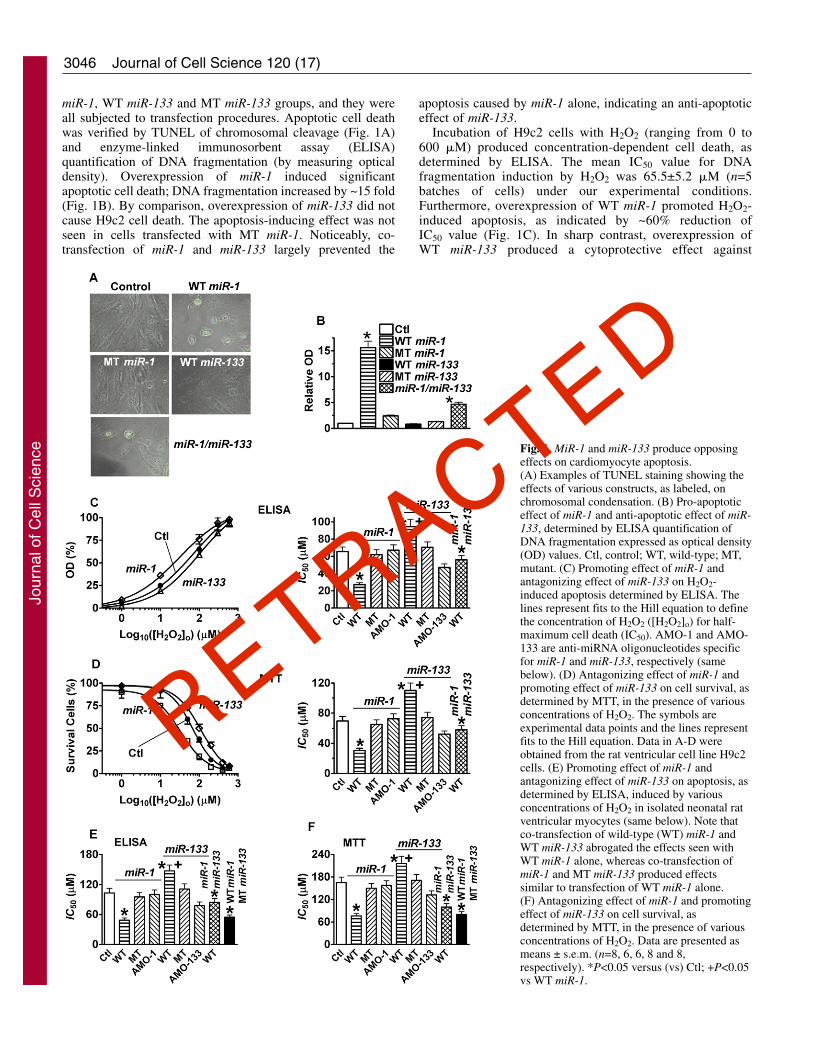
miR-1, WT miR-133 and MT miR-133 groups, and they wereall subjected to transfection procedures. Apoptotic cell deathwas verified by TUNEL of chromosomal cleavage (Fig. 1A)and enzyme-linked immunosorbent assay (ELISA)quantification of DNA fragmentation (by measuring opticaldensity). Overexpression of miR-1 induced significantapoptotic cell death; DNA fragmentation increased by ~15 fold(Fig. 1B). By comparison, overexpression of miR-133 did notcause H9c2 cell death. The apoptosis-inducing effect was notseen in cells transfected with MT miR-1. Noticeably, co-transfection of miR-1 and miR-133 largely prevented the
apoptosis caused by miR-1 alone, indicating an anti-apoptoticeffect of miR-133.
Incubation of H9c2 cells with H2O2 (ranging from 0 to600 �M) produced concentration-dependent cell death, asdetermined by ELISA. The mean IC50 value for DNAfragmentation induction by H2O2 was 65.5±5.2 �M (n=5batches of cells) under our experimental conditions.Furthermore, overexpression of WT miR-1 promoted H2O2-induced apoptosis, as indicated by ~60% reduction ofIC50 value (Fig. 1C). In sharp contrast, overexpression ofWT miR-133 produced a cytoprotective effect against
Journal of Cell Science 120 (17)
Fig. 1. MiR-1 and miR-133 produce opposingeffects on cardiomyocyte apoptosis.(A) Examples of TUNEL staining showing theeffects of various constructs, as labeled, onchromosomal condensation. (B) Pro-apoptoticeffect of miR-1 and anti-apoptotic effect of miR-133, determined by ELISA quantification ofDNA fragmentation expressed as optical density(OD) values. Ctl, control; WT, wild-type; MT,mutant. (C) Promoting effect of miR-1 andantagonizing effect of miR-133 on H2O2-induced apoptosis determined by ELISA. Thelines represent fits to the Hill equation to definethe concentration of H2O2 ([H2O2]o) for half-maximum cell death (IC50). AMO-1 and AMO-133 are anti-miRNA oligonucleotides specificfor miR-1 and miR-133, respectively (samebelow). (D) Antagonizing effect of miR-1 andpromoting effect of miR-133 on cell survival, asdetermined by MTT, in the presence of variousconcentrations of H2O2. The symbols areexperimental data points and the lines representfits to the Hill equation. Data in A-D wereobtained from the rat ventricular cell line H9c2cells. (E) Promoting effect of miR-1 andantagonizing effect of miR-133 on apoptosis, asdetermined by ELISA, induced by variousconcentrations of H2O2 in isolated neonatal ratventricular myocytes (same below). Note thatco-transfection of wild-type (WT) miR-1 andWT miR-133 abrogated the effects seen withWT miR-1 alone, whereas co-transfection ofmiR-1 and MT miR-133 produced effectssimilar to transfection of WT miR-1 alone.(F) Antagonizing effect of miR-1 and promotingeffect of miR-133 on cell survival, asdetermined by MTT, in the presence of variousconcentrations of H2O2. Data are presented asmeans ± s.e.m. (n=8, 6, 6, 8 and 8,respectively). *P<0.05 versus (vs) Ctl; +P<0.05vs WT miR-1.
Jour
nal o
f Cel
l Sci
ence

3047miR-1 and miR-133 regulate apoptosis
H2O2-induced apoptosis; the IC50 was increased by ~40%.Consistently, overexpression of miR-133 markedly increasedcell viability, whereas that of miR-1 did the opposite, asdetermined by MTT assay (Fig. 1D). These effects were notseen when MT miR-1 or MT miR-133 was transfected. Tofurther verify the opposing effect of miR-1 and miR-133, weperformed reciprocal experiments wherein we transfectedH9c2 cells with 2�-O-methyl antisense inhibitoryoligonucleotides (AMOs) against miR-1 (AMO-1) or miR-133 (AMO-133) (Krutzfeldt et al., 2005; Cheng et al.,2005). AMO-1 reduced H2O2-induced apoptosis, whereasAMO-133 facilitated it (Fig. 1C,D). Most strikingly, co-transfection of WT miR-1 and WT miR-133 failed to alterH2O2-induced apoptosis, indicating that they counteract eachother.
To confirm the above results obtained with the H9c2 cellline, the same experiments were conducted in neonatal ratventricular myocytes, which are known to be terminallydifferentiated cardiomyocytes. Quantitatively the same resultswere obtained with these cells (Fig. 1E,F).
Post-transcriptional repression of HSP60 and HSP70 bymiR-1 and of Casp9 by miR-133The above results suggest that miR-1 is pro-apoptotic whereasmiR-133 is anti-apoptotic and these opposing actions areprobably due to regulation of distinct apoptotic and survivalfactors by these miRNAs. To test this notion, we first used acomputational and bioinformatics-based approach to predictthe putative targets related to apoptosis versus survival usingTargetScan hosted by the Wellcome Trust Sanger Institute(Griffiths-Jones, 2004). In this way, we identified twoimportant candidate targets for miR-1: HSP60 and HSP70 (heatshock proteins). We also identified several targets sites for miR-133 in the caspase-9 (Casp9) gene by complementarity (Fig.2).
To verify that HSP60, HSP70 and Casp9 are indeed thecognate targets of miR-1 and miR-133, respectively, for post-transcriptional repression, we took the following approaches.We first determined the effects of the two miRNAs on proteinlevels, by western blotting and caspase activity assays. miR-1remarkably reduced the levels of HSP60 proteins by ~73% inH9c2 cells both in the absence and presence of H2O2 (Fig. 3A).HSP70 protein level was similarly reduced, albeit to a lessextent (~53%; Fig. 3B). Co-application of miR-1 and AMO-1almost abolished the effects. By comparison, miR-133decreased total Casp9 protein level by ~89% (Fig. 3C), aneffect eliminated by co-transfection with AMO-133. Whentransfected with MT miR-1 and MT miR-133, decreases inHSP60, HSP70 and Casp9 were hardly seen. Since reductionof Casp9 protein level may well result in reduction of Casp9activation, Casp9 activities were determined. As shown in Fig.3D, miR-133 diminished basal Casp9 activity and preventedH2O2-induced increase in Casp9 activity. These effects of miR-133 were antagonized by AMO-1. Moreover, application ofAMOs alone increased the levels of HSP60, HSP70 and Casp9in H9c2 cells (Fig. 3E), indicating the roles of basal miR-1 andmiR-133 in cardiac cells.
We subsequently investigated the effects of the miRNAs onmRNA levels of HSP60, HSP70 and Casp9 in H9c2 cells. ThemRNA levels of HSP60, HSP70 were unaffected by miR-1,indicating that miR-1 and miR-133 do not affect their mRNAstability. Casp9 mRNA was reduced by miR-133 (Fig. 3F).
Effects of miR-1 and miR-133 on caspase-3 (Casp3)activitiesCasp3 genes do not contain any domains bearing putativetarget sites for miR-1 or miR-133. Thus, miR-1 or miR-133 isnot expected to alter Casp3 protein levels. This was indeedverified by our experiments using western blot analysis withthe antibody against the total Casp3 protein (Fig. 4A).However, miR-1 and miR-133 may indirectly affect Casp3activities because these miRNAs repress HSP60, HSP70 andCasp9, which could in turn alter Casp3 activities (but not totalprotein levels). To test this notion, we determined Casp3activities in H9c2 cells with or without H2O2 treatments, miR-1, miR-133 and their AMOs. Transfection of cells with miR-1 or challenge of cells with H2O2 both robustly increasedCasp3 activities and the effects were abolished by co-application of AMO-1. By contrast, transfection of cells withmiR-133 diminished Casp3 activity, an effect prevented byco-application of AMO-133. Moreover, H2O2 enhancedCasp3 activity and co-application with AMO-133 further
3'-AAUGUAUGAAGAAAUGUAAGGU-5' 3'-UAUGUGUGAAGAAAUGUAAGGU-5' | | | || |:| |||||||| ||| | || |:| ||||||||1988-UAAAAAACAUUUGUACAUUCCU-2009 1997-AUAAAGACAUUUGUACAUUCCU-2018
H-HSP60 3'UTR (BC073746) R-HSP60 3'UTR (BC086507)
3'-AAUGUAUGAAGAAAUGUAAGGU-5' 3'-UAUGUGUGAAGAAA-UGUAAGGU-5' : | ||||:||||:| : ::: ||| |||||| |2137-AGCUUCAAGACUUUGCAUUUCC-2128 2418-UCUUUUGUAACUUAAACAUUCAA-2439
H-HSP70 3'UTR (BC0002453) R-HSP70 3'UTR (NM_031971)
3'-UGUCGACCAACUUCCCCUGGUU-5' 3'-UGUCGACCAACUUCCCCUGGUU-5' : | | || || |||||||: ::: ||| |||||||| 57-GGAACAUCUUCAAUGGGACCAG-78 377-GTGCCUGUGGUCCUGGGACCAA-398
3'-UGUCGACCAACUUCC-CCUGGUU-5' 3'-UGUCGACCAACUUCCCCUGGUU-5' : |||||| | || |||||| | : | ||||||: 657-GGAGCUGG-CGCAGCAGGACCAC-678 1125-AAGCCCAAGGACCUUGGACCAG-1147
3'-UGUCGACCAACUUCCCCUGGUU-5' 3'-UGUCGACCAACUUCCCCUGGUU-5' | | || || ||||||| :| ||| |: ||||| || 797-UGAACAUCUUCAAUGGGACCAG-818 1581-UGGGACGGUGGGAGGGGAGGAA-1602
3'-UGUCGACCAACUUCCCCUGGUU-5' 3'-UGUCGACCAACUUCCCCUGGUU-5' : |:| ||:| |||||| | :| || | ||||||1057-UUUGUUUCCUGGA-GGGACCCC-1077 1644-AGGGACUGUGGCCUGGGACCCC-1665
3'-UGUCGACCAACUUC-CCCUGGUU-5' 3'-UGUCGACCAACUUCCCCUGGUU-5' | |||| |||| ||||| :| : : | : |||||||1438-AACAGUGGAGGAAGAGGGACAGA-1460 2211-UGGUUACCCUAGUGGGGACACC-2234
H-Caspase 9 (BC002452)U-'3 GUCGACCAACUUCCCCUGGUU-5'
|||| |||||| |||| | U-7132 CCCCUGG--GAAGGGUACCAU-2337
R-Caspase 9 (NM_031632)
A miR-1
Human miR-1 Rat miR-1
Caspase 9 Rat miR-133Human miR-133
HSP60 3'UTR
HSP70 3'UTR
B miR-133
Fig. 2. The sequences showing the unique sites of miRNA::mRNAcomplementarity between miR-1 and HSP60 or HSP70, and betweenmiR-133 and caspase-9 (Casp9) for both human (H) and rat (R)genes. The matched base pairs are bold and connected by a verticalline and the G:U/U:G wobble is indicated by bold letters connectedby dots. The GenBank accession numbers of the genes are indicatedin the brackets and the positions of the target sites are numbered.Note that there is only a single target site for miR-1 in HSP60 orHSP70 and the complementarity is limited to the 3�UTRs of thesegenes, whereas there are multiple target sites in Casp9 and the sitesare distributed throughout the whole mRNA sequence.
Jour
nal o
f Cel
l Sci
ence

3048
increased Casp3 activity (Fig. 4B), in line with the notion thatmiR-133 diminishes Casp3 activation as a result of repressionof Casp9.
Verification of interactions between miR1, miR-133 andtheir target genesWe placed the 3�UTRs of HSP60 and HSP70, or the full-lengthcDNA of Casp9 into the 3�UTR of a luciferase reporter plasmidto construct chimeric vectors. Co-transfection of the chimericconstructs with miR-1 or miR-133 (Fig. 5) into HEK293 cells,
consistently resulted in smaller luciferase activity relative totransfection of the chimeric plasmid alone. Co-application ofmiR-1 or miR-133 with their respective AMOs eliminated thesilencing effects. A mutated target sequences of HSP60 orHSP70 fused to the 3�UTR of luciferase was not responsive tomiR-1 or miR-133, suggesting specificity of the repressioneffect. Furthermore, MT miR-1 or MT miR-133 had no effecton the WT target sequences, but could efficiently repressluciferase activities with the constructs containing the MT3�UTRs of HSP60 or HSP70 complementary to MT miR-1.
Journal of Cell Science 120 (17)
Fig. 3. Post-transcriptional repression of HSP60 and HSD70 by miR-1 and caspase-9 (Casp9) by miR-133. (A-C) Western blot analysis ofHSP60, HSP70 and Casp9 (total) under various conditions with protein samples from H9c2 cells. The bar charts in the lower panels representthe densitometric measurements of western blots of HSP60, HSP70 and Casp9 expression. (D) Effects of miR-133 on Casp9 activity.(E) Effects of AMO-1 and AMO-133, respectively, on protein levels of HSP60, HSP70 and Casp9 in H9c2 cells. (F) Effects of miR-1 and miR-133 on mRNA levels of HSP60, HSP70 and Casp9 in H9c2, as determined by real-time RT-PCR. Data presented as means ± s.e.m. (n=8, 6, 6,5, 6, and 6, for A-F, respectively). *P<0.05 vs Ctl; +P<0.05 vs WT miR-1 or WT miR-133.
Jour
nal o
f Cel
l Sci
ence

3049miR-1 and miR-133 regulate apoptosis
We then used miR-1 and miR-133 standards in which thecomplementary sequences of miR-1 and miR-133 were cloneddownstream of the luciferase gene in the pMIR-REPORTplasmid (Chen et al., 2006; Krutzfeld et al., 2005). With theseconstructs, we were able to confirm the uptake and activitiesof transfected miRNAs. Real-time RT-PCR analyses on theeffects of AMO-1 and AMO-133 on miR-1 and miR-133 levelsin H9c2 cells, to verify the efficacy and specificities against theexogenous miRNAs, have been reported in our previous study(Yang et al., 2007; Luo et al., 2007).
Successful delivery of miR-1 or miR-133 and AMO-1 orAMO-133 into the cells was further verified by comparing themiR-1 or miR-133 levels before and 48 hours after transfectionof the constructs in cultured neonatal ventricular myocytes. Asshown in Fig. 6A, transfection resulted in approximatelythree- to fourfold increases in miR-1 and miR-133 levels. Forcontrol purpose, transfection of miR-1 or miR-133 did notsignificantly alter the level of miR-133 or miR-1. It should bementioned that the miR-1 and miR-133 levels are dynamicwith transfection. Our data were collected at a specific time,48 hours after transfection (because all our measurementswere performed at this time) and the levels do not apply toother times. Coincidently, incubation of cells with H2O2 (150�M) caused an approx. threefold elevation of miR-1 (Fig. 6B).By comparison, H2O2 induced only 70% increase in miR-13.
The results are consistent with the fact that H2O2 inducesapoptosis.
The H9c2 rat ventricular cell line and the HEK293 humanembryonic kidney cell line were used in our study for differentspecific objectives. The former was used for experimentsinvolving endogenous miR-1 or miR-133, such as theexperiments involving application of AMO-1 alone, and thelatter was used for experiments involving only exogenouslydelivered miR-1 or miR-133 by transfection, such as theluciferase reporter gene experiments. We quantified the levelsof miR-1 and miR-133 in these cell lines as well as in the A549human lung cancer cell line. Our data confirmed that H9c2expresses endogenous miR-1 and miR-133, which are knownto be muscle-specific, whereas the non-muscle cells HEK293
Fig. 4. Effects of miR-1 and miR-133 on caspase 3 (Casp3) proteinlevels and activities in H9c2 cells. (A) Immunoblotting analysis ofCasp3 protein levels with and without miR-1 or miR-133 treatment.The antibody against the total Casp3 recognized the 35 kDa bandrepresenting Casp3. AP+: antibody pretreated with its antigenicpeptide; Ctl: cells treated with Lipofectamine 2000 only; miR-1 andmiR-133: cells transfected with miR-1 and miR-133, respectively andwith Lipofectamine 2000. n=8 experiments for each group.(B) Regulation of Casp3 activity by miR-1 and miR-133.Transfection was performed with Lipofectamine 2000 andmeasurements were made 24 hours after transfection. *P<0.05 vsCtl; +P<0.05 vs miR-1 alone or miR-133 alone or H2O2 alone; n=5for each group.
0
40
80
120
*
+
*WT 3'UTRMT 3'UTRWT miR-1MT miR-1 AMO-1
+----
+-+--
+-+-+
+--+-
-++--
-+---
-+-+-
Rel
ativ
e A
ctiv
ity
0
40
80
120
*
+
*WT 3'UTRMT 3'UTRWT miR-1MT miR-1 AMO-1
+----
+-+--
+-+-+
+--+-
-++--
-+---
-+-+-
Rel
ativ
e A
ctiv
ity
A
B
C
HSP60
HSP70
0
40
80
120
*
+
WT miR-133MT miR-133 AMO-133 WT miR-1
----
+---
+-+-
-+--
+--+
Rel
ativ
e A
ctiv
ity
Casp9
Fig. 5. Verification of HSP60 (A), HSP70 (B) and Casp9 (C) ascognate targets of miR-1 and miR-133, respectively, for post-transcriptional repression. Data on luciferase reporter activities showthe interaction between miR-1 and HSP60 and HSP70 3�UTRs andbetween miR-133 and Casp9 mRNA. WT, wild type; MT, mutant,AMO-1 and AMO-133, miR-1- and miR-133-specific antisenseinhibitors, respectively (see Fig. 2). Shown are means ± s.e.m. (n=5batches of cells for each bar in A-C). *P<0.05 vs Ctl; +P<0.05 vsWT miR-1 or WT miR-133.
Jour
nal o
f Cel
l Sci
ence

3050
and A549 express only minimal levels (~1/100-1/1000 ofH9c2) of endogenous miR-1 and miR-133 (Fig. 6C).
DiscussionThe study reported by Chen et al. (Chen et al., 2006) revealedthat miR-1 and miR-133 are clustered on the samechromosomal loci and transcribed together as a singletranscript which becomes two independent, mature miRNAswith distinct biological functions. One important characteristicof miR-1 and miR-133 regulation of cardiomyocyte apoptosis,revealed in this study, is that they produce opposing effects;miR-1 is pro-apoptotic whereas miR-133 is anti-apoptotic. Thissuggests that the relative levels of miR-1 and miR-133 is moreimportant than their absolute levels, to determining the fate(apoptosis and survival) of cardiac cells. This notion issupported by the fact that co-transfection of miR-1 and miR-133 failed to induce apoptosis or to affect oxidative stress-induced apoptosis. In addition, in the presence of oxidative
stress to induce apoptosis, both miR-1 and miR-133 levels wereincreased relative to those in cells under normal conditions, butthe increase in miR-1 predominantly overweighed that in miR-133, favoring the occurrence of apoptosis. However, at thisstage the notion is merely a speculation that needs furtherstudies to verify. Moreover, it should also be noted that thisstudy merely provided indirect evidence for the interactionsbetween miR-1, miR-133 and their target genes, and morerigorous experimentation is required to fully establish therelationships.
Mitochondrial death pathway is one of the majormechanisms for apoptosis, which involves selective disruptionof the outer membrane as a result of mitochondrial matrixhyperpolarization and/or matrix swelling, pore formation byproteins such as Bax and Bcl-xS, or rapid loss of �� followingpermeability transition (Latchman, 2001; Gupta and Knowlton,2005). HSPs are expressed both constitutively (cognateproteins) and under stressful conditions (inducible forms), withconstitutive expression being most prominent in mammaliantissues. HSPs are primarily anti-apoptotic and different HSPshave been shown to inhibit the mitochondrial death pathway atdifferent points. HSP60 in the heart has key anti-apoptoticfunctions because of its ability to form complexes with Bax,Bak and Bcl-xS (Lin et al., 2001; Kirchhoff et al., 2002; Shanet al., 2003; Marber et al., 1995), but not with Bcl-2. Bindingof HSP60 in the normal cardiac cells prevents Bax fromoligomerizing and inserting into the mitochondrial membrane.Reduction of HSP60 is associated with an overall decrease inBcl-2 along with an increase in Bax and Bak and is sufficientto precipitate apoptosis (Lin et al., 2001; Kirchhoff et al., 2002;Shan et al., 2003; Marber et al., 1995). HSP70 exerts its anti-apoptotic effect by preventing oligomerized Apaf-1 fromrecruiting pro-Casp9 (Latchman, 2001; Marber et al., 1995).HSP70 can also inhibit apoptosis in a caspase-independentmanner by inhibiting the c-Jun N-terminal kinase (JNKkinase). However, Casp9 is a critical regulator of mitochondria-mediated apoptosis; it forms a multimeric complex withcytochrome c and Apaf-1 to activate downstream caspases suchas caspase-3 leading to apoptotic cell death (Han et al., 2006;Bialik et al., 1999; Kannan and Jain, 2000). The data in thepresent study showing repression of HSP60 and HSP70 bymiR-1 and Casp9 by miR-133 and the opposing actions of thesetwo miRNAs on apoptosis are in line with these previousfindings.
Our data demonstrated silencing of HSP60 and HSP70 bymiR-1 only at the protein level, and knockdown of Casp9 bymiR-133 at both protein and mRNA levels. Earlier, miRNAswere though to primarily repress their targets at the proteinlevel without affecting mRNA stability (Meister and Tuschl,2004; Lewis et al., 2003). Increasing evidence, however,indicate that miRNAs silence genes by multiple mechanismsincluding degrading their target mRNAs (Kannan and Jain,2000; Nilsen, 2007; Pillai et al., 2007). Our observations seemto be in line with multiple mechanisms of the action. However,it is presently unclear what determines the exact mechanismsof miRNA actions.
Collectively, our study revealed a novel aspect of cellularfunctions of the muscle-specific miRNAs miR-1 and miR-133,i.e. regulation of apoptosis and survival in cardiomyocytes. Aunique feature of this regulation is the opposing actions withmiR-1 being pro-apoptotic and miR-133 being anti-apoptotic,
Journal of Cell Science 120 (17)
miR-1 miR-1330
1
2
3
4
Non-transfectedmiR-1 TransfectedmiR-133 Transfected
**
Rel
ativ
e L
evel
miR-1 miR-1330
1
2
3
4 CtlH2O2H2O2/AMO*
*++
Rel
ativ
e L
evel
A
B
C
miR-1 miR-1330.00.20.40.60.81.0
* * * *
H9c2A549HEK293
Rel
ativ
e L
evel
Fig. 6. Comparison of miR-1 and miR-133 expression levels undervarious conditions, measured by real-time RT-PCR. (A) miR-1 andmiR-133 levels without and with transfection of exogenous miR-1and miR-133. The data are averaged from three batches of H9c2cells. *P<0.05 vs non-transfected cells. (B) Enhanced expression ofmiR-1 and miR-133 induced by H2O2 (150 �M) in H9c2 cells (n=5batches of cells for each group). *P<0.05 vs Ctl; +P<0.05 vs miR-1or miR-133. (C) Comparison of miR-1 and miR-133 expression levelsin various cell lines indicated. H9c2, rat ventricular cell line (n=6batches of cells); A549, human lung cancer cell line (n=7 batches ofcells); HEK293, human embryonic kidney cell line (n=7 batches ofcells). MiR-1 and miR-133 levels are expressed as relative levels bynormalizing to the values obtained from H9c2 cells. *P<0.05 vsH9c2.
Jour
nal o
f Cel
l Sci
ence

3051miR-1 and miR-133 regulate apoptosis
suggesting a possible role of relative miR-1 and miR-133 levelsin regulating the cell fate. Post-transcriptional repression ofHSP60 and HSP70 by miR-1 and of Casp9 by miR-133 isprobably one of the mechanisms underlying their regulation ofapoptosis versus survival. Our present and previous studiesrevealed the pathological elevations of miR-1 levels incardiomyocytes in conditions favoring apoptosis (ischemia andoxidative stress). However, how these conditions lead tooverexpression of miR-1 remains unclear.
Materials and MethodsCell cultureThe cell lines used in this study were all purchased from American Type CultureCollection (ATCC, Manassas, VA). H9c2 (rat ventricular cell line) and HEK293(human embryonic kidney cell line) were cultured in Dulbecco’s Modified EagleMedium (DMEM). The cultures were supplemented with 10% fetal bovine serumand 100 �g/ml penicillin/streptomycin.
Synthesis of miRNAs and sequences of miRNA inhibitorsmiR-1 and miR-133 and their respective mutant constructs were synthesized byIntegrated DNA Technologies (IDT) (1). The sequences of miR-1 and miR-133inhibitors (AMOs; anti-miRNA oligonucleotides) used in our studies are the exactantisense copies of their respective mature miRNA sequences: 3�-AAUGUAUGAAGAAAUGUAAGGU-5� for human miR-1 (GenBank acc. no.:HSM808714), 3�-AAUGUAUGAAGAAAUGUAAGGU-5� for rat miR-1(GenBank ac. no.: DQ066650), and 3�-UGUCGACCAACUUCCCCUGGUU-5�for both human and rat miR-133 [the sequences of miR-133 are identical in human(HSM808714) and rat (RATNCRNAB)]. All the nucleotides in the AMOs (AMO-1 for miR-1 and AMO-133 for miR-133) contain 2�-O-methyl modifications at everybase and a 3� C3-containing amino linker. The antagomers were also synthesizedby IDT.
Construction of the chimeric miRNA binding site – luciferasereporter vectors and mutagenesisTo generate reporter vectors bearing miRNA-binding sites, we generated directmatch miR-1 and miR-133 sites (synthesized by Invitrogen), respectively, and thesequences around the putative target sites for these miRNAs in the 3� UTRs ofHSP60 and HSP70, and the full-length Casp9 mRNA (1). These inserts were clonedinto the multiple cloning sites in the pMIR-REPORTTM luciferase miRNAexpression reporter vector (Ambion, Inc.). The sense and antisense strands of theoligonucleotides were annealed by adding 2 �g of each oligonucleotide to 46 �l ofannealing solution (100 mM potassium acetate, 30 mM Hepes-KOH, pH 7.4 and 2mM magnesium acetate) and incubating at 90°C for 5 minutes and then at 37°C for1 hour. The annealed oligonucleotides were digested with HindIII and SpeI and usedto ligate into HindIII and SpeI sites.
Nucleotide-substitution mutations (MT) were carried out using direct oligomersynthesis for miR-1 and miR-133, and PCR-based methods for the 3� UTRs ofHSP60 and HSP70 genes. The substitution nucleotides were so designed to avoidproducing new binding sites for other miRNAs potentially existing in HEK293 cells.All constructs were sequencing verified. See Fig. S1 in supplementary material fordetails of the mutations.
Transfection of miRNAs and luciferase assayAfter 24 hours starvation in serum-free medium, cells (1�105 per well) weretransfected with 1 �g miR-1, miR-133, or other constructs, with Lipofectamine 2000(Invitrogen), according to the manufacturer’s instructions.
For luciferase assay, cells were transfected with 1 �g PGL3-target DNA (fireflyluciferase vector) and 0.1 �g PRL-TK (TK-driven Renilla luciferase expressionvector) with Lipofectamine 2000. Luciferase activities were measured 48 hours aftertransfection with a dual luciferase reporter assay kit (Promega) on a luminometer(Lumat LB9507) (Yang et al., 2007; Luo et al., 2007).
Quantification of mRNA and miRNA levelsFor quantification of HSP60, HSP70 and Casp9 transcripts, conventional real-timeRT-PCR was carried out with total RNA samples extracted from H9c2 cells andneonatal rat ventricular cells 48 hours after transfection. TaqMan quantitative assaywas performed with the expression level of GAPDH as an internal control.
The mirVanaTM qRT-PCR miRNA Detection Kit (Ambion) was used inconjunction with real-time PCR with SYBR Green I for quantification of miR-1 andmiR-133 transcripts, as detailed elsewhere (Yang et al., 2007; Luo et al., 2007). Foldvariations in expression of miR-1 and miR-133 between RNA samples werecalculated after normalization to 5s rRNA.
Western blot analysisThe protein samples were extracted from H9c2 cells and cultured neonatal rat
ventricular cells, with the procedures essentially the same as described in detailelsewhere (Han et al., 2001; Han et al., 2004a; Han et al., 2004b; Luo et al., 2007;Wang et al., 2002). Protein samples (~50 �g) were fractionated by SDS-PAGE (7.5-10% polyacrylamide gels). The primary antibodies against HSP60 (StressgenBioreagents, Ann Arbor, MI; rabbit polyclonal), HSP70 (Cell Signaling; rabbitpolyclonal) and total caspase-3 and total caspase-9 (Cell Signaling; rat specific,rabbit polyclonal) were used, with GAPDH (anti-GAPDH antibody from ResearchDiagnostics, Concord, MA) as an internal control.
Caspase-9 and caspase-3 activity assayThe procedures were the same as previously described in detail (Han et al., 2001;Han et al., 2004a; Han et al., 2004b; Wang et al., 2002).
MTT assay for cell viabilityCell Proliferation Kit I [3-(4,5-dimethylthiazol-2-yl)-2,5-diphenyl tetrazoliumbromide (MTT); Roche Molecular Biochemicals, Laval, PQ, Canada] was used toquantify survival of cells from oxidative stress (Han et al., 2001; Han et al., 2004a;Han et al., 2004b; Wang et al., 2002).
Enzyme-linked immunosorbent assay (ELISA)The Cell Death Detection ELISA kit (Roche Molecular Biochemicals) wasemployed to quantify DNA fragmentation on the basis of antibody detection of freehistone and fragmented DNA (Han et al., 2001; Han et al., 2004a; Han et al., 2004b;Wang et al., 2002).
Terminal deoxyribonucleotide transferase-mediated dUTP nickend labeling (TUNEL)DNA fragmentation of individual cells was detected in situ by TUNEL with the InSitu Cell Death Detection kit, Fluorescein (Roche Molecular Biochemicals) (Han etal., 2001; Han et al., 2003; Han et al., 2004a; Han et al., 2004b; Wang et al., 2002).
Data analysisGroup data are expressed as mean ± s.e.m. Statistical comparisons (performed usingANOVA followed by Dunnett’s method) were carried out using Microsoft Excel. Atwo-tailed P<0.05 was taken to indicate a statistically significant difference.
The authors thank XiaoFan Yang for excellent technical support.This work was supported in part by the Natural Sciences andEngineering Research Council of Canada and Fonds de la Recherchede l’Institut de Cardiologie de Montreal, awarded to Z. Wang, and bythe National Nature Science Foundation of China (30430780), theFoundation of National Department of Science and Technology ofChina (2004CCA06700), and National Basic Research Program ofChina (973 Program; 2007CB512000/2007CB512006) awarded to B.Yang. Z. Wang is a senior research scholar of the Fonds de Rechercheen Sante de Quebec.
ReferencesAmbros, V. (2004). The functions of animal microRNAs. Nature 431, 350-355.Bialik, S., Cryns, V. L., Drincic, A., Miyata, S., Wollowick, A. L., Srinivasan, A. and
Kitsis, R. N. (1999). The mitochondrial apoptotic pathway is activated by serum andglucose deprivation in cardiac myocytes. Circ. Res. 85, 403-414.
Chen, J. F., Mandel, E. M., Thomson, J. M., Wu, Q., Callis, T. E., Hammond, S. M.,Conlon, F. L. and Wang, D. Z. (2006). The role of microRNA-1 and microRNA-133in skeletal muscle proliferation and differentiation. Nat. Genet. 38, 228-233.
Cheng, A. M., Byrom, M. W., Shelton, J. and Ford, L. P. (2005). Antisense inhibitionof human miRNAs and indications for an involvement of miRNA in cell growth andapoptosis. Nucleic Acids Res. 33, 1290-1297.
Griffiths-Jones, S. (2004). The microRNA Registry. Nucleic Acids Res. 32, D109-D111.Gupta, S. and Knowlton, A. A. (2005). HSP60, Bax, apoptosis and the heart. J. Cell
Mol. Med. 9, 51-58.Hammond, S. M. (2006). MicroRNAs as oncogenes. Curr. Opin. Genet. Dev. 16, 4-9.Han, H., Wang, H., Long, H., Nattel, S. and Wang, Z. (2001). Oxidative
preconditioning and apoptosis in L-cells: Roles of protein kinase B and mitogen-activated protein kinases. J. Biol. Chem. 276, 26357-26364.
Han, H., Long, H., Wang, H., Wang, J., Zhang, Y. and Wang, Z. (2004a). Cellularremodeling of apoptosis in response to transient oxidative insult in rat ventricular cellline H9c2: a critical role of the mitochondria death pathway. Am. J. Physiol. 286,H2169-H2182.
Han, H., Wang, J., Zhang, Y., Long, H., Wang, H., Xu, D. and Wang, Z. (2004b).HERG K+ channel conductance promotes H2O2-induced apoptosis in HEK293 cells:cellular mechanisms. Cell. Physiol. Biochem. 14, 121-134.
Han, Y., Chen, Y. S., Liu, Z., Bodyak, N., Rigor, D., Bisping, E., Pu, W. T. and Kang,P. M. (2006). Overexpression of HAX-1 protects cardiac myocytes from apoptosisthrough caspase-9 inhibition. Circ. Res. 99, 415-423.
Hwang, H. W. and Mendell, J. T. (2006). MicroRNAs in cell proliferation, cell death,and tumorigenesis. Br. J. Cancer 94, 776-780.
Jour
nal o
f Cel
l Sci
ence
3052
Jackson, R. J. and Standart, N. (2007). How do microRNAs regulate gene expression?Sci. STKE 23, 243-249.
Kannan, K. and Jain, S. K. (2000). Oxidative stress and apoptosis. Pathophysiology 7,153-163.
Kirchhoff, S. R., Gupta, S. and Knowlton, A. A. (2002). Cytosolic HSP60, apoptosis,and myocardial injury. Circulation 105, 2899-2904.
Krutzfeldt, J., Rajewsky, N., Braich, R., Rajeev, K. G., Tuschl, T., Manoharan, M.and Stoffel, M. (2005). Silencing of microRNAs in vivo with ‘antagomirs’. Nature438, 685-689.
Kwon, C., Han, Z., Olson, E. N. and Srivastava, D. (2005). MicroRNA1 influencescardiac differentiation in Drosophila and regulates Notch signaling. Proc. Natl. Acad.Sci. USA 102, 18986-18991.
Latchman, D. S. (2001). Heat shock proteins and cardiac protection. Cardiovasc. Res.51, 637-646.
Lewis, B. P., Shih, I., Jones-Rhoades, M. W., Bartel, D. P. and Burgel, C. B. (2003).Prediction of mammalian microRNA targets. Cell 115, 787-798.
Lim, L. P., Lau, N. C., Garrett-Engele, P., Grimson, A., Schelter, J. M., Castle, J.,Bartel, D. P., Linsley, P. S. and Johnson, J. M. (2005). Microarray analysis showsthat some microRNAs downregulate large numbers of target mRNAs. Nature 433, 769-773.
Lin, K. M., Lin, B., Lian, I. Y., Mestril, R., Scheffler, I. and Dillmann, W. H. (2001).Combined and individual mitochondrial HSP60 and HSP10 expression in cardiacmyocytes protects mitochondrial function and prevents apoptotic cell deaths inducedby simulated ischemia-reoxygenation. Circulation 103, 1787-1792.
Luo, X., Xiao, J., Lin, H., Li, B., Lu, Y., Yang, B. and Wang, Z. (2007). Transcriptionalactivation by stimulating protein 1 and post-transcriptional repression by muscle-
specific microRNAs of IKs-encoding genes and potential implications in regionalheterogeneity of their expressions. J. Cell. Physiol. 212, 358-367.
Marber, M. S., Mestril, R., Chi, S. H. and Sayen, M. R. (1995). Overexpression of therat inducible 70 kDa heat shock protein in a transgenic mouse increases the resistanceof the heart to ischemic injury. J. Clin. Invest. 95, 1446-1456.
Meister, G. and Tuschl, T. (2004). Mechanisms of gene silencing by double-strandedRNA. Nature 431, 343-349.
Nilsen, T. W. (2007). Mechanisms of microRNA-mediated gene regulation in animalcells. Trends Genet. 23, 243-249.
Pillai, R. S., Bhattacharyya, S. N. and Filipowicz, W. (2007). Repression of proteinsynthesis by miRNAs: how many mechanisms? Trends Cell Biol. 17, 118-126.
Rao, P. K., Kumar, R. M., Farkhondeh, M., Baskerville, S. and Lodish, H. F. (2006).Myogenic factors that regulate expression of muscle-specific microRNAs. Proc. Natl.Acad. Sci. USA 103, 8721-8726.
Shan, Y. X., Liu, T. J., Su, H. F., Samsamshariat, A., Mestril, R. and Wang, P. H.(2003). Hsp10 and Hsp60 modulate Bcl-2 family and mitochondria apoptosis signalinginduced by doxorubicin in cardiac muscle cells. J. Mol. Cell. Cardiol. 35, 1135-1143.
Wang, H., Zhang, Y., Cao, L., Han, H., Wang, J., Yang, B., Nattel, S. and Wang, Z.(2002). HERG K+ channel: a regulator of tumor cell apoptosis and proliferation.Cancer Res. 62, 4843-4848.
Yang, B., Lin, H., Xiao, J., Lu, Y., Luo, X., Li, B., Zhang, Y., Xu, C., Bai, Y., Wang,H. et al. (2007). The muscle-specific microRNA miR-1 causes cardiac arrhythmias bytargeting GJA1 and KCNJ2 genes. Nat. Med. 13, 486-491.
Zhao, Y., Samal, E. and Srivastava, D. (2005). Serum response factor regulates amuscle-specific microRNA that targets Hand2 during cardiogenesis. Nature 436, 214-220.
Journal of Cell Science 120 (17)
Jour
nal o
f Cel
l Sci
ence