the major protein of bull seminal plasma: biosynthesis … · bioscience reports, vol. 8, no. 6,...
TRANSCRIPT

Bioscience Reports, Vol. 8, No. 6, 1988
The Major Protein of Bull Seminal Plasma: Biosynthesis and Biological Function
Karl Heinz Scheit, Michael Kemme, Gerhard Aumiiller ~, Jiirgen Seitz ~, Gerd Hagendorff and Michael Zimmer
We isolated the major protein of apparent Mr of 15,000-16,000 from seminal plasma as well as from seminal veiscle secretion of bull and proved by amino acid analysis and tryptic peptide mapping that the two proteins were identical. An antiserum against this major protein was employed to quantitate and identify the major protein in seminal plasma as well as seminal vesicle secretion. The antiserum did not cross-react with proteins from bovine or human plasma or follicular fluid respectively.
Cell-free translation of poly(A)RNA from seminal vesicle tissue and immunoprecipitation yielded one major species with apparent M r of 18,000. Using the anti-major protein antiserum, this major species was specifically immuno absorbed. Cloning and sequencing of a major protein-specific cDNA led to the identification of clone pMP17, encoding a precursor of the major protein of 128 amino acid residues. We proved that the major protein is identical to protein PDC 109 (Esch et al., Biochem. Biophys. Res. Comm. 113:861-867, 1983).
The seminal vesicles synthesize major protein in an androgen-dependent fashion. In addition to intraluminal secretion of the vas deferens, ampullary spermatozoa revealed an intense immunoreac- tion which was restricted to the neck region of the sperm head and the middle piece, while the principal piece of the tail as weU as the sperm head were devoid of immunoreactive material. Epididymal epithelium (as well as calf seminal vesicle epithelium) showed no immunoreactivity with major protein antiserum. Immunoelectron microscopy demonstrated that only spermatozoa devoid of a plasma membrane around the middle piece were able to bind the antiserum against major protein. After removal of the plasma membrane from epididymal spermatozoa, binding of major protein to subplasmalemmal binding sites was visualised using gold-labeled MP.
Transblotting with gold-labeled MP demonstrated a protein of about 66 kDa which appears to represent the major protein-receptor. Binding of major protein to the receptor (after loss of the plasma membrane in the mid-piece region of the spermatozoa after contact with secretions from seminal vesicles) is interpreted as a phyisological process presumably related to the onset of sperm motility.
KEY WORDS: seminal plasma protein
INTRODUCTION
The surface of spermatozoa is considerably modified during the process of sperm maturation and the moment of ejaculation (Koehler, 1976; Friend et al., 1977). This latter event is the initiation of dramatic changes in sperm structure
Max-Planck-Institut for Biophysikalische Chemie, Grttingen. Institut ftir Anatomie und Zellbiologie, Universit~it Marburg.
589
0144-8463/88/1200-0589506.00/0 ~) 1988 Plenum Publishing Corporation

590 Scheit e t al.
and activity resulting in increased motility, capacitation and acrosomal reaction (for review see Bedford, 1983). While the modification of sperm cell surface during the transit of the cells through the epididymis is well established (Gaddum, 1968; Bedford, 1963), and a number of specific proteins with discrete functions has been identified (e.g. by Acott and Hoskins, 1981; Rifkin and Olson, 1985), no such detailed knowledge exists about the action on spermatozoa of the several proteins secreted by the accessory sex glands, i.e. prostate and seminal vesicles (David et al., 1985). Lindholmer (1974) showed that human seminal plasma is capable of inducing forward motion in vibratile human sperm that had been removed from thecaput epididymis of patients with obstructive azoospermia.
In the bovine genital tract, seminal plasma is the second most active fluid producing forward motility by increasing the number of moving epididymal spermatozoa (Brandt et al., 1978). Hoskins et al. (1979) have performed a series of elegant studies of motility induction in bovine epididymal spermatozoa which resulted in the identification of a heat stable glycoprotein with a molecular weight of 37 kDa in its monomeric form, which is produced in the epididymis and acts as a forward motility protein (FMP). Recently, Acott et al. (1983) demonstrated that forward motility protein in bovine epididymal fluid induced progressive motility to the same extent as FMP in bovine seminal plasma.
We recently reported on the characterisation of basic proteins of bull seminal plasma (Kemme et al., 1984) as well as of seminal vesicle secretion from bull (Scheit et al., 1986). The latter has been shown to contain a number of basic proteins (Scheit, 1986) with highly specialized biological activities such as a calcium transport inhibitor (Rufo et al., 1982), ribonuclease (d'Alessio et al., 1972), protinease inhibitors (Cechova et al., 1979), a nerve growth factor (Harper et al., 1982), and an antimicrobial protein (Scheit et al., 1985), in addition to several others such as 5'nucleotidase (Fini et al., 1983).
It was shown by immuno-histochemical methods that most of these proteins are synthesized in and secreted from the seminal vesicles and ampulla of the vas deferens of the bull (Aum/iller and Scheit, 1987).
During the course of this work we became aware of the presence of a major protein in bull seminal plasma. In the following, we present experimental evidence that the major protein of bull seminal plasma is a secretory product of seminal vesicles. Furthermore, this protein appeared to be closely related to a major protein constituent of bull seminal plasma, the amino acid sequence of which was reported by Esch et al. (1983). This protein of unknown biological function, named PDC109 by those authors, possessed a Me of 12,774 for the polypeptide chain.
PROTEINCHEMICAL CHARACTERISATION OF THE MAJOR PROTEIN OF BULL SEMEN
Isolation of Major Protein from Bull Seminal Plasma and Seminal Vesicle Secretion
SDS-polyacrylamide gel electrophoresis of bull seminal plasma as well as seminal vesicle secretion revealed a protein with an apparent Mr of approx-

Seminal Plasma Protein 591
Fig. 1. Characterization of major protein by polyacrylamide gel electrophoresis. Aliquots were denatured by SDS in the presence of mercaptoethanol and subjected to polyacrylamide gel electrophoresis on a 12.5% gel. Lane 1, CM-Sephadex C25-flow-through fraction from seminal vesicle secretion; lane 2, bull seminal plasma; lane 3, seminal veiscle secretion. Lanes 4-6 contain fractions of different retention times from the gel permeation experiment of Fig. 2. Lane 4, fraction 19 min; lane 5, fraction 21 min; lane 6, fraction 22 min; lane 7, major protein from pooled fractions 21 and 22rain after repeated gel-permeation chromatography.
imately 16,000 for the polypeptide chain as major component (Fig. 1). After removal of basic proteins by passage over CM-Sephadex C25, SDS- polyacrylamide gel electrophoresis of the flow-through-fraction indicated the presence of this protein (Fig. 1). Gel-filtration on Superose 6TM employing 0.5 M NaC1, 10 mM Tris/HC1, pH 7.4 containing 4 M urea led to fractionation of the protein mixture of the CM-Sephadex flow-through fraction. The protein appeared to be 90% pure as judged by SDS-polyacrylamide gel electrophoresis; it was contaminated by a higher molecuar weight component. Repetition of the gel-permeation chromatography led to homogeneous material as judged by SDS-polyacrylamide gel electrophoresis. Originally, it was attempted to carry out gel-permeation chromatography in the absence of urea. However, the protein precipitated during chromatography. This was not observed in presence of 4 M urea. The yield of major protein from seminal plasma based on A280-units applied for gel-filtration was approximately 40%; that for major protein from seminal ,vesicle secretion was in the same range.
The CM-Sephadex flow-through fraction from either seminal plasma or seminal vesicle secretion could also be fractionated by reversed phase-HPLC on a C18-support. Antiserum against major protein from bull seminal plasma, purified by reversed phase-HPLC, was raised in rabbits. Double immunodiffusion experiments showed that major protein from bull seminal plasma and major protein from seminal vesicle secretion cross reacted with fusion of precipitin arcs; bull seminal plasma and seminal vesicle secretion behaved similarly, lmmuno- blotting of an electrophoreticaUy transferred SDS-polyacrylamide gel of bull seminal plasma and seminal vesicle secretion revealed the doublet at the position

592 Scheit et al.
Fig. 2. lmmunoblotting of seminal plasma and seminal vesicle secretion employing anti-major protein-antiserum. Polyacrylamide gel electrophoresis for immunoblotting was performed on a 12.5% gel. (A) SDS-polyacrylamide gel electrophoresis; lane 1, seminal plasma; lane 2, seminal vesicle secretion; lane 3, major protein from seminal vesicle secretion; lane 4, protein standards of molecular weights 14,200, 20,100, 24,000, 29,000, 36,000, 45,000 and 66,000. (B) Electrophoretic transfer of proteins from poly- acrylamide gel electrophoresis to nitrocellulose membrane. The membrane was stained for proteins. Lane 1, seminal plasma; lane 2, seminal vesicle secretion; lane 3, major protein from seminal vesicle secretion. (C) Immunobiot: Lane 1, seminal plasma; lane 2, seminal vesicle secretion; lane 3, major protein from seminal vesicle secretion.
of major protein as the only immuno-reactive polypeptides (Fig. 2A, B). Similar experiments with bovine as well as human serum and follicular fluid provided no evidence for the presence of major protein immunolike proteins. The im- munochemical results suggested that major protein preparations from bull seminal plasma and seminal vesicle secretion were identical. Furthermore, the major protein may perhaps represent a species as well as sex specific protein.
Employing anti-major protein antiserum in single radial immuno diffusion we determined the content of major protein in bull seminal plasma and seminal vesicle secretion respectively to be 16 mg/ml and 24.8 mg/ml.
Chemical Characterisation
The amino acid composition for the major protein isolated from seminal plasma and seminal vesicle secretion respectively was determined under different conditions. The experimental values are depicted in Table 1 in a normalised manner, in order to allow easy comparison with the amino acid composition for the protein PDC 109 (Esch et al., 1983). The compositions, determined under identical conditions, for the major protein isolated from the two different sources were found to be in good agreement. The ultraviolet absorption spectra of the

Serainal Plasma Protein 593
Table 1. Amino acid composition of the major protein from bull seminal plasma and seminal vesicle secretion. Hydrolysis of non-carboxymethylated (A) or carboxymethylated (C) protein samples was carried out in 6N HCI in the presence of 0.1% mercaptoethanol for 24 h at 110~ Samples were carboxymethylated after denaturation by urea. Aromatic amino acids were determined by hydrolysis in 4 M methane-sulfonic acid (B). The ex- perimental values were normalised to a number of 10 glutamic acid or 5 alanine residues per polypeptide chain respectively. The composition of PDC 109 (D) was taken from the amino acid sequence (Esch et al. 1983).
Major-protein Amino Seminal plasma Vesicle secretion PDC109 acid (A) (B) (A) (C) (B) (D)
Asp 11.3 11.8 11.9 12 Thr 5.3 4.9 5.2 5 Ser 7.7 7.3 7.0 7 Glu 10 10 10 10 Gly 8.1 9.6 7.6 7 Ala 5.4 5 5.9 5.7 5 5 Cys n.d. n.d. 7.7 8 Val 6.0 5.8 6.5 6 Met 1.5 1.9 1.2 2 Ile 2.5 2.3 1.9 2 Leu 4.7 4.5 3.7 4 Tyr 6.8 8.5 6.6 5.0 8.6 8 Phe 4.9 6.1 5.2 6.0 6.0 6 Lys 6.7 6.2 8.4 8 His 1.9 1.9 2.2 2 Arg 4.4 5.1 4.6 5 Trp n.d. 3.8 n.d. n.d. 4.9 5 Pro 7.1 8,1 6.8 7
two protein preparat ions were identical; the ratios A275/A290 were 1.34 for the protein f rom seminal plasma and 1.35 for the preparat ion f rom seminal vesicle. The spectra were found to be indicative for a protein of high t ryptophan content. The extinction coefficient based upon amino acid analysis was determined to E1~(280 n m ) = 7.2. The pat tern of tryptic peptides, obtained by reversed phase- HPLC, for the preparat ion of the major protein f rom seminal p lasma and seminal vesicle secretion respectively were identical (Fig. 3). F rom the identity of amino acid composit ion and tryptic peptide pat tern we conclude that the major protein preparat ions, isolated f rom seminal p lasma or seminal vesicle secretion respec- tively, were identical.
Fur thermore , the agreement between the composit ions of the respective preparat ions of the major protein with that of P D C 109 was striking. The only discrepancy was observed with respect to the tyrosine and methionine content, 'where we obtained to low values. However , comparison with the composit ions .determined with non-carboxymethyla ted protein samples revealed that the reason :for this very likely resulted f rom partial decomposi t ion of tyrosine and meth- ionine during carboxymethylat ion. Indeed, determinat ion of aromatic amino acids ,of major protein samples under appropr ia te conditions yielded bet ter agreement . On the basis of these results we assume that the major protein isolated f rom seminal plasma and seminal vesicle secretion might be closely related to protein

594 Scheit et al.
Mp'svs'
0 x o
60
40
90
20Time 40 (min'p 20Time 40 (min)
Fig. 3. Separation of tryptic peptides of major protein and major protein by reversed phase-HPLC, Solvent A, 0.1% trifluoroacetic acid; solvent B, acetonitrile (60%, v/v) in 0.1% trifluoroacetic acid. Support Nucleosil C18. Column dimension 4 • 250 mm.
PDC 109. First attempts to prove identity by determination of the partial amino acid sequence failed, because the major protein preparations described above possessed blocked N-termini. Due to the similarity between the amino acid compositions of the major protein preparations compared to that of protein PDC 109 there may be only minor differences at the N-terminal part of the polypeptide, perhaps caused by limited proteolysis. This appeared likely, since the major protein, as described above, was isolated from fresh seminal plasma or vesicle secretion, in contrast to PDC 109, which was obtained from a large batch of ethanol precipitated protein (Esch et al . , 1983).
The apparent M r of the major protein as determined by SDS-polyacrylamide gel electrophoresis was between 15,000-16,000 and in agreement with that reported by Esch et al. (1983) for protein PDC 109. The major protein did not contain free SH-groups. SDS-polyacrylamide gel electrophoresis in the absence of reducing agent gave no indication of the existence of intermolecular disulfide bonds. Esch et al. (1983) also noted that protein PDC 109, like the major protein, existed in two forms, separated by polyacrylamide gel electrophoresis. Those authors explained this behaviour by partial oxidation of methionine residues.
BIOSYNTHESIS OF THE MAJOR PROTEIN
Ceil-free Translation of Poly(A)RNA from Seminal Vesicles
The isolation of major protein from seminal vesicle secretion is a necessary but not sufficient condition for the statement that the biosynthesis of the major

Seminal Plasma Protein 595
protein occurs in seminal vesicles. To clarify this point, we attempted the cell-free synthesis of major protein.
The preparation of poly(A)RNA from seminal vesicle tissue was a difficult task because this tissue is very rich in a highly active ribonuclease. Recently Furia et al. reported the cell-free synthesis of the bull seminal ribonuclease precursor employing poly(A)RNA from bull seminal vesicles (Furia et al., 1983). The protocol for the isolation of poly(A)RNA given by those workers, although principally reproducible by us, yielded less satisfactory results than the procedure described by us (Kemme et al., 1986).
Cell-free translation of poly(A)RNA from seminal vesicles led to the formation of a number of [35S]methionine labelled proteins (Fig. 4a). Clearly, the polypeptide with an apparent Mr of 18,000 represented the major species amongst the cell-free translation products. When the mixture of translation products was subjected to immuno affinity absorption with anti-major protein antiserum, a
Fig. 4. Cell-free translation of poly(A)RNA from seminal vesicle. Analysis by immunoabsorption employing anti-major protein antiserum. (a) Separation of proteins by SDS-polyacrylamide gel electrophoresis on a 12.5% gel. Lane A, without poly(A)RNA; lane B, molecular weight markers 1.2,500, 21,000, 30,000 with positions indicated by arrows; lane C, complete reaction mixture, the arrow indicating the position of the precursor of the major protein. (b) Separation of proteins obtained by immuno absorption on a 12.5% SDS-gel. Lane A, without poly(A)RNA; lane B, complete reaction mixture without anti-major protein antiserum; lane C, complete reaction mixture; lane D, complete reaction mixture with added major protein. The arrows indicate the position of molecular weight markers with masses of 30,000, 21,500 and 12,500 respectively. Further experimental details are given in Methods. (c) Separation of proteins obtained by immuno absorption on a SDS-gradient gel from 15 to 20%. Lane A, poly(A)RNA translation mixture; lane B, poly(A)RNA translation mixture treated with microsomal membrane pre- paration; lane C, molecular weight markers with masses of 30,000, 21,500, 12,500, 6,500 and 2,400 respectively.
596 Scheit et al.
single labelled polypeptide chain with Mr of approx. 18,000 was obtained (Fig. 4b). Major protein from seminal vesicle secretion, when added to the translation mixture, effectively competed with this labelled polypeptide in binding to anti-major protein antiserum (Fig. 4b). We thus have reason to conclude that this polypeptide represents the precursor of the major protein. The Me of this precursor was more accurately determined to 18,000 by means of a gradient polyacrylamide gel electrophoresis (Fig. 4c). Cell-free translation in the presence of a microsomal membrane preparation from dog pancreas, a system known to process mammalian precursor polypeptide molecules, led to the formation of a polypeptide, immunoreactive with anti-major protein, exhibiting an apparent Mr identical with that of major protein.
The existence of one precursor protein would imply that the major protein does not consist of two polypeptide chains differing in sequence. Thus the observed doublet for the preparations of the major protein might indeed be the result of chemical modification as already hypothesised by Esch et al. (1983).
The cell-free synthesis of a precursor of major protein by translation of poly(A)RNA obtained from seminal vesicles definitively established that this gland is involved in the synthesis of the major protein found in bull seminal plasma.
Northern Analysis and Cloning of Seminal Veside Poly(A)RNA
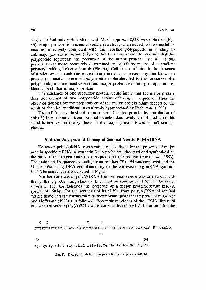
To screen poly(A)RNA from seminal vesicle tissue for the presence of major protein-specific mRNA, a synthetic DNA probe was designed and synthesised on the basis of the known amino acid sequence of the protein (Esch et al. , 1983). The amino acid sequence extending from residues 78 to 94 was employed and the 51 nucleotide long DNA complementary to the corresponding mRNA synthes- ized. The sequences are depicted in Fig. 5.
Northern analysis of poly(A)RNA from seminal vesicle was carried out with the synthetic probe using standard hybridisation conditions at 51~ The result shown in Fig. 6A indicates the presence of a major protein-specific mRNA species of 750 bp. For the synthesis of its cDNA from poly(A)RNA of seminal vesicle tissue and the construction of recombinant pBR322 the protocol of Gubler and Hoffmann (1983) was followed. Recombinant clones of the cDNA library of bull seminal vesicle poly(A)RNA were screened by colony hybridisation using the
C C C G
TTTTTTATGCTCTGGACGTGGTTTTAGCCCAGGTACACCTACAGGACCACG 5' probe
C
78 94
LysLysTyrGluThrCysThrLysIleGlySerMetTrpMetSerTrpCys
~g. 5. Designofhybridisationprobe ~r m~orprotein mRNA.

Seminal Plasma Protein 597
Fig. 6. Northern analysis of poly(A)RNA from seminal vesicle tissue. (A) Hydridisation carried out with synthetic DNA probe (see Fig. 5). (B)
Hybridisation with PstI insert of pMP17.
radioactively labelled synthetic probe. A number of positive clones were identified; the one containing the longest cDNA insert was subjected to sequencing employing both chemical (Maxam and Gilbert, 1977) as well as enzymatic sequencing (Sanger et al., 1977). The sequencing strategy of the major protein specific cDNA clone pMP17 containing a 627 pb long insert is depicted in Fig. 7.
Northern analysis of seminal vesicle poly(A)RNA with the Pstl insert of
i00 200 300 400 500 600
PstI
0 Sau3A
Fig. 7. Partial restriction map and sequencing strategy for cDNA clone pMP17.

598 Seheit et al.
1 CTC TTT CTC ATT TGG GCT GGC GTG TCT GTG TTT CTA CAA CTG GAC CCT GTG AAT GGA GAT
L F L I W A G V S V F L Q L D P V N G D
61 CAG GAC G~ GGT GTT TCT ACT GAA CCT ACC CAA GAC GGT CCT GCT GAA TTA CCT GAA GAC
2 Q D E G V S T E P T Q D G P A E L P E D
121 GAA GAA TGC GTT TTC CCA TTC GTC TAT AGA AAC AGA AAG CAT TTT GAC TGC ACA GTG CAT
22 E E C V F P F V Y R N R K H F D C T V H
181 GGT TCC TTA TTC CCG TGG TGT TCC CTC GAT GCA GAC TAT GTA GGA AGA TGG A~TAC TGT
42 G S L F P W C S L D A D Y V G R W K Y C
241 GCT CAG AGA GAC TAT GCT AAA TGT GTC TTC CCA TTT ATC TAT GGA GGC AAG A~ TAT GAG
62 A Q R D Y A K C V F P F I Y G G K K Y E
301 ACT TGC ACA AAA ATT GGG AGT ATG TGG ATG TCT TGG TGC TCA CTC TCT CCA AAC TAC GAC
82 T C T K I G S M W M S W C S L 5 P N Y D
361 AAG GAC AGA GCT TGG AAG TAT TGC TAGCCAT~AAAGAGCTATGTTCAGTCACTGTCCAGTGCATCCACCC
102 K D R A W K Y C *
432 TTGGCCATGGATGCAATCCAGCCTTTG~ACCTCAGTGAAGAAAATCAAGAACTCTC~GCATGAAGATGATGCTCTGA
511 AAGCCAGAGGTGAAATCCTTTCCCTACATCTCCACCATGTTCCCCCTATGCGTGGATCCAATC~TAAACCACTTTCTC
590 AGCAAAAAA~AAAA
cDNA Clone pMP17
Fig, 8. Nucleotide sequence and deduced amino acid sequence of clone pMP17. The amino acid sequence of major protein (PDC109) is indicated by a dotted line. The amino acid sequence extending from the N-terminus is marked by asterisks. The polyadenylation signal is underlined.
clone pMP17 furnished a mRNA species of 750 bp (Fig. 6B) similar to the results obtained with the synthetic DNA probe. This indicated that clone pMP17 cannot comprise the sequence of the full mRNA species. The obtained sequence of the Pstl insert of pMP17 (Fig. 8) contained an open reading frame extending over 387 nucleotides and coding for a polypeptide of 128 amino acid residues. The derived polypeptide sequence included in the C-terminal part of 109 amino acid residue long sequence of the major protein as published by Esch e t al. (1983). The N-terminus of this polypeptide sequence was extended by 19 amino acid residues. The Mr of the 128 amino acid residue polypeptide is 14,850 and it is obvious from the nucleotide sequence that pMP17 does not represent the full length mRNA which was found to be 650 kb. The precursor sequence has a hydrophobic character, but a signal sequence for processing is not obvious. The Pstl insert of pMP17 possesses a poly(A) tail and the putative poly-adenylation signal A A A T A A 13 nucleotides downstream of the poly(A) tail.
BIOLOGICAL PROPERTIES OF THE MAJOR PROTEIN
Antibody Specificity
The experiments described in the following have been carried out employing the anti-MP antiserum described above. Separation of seminal fluid proteins by SDS-polyacrylamide electrophoresis, transblotting of the proteins on nitro- cellulose and immunostaining with the antiserum against major proteins resulted

Seminal Plasma Protein 599
Fig. 9. Immunological identification of major protein on spermatozoa. Spermatozoa were ex- tracted with 0.1 N NaOH. Sperm extracts and seminal vesicle secretion were separated by SDS- PAGE, transblotted onto nitrocellulose and re- acted with anti-MP antibody. Lane 1, seminal vesicle secretions; lane 2, epididymal sper- matozoa; lane 3, ampullary spermatozoa; lane 4, urethral spermatozoa.
in one band at about 15 kDa and a considerably weaker immunoreaction slightly above 15 kDa, indicating some microheterogeneity in the antigen (Fig. 9, lane 1). Immunoprint analysis of stringently washed epididymal, ampullary and urethral spermatozoa showed a strong immunoreaction in urethral and ampullary sper- matozoa (Fig. 9, lanes 3 and 4) while no immunoreaction was found in
Fig. 10. Immunohistochemical demonstration of MP in tissue. (2) Bovine seminal vesicle: ST stroma; EP, epithelium; S, secretion. (3) Calf seminal vesicle: GL, undifferentiated glandular structure. (4) Epididymal tail of bull: EP, epithelium surrounding intraluminal spermatozoa, SP.

600 Scheit et al.
epididymal spermatozoa. This is suggested to indicate the association of major protein with spermatozoa that had passed the secretions of the seminal vesicles.
Tissue Distribution of Major Protein
As already described (Aumiiller and Scheit, 1987) the epithelium of the seminal vesicles as well as intraluminal secretion (Fig. 10) demonstrate an intense immunoreaction. To prove the androgen-dependence of this secretory protein, the seminal vesicles of a 14 days old bull calf were treated with the antiserum (Fig. 10). No immunoreaction could be detected within the lumen and the epithelium. The same applied for the epididymis (Fig. 10), where neither the epithelium nor the intraluminal fluid showed a positive immunoreaction.
Smears of ampullary (and urethral) spermatozoa (Fig. 11) immunostained with major protein antiserum revealed a strong immunofiuorescence of the mid-piece as well as the implantation fossa at the neck region. Both the serum head and the sperm tail distal from the mid-piece were devoid of immunoreactive material. Urethral spermatozoa that had not or insufficiently b e e n washed
Fig. U . Detection by indirect immunofluorescence of major protein bound to spermatozoa. (5) Immunoreaction of ampullary spermatozoa with anti-MP is restricted to the mid-piece, the neck and the implantation fossa of the head (magn. x 825); labelling is indicated by an arrow head; (6) immunofluorescence is lacking in an epididymal sperm reacted with anti-MP (magn. • 1200).

Seminal Plasma Protein 601
contained some spots of immunoreactive material in varying distribution on either heads.
Spermatozoa taken from the caput or the corpus of the epididymis never showed a positive immunoreaction (Fig. 11). In the most distal portions of the epididymal cauda, however, a certain number (up to - 2 0 % ) of the spermatozoa could be detected, which had a positive immunoreaction.
Incubation Experiments and Immuno Electron Microscopy
Immuno electron microscopy clearly demonstrated that the antigen sites, i.e. bound major protein, were restricted to the mid-piece region of tail (Figs. 12), the neck region and the implantation fossa of the head. As seen in Fig. 12 cross sections of the principal piece and the distal portions of the sperm tail are unlabeled.
On tangential longitudinal sections (Fig. 13), a preferential labeling of the intersections between adjacent mitochondria was recognized. At higher mag- nifications the spermatozoa were seen to lack a plasma membrane just in those areas, where immunostaining was observed.
These results indicated that a positive immunoreaction of the mid-piece occurred only when the plasma membrane was removed and spermatozoa had
Fig. 12. Binding of MP to ampuUary spermatozoa. Immunogold labelling after incubation with anti-MP: cross section (magn. • 3000); labelling is restricted to the mid-piece region.

602 Scheit et al.
Fig. 13. Binding of MP to ampullary spermatozoa. Tangential section through the mid-piece of an ampuUary sperm (magn. x 3000); bound major protein is labelled (arrow).
been in contact with seminal fluid. As an experimental approach immunoreac- tions of Triton-treated (0.1%) and of glutaraldehyde-prefixed spermatozoa incubated with dilute seminal plasma were performed. As expected, only in unfixed or Triton-treated spermatozoa, where the plasma membrane was lacking, a positive immunolabeling was found.
Most prefixed specimens contained mid-pieces surrounded by a plasma membrane (Fig. 14). In some instances, the plasma membrane appeared labilized and occasionally it was missing. The number of mid-pieces with altered (or without) plasma membrane was greatly increased, when unfixed spermatozoa were incubated with concentrated seminal vesicle fluid. Under these conditions immunolabeled mid-pieces with partially removed plasma membrane (Fig. 15) were observed. This was still more pronounced in cases where epididymal spermatozoa had been incubated with 10 -2 M EGTA in the presence of seminal fluid. Membrane fragments, partially adherent to the mid-piece, were labeled, indicating that the component binding the major protein is easily detached from underlying mitochondria in the absence of calcium. If in such an incubation experiment EGTA was replaced by 10-2M DTE, similar membrane alterations did not develop. The same was true when purified major protein was added in the place of seminal vesicle secretion. Zinc ions added at high concentrations had a

Seminal Plasma Protein 603
Fig. 14. Binding of major protein to prefixed epididymal spermatozoa. Immuno-gold labelling after incubation with seminal vesicle secretion followed by anti-MP. The plasma membrane is preserved, no binding of MP detectable (magn. x 32250).
strong cytotoxic effect on spermatozoa with marked ultra-structural alterations (results not shown).
Treatment of epididymal spermatozoa with phospholipase A2 (or C) and calcium ions in the presence of seminal vesicle fluid completely detached the mid-piece plasma membrane as well as the subplasmalemmal binding structures of major protein from the sperm surface. In some cases small membrane fragments with strong immunogold labeling were seen close to the mid-pieces.
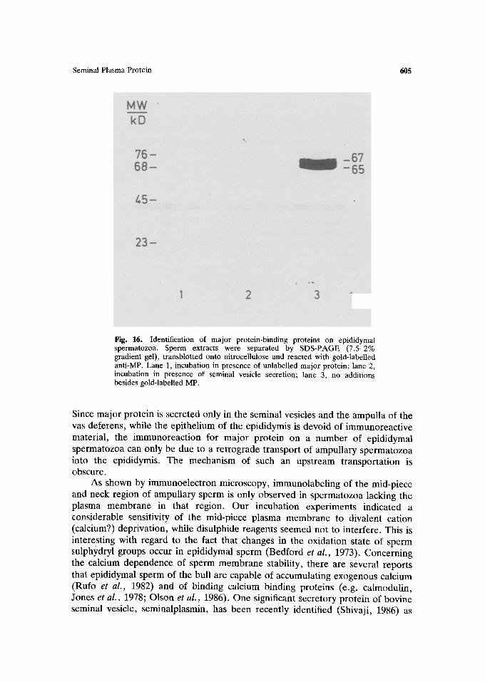
Identification of the Major Protein Receptor
Unfixed epididymal spermatozoa were treated for 10-30 min with 10-4M EGTA in TBS containing gold-labeled major protein. As anticipated, in a number of cases the mid-pieces of spermatozoa had bound the gold-labeled antigen. In some instances, subplasmalemmal material surrounding the sperm head as well as isolated plasma membrane fragments were labeled. The most intense labeling usually occurred in the neck region of the spermatozoa.
To gain some information on the biochemical nature of the major protein receptor, extracts from epididymal spermatozoa were separated on SDS-gels

604 Scheit et al.
Fig. 15. Binding of major protein to unfixed epididymal spermatozoa. Immuno-gold labelling after incubation with seminal vesicle secretion followed by anti-MP. The plasma membrane is lost and binding of MP is detectable (arrows); magn. • 32250).
(7.5-1.5%), transblotted on nitrocellulose and incubated with the gold-labeled antigen. Two polypeptide species with molecular weights of 67 and 65 kDa respectively, exclusively bound to the antigen (Fig. 16).
DISCUSSION
We have shown by immunohistochemical methods that major protein is synthesized in the seminal vesicles of the bull, while this is not the case in the calf. This indicates the androgen-dependence of the synthesis and secretion of major protein. Androgen dependence of synthesis and secretion is characteristic for secretory proteins in the male accessory sex glands, as has been shown previously in the rat (Aumtiller et al., 1982; Aumiiller and Seitz, 1986). Immunocytochem- istry has demonstrated major protein to be bound to the mid-piece of the sperm tail and to the neck region (where immunolabeling is usually most prominent).
Binding of proteins secreted by the accessory sex glands to spermatozoa on ejaculation has been established in the rat (Weil, 1965; Dravland and Joshi, 1981), rabbit (Oliphant and Singhas, 1979), mouse (Herr and Eddy, 1980), bull (Rufo et al., 1984; Shivaji, 1986) and man (Herr et al., 1986; Foresta et al., 1986).
Seminal Plasma Protein 605
Fig. 16. Identification of major protein-binding proteins on epididymal spermatozoa. Sperm extracts were separated by SDS-PAGE (7.5-2% gradient gel), transblotted onto nitrocellulose and reacted with gold-labelled anti-MP. Lane 1, incubation in presence of unlabelled major protein; lane 2, incubation in presence of seminal vesicle secretion; lane 3, no additions besides gold-labelled MP.
Since major protein is secreted only in the seminal vesicles and the ampulla of the vas deferens, while the epithelium of the epididymis is devoid of immunoreactive material, the immunoreaction for major protein on a number of epididymal spermatozoa can only be due to a retrograde transport of ampullary spermatozoa into the epididymis. The mechanism of such an upstream transportation is obscure.
As shown by immunoelectron microscopy, immunolabeling of the mid-piece and neck region of ampullary sperm is only observed in spermatozoa lacking the plasma membrane in that region. Our incubation experiments indicated a considerable sensitivity of the mid-piece plasma membrane to divalent cation (calcium?) deprivation, while disulphide reagents seemed not to interfere. This is interesting with regard to the fact that changes in the oxidation state of sperm sulphydryl groups occur in epididymal sperm (Bedford et al., 1973). Concerning the calcium dependence of sperm membrane stability, there are several reports that epididymal sperm of the bull are capable of accumulating exogenous calcium (Rufo et al., 1982) and of binding calcium binding proteins (e.g. calmodulin, Jones et al., 1978; Olson et al., 1986). One significant secretory protein of bovine seminal vesicle, seminalplasmin, has been recently identified (Shivaji, 1986) as

Scheit et al.
the calcium transport inhibitor Caltrin (Lewis et al. , 1985), which appears to act as an antagonist of calmodulin (Gietzen and Galla, 1985). Obviously, the different compartments of the plasma membrane of bovine spermatozoa are differentially sensitive to changes in calcium concentrations, since in all cases, where spermatozoa were incubated in calcium-free solutions, electron microscopy demonstrated an intact outer arosomal and plasma membrane, while the mid-piece plasma membrane was lost. These different responses of the sperm plasma membrane must be related with specific functions of the mature spermatozoon such as the onset of motility and the acrosomal reaction (see Bedford, 1983).
Taking together the observations of a lacking plasma membrane on mid-pieces of (most) bovine ampullary spermatozoa, our studies indicate this not to be an artifact, but rather a physiological event required for the binding of major protein as a subplasmalemmal perimitochondrial binding site. Analysing the surface of ejaculated bovine spermatozoa, Ji et al. (1981) found two different proteins restricted to the mid-piece, their molecular weights being 15 kDa and 29 kDa, respectively. P15 was the smallest molecule found by Ji et aL (1981) and was a major protein of the sperm surface, accounting for almost 10% of total membrane protein. It was insensitive to trypsin and chymotrypsin and must have an amino terminus or a lysine residue at the sperm surface. The molecular weight of this protein is in the same range as the major protein described here. The possibility cannot be excluded that P15 of Ji et al. (1981) is identical with the major protein. Other protein candidates binding to bovine spermatozoa would be forward motility protein (Acott and Hoskins, 1981) or the so-called HIS- proteins of Rifkin and Olson (1985). Their respective molecular weights and the immunocytochemical localization on the sperm surface, as well as their epididy- mal origin, are strong arguments against a relationship of major protein with these factors.
Major protein from seminal vesicles has been proven to specifically bind to a subplasmalemmal receptor protein of -66 kDa that can be removed from the sperm surface by phospholipase A2 treatment. As far as we see, this is the first case described where a ligand together with its receptor on the sperm surface, located in a strategic position close to the mitochondria, has been identified. This system should provide an interesting experimental model for studies of molecular events in spermatozoa occurring during the interaction of seminal fluid with the sperm surface.
REFERENCES
Acott, T. S. and Hoskins, D. D. (1981) Bovine sperm forward motility protein: binding to epididymal spermatozoa. Biol. Reprod. 24:234-240.
Acott, T. S., Katz, D. F. and Hoskins, D. D. (1983) Movement characteristics of bovine epididymal spermatozoa: effects of forward motility protein and epididymai maturation. Biol. Reprod. 29: 389-399.
Aumiiller, G. and Scheit, K. H. (1987). Immunohistochemistry of secretory proteins in the bull seminal vesicle. J. Anat. (London) 1511:43-48.

Seminal Plasma Protein 607
Aumiiller, G. and Seitz, J. (1986) Immunoelectron microscopic evidence for different compartments in the secretory vacuoles of the rat seminal vesicles. Histochem. J. 18:15-23.
Aumiiller, G., Seitz, J., Heyns, W. and Flickinger, C. J. (1982) Intra-cellular localization of prostatic binding protein (PBP) in rat prostate by light and electron microscopic immunocytochemistry. Histochemistry 76: 497-516.
Bedford, J. M. (1963) Changes in the electrophoretic properties of rabbit spermatozoa during passage through the epididymis. Nature 200:1178-1180.
Bedford, J. M. (1983) Significance of the need for sperm capacitation before fertilization in eutherian mammals. Biol. Reprod. 28:108-120.
Bedford, J. M., Calvin, H. and Cooper, G. W. (1973) The maturation of spermatozoa in the human epididymis. J. Reprod. Fertil. Suppl. 18: 199-213.
Bradford, M. M. (1976) A rapid and sensitive method for the quantitation of microgram quantities of protein utilizing the principle of protein dye-binding. Anal. Biochem. 72: 248-254.
Brandt, H., Acott, T. S., Johnson, D. J. and Hoskins, D. D. (1978) Evidence for an epididymal origin of bovine sperm forward motility protein. Biol. Reprod. 19: 830-835.
Cecbova, D., Jonakova, V., Sedlakova, E. and Mach, O. (1979) Isolation of basic acrosin inhibitor from bull seminal plasma (BUSI II). Hoppe-Seyler's Z. Phyisol. Chem. 360:1753-1758.
D'Alessio, G., Floridi, A., De Proseo, R., Piguero, A. and Leone, E. (1972) Bull semen ribonuclease. 1. Purification and physico-chemical properties of the major component. Eur. J. Biochem. 26:153-161.
David, G. F. X., Koehler, J. K., Brown, J. H., Petra, P. H. and Farr, A. G. (1985) Light and electron microscopic studies on the localization of steroid-binding protein (SBP) in rabbit spermatozoa. Biol. Reprod. 33:503-514.
Dravland, E. and Joshi, M. S. (1981) Sperm-coating antigens secreted by the epididymis and seminal vesicle of the rat. Biol. Reprod. 25, 649-658.
Esch, F. S., Ling, N. C., Bohlen, P., Ying, S. Y. Guillemin, R. (1983) Primary structure of PDC-109, a major protein constitutent of bovine seminal plasma. Biochem. Biophys. Res. Comm. 113: 861-867.
Fini, C., Ipata, P. L., Palmieri, C. A. and Floridi, A. (1983) 5'nucleotidase from bull seminal plasma. Biochim. Biophys. Acta 748:405-412.
Furia, A., Palmieri, M. and Libonati, M. (1983) Biochim. Biophys. Acta 741:303-307. Foresta, C., Caretto, A., Indino, M., Betterle, C. and Scandellari, C. (1986) Calcitonin in human
seminal plasma and its localization on human spermatozoa. Andrologia 18:470-473. Friend, D. S., Orci, L., Perrelet, A. and Yanagimachi, R. (1977) Membrane particle changes
attending the acrosome reaction. J. Cell. Biol. 74:561-577. Gaddum, P. (1968) Sperm maturation in the male reproductive tract: development of motility. Anat.
Rec. 161:471-482. Gietzen, K. and Galla, H. J. (1985) Seminal plasmin. An endogenous calmodulin antagonist.
Biochem. J. 230: 277-280. Gubler, U. and Hoffman, B. J. (1983) A simple and very efficient method for generating cDNA
libraries. Gene 15: 263-269. Harper, G. P., Glanville, R. W. and Thoenen, H. (1982) The purification of nerve growth factor from
bull seminal plasma. J. Biol. Chem. 257:8541-8548. Herr, J. C. and Eddy, E. M. (1980) Evidence for a sperm component acquired in the vas deferens.
Anat. Rec. 196:77A-78A. Herr, J. C., Summers, T. A., McGee, R. S., Sutherland, W. M., Sigman, M. and Evans, R. J. (1986)
Characterization of a monoclonal antibody to a conserved epitope on human seminal vesicle- specific peptides: a novel probe/marker system for semen identification. Biol. Reprod. 35:773- 784~
Hoskins, D. D., Johnson, D. J., Brandt, H. and Acott, T. S. (1979) Evidence for a role for forward motility protein in the epididymal development of sperm motility. In: The Spermatozoon (eds. D. W. Fawcett, J. M. Bedford), Urban and Schwarzenberg, Miinchen-Baltimore, pp. 43-53.
][to, S. and Karnovsky, M. J. (1968) Formaldehyde-glutaraldehyde fixatives containing trinitro compounds. J. Cell. Biol. 39: 168A.
Ji, I., Yoo, B. Y. and Ji., T. H. (1981) Surface proteins and glycoproteins of ejaculated bovine spermatozoa. II. Molecular composition of the mid-piece and main piece. Biol. Reprod. 24: 627-636.
Khandian, E. W. (1980) UV-cross linking of RNA to nylon membrane enhances hybridisation signals. Molec. Biol. Rep. 11:107-115.
Kemme, M., Theil, R., Madiraju, M. V. V. S., Scheit, S. and Scheit, K. H. (1984) Hoppe Seyler's Z. Phyisol. Chem. 365:1173-1181.

608 Scheit et al.
Maxam, A. M. and Gilbert, W. (1977) A new method for sequencing DNA. Proc. Natl. Acad. Sci. USA 74: 560-564.
Sanger, F., Nicklen, S. and Coulson, A. R. (1977) DNA sequencing with chain-terminating inhibitors. Proc. Natl. Acad, Sci. USA 74"5463-5467.
Scheit, K. H. (1986) Biol. Chem. Hoppe-Seyler 36X:229-233. Shivaji, S., Scheit, K. H. and Bhargava, P. M. (1984) Biol. Reprod. 30:1237-1241.