the international consensus on standardized nomenclature … · the icap home page provides a short...
TRANSCRIPT

282 Methodical Aspects and Diagnostic Strategies
The international consensus on standardized nomenclature of antinuclear antibody HEp-2 Cell patterns (ICAP) initiative - Current state and perspectives
Edward K.L. Chan1, Wilson de Melo Cruvinel2, and Luis E.C. Andrade3
1) Department of Oral Biology, University of Florida, Gainesville, Florida, USA 2) Pontifícia Universidade Católica de Goiás, Goiânia, GO, Brazil 3) Rheumatology Division, Escola Paulista de Medicina, Universidade Federal de São Paulo;
Immunology Division, Fleury Medicine and Health Laboratories, São Paulo, Brazil
Email: [email protected]
Keywords
antinuclear antibodies, autoantibody, autoimmunity, consensus, standardization
Abbreviations
anti-cell (AC), antinuclear antibody (ANA), Centers for Disease Control and Prevention (CDC), indirect immunofluorescence (IIF), International Union of Immunological Societies (IUIS), rods and rings (RR)
Introduction
The determination of antinuclear antibodies (ANA) is frequently used for the screening of autoantibodies in systemic autoimmune diseases and indirect immunofluorescence (IIF) has been the main methodology for ANA determination.1,2 Since the early 1970’s, the immortal HEp-2 cell line has progressively replaced the initially used rodent tissue slices as the substrate for the assay. HEp-2 cells grow easily as a monolayer and exhibit relatively large intracellular structures. These features contribute for optimal visibility and ready recognition of several subcellular structures. In addition, it is quite a stable cell line what favors uniformity of substrate for universal application.
The establishment and distribution of autoantibody reference reagents was a cornerstone in the development of autoantibody standardization3. Over the past 35 years, the Autoantibody Standardization Committee (www.AutoAb.org), a subcommittee of the International Union of Immunological Societies (IUIS) Quality Assessment and Standardization Committee, has coordinated a joint initiative with the Centers for Disease Control and Prevention (CDC) and other agencies to provide autoantibody reference standards (also known as CDC ANA reference standards, or IUIS ANA reference standards). Currently, 17

Methodical Aspects and Diagnostic Strategies 283
reference standards are available free of charge to any qualified clinical or commercial laboratory and research investigator4. These efforts have largely contributed for education, training, and laboratory quality control of ANA and related autoantibody testing. Despite substantive contribution of these reference standards in promoting inter-laboratory standardization in ANA and disease-specific autoantibody testing, there is a critical need for improving standardization in ANA pattern nomenclature and reporting. This point is particularly important in view of recent discussions on the appropriate use of the ANA assay5-7.
During the 12th International Workshop on Autoantibodies and Autoimmunity (IWAA) held in São Paulo, Brazil, a full day workshop was promoted to build up the first International Consensus on ANA staining Patterns (ICAP). Brazil has pioneered the initiative of ANA pattern consensus with their first workshop held in Goiania in August, 2000. Reports on the successive rounds of ANA consensus were published in 20018, 20039, 200910 and 201311. The first ICAP initiative was framed mainly on the template of the Brazilian consensus on ANA nomenclature, but other initiatives with focus on nomenclature consensus in ANA on HEp-2 cells12-15 were also taken into consideration.
The ICAP agenda was focused on the discussion of ANA patterns regarding four cell compartments: nucleus, nucleolus, cytoplasm, and mitotic apparatus. Each session was coordinated by two experts, who presented a preliminary proposal that was subsequently discussed by the assembly that counted with 63 participants from several countries. The report on the first ICAP has been recently published in Frontiers in Immunology16 and is available online at the IWAA 2014 website with a link to the ICAP website: (www.ANApatterns.org). It has been acknowledged that the establishment of ANA consensus must be a continuous process seeking universal harmonization and improvement in the standard of ANA testing. Therefore, successive ICAP rounds are expected in the next years and the second ICAP workshop is scheduled for September 22, 2015, one day before the 12th Dresden Symposium on Autoantibodies.
ICAP home page: www.ANApatterns.org
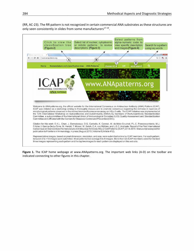
The ICAP home page provides a short introduction to the history of this initiative as well as briefly describes how the consensus on pattern classification and nomenclature was constructed (figure 1). The key toolbar shows links to the nomenclature and classification tree (figure 2), listings of designated nuclear, cytoplasmic, or mitotic patterns (figure 3), access to patterns based on the alphanumeric codes (figure 4), and a search function for patterns based on keywords. The classification tree is a summary of all the consensus patterns based on their main staining feature fitting best to the nuclear, cytoplasmic, or mitotic compartments (figure 2). All 28 ICAP patterns are shown in the classification tree as designated from AC-1 to AC-28. Boxes with amber background are recommended as competent-level reporting, whereas those with olive green background are considered for expert-level reporting. Competent-level patterns are those that should be readily recognized versus patterns that would be more challenging and distinguishable only when observers or technologists have attained the expert-level. The distinction between competent-level versus expert-level patterns is based on at least two considerations. First, clinical relevance is a major consideration to ensure that important clinical implications are recognized. Second, easily recognizable patterns should be included even when the clinical relevance is less clear at this time. The competent-level patterns are placed at the top levels starting from the left. The assignment of the different AC codes generally flows from left to right, and top to bottom. Thus, the classification tree shows 11 competent-level reportable patterns. The six competent-level reportable nuclear patterns include homogeneous (AC-1), speckled (AC-2, 4, 5), dense fine speckled (AC-2), centromere (AC-3), discrete nuclear dots (AC-6,7), and nucleolar (AC-8,9,10). Five competent-level reportable cytoplasmic patterns are fibrillar (AC-15, 16, 17), speckled (AC-18,19,20), reticular/mitochondrion-like (AC-21), polar/Golgi-like (AC-22), and rods and rings
284 Methodical Aspects and Diagnostic Strategies
(RR, AC-23). The RR pattern is not recognized in certain commercial ANA substrates as these structures are only seen consistently in slides from some manufacturers17-19.
Figure 1. The ICAP home webpage at www.ANApatterns.org. The important web links (A-D) on the toolbar are indicated connecting to other figures in this chapter.

Methodical Aspects and Diagnostic Strategies 285
Figure 2. The nomenclature and classification tree for all HEp-2 cell patterns as shown at www.ANApatterns.org. There are total of 28 ICAP patterns designated with alphanumeric AC code for each from AC-1 to AC-28. Boxes with amber background are recommended as competent-level reporting, whereas those with olive green background are considered for expert-level reporting. AC, anti-cell.

286 Methodical Aspects and Diagnostic Strategies
Figure 3 shows an example of the list of ICAP nuclear patterns. Similar lists are provided for ICAP cytoplasmic and mitotic patterns. Each pattern is shown with the AC code, name of the pattern, other common names in use (synonym), a general description, and two small IIF icons of full representative images. Selecting each pattern links to information as shown in figure 4, which also provides additional information on antigen association and disease association. In figure 4, when one of the two images is selected, the corresponding full image is shown.
Figure 3. An image of the webpage providing a partial list of the ICAP nuclear patterns. Two representative images for each pattern are shown as icons on the right column. Click each pattern to view its synonyms, antigen associations, disease associations, and detail description.

Methodical Aspects and Diagnostic Strategies 287
Figure 4. Example of information shown on the web page for the nuclear homogeneous pattern AC-1. A click of the smaller image links to the full ICAP image.
Agenda for second ICAP meeting
An aggressive agenda with several items is planned for the second ICAP meeting to be held in Dresden, Germany. It is acknowledged that the first ICAP report should be followed up with actions to ensure that the consensus is progressively adopted worldwide. In addition, it is recognized that several points in the first ICAP need further discussion. What are the practical limitations for laboratories to meet the recommended competent-level reporting? What is needed to encourage laboratories to gain ability to reach the expert-level? The www.ANApatterns.org site will contribute towards this goal. Should the ultimate target be global standardization so that everyone is capable to identify all patterns? It has been proposed that an on-line assessment tool for users of www.ANApatterns.org will help to train technicians and investigators to develop technical competence. Are there available tools ready to be adapted?
In the first ICAP report, one limitation is the lack of consideration for composite patterns as a separate category. Discussion will focus on what are the most important composite patterns and how to distinguish them effectively. ANA patterns association with diseases is listed in three of the tables in the Frontiers in Immunology ICAP report. What do clinicians want to know from the HEp-2 patterns? Can we come up with a more practical presentation for what comes after the pattern is reported?
Recommendations for standardized ANA pattern reporting will be discussed. This can be an important development to establish an international standard. To become a true international consensus, plans are needed to translate ICAP website into other languages. Appropriate discussion is needed regarding the strategy and guidelines to achieve an effective outcome.
References
Tan, E.M. Antinuclear antibodies: diagnostic markers for autoimmune diseases and probes for cell biology. Adv. Immunol. 44, 93-151 (1989).
Satoh, M., et al. Clinical implication of autoantibodies in patients with systemic rheumatic diseases. Expert review of clinical immunology 3, 721-738 (2007).
Tan, E.M., et al. Reference reagents for antinuclear antibodies. Arthritis Rheum. 31, 1331 (1988). Chan, E.K.L., et al. Autoantibody Standardization Committee in 2006. Autoimmun Rev 6, 577-580 (2007). Meroni, P.L. & Schur, P.H. ANA screening: an old test with new recommendations. Ann. Rheum. Dis. 69, 1420-
1422 (2010).

288 Methodical Aspects and Diagnostic Strategies
Agmon-Levin, N., et al. International recommendations for the assessment of autoantibodies to cellular antigens referred to as anti-nuclear antibodies. Ann. Rheum. Dis. 73, 17-23 (2014).
Meroni, P.L., Chan, E.K.L., Tincani, A., Garcia de la Torre, I. & Andrade, L.E.C. Antinuclear antibody test: when to order? Am. J. Med. 126, e17 (2013).
Dellavance, A., et al. The first Brazilian Consensus for Standardization of ANA in HEp-2 Cells. Jornal Brasileiro de Patologia e Medicina Laboratorial 38, 207-216 (2001).
Dellavance, A., et al. II. Brazilian consensus on antinuclear antibodies in HEp-2 cells. Definitions for standardization of autoantibody testing against the nucleus (ANA HEP-2), nucleolus, cytoplasm and mitotic apparatus, as well as its clinical associations. Revista brasileira de reumatologia 43, 129-140 (2003). Dellavance, A., et al. 3º Consenso Brasileiro para pesquisa de autoanticorpos em células HEp-2 (FAN). Recomendações para padronização do ensaio de pesquisa de autoanticorpos em células HEp-2, controle de qualidade e associações clínicas. Revista brasileira de reumatologia 49, 89-109 (2009). Francescantonio, P.L., et al. IV Brazilian guidelines for autoantibodies on HEp-2 cells. Rev Bras Reumatol 54, 44-50 (2014). Wiik, A.S., Hoier-Madsen, M., Forslid, J., Charles, P. & Meyrowitsch, J. Antinuclear antibodies: a contemporary nomenclature using HEp-2 cells. Journal of autoimmunity 35, 276-290 (2010). Sack, U., et al. Autoantibody detection using indirect immunofluorescence on HEp-2 cells. Annals of the New York Academy of Sciences 1173, 166-173 (2009). Carballo, O.G., et al. First Argentine consensus for standardization of antinuclear antibodies by indirect immunofluorescence–HEp-2. Acta Bioquím Clín Latinoam 46, 3-13 (2012). Mimori, T. Atlas of Antinuclear Antibodies, (Medical and Biological Laboratories Co., Ltd., Nagoya, Japan, 1999). Chan, E.K.L., et al. Report of the First International Consensus on Standardized Nomenclature of Antinuclear Antibody HEp-2 Cell Patterns (ICAP) 2014-2015. Frontiers in immunology, in press (2015). Covini, G., et al. Cytoplasmic rods and rings autoantibodies developed during pegylated interferon and ribavirin therapy in patients with chronic hepatitis C. Antivir Ther 17, 805-811 (2012). Keppeke, G.D., et al. Longitudinal study of a human drug-induced model of autoantibody to cytoplasmic rods/rings following HCV therapy with ribavirin and interferon-alpha. PLoS One 7, e45392 (2012). Stinton, L.M., Myers, R.P., Coffin, C.S. & Fritzler, M.J. Clinical associations and potential novel antigenic targets of autoantibodies directed against rods and rings in chronic hepatitis C infection. BMC gastroenterology 13, 50 (2013).