the interglobular domain of cartilage aggrecan is cleaved by
TRANSCRIPT

THE JOURNAL OF BIOLOGICAL CHEMISTRY 0 1992 by The American Society for Biochemistry and Molecular Biology, Inc. Vol. 267, No. 27, Issue of September 25, pp. 19470-19474,1992
Printed in LISA.
The Interglobular Domain of Cartilage Aggrecan Is Cleaved by PUMP, Gelatinases, and Cathepsin B*
(Received for publication, April 13, 1992)
Amanda J. Fosang$Q, Peter J. Neamell, Karena Last$, Tim E. Hardinghamll, Gillian Murphy**$$, and John A. Hamilton$ From the $Department of Medicine, University of Melbourne, Royal Melbourne Hospital, Parkville, 3050, Australia, the Whriners Hospital for Crippled Children, Tampa, Florida, 33612, the [)Kennedy Institute of Rheumatology, 6 Bute Gardens, Hammersmith. W6 70 W, United Kingdom. and the **Strangeways Research Laboratory, Worts’ Causeway, Cambridge, CBl4RN, United Kingdom
”
The action of three matrix metalloproteinases (MMPs), 72- and 95-kDa gelatinases (MMP-2 and MMP-9) and PUMP (MMP-7), and a cysteine protein- ase, cathepsin B, were investigated on aggrecan the major proteoglycan of cartilage. All the enzymes cleaved aggrecan although the activity of the 95-kDa gelatinase was very low. Specific cleavage sites were investigated following incubation with a purified ag- grecan G1-G2 domain fragment (150 kDa). Both ge- latinases produced 110-kDa G2 and 56-kDa G 1 prod- ucts by a single cleavage at an Asn-Phe bond within the interglobular domain close to the G 1 domain. This was similar to the action of stromelysin (MMP-3) (Fos- ang, A. J., Neame, P. J., Hardingham, T. E., Murphy, G., and Hamilton, J. A. (1991) J. Biol. Chem. 266, 15579-15582). Cathepsin B also produced two frag- ments from a single cleavage at a Gly-Val bond only three amino acids C-terminal to the metalloproteinase cleavage site. PUMP cleaved at the metalloproteinase Asn-Phe site, but in addition produced a low yield of a smaller G2 fragment (56 kDa) corresponding to cleav- age between Asp4“ and (human sequence), within the interglobular domain, close to the G2 do- main. The apparent difference in size between the two 6 2 fragments released by PUMP (110 and 56 kDa) was much greater than predicted from the peptide length between the cleavage sites (100 amino acids). However, keratanase digestion greatly reduced the size of the 110-kDa G2 fragment, while producing only a small reduction in size of the 56-kDa product, show- ing that there was approximately 30-40 kDa of kera- tan sulfate attached to the interglobular domain be- tween the PUMP cleavage sites. This new structural information on aggrecan may account for the previ- ously observed stiffness of the interglobular domains when viewed by rotary shadowing electron microscopy (Paulsson, M., Morgelin, M., Wiedemann, H., Beard- more-Gray, M., Dunham, D. G., Hardingham, T. E., Heinegard, D., Timpl, R., and Engel, J. (1987)
* This work was supported by program grants from the National Health and Medical Research Council (Australia) and the Victorian Health Promotion Foundation (Australia), and by the Arthritis and Rheumatism Council (United Kingdom). The costs of publication of this article were defrayed in part by the payment of page charges. This article must therefore be hereby marked “advertisement” in accordance with 18 U.S.C. Section 1734 solely to indicate this fact.
f Supported by the Arthritis Foundation of Australia. To whom correspondence should be addressed Dept. of Medicine, University of Melbourne, Royal Melbourne Hospital, Royal Parade, Parkville, 3050, Australia. Tel.: 03-344-5201; Fax: 03-347-1863.
$4 Supported by the Arthritis and Rheumatism Council, United Kingdom.
Biochem. J. 245, 763-772). These results show that in spite of a high keratan sulfate content the interglob- ular domain provides important sites for cleavage by different proteinases, including several members of the matrix metalloproteinase family.
Aggrecan is the major proteoglycan in cartilage. I t is present a t high concentration and is responsible for the compressive resilience of the tissue. One of the mechanisms for holding aggrecan within the cartilage matrix is the formation of high molecular weight aggregates in which up to a hundred or more proteoglycan monomers are bound by their G1 domains to a hyaluronan chain. Investigations of aggrecan turnover have shown that large fragments are slowly released from the tissues (l), which have lost their ability to bind to hyaluronan, suggesting a cleavage close to the G1 domain (2-5). We showed previously (6) that the metalloproteinase stromelysin makes a single cleavage between the aggrecan G1 and G2 domains at an Asn-Phe bond. Other studies have identified in interleukin-l-stimulated degradation, a different cleavage site a t a Glu-Ala bond 30 amino acids C-terminal to the stromelysin site (7). The enzyme responsible for this cleavage, “aggrecanase,” is as yet unidentified. I t is significant that aggrecan fragments corresponding to aggrecanase action as well as the stromelysin action have been detected in extracts of human articular cartilage (8). Both are therefore active in uiuo, but which enzymes have a major role in normal turnover and whether they differ from those most active in cartilage pathology remains to be determined.
Several classes of proteinases have been identified in car- tilage including the metalloproteinases which are secreted enzymes active at acid and neutral pH. The expression of stromelysin and collagenase (both metalloproteinases) by chondrocytes and its modulation by inflammatory mediators such as IL-1’ have been extensively studied (9-14). The 72- and 95-kDa gelatinases have also been identified in cartilage extracts, and the latter is induced by IL-1 and tumor necrosis factor-a in chondrocytes (15, 16). Cathepsin B, a cysteine proteinase, may also be active in cartilage. It has been iden- tified in rheumatoid arthritic synovium (17) and in synovial fluids from rheumatoid arthritis and osteoarthritic patients (18). Although cysteine proteinases have pH optima at acid pH, they also have some activity close to neutral pH (17, 19).
Which proteinases are active in normal turnover of aggre- can and which are responsible for the enhanced rates of
‘The abbreviations used are: IL-1, interleukin-1, HPLC, high performance liquid chromatography.
19470
PUMP, Gelutinases, and Cathepsin B Cleave Aggrecan 19471
degradation induced by IL-1 and tumor necrosis factor-a remains unknown. It is therefore important to investigate the actions of physiologically relevant enzymes such as the gela- tinases and cathepsin B. We have also investigated PUMP, as this is closely related to the gelatinases and provides further information on what determines substrate specificity among the metalloproteinases.
EXPERIMENTAL PROCEDURES
Materials-Na'261 (IMS 30), Hyperfilm, and enhanced chemilu- minescence reagents were from Amersham (Australia). Trypsin (di- phenylcarbamoyl chloride treated) (EC 3.4.21.4), soybean trypsin inhibitor, 4-aminophenylmercuric acetate, 1,lO-phenanthroline, 3- dimethylaminopropionitrile, 6-aminohexanoic acid, phenylmethane- sulfonyl fluoride, benzamidine hydrochloride, 7-amino-4-methylcou- marin, and swine anti-rabbit-horseradish peroxidase were from Sigma. Agarose type HSC was from PS Park Scientific (Northamp- ton, United Kingdom (U. K.)). A Bio-Si1 SEC-400 HPLC column was from Bio-Rad (Sydney, Australia). Human umbilical chord hyalu- ronan was from RDH. Immobilon was from Millipore-Waters (Syd- ney, Australia). Keratanase (Pseudomonos sp) was from ICN Biomed- icals Australasia (New South Wales, Australia). 2-Phe-Arg-NHMec was from Bachem Feinchemikalien (Bubendorf, Switzerland). All other reagents were of analytical grade except guanidine HCI (Sigma) which was purified with activated charcoal and diatomaceous earth (Sigma).
Preparation of Cartilage Proteoglycan Aggregates, Proteoglycan Globular Domains, and Rabbit Antisera-Proteoglycan aggregates ex- tracted from pig laryngeal cartilage in 4 M guanidinium hydrochloride, 50 mM sodium acetate buffer, pH 5.8, containing the proteinase inhibitors disodium EDTA, 6-aminohexanoic acid, benzamidine hy- drochloride, and phenylmethanesulfonyl fluoride were purified on a CsCl density gradient (20). Proteoglycan aggregates from human (43- year-old male) were extracted in the same buffer and purified as described previously (21). Proteoglycan G I G 2 fragment was isolated from pig proteoglycan aggregates by mild trypsin digestion (22). The preparation of rabbit antisera against pig G1 and pig G2 core protein domains has been described elsewhere (22, 23).
Preparation and Activation of Enzymes-Recombinant human proPUMP cDNA (24) (a generous gift of Dr. Richard Breathnach) was expressed following transfection in a pEE12 vector into NSO (nonsecreting) mouse myeloma cells. Recombinant proPUMP was purified from culture medium by binding to S-Sepharose and elution with 200 mM sodium chloride followed by gel chromatography on a column of S-200 (25). Human 95- and 72-kDa progelatinases were prepared from culture media conditioned by U937 cells and human gingival fibroblasts, respectively (26). ProPUMP and progelatinases were activated by incubation for 1 h a t 37 "C with 2 mM 4-amino- phenylmercuric acetate, and the resulting activities were as described previously (25). Recombinant rat cathepsin B, expressed in Saccha- romyces cereuisiae and purified by chromatography on DEAE-cellu- lose and Sephadex (3-75 (27) was a generous gift of Dr. John Mort, Montreal, Canada. The activity of the cathepsin B, assayed with Z- Phe-Arg-NHMec as substrate at 30 "C and pH 5.5 (28) was 7.58 milliunits/mg.
Enzyme Digestions-Metalloproteinase digestions were carried out in buffer containing 10 mM calcium chloride, 100 mM sodium chloride, 50 mM Tris-HCI, pH 7.5, a t 37 "C. The digests were stopped by the addition of EDTA and 1,lO-phenanthroline (10 and 2 mM final concentration, respectively). Cathepsin B digests were done in buffer containing 0.2 M sodium chloride, 1 mM EDTA, 10 mM dithiothreitol, 0.25 M sodium acetate, pH 5.5, a t 37 "C. The digests were stopped by boiling for 5 min. Keratanase digestion of G1-G2 and G1-G2 frag- ments was done in 50 mM-Tris-acetate buffer, pH 7.5, at 37 "C for 18 h with 0.02 units of keratanase, 2 pg of substrate/60 pl.
Gel Electrophoresis and N-terminal Sequencing"Gl-G2 digest products were analyzed on 5% sodium dodecyl sulfate-polyacrylamide gels (29), and protein bands were visualized by silver staining (30). For immunolocalization experiments, proteins were transferred onto Immobilon membranes, and G1 and G2 epitopes were identified using specific rabbit antisera, a swine-anti-rahbit antibody conjugated to horseradish peroxidase, followed by enhanced chemiluminescence using luminol substrate. Composite gels containing 1.2% polyacryl- amide and 0.6% agarose were electrophoresed in a dissociative buffer containing 4 M urea (31). After electrophoresis proteoglycans were visualised by Toluidine Blue staining. Iodination of G I 4 2 fragments
and localization of digest product9 on gels were as described previously (22, 32). N-terminal sequencing of isolated G2 products was as described by Sandy et al. (33).
RESULTS
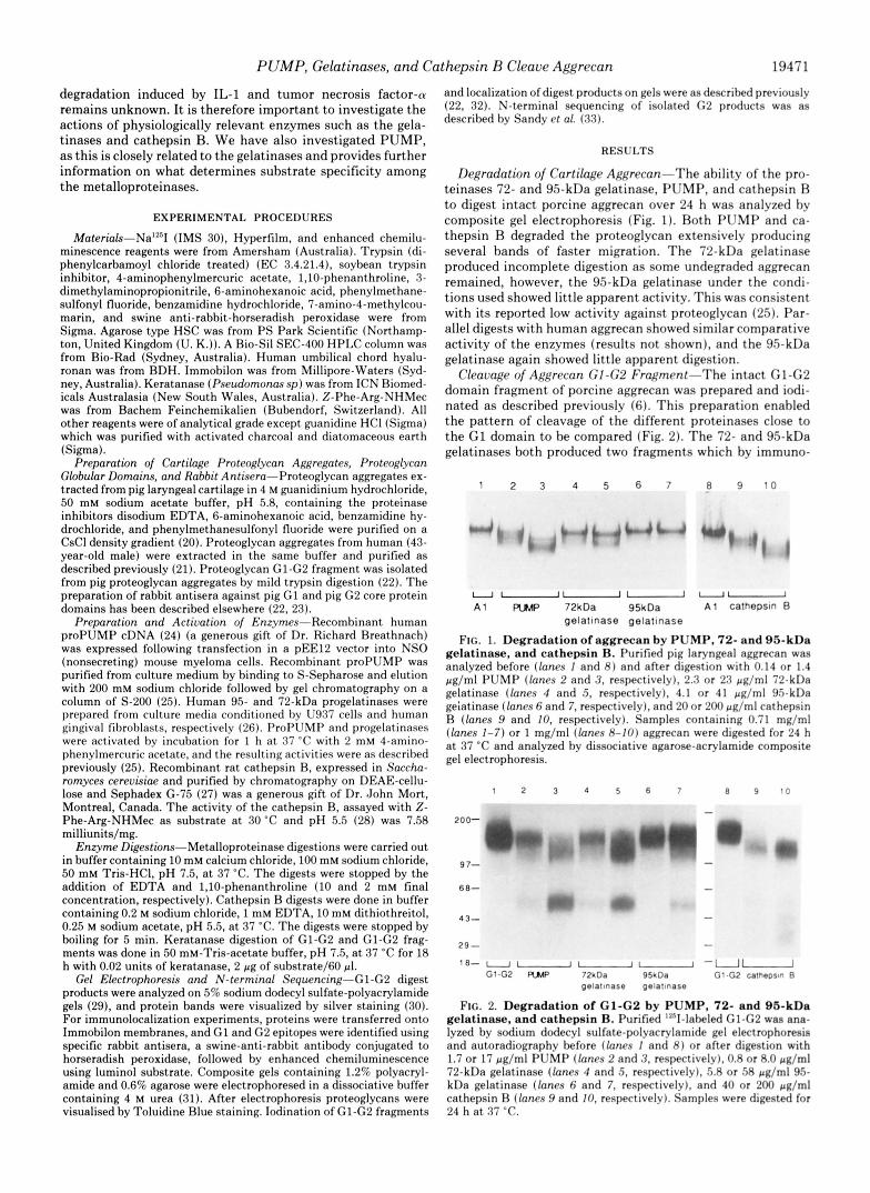
Degradation of Cartilage Aggrecan-The ability of the pro- teinases 72- and 95-kDa gelatinase, PUMP, and cathepsin B to digest intact porcine aggrecan over 24 h was analyzed by composite gel electrophoresis (Fig. 1). Both PUMP and ca- thepsin B degraded the proteoglycan extensively producing several bands of faster migration. The 72-kDa gelatinase produced incomplete digestion as some undegraded aggrecan remained, however, the 95-kDa gelatinase under the condi- tions used showed little apparent activity. This was consistent with its reported low activity against proteoglycan (25). Par- allel digests with human aggrecan showed similar comparative activity of the enzymes (results not shown), and the 95-kDa gelatinase again showed little apparent digestion.
Cleavage of Aggrecan G I 4 2 Fragment-The intact G1-G2 domain fragment of porcine aggrecan was prepared and iodi- nated as described previously (6). This preparation enabled the pattern of cleavage of the different proteinases close to the G1 domain to be compared (Fig. 2). The 72- and 95-kDa gelatinases both produced two fragments which by immuno-
1 2 3 4 5 6 7 8 9 1 0
U " U U U
A 1 RMP 72kDa 95kDa A 1 cathepsm B gelatmase gelatlnase
FIG. 1. Degradation of aggrecan by PUMP, 72- and 95-kDa gelatinase, and cathepsin R. Purified pig laryngeal aggrecan was analyzed before (lanes I and H ) and after digestion with 0.14 or 1.4 pg/ml PUMP (lanes 2 and 3, respectively). 2.3 or 23 pg/ml 72-kDa gelatinase (lanes 4 and 5, respectively), 4.1 or 41 pg/ml 95-kDa geiatinase (lanes 6 and 7, respectively). and 20 or 200 pg/ml cathepsin R (lanes 9 and IO, respectively). Samples containing 0.71 mg/ml (lanes 1-7) or 1 mg/ml (lanes 8-10) aggrecan were digested for 24 h a t 37 "C and analyzed by dissociative agarose-acrylamide composite gel electrophoresis.
1 2 3 4 5 6 7 8 9 1 0
200- m 9 7-
6 8-
43-
29- - 18-
G1-G2 RMP 72kDa 95kDa Gl-G2 celhepstn B ""UU
pelatmese pelstmase
FIG. 2. Degradation of G1-G2 by PUMP, 72- and 95-kDa gelatinase, and cathepsin B. Purified 12'II-labeled G1-G2 was ana- lyzed by sodium dodecyl sulfate-polyacrylamide gel electrophoresis and autoradiography before (lanes I and 8) or after digestion with 1.7 or 17 pg/ml PUMP (lanes 2 and 3, respectively), 0.8 or 8.0 pg/ml 72-kDa gelatinase (lanes 4 and 5, respectively). 5.8 or SR pg/ml 95- kDa gelatinase (lanes 6 and 7, respectively), and 40 or 200 pg/ml cathepsin R (lanes 9 and IO. respectively). Samples were digested for 24 h a t 37 "C.

19472 PUMP, Gelatinases, and Cathepsin B Cleave Aggrecan
blotting (22) were identified as a 110-kDa G2 fragment and a 56-kDa G1 fragment. PUMP also produced two major bands. The 110-kDa band was similar to that produced by the gela- tinase, but the 56-kDa band was broader. Immunolocalization experiments showed both G1 and G2 domain fragments were present in the 56-kDa band, whereas the 110-kDa band con- tained G2 only, as with the gelatinases (Fig. 3). These results were compatible with PUMP cleaving a t two sites, one similar to the gelatinase cleavage site, and the second cleaving some of the 110-kDa G2 fragment to release a 56-kDa G2 fragment.
Cathepsin B produced several cleavage products from the G1-G2 preparation (Fig. 2, lanes 9 and 10). A major 110-kDa band was again identified as containing G2 and no G1 domain. There were several diffuse bands of 56 kDa and less, which probably represented degraded G1 domains, although they were only poorly detected with anti-G1 antibodies. The re- ducing conditions used to promote cathepsin B activity are likely to have reduced some of the disulfide bonds in the G1 domain and this may have made more sites available for cathepsin B attack. Under reducing conditions we have pre- viously found that stromelysin cleaves the G1 domain at sites that are not susceptible under non-reducing conditions.
Isolation and N-terminal Sequencing of Gelatinme and Ca- thepsin B-derived G2 Fragments-G2 fragments derived from gelatinase and cathepsin B digests were isolated by size exclu- sion chromatography on HPLC after mixing with hyaluronan to bind all G1 fragments and elute them in the void of the column (6, 22). For each digest, only one included peak corresponding to a G2 fragment was detected. Sequence analy- sis gave unambiguous results for both gelatinase digests. The G2 fragment had an N-terminal sequence Phe-Phe-Gly-Val. This showed that both gelatinases cleaved at the Asn-Phe site cleaved by stromelysins and shows this to be a site favored by several metalloproteinases. The G2 fragment produced by cathepsin B digestions had a different N-terminal sequence Val-Gly-Gly-Glu. This corresponds to a sequence three amino acids C-terminal to the gelatinase cleavage site. The proximity to the metalloproteinase site explains the similarity in size of this major G2 product of cathepsin B.
Isolation and N-terminal Sequencing of PUMP Digest Prod-
. . . .
. . . . . . . . . . . . . . . . . . . . . . . . . . . . . . . . . . . . . . . . . . . . . .
........ Undigested GI-G2 -PUMP digested G l - G 2
Dislance mbraled
FIG. 3. Immunolocalization of G 2 products following diges- tion of G1-G2 with PUMP. Purified G1-G2 (1.4 pg) was digested with 0.2 pg of PUMP in a total volume of 30 pl for 24 h at 37 "C. Undigested (dotted line) and digested (solid l i n e ) G1-G2 were electro- phoresed on a 5% gel and transferred to immobilon membranes for immunodetection with specific anti-G2 antisera. Bands were visual- ized on x-ray film by enhanced chemiluminescence using luminol, and the film was scanned on a laser densitometer (Pharmacia, Aus- tralia).
ucts-After mixing with hyaluronan and running on HPLC as described above, two included peaks of PUMP digests were identified (Fig. 4). Isolation and N-terminal sequencing of the larger 110-kDa fragment showed the same sequence as the 110-kDa G2 fragment produced by the gelatinases. The smaller 56-kDa peak also contained G2 but did not usually represent more than 20-30% of the total G2 detected. Se- quence analysis showed two separate products to be present in the 56-kDa peak. One had an N-terminal sequence identical to the 110-kDa fragment (Phe-Phe-Gly-Val) whereas the other had an N-terminal sequence corresponding to a cleavage between Asp-Leu, where the aspartate represents residue 441 and 444 in the human and rat core protein sequences, respec- tively (34,35) (Fig. 5). The latter sequence corresponded to a site in the interglobular domain close to G2. These results showed that PUMP cleaved with greatest activity at the main metalloproteinase site near G1, but also cleaved less fre- quently a t a second site in the interglobular domain much closer to G2.
The second PUMP cleavage site cleaved the 110-kDa G2 product into two fragments of equal size (-56 kDa). This was surprising as the sequence of the interglobular domain be- tween the two PUMP cleavage sites is only about 100 amino acids (-10 kDa). The explanation for this was provided by keratanase digestions, which reduced the apparent size of the 110-kDa G2 fragment to 70 kDa, but only decreased the size of the 56-kDa G2 fragment by -10 kDa. A large proportion of the total keratan sulfate (-30-40 kDa) was therefore at- tached to the interglobular domain between the two PUMP cleavage sites (Fig. 6).
DISCUSSION
Our present results provide further evidence that the inter- globular domain is a key site for the attack of aggrecan by proteinases. Cleavage in this region is important as it releases a major proteoglycan fragment from the G1 domain which binds it to aggregates. The results show that the gelatinases (MMP-2 and -9) and PUMP (MMP-7) cleave at the Asn-Phe site close to the G1 domain. We previously showed that stromelysins cleaved at this same site (6). Cathepsin B was also found to cleave very close to this site a t a Gly-Val bond. Our results show that these metalloproteinases and cathepsin
24 32 40 48 16
TIME (minutes)
FIG. 4. Isolation of G 2 and interglobular domain fragments produced by PUMP digestion of Gl-G2. Purified G1-G2 (200 pg) was digested with (0.4 pg) PUMP in a total volume of 40 pl for 24 h at 37 "C. The digestion products were mixed with 50 pg of hyaluronan overnight at 4 "C, and the products were separated by size exclusion chromatography on a BioSil SEC 400 HPLC column. The column was eluted in buffer containing 0.1 M sodium sulfate, 0.123 M sodium chloride, 0.1 M sodium dihydrogen orthophosphate, pH 6.8, at 0.3 ml/ min, and 0.4-ml fractions were collected. The inset shows aliquots of column fractions analyzed on 5% sodium dodecyl sulfate gels and visualized by silver stain.

PUMP, Gelatinases, and Cathepsin B Cleave Aggrecan 19473
95 kDa gelatinase 0 Threonine &I Serine 0 Asparagine
the G1, G2, and G3 globular domains, the interglobular domain (ZGD) between G1 and G2, and the extended keratan sulfate ( K S ) and FIG. 5. Cleavage sites within the interglobular domain of aggrecan. a, schematic representation of aggrecan core protein showing
chondroitin sulfate (CS) attachment region between G2 and G3. b, schematic representation of the amino acid sequence in human aggrecan IGD showing the cleavage sites identified for cathepsin B, gelatinases, PUMP, stromelysin (6, a), leucocyte elastase (43), and aggrecanase, a cleavage site identified following IL-1 stimulation of articular cartilage (7). Filled, hatched, and blank circles designate potential glycosylation sites and represent threonine, serine, and asparagine residues, respectively. The horizontal arrows mark the interglobular domain boundaries (35,44).
(a) G1 and G2 fragments produced by stromelysin, gelatinases and PUMP
n
(b) Minor G2 and IGD fragmenfs produced by PUMP
Interglobular domain (approx 56 kDa)
G2 domain (56 kDa)
FIG. 6. Fragments produced by metalloproteinase digestion of Gl -G2 . Panel a, stromelysin, gelatinases, and PUMP cleave aggrecan Gl-G2, yielding a 56-kDa G1 fragment and a 110-kDa G2 fragment. After keratanase treatment, the size of the G2 fragment was reduced to -70 kDa. Panel b, PUMP made an additional cleavage in a small proportion (-20%) of 110-kDa G2 fragments, yielding a G2 fragment with mass 56 kDa, and an interglobular domain fragment of similar electophoretic mobility. After keratanase treatment, the mass of the small G2 was reduced to only -46 kDa. Thus, the bulk of the keratan sulfate chains present on the major 110-kDa G2 product are located on the 100 amino acid peptide derived from the interglobular domain.
B are not responsible for the aggrecanase cleavage site iden- tified in IL-1-stimulated cartilage degradation (7) (Fig. 5 ) . This implies that there is another unidentified enzyme in- duced by IL-1, which must be particularly active in cleaving the interglobular domain of aggrecan. However, there may still be participation of metalloproteinases in aggrecan turn- over, as some G1 fragments in human cartilage have been shown to contain the C terminus of the metalloproteinase site
(8). Which of the metalloproteinases is responsible for cleav- age at the Asn-Phe site i n vivo is difficult to predict. Several of the metalloproteinases with the exception of PUMP are produced by chondrocytes. In culture the 72-kDa gelatinase is constitutively expressed, whereas the 95-kDa gelatinase (15, 16, 36), stromelysin (11-14), and collagenase (9, 10) are induced by cytokines. However, chondrocytes appear to be- have differently in cartilage where separate expression of individual metalloproteinases has been observed, such as in the developing growth plate (37). Evidence in human articular cartilage suggested stromelysin to be most abundantly ex- pressed and to increase with age, whereas gelatinase and collagenase were less abundant (38, 39). There are many factors that control metalloproteinase activity i n vivo and the extent to which they are activated and how rapidly they become inhibited are difficult to determine experimentally, but are of key importance. The generation of plasmin by urokinase-type plasminogen activator is thought to be the key to stromelysin, collagenase, and 95-kDa gelatinase activation, while 72-kDa gelatinase may be regulated by a membrane activator (40).' A more complete analysis of all these param- eters is therefore necessary before it can be determined which metalloproteinases are most active in normal and pathological situations in cartilage and how their activities complement those of other proteinases.
Very little is known about what determines substrate spec- ificity among the metalloproteinases, and it is interesting that four different metalloproteinases with different substrate specificities all cleaved at the same Asn-Phe site in the interglobular domain. This included PUMP which differs from the others in lacking the large C-terminal hemopexin domain. In fibroblast collagenase the hemopexin domain has been shown to be important for binding to native triple helical collagen, and collagenase activity, but not caseinase activity, was lost on its removal (41). The results with PUMP show
S. Atkinson and G. Murphy unpublished results.

19474 PUMP, Gelatinases, and Cathepsin B Cleave Aggrecan
that the hemopexin domain is not essential for determining activity at the Asn-Phe bond in the interglobular domain. PUMP also showed activity at a site not shared with the other metalloproteinases. This may reflect a less restricted substrate specificity of this metalloproteinase, or possibly greater access of the smaller enzyme lacking the hemopexin domain to a site inaccessible to the larger metalloproteinases.
The interglobular domain appears as an extended and stiff- ened segment 30 f 5 nm long by rotary shadowing electron microscopy (42). This is shorter than the calculated fully extended sequence (-45 nm) and secondary structure predic- tion suggests it to be predominantly p sheet or random Glycosylation is an important factor that may influence the protein configuration of the interglobular domain, its appar- ent stiffness, and the susceptibility to proteinases. There are 14 threonine, 4 serine, and 2 asparagine residues in the human sequence that may be glycosylated. Our analysis of the second PUMP cleavage site provides new evidence that up to 40 kDa of keratan sulfate is attached to the interglobular domain between the two PUMP sites (Fig. 6). This localizes most of the keratan sulfate attached to the G1-G2 region of aggrecan to a short 100 amino acid sequence separating the globular G1 and G2 domains. The keratan sulfate is likely to be in five to eight chains each 10-15 nm long, and they may make a major contribution to the apparent stiffness of this segment of the protein core. There may also be other 0-linked or N - linked oligosaccharides. The precise distribution of these car- bohydrate substitutions is not yet known, but amino acid sequencing of peptides from the interglobular domain has shown blank cycles at the position of several threonine and serine residues which suggests that there is carbohydrate attached to them.4
The sites of proteolytic cleavage reported within the inter- globular domain appear to be grouped in short sequences that are free of sites for carbohydrate attachment and also of lower proline content (Fig. 5). However, variable glycosylation may influence the access of enzymes to some sites and the rather low efficiency of cleavage at the second PUMP site may be caused by glycosylation on some molecules at the Thr-Ser dipeptide (human residues 338-339) close to this cleavage site. The glycosylation of the interglobular domain may therefore under some circumstances influence the proteoglycans sus- ceptibility to the proteolytic enzymes involved in its turnover.
Acknowledgments-We thank Dr. S. J. Perkins, Dept. of Biochem- istry, Royal Free Hospital, School of Medicine, London, U. K., for an assessment of the secondary structure of the interglobular domain. We thank Mark Cockett and Andrew Docherty for help with the expression system and Robin Ward for preparation of the progelati- nases. ~
S. J. Perkins, personal communication. P. J. Neame, unpublished results.
REFERENCES
2. Sandy, J. D., Brown, H. L. G., and Lowther, D. A. (1978) Biochim. Biophys. 1. Tyler, J. A. (1985) Biochem. J. 225,493-507
3. Ratcliffe, A., Tyler, J. A., and Hardingham, T. E. (1986) Biochem. J. 238 ,
4. Campbell, I. K., Roughley, P. J., and Mort, J. S. (1986) Biochem. J. 237 ,
5. Poole, A. R., Witter J., Roberts, N., Roughley, P. J., Wehber, C., and
6. Fosang A. J. Neame P. J. Hardingham, T. E. Murphy, G., and Hamilton,
7. Sandy, J. D., Neame, P. J., Boynton, R. E., and Flannery, C. R. (1991) J.
8. Flannery, C. R., Lark, M. W., and Sandy, J. D. (1992) J. Biol. Chem. 2 6 7 ,
9. Trechsel, U., Dew, G., Murphy, G., and Reynolds, J. J. (1982) Biochim.
10. Saklatvala, J., Pilsworth, L. M. C., Sarsfield S. J., Gavrilovic, J., and
11. Murphy, G., Hembry, R. M., and Reynolds, J. J. (1986) Collagen Rel. Res.
12. Frisch, S. M., and Ruley, H. E. (1987) J. Biol. Chem. 262 , 16300-16304 13. Hasty, K. A,, Reife, R. A., Kang, A. H., and Stuart, J. M. (1990) Arthritis
14. Case, J. P., Lafyatis, R., Kumkumian, K., Remmers, E. F., and Wilder, R.
15. Lefebvre, V., Peeters-Joris, C., and Vaes, G. (1991) Biochim. Biophys. Acta
16. Ogata, Y., Enghild, J. J., and Nagase, H. (1992) J. Biol. Chem. 267 , 3581-
17. Mort, J. S., Recklies, A. D., and Poole, A. R. (1984) Arthritis Rheum. 2 7 ,
18. Maciewicz, R. A,, and Etherington, D. J. (1988) Biochem. SOC. Trans. 16,
19. Maciewicz, R. A,, Wotton, S. F., Etherington, D. J., and Duance, V. C.
Acta 543,536-544
571-580
117-122
Campbell, I. K. (19b7) J. Rheumatol. 14,80-82
J. A.’(199i) J. Bioi Cheh. 266,15579-1558’2
Bwl. Chem. 266,8683-8685
1008-1014
Biophys. Acta 720,364-370
Heath, J. K. (1984) Biochem. J. 244,461-466
6,351-364
Rheum 33,388-397
L. (1990) J. Immunol. 145 , 3755-3761
1094,8-18
3584
509-515
812-813
(1990) FEES Lett. 2 6 9 . 189-193 20. Hardingham, T. E. (1979)’Biochem. J. 177,237-247 21. Bayliss, M. T. and Roughley, P. J. (1985) Biochem. J. 232,111-117 22. Fosang A. J. Lnd Hardin ham T. E (1989) Biochem. J. 261,801-809 23. Ratclifie, A,, ’and Hardin&am,’T. E.’(1983) Biochem. J. 213 , 371-378 24. Muller, D., Quantin, B., Gesnel, M. C., Millon-Collard, R., Abecassis,
and Breathnach. R. (1988) Biochem. J. 253.187-192 J.,
25. Murph G., Cockett, M . I.,’ Ward, R. V.: and Docheky, A. J. P. (1991) Bloc&m. J. 2 7 7 , 277-279
26. Ward, R. V., Hembry, R. M., Reynolds, J. J., and Murphy, G. (1991) Biochem. J. 278,179-187
27. Lee, X., Ahmed, F. R., Hirama, T., Huber, C. P., Rose, D. R., To, R.,
5951 Hasnain, S., Tam, A,, and Mort, J. S. (1990) J. Biol. Chem. 265 , 5950-
28. Bafreit, A. J., and Kirschke, H. (1981) Methods Enzymol. 80, 535;561 29. Fairbanks, G., Steck, T. L., and Wallach, D. F. H. (1971) Biochemlstry 10,
30. Morrissey, J. H. (1981) Anal. Biochem. 117,307-310 31. Carnev. S. L.. Bavliss. M. T.. Collier. J. M.. and Muir, H. (1986) Anal.
2606-2616
Bioihem. 156 , 38-44 32. Fosang, A. J., and Hardingham, T. E. (1991) Biochem. J. 273,369-373 33. Sandy, J. D., Flannery, C. R. Boynton, R. E., and Neame, P. J. (1990) J.
34. Doege, K. J., Sasaki, M., Horigan, E., Hassell, J. R., and Yamada, Y. (1987) Biol. Chem. 2 6 5 , 21108-21’113
35. Doege, K. J., Sasaki, M., Kimura, T., and Yamada, Y. (1991) J. B i d . Chem. J. Biol. Chem. 2 6 2 , 17757-17767
36. Van Ranst, M., Norga, K., Masure, S., Proost, P., Vandekerckhove, F., 266,894-902
Auwerx, J., Van Damme, J., and Opdenakker, G. (1991) Cytokine 3,231- 239
37. Brown, C. C. Hemhry, R. M., and Reynolds, J. J. (1992) J. Bone Joint Surg. 71-A, 580-593
38. Allan, J. A., Hembry, R. M., Angal, S., Reynolds, J. J., and Murphy, G. (1991) J. Cell Sci. 9 9 , 789-795
39. Nguyen, Q., Mort, J. S., and Roughley, P. J. (1992) J. Clin. Inuest. 8 9 , 1189-1197
40. Ward, R. V., Atkinson, S. J., Slocombe, P. M., Docherty, A. J. P., Reynolds, J. J. and Mu hy G. (1991) Biochim. Biophys. Acta 1079 , 242-246
41. Clark, \. M., an?Ca&ston, T. E. (1989) Biochem. J. 263,201-206 42. Paulsson, M., Mor elin, M., Wiedemann, H., Beardmore-Gray, M., Dun-
ham D. G. Harfin ham T. E. Heinegard, D., Timpl, R., and Engel, J. (198:) Biodhem. J . 8 4 5 , F63-7i2
43. Mok, M. T., Ilic, M. Z., Handley, C. J., and Robinson, H. C. (1992) Arch. Biochem. Bio hys 292,442-447
44. Perkins, S. J., fiealis, A. S., Dudhia, J., and Hardingham, T. E. (1989) J. Mol. Biol. 2 0 6 , 737-754
. . .