aggrecan degradation in human cartilage
TRANSCRIPT

Aggrecan Degradation in Human Cartilage
93
J. Clin. Invest.© The American Society for Clinical Investigation, Inc.0021-9738/97/07/0093/14 $2.00Volume 100, Number 1, July 1997, 93–106
Aggrecan Degradation in Human Cartilage
Evidence for both Matrix Metalloproteinase and Aggrecanase Activity in Normal, Osteoarthritic,and Rheumatoid Joints
Michael W. Lark,* Ellen K. Bayne,* Jamie Flanagan,* Coral F. Harper,* Lori A. Hoerrner,* Nancy I. Hutchinson,* Irwin I. Singer,* Susan A. Donatelli,* Jeffrey R. Weidner,* Hollis R. Williams,* Richard A. Mumford,* and L. Stefan Lohmander
‡
*
Department of Inflammation Research, Merck Research Laboratories, Rahway, New Jersey 07065; and
‡
Department of Orthopedics, University Hospital, 22185 Lund, Sweden
Abstract
To examine the activity of matrix metalloproteinases (MMPs)
and aggrecanase in control and diseased human articular
cartilage, metabolic fragments of aggrecan were detected
with monospecific antipeptide antibodies. The distribution
and quantity of MMP-generated aggrecan G1 fragments
terminating in VDIPEN
341
were compared with the distri-
bution of aggrecanase-generated G1 fragments terminating
in NITEGE
373
. Both types of G1 fragments were isolated
from osteoarthritic cartilage. The sizes were consistent with
a single enzymatic cleavage in the interglobular domain re-
gion, with no further proteolytic processing of these frag-
ments. Both neoepitopes were also detected by immunohis-
tochemistry in articular cartilage from patients undergoing
joint replacement for osteoarthritis (OA), rheumatoid arthritis
(RA), and in cartilage from adults with no known joint
disease.
In control specimens, the staining intensity for both G1
fragments increased with age, with little staining in carti-
lage from 22-wk-old fetal samples. There was also an in-
crease with age in the extracted amount of MMP-generated
neoepitope in relation to both aggrecan and collagen con-
tent, confirming the immunohistochemical results. After the
age of 20–30 yr this relationship remained at a steady state.
The staining for the MMP-generated epitope was most
marked in control cartilage exhibiting histological signs of
damage, whereas intense staining for the aggrecanase-gen-
erated fragment was often noted in adult cartilage lacking
overt histological damage
.
Intense staining for both neo-
epitopes appeared in the more severely fibrillated, superfi-
cial region of the tissue.
Intense immunostaining for both VDIPEN- and NITEGE-
neoepitopes was also detected in joint cartilage from pa-
tients with OA or RA. Cartilage in these specimens was sig-
nificantly more degraded and high levels of staining for
both epitopes was always seen in areas with extensive carti-
lage damage. The levels of extracted VDIPEN neoepitope
relative to collagen or aggrecan in both OA and RA samples
were similar to those seen in age-matched control speci-
mens.
Immunostaining for both types of aggrecan fragments
was seen surrounding the cells but also further removed in
the interterritorial matrix. In some regions of the tissue,
both neoepitopes were found while in others only one was
detected. Thus, generation and/or turnover of these specific
catabolic aggrecan fragments is not necessarily coordinated.
Our results are consistent with the presence in both normal
and arthritic joint cartilage of proteolytic activity against
aggrecan based on both classical MMPs and “aggrecanase.”
(
J. Clin. Invest.
1997. 100:93–106.) Key words: aggrecan
•
matrix metalloproteinases
•
aggrecanase
•
cartilage
•
os-
teoarthritis
Introduction
The destruction of joint cartilage is of central importance inhuman arthritic disease. Advances are now being made in ourunderstanding of the events which initiate joint diseases, aswell as of the processes involved in the degradation of articularcartilage in osteoarthritis (OA)
1
and RA. For example, in pa-tients with RA and OA, there is clear evidence for increasedexpression and synthesis of enzymes, including matrix metallo-proteinases (MMPs), in synovial tissue and cartilage, and of in-creased concentrations of the same proteases in joint fluids(1–9). These enzymes are synthesized as proenzymes and themajority of the enzyme that has been described in cartilage orjoint fluid is inactive proenzyme (1, 5). This has left us with thequestion as to what role, if any, these enzymes play in cartilagematrix degradation in joint disease.
One of the initial approaches taken to address whetherMMPs have the capacity to degrade matrix components in situ,was to identify and characterize specific enzyme-generatedcleavage products in vitro (10–14). Cartilage extracts were pre-pared and the cleavage sites identified using NH
2
-terminal se-quence analysis. Using this approach, evidence for MMP-gen-erated degradation of link protein (10) and aggrecan (11) havebeen found. To further define the identity, distribution andquantity of such fragments, monospecific antibodies were pro-duced that recognize enzyme-generated neoepitopes that arenot present in the native, uncleaved molecules. The MMP-gen-erated neoepitope in link protein was thus detected in youngcartilage, and with age and joint disease there was a reductionin this epitope (15). These data suggested that MMP degrada-tion of link protein may be involved in normal matrix turnover
Michael W. Lark and Ellen K. Bayne contributed equally to this work.Address correspondence to Michael W. Lark, SmithKline Beecham
Pharmaceuticals, 709 Swedeland Road, uw2109, King of Prussia, PA19406. Phone: 610-270-4914; FAX: 610-270-5598; E-mail: [email protected]
Received for publication 2 December 1996 and accepted in revised
form 8 April 1997.
1.
Abbreviations used in this paper:
G1 fragments, the amino-termi-nal, hyaluronan-binding globular domain of aggrecan; GuHCl, guani-dine hydrochloride; HA, hyaluronan; MMP, matrix metalloprotein-ase; OA, osteoarthritis.

94
Lark et al.
and that further catabolic processes may be involved in linkprotein degradation with age.
More recently a monoclonal antibody was generated againsta neoepitope in type II collagen (16). This antibody does notdetect native intact type II collagen but does detect cleavedcollagen once the triple helix has unwound. With the antibody,it was shown that there is a regional elevation in this neo-epitope that correlates with osteoarthritic disease severity asdefined by histopathological score (16).
It has been reported that aggrecan G1 fragments increasein cartilage with age (17, 18). Stromelysin-1 (MMP-3), as wellas other MMPs, cleave aggrecan in the interglobular domainbetween Asn
341
and Phe
342
to generate a G1 fragment with theCOOH terminus VDIPEN
341
(11–13). This fragment has beenisolated and identified by NH
2
-terminal sequence analysisfrom human OA cartilage (11). A second proteolytic activityidentified as “aggrecanase” also cleaves aggrecan in the inter-globular domain, but between Glu
373
and Ala
374
(19–24), gen-erating a G1 fragment with a COOH terminus of NITEGE
373
(Please refer to Fig. 9 for a schematic representation of theCOOH and NH
2
termini thus generated in the aggrecan inter-globular domain).
Large aggrecan fragments which have an NH
2
terminus of
374
ARGSV, consistent with aggrecanase cleavage, have beendetected in human joint fluids by NH
2
-terminal sequence anal-ysis (25, 26). However, no G1 fragments terminating inNITEGE
373
have been identified in human cartilage to date. Incontrast, it has been shown that the NITEGE-positive G1 frag-ment does accumulate in the culture medium of both retinoicacid and IL-1–stimulated rat chondrosarcoma cells withoutany accumulation of the VDIPEN-positive fragment (23). Thisindicates that aggrecanase-mediated aggrecan cleavage cantake place independent of MMP-mediated degradation. Theonly isolated enzyme which has been shown to cleave betweenGlu
373
and Ala
374
is neutrophil collagenase (MMP-8) (27). How-ever, this enzyme preferentially cleaves at the MMP site, be-tween Asn
341
and Phe
342
.To define the distribution and quantity of these aggre-
can G1 fragments in cartilage, we have developed antiserawhich detect either the MMP-generated G1 fragment (anti-VDIPEN) (28) or the aggrecanase-generated G1 fragment(anti-NITEGE) (23). Neither antibody detects intact aggrecan(23, 28). In this study, we have used both the anti-VDIPENand anti-NITEGE antisera to evaluate the distribution andquantities of these aggrecan-related epitopes in human articu-lar cartilage. Specimens from patients undergoing joint re-placement for OA or RA as well as specimens from individu-als with no known joint disease were evaluated for thedistribution of both neoepitopes.
2
Methods
Materials.
Materials were purchased from the following sources: ben-zamidine hydrochloride, 6-amino-
n
-hexanoic acid,
N
-ethylmaleimide(NEM), PMSF, and papain from Sigma Chemical Co., St. Louis, MO;Guanidine hydrochloride (GuHCl) from Pierce Chemical Co., Rock-ford, IL; EDTA and Alcian blue 8GX from Aldrich Chemical Co.,Milwaukee, WI; protease-free chondroitinase ABC and keratanase II
from Seikagaku Corp., Tokyo, Japan;
L
-Hydroxyproline from East-man Kodak Co., Rochester, NY; dextran T-70 from Pharmacia, Upp-sala, Sweden; charcoal from Mallinckrodt, Paris, KY; PBS fromGibco BRL, Grand Island, NY; SDS-polyacrylamide Tris-glycine gelsfrom Integrated Separation Systems (ISS), Natick, MA; nitrocellu-lose membranes from Schleicher & Schuell, Keene, NH; biotinylatedgoat anti–rabbit IgG (H
1
L), alkaline phosphatase streptavidin, and5-bromo-4-chloro-3-indolyl-phosphate/nitroblue tetrazolium (BCIP/NBT) substrate systems are from Kirkegaard & Perry Laboratories(KPL), Gaithersburg, MD; Na
125
I from Amersham Corp., ArlingtonHeights, IL; avidin/biotin blocking kit and Vectastain Elite ABC kit,from Vector Laboratories, Inc., Burlingame, CA. Recombinant hu-man stromelysin-1 was obtained from an expressing mammalian cellline generated by Celltech Ltd., Slough, United Kingdom.
Human cartilage samples.
Samples were obtained from patientsundergoing joint replacement surgery for OA or RA. Surgical sam-ples were kept on ice and cartilage dissected from the tibial condyleswithin 1 h of surgery. For immunohistochemistry, full thickness jointcartilage samples were immersed in OCT embedding medium andsnap frozen on liquid nitrogen. Where anatomy allowed, continuouscartilage samples were dissected from the periphery of the jointthrough the loaded area towards the tibial spines. Parallel samples forextraction were dissected and put into plastic vials and snap frozen inliquid nitrogen. All samples were stored at
2
70
8
C. For details on pa-tient history, diagnosis, age and current medication, see Table I. Con-trol cartilage was obtained at the time of lower extremity tumor re-section from patients with no known joint disease, and processed asabove. In no case were these patients treated with chemotherapy be-fore surgery. Finally, cartilage was also obtained within 24 h of deathfrom individuals with no known history of joint disease. Here, the tib-ial plateaus were collected by the International Institute for the Ad-vancement of Medicine (71 West Uwchlan Avenue, Suite 120, Exton,PA 19341) and processed for either immunohistochemistry or radio-immunoassay. In some cases only one specimen per patient was pre-pared; however, in the majority of the cases, multiple specimens weretaken from the cartilage of single individuals. This allowed for ananalysis of the presence of epitope regionally within the joint as wellas the ability to directly compare immunochemical quantitation withimmunohistochemical analysis of parallel samples.
Extraction of human cartilage samples.
Cartilage samples,
z
1 mmin cube, were extracted with a 20-fold volume of 4.0 M guanidine hy-drochloride in a proteinase inhibitor cocktail consisting of 100 mM6-amino-
n
-hexanoic acid, 10 mM ethylenediamine tetraacetic acid, 10mM benzamidine hydrochloride, 100 mM
n
-ethylmaleimide, and 10mM phenylmethyl sulfonyl fluoride at 4
8
C for 48 h (30). After extrac-tion, the samples were centrifuged at 3,000
g
for 10 min to separatethe extracted collagen-rich cartilage pellet from the supernatant con-taining the proteoglycans and proteoglycan fragments. The collagencontent was determined after acid hydrolysis of the pellet (31). Thesupernatant was removed and stored at
2
70
8
C and used for quantita-tive assessment of VDIPEN by radioimmunoassay.
Isolation of aggrecan fragments for Western blotting, using cesium
chloride density gradient centrifugation.
The supernatant containingaggrecan and aggrecan fragments was brought to 50
m
g/ml with hu-man umbilical cord hyaluronan and dialyzed in a 3,000-kD cut-offmembrane for 24 h at 4
8
C against 0.1 M sodium acetate, pH 6.0, con-taining the proteinase inhibitor cocktail. After dialysis, the samplewas clarified by centrifugation and the aggrecan-hyaluronan complexin the supernatant fractionated in an associative cesium chloride den-sity gradient (starting density 1.5 grams/ml). The bottom one-fourthof the gradient (A1) was harvested and brought to 4 M with solidGuHCl and fractionated on a dissociative cesium chloride densitygradient (starting density 1.5 grams/ml). The top one fourth of thisgradient (A1D4) was harvested , dialyzed against water, lyophilized,and evaluated by Western blotting using the anti-VDIPEN and anti-NITEGE antisera (23, 28).
RIA to quantify VDIPEN neoepitope.
The MMP-generated neo-epitope, VDIPEN, was quantitated in the 4 M GuHCl extracts using
2. Residue numbers throughout the manuscript are based on humansequence analysis (29) assuming that Val
20
of that sequence is theNH
2
terminus of the mature protein and is therefore numbered asresidue 1 [Val
1
].
Aggrecan Degradation in Human Cartilage
95
the radioimmunoassay previously described (28). Briefly, cartilageextracts were initially diluted 1:100 into a total volume of 100
m
l ofRIA buffer (PBS, 0.1% gelatin, 0.01% thimerasol, and 1 mM ethyl-enediamine tetraacetic acid). At this concentration of 0.4 M GuHCland proteinase inhibitors, there is no interference with neoepitopedetection by the assay. The samples were incubated with 100
m
l of a1:4,000 dilution of the rabbit anti-VDIPEN antiserum for 24 h at 4
8
C.Samples were then incubated with 100
m
l of the
125
I-YTGEDF-VDIPEN peptide probe at 4
8
C for 24 h. Unbound radiolabeled probewas removed from the solution by adding 1 ml of dextran-coatedcharcoal for 10 min on ice followed by centrifugation at 900
g
for 10min. The supernatant was decanted and the amount of radioactivitydetermined in a gamma counter.
Stromelysin digestion of the cartilage extracts.
Aliquots of the sam-ple extracts were dialyzed against water, lyophilized, and reconsti-tuted in a buffer containing 25 mM Tris, 10 mM CaCl
2
, 0.02% sodiumazide, and 0.15 M NaCl. A volume of 100
m
l of sample was in-cubated with 13
m
g of trypsin-activated recombinant human stromel-ysin-1 for 24 h at 37
8
C. The digestion was terminated with the addi-tion of 10 mM EDTA. The VDIPEN content was quantified in eachof the samples using the RIA.
Immunohistochemical detection of neoepitopes.
6-
m
m thick cryo-sections were cut on a model OTF Bright Cryotome (Hacker Instru-ments, Inc., Fairfield, NJ) equipped with a cryostat frozen sectioningaid (Instrumedics, Inc., Teaneck, NJ), mounted on glass slides andpermeabilized by digestion with protease-free chondroitinase ABC
(0.02 U/50 ml 0.1 M Tris-HCl pH 8.0) for 20 min at 37
8
C. After wash-ing in PBS, the sections were fixed with periodate-lysine-paraformal-dehyde fixative (32) and treated with 3% H
2
O
2
in methanol to inacti-vate endogenous peroxidases, followed by treatment with 0.1%Triton-X 100 in PBS (4°C for 5 min). Biotin and avidin binding siteswithin the sections were blocked with biotin and avidin solutions asspecified in the vector blocking kit. Sections were then treated withpreimmune, anti-VDIPEN or anti-NITEGE IgG and bound antibod-ies detected via immunoperoxidase microscopy using the avidin-biotincomplex method. Peroxidase reaction product was developed with aglucose oxidase/diaminobenzidine/nickel method (33). A 1% solutionof eosin was used as a counterstain. For specificity controls the pri-mary antibody was preincubated for 3 h at room temperature with 1to 100-
m
mol concentrations of synthetic peptide terminating with theneoepitope sequence or spanning the cleavage site (YTGEDF-VDIPEN, YTGEDFVDIPENFFGV, YPLPRNITEGE, or YPLPR-NITEGEAR) and clarified before use.
Parallel sections were stained with and without previous chon-droitinase digestion and similar patterns of staining were observed.Chondroitinase digestion did, however, intensify the signal, and wastherefore routinely used. Additional parallel sections of control andOA cartilages were stained with toluidine blue or hematoxylin andeosin and scored using the Mankin scoring system (34).
Stromelysin digestion of tissue sections.
In some cases, as a posi-tive control, unfixed cryosections were digested with trypsin-acti-vated recombinant human stromelysin-1 (100
m
g/ml in 25 mM Tris-
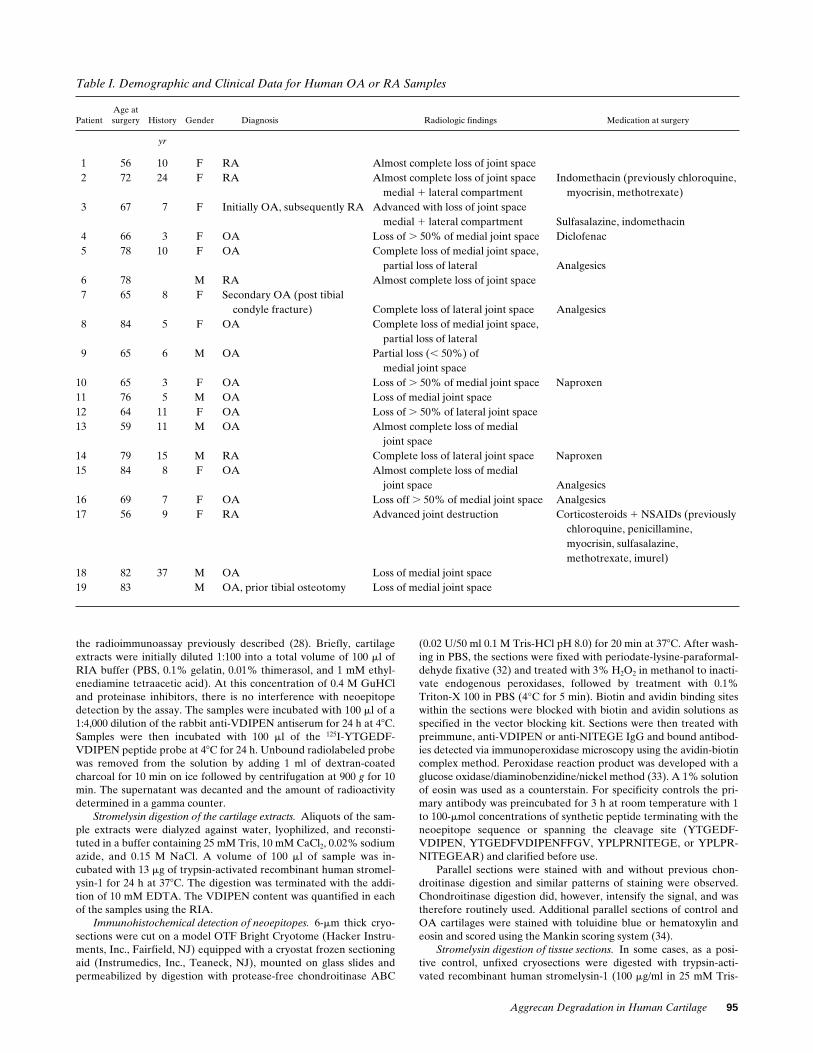
Table I. Demographic and Clinical Data for Human OA or RA Samples
PatientAge atsurgery History Gender Diagnosis Radiologic findings Medication at surgery
yr
1 56 10 F RA Almost complete loss of joint space
2 72 24 F RA Almost complete loss of joint space Indomethacin (previously chloroquine,
medial
1
lateral compartment myocrisin, methotrexate)
3 67 7 F Initially OA, subsequently RA Advanced with loss of joint space
medial
1
lateral compartment Sulfasalazine, indomethacin
4 66 3 F OA Loss of
.
50% of medial joint space Diclofenac
5 78 10 F OA Complete loss of medial joint space,
partial loss of lateral Analgesics
6 78 M RA Almost complete loss of joint space
7 65 8 F Secondary OA (post tibial
condyle fracture) Complete loss of lateral joint space Analgesics
8 84 5 F OA Complete loss of medial joint space,
partial loss of lateral
9 65 6 M OA Partial loss (
,
50%) of
medial joint space
10 65 3 F OA Loss of
.
50% of medial joint space Naproxen
11 76 5 M OA Loss of medial joint space
12 64 11 F OA Loss of
.
50% of lateral joint space
13 59 11 M OA Almost complete loss of medial
joint space
14 79 15 M RA Complete loss of lateral joint space Naproxen
15 84 8 F OA Almost complete loss of medial
joint space Analgesics
16 69 7 F OA Loss off
.
50% of medial joint space Analgesics
17 56 9 F RA Advanced joint destruction Corticosteroids
1
NSAIDs (previously
chloroquine, penicillamine,
myocrisin, sulfasalazine,
methotrexate, imurel)
18 82 37 M OA Loss of medial joint space
19 83 M OA, prior tibial osteotomy Loss of medial joint space
96
Lark et al.
HCl, 150 mM NaCl, 10 mM CaCl
2
, 0.02% NaN
3
) for 30 min at 37
8
C.The tissue sections were then stained for the neoepitopes describedabove. Trypsin was removed from all activated stromelysin samplesusing soybean trypsin inhibitor-agarose (35). The only proteolytic ac-tivity remaining in the sample was due to stromelysin. This was con-firmed by using transferrin as substrate, and showing that all pro-teolytic activity was inhibited by tissue inhibitor of metalloproteinases(TIMP) and 1,10-phenanthroline and not inhibited using PMSF. Fur-ther, second-order rate constants using a peptide substrate wereshown to be similar to those published for stromelysin (36).
Results
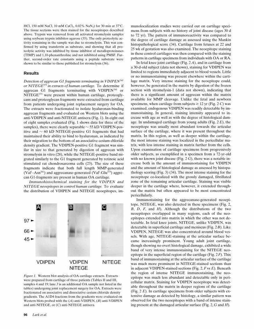
Detection of aggrecan G1 fragments terminating in VDIPEN
341
or NITEGE
373
in extracts of human cartilage.
To determine ifaggrecan G1 fragments terminating with VDIPEN
341
orNITEGE
373
were present within human cartilage, proteogly-cans and proteoglycan fragments were extracted from cartilagefrom patients undergoing joint replacement surgery for OA.The extracts were fractionated to isolate hyaluronan-bindingaggrecan fragments and evaluated on Western blots using theanti-VDIPEN and anti-NITEGE antisera (Fig. 1). In eight outof eight samples evaluated (Fig. 1 shows data for three of thesamples), there were clearly separable
z
55 kD VDIPEN-pos-itive and
z
60 kD NITEGE-positive G1 fragments that hadmaintained their ability to bind to hyaluronan, as indicated bytheir migration to the bottom of an associative cesium chloridedensity gradient. The VDIPEN-positive G1 fragment was sim-ilar in size to that generated by digestion of aggrecan withstromelysin in vitro (28), while the NITEGE-positive band mi-grated similarly to the G1 fragment generated by retinoic acidstimulated rat chondrosarcoma cells (23). The size of thesefragments indicate that both full length MMP-generated(Val
1
–Asn
341
) and aggrecanase-generated (Val
1
-Glu
373
) aggre-can G1-fragments are present in human OA cartilage.
Immunohistochemical staining for the VDIPEN and
NITEGE neoepitopes in control human cartilage.
To evaluatethe distribution of VDIPEN and NITEGE neoepitopes, im-
munolocalization studies were carried out on cartilage speci-mens from subjects with no history of joint disease (ages 30 dto 72 yr). The pattern of immunoreactivity was compared tothe degree of cartilage damage as assessed using the Mankinhistopathological score (34). Cartilage from fetuses at 22 and25 wk of gestation was also examined. The neoepitope stainingin these control cartilages was then compared with the stainingpatterns in cartilage specimens from individuals with OA or RA.
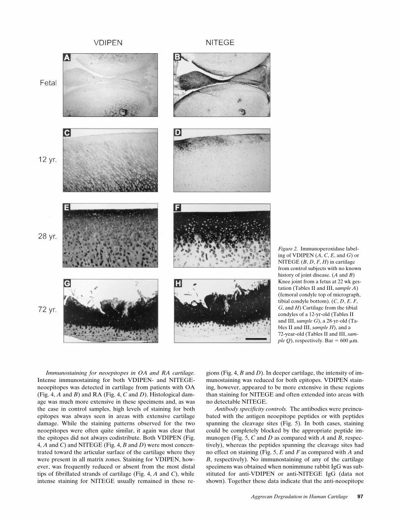
In fetal knee joint cartilage (Fig. 2
A
), and in cartilage froma 30-d-old subject (data not shown), staining for VDIPEN waslimited to regions immediately adjacent to blood vessels. Littleor no immunostaining was present elsewhere within the carti-lage matrix. Very intense staining for the neoepitope could,however, be generated in the matrix by digestion of the frozensection with stromelysin-1 (data not shown), indicating thatthere is a significant amount of aggrecan within the matrixavailable for MMP cleavage. Unlike the fetal and newbornspecimens, when cartilage from subjects
$
12 yr (Fig. 2
C
) wasexamined, endogenous VDIPEN was readily detectable by im-munostaining. In general, staining intensity appeared to in-crease with age as well as with the degree of histological dam-age. In undamaged cartilage from young adults (Fig. 2
E
), theneoepitope was usually most abundant towards the articularsurface of the cartilage, where it was present throughout thematrix. In this region, as well as deeper within the cartilage,the most intense staining was localized in the pericellular ma-trix, with less intense staining in matrix further from the cells.Upon examination of cartilage specimens from progressivelyolder subjects, as exemplified in a specimen from a 72-yr-oldwith no known joint disease (Fig. 2
G
), there was a notable in-crease both in the amount of immunostaining for VDIPENand the amount of histological damage as assessed by histopa-thology scoring (Fig. 3) (34). The most intense staining for theneoepitope co-localized with the grossly damaged, fibrillatedzone of the remaining articular cartilage. Staining was visibledeeper in the cartilage where, however, it extended through-out the matrix but often appeared to be most concentratedpericellularly.
Immunostaining for the aggrecanase-generated neoepi-tope, NITEGE, was also detected in these specimens (Fig. 2,
B
,
D
,
F
, and
H
). Although the distributions of the twoneoepitopes overlapped in many regions, each of the neo-epitopes extended into matrix in which the other was not de-tectable. In fetal knee joints, NITEGE, unlike VDIPEN, wasdetectable in superficial cartilage and meniscus (Fig. 2
B
). LikeVDIPEN, NITEGE was also concentrated around blood ves-sels. With age, NITEGE-staining at the articular surface be-came increasingly prominent. Young adult joint cartilage,though showing no overt histological damage, exhibited a wideband of very intense immunostaining for the NITEGE neo-epitope in the superficial region of the cartilage (Fig. 2
F
). Thisband of immunostaining at the articular surface of the cartilagewas much more prominent in NITEGE-stained sections thanin adjacent VDIPEN-stained sections (Fig. 2,
F
vs
E
). Beneaththe region of intense NITEGE immunostaining, the neo-epitope was much less abundant and detectable only in peri-cellular matrix. Staining for VDIPEN neoepitope was detect-able throughout the matrix in deeper regions of the cartilage(Fig. 2
E
). In cartilage specimens from older subjects with ex-tensive damage as detected by histology, a similar pattern wasobserved for the two neoepitopes with a band of intense stain-ing present at the damaged articular surface (Fig. 2,
G
and
H
).
Figure 1. Western blot analysis of OA cartilage extracts. Extracts were prepared from cartilage of three patients (Tables II and III, samples 4 and 19; lane 3 is an additional OA sample not listed in the tables) undergoing joint replacement surgery for OA. Extracts were fractionated on associative and dissociative cesium chloride density gradients. The A1D4 fractions from the gradients were evaluated on Western blots probed with the (A) anti-VDIPEN, (B) anti-VDIPEN and anti-NITEGE, or (C) anti-NITEGE antisera.
Aggrecan Degradation in Human Cartilage
97
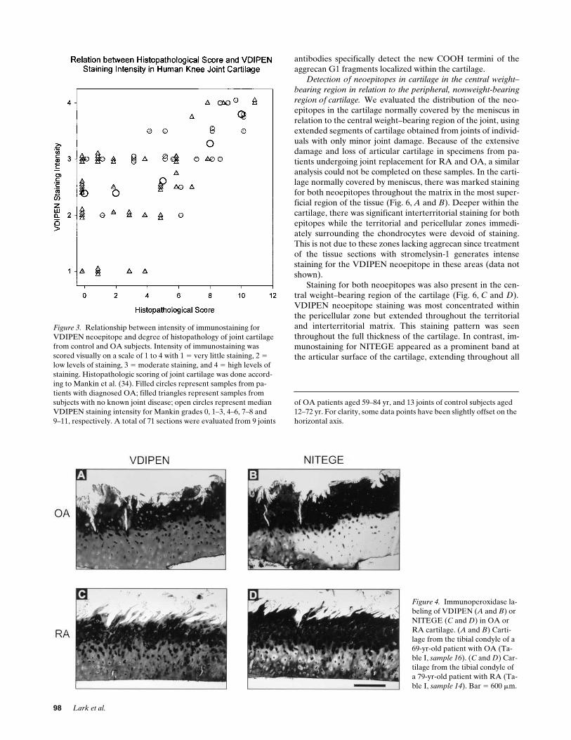
Immunostaining for neoepitopes in OA and RA cartilage.
Intense immunostaining for both VDIPEN- and NITEGE-neoepitopes was detected in cartilage from patients with OA(Fig. 4,
A
and
B
) and RA (Fig. 4,
C
and
D
). Histological dam-age was much more extensive in these specimens and, as wasthe case in control samples, high levels of staining for bothepitopes was always seen in areas with extensive cartilagedamage. While the staining patterns observed for the twoneoepitopes were often quite similar, it again was clear thatthe epitopes did not always codistribute. Both VDIPEN (Fig.4,
A
and
C
) and NITEGE (Fig. 4,
B
and
D
) were most concen-trated toward the articular surface of the cartilage where theywere present in all matrix zones. Staining for VDIPEN, how-ever, was frequently reduced or absent from the most distaltips of fibrillated strands of cartilage (Fig. 4,
A
and
C
), whileintense staining for NITEGE usually remained in these re-
gions (Fig. 4, B and D). In deeper cartilage, the intensity of im-munostaining was reduced for both epitopes. VDIPEN stain-ing, however, appeared to be more extensive in these regionsthan staining for NITEGE and often extended into areas withno detectable NITEGE.
Antibody specificity controls. The antibodies were preincu-bated with the antigen neoepitope peptides or with peptidesspanning the cleavage sites (Fig. 5). In both cases, stainingcould be completely blocked by the appropriate peptide im-munogen (Fig. 5, C and D as compared with A and B, respec-tively), whereas the peptides spanning the cleavage sites hadno effect on staining (Fig. 5, E and F as compared with A andB, respectively). No immunostaining of any of the cartilagespecimens was obtained when nonimmune rabbit IgG was sub-stituted for anti-VDIPEN or anti-NITEGE IgG (data notshown). Together these data indicate that the anti-neoepitope
Figure 2. Immunoperoxidase label-ing of VDIPEN (A, C, E, and G) or NITEGE (B, D, F, H) in cartilage from control subjects with no known history of joint disease. (A and B) Knee joint from a fetus at 22 wk ges-tation (Tables II and III, sample A) (femoral condyle top of micrograph, tibial condyle bottom). (C, D, E. F, G, and H) Cartilage from the tibial condyles of a 12-yr-old (Tables II and III, sample G), a 28-yr-old (Ta-bles II and III, sample H), and a72-year-old (Tables II and III, sam-
ple Q), respectively. Bar 5 600 mm.
98 Lark et al.
antibodies specifically detect the new COOH termini of theaggrecan G1 fragments localized within the cartilage.
Detection of neoepitopes in cartilage in the central weight–
bearing region in relation to the peripheral, nonweight-bearing
region of cartilage. We evaluated the distribution of the neo-epitopes in the cartilage normally covered by the meniscus inrelation to the central weight–bearing region of the joint, usingextended segments of cartilage obtained from joints of individ-uals with only minor joint damage. Because of the extensivedamage and loss of articular cartilage in specimens from pa-tients undergoing joint replacement for RA and OA, a similaranalysis could not be completed on these samples. In the carti-lage normally covered by meniscus, there was marked stainingfor both neoepitopes throughout the matrix in the most super-ficial region of the tissue (Fig. 6, A and B). Deeper within thecartilage, there was significant interterritorial staining for bothepitopes while the territorial and pericellular zones immedi-ately surrounding the chondrocytes were devoid of staining.This is not due to these zones lacking aggrecan since treatmentof the tissue sections with stromelysin-1 generates intensestaining for the VDIPEN neoepitope in these areas (data notshown).
Staining for both neoepitopes was also present in the cen-tral weight–bearing region of the cartilage (Fig. 6, C and D).VDIPEN neoepitope staining was most concentrated withinthe pericellular zone but extended throughout the territorialand interterritorial matrix. This staining pattern was seenthroughout the full thickness of the cartilage. In contrast, im-munostaining for NITEGE appeared as a prominent band atthe articular surface of the cartilage, extending throughout all
Figure 4. Immunoperoxidase la-beling of VDIPEN (A and B) or NITEGE (C and D) in OA or RA cartilage. (A and B) Carti-lage from the tibial condyle of a 69-yr-old patient with OA (Ta-ble I, sample 16). (C and D) Car-tilage from the tibial condyle of a 79-yr-old patient with RA (Ta-ble I, sample 14). Bar 5 600 mm.
Figure 3. Relationship between intensity of immunostaining for VDIPEN neoepitope and degree of histopathology of joint cartilage from control and OA subjects. Intensity of immunostaining was scored visually on a scale of 1 to 4 with 1 5 very little staining, 2 5 low levels of staining, 3 5 moderate staining, and 4 5 high levels of staining. Histopathologic scoring of joint cartilage was done accord-ing to Mankin et al. (34). Filled circles represent samples from pa-tients with diagnosed OA; filled triangles represent samples from subjects with no known joint disease; open circles represent median VDIPEN staining intensity for Mankin grades 0, 1–3, 4–6, 7–8 and9–11, respectively. A total of 71 sections were evaluated from 9 joints
of OA patients aged 59–84 yr, and 13 joints of control subjects aged 12–72 yr. For clarity, some data points have been slightly offset on the horizontal axis.

Aggrecan Degradation in Human Cartilage 99
matrix zones. In deeper regions of the cartilage there was verylittle immunostaining for NITEGE and this was limited to thepericellular zone.
Specimens closer to the tibial spine showed a band of in-tense immunostaining for the NITEGE neoepitope extendingin from the articular surface of the cartilage (Fig. 6 F). Beneaththis, immunostaining for NITEGE was concentrated in thepericellular matrix, with little or no staining in the territorial orinterterritorial matrix. Staining for VDIPEN, on the otherhand, did not appear to be preferentially concentrated towardthe articular surface of the cartilage (Fig. 6 E). Again, the dis-tribution of this neoepitope was similar throughout the thick-ness of the cartilage, with immunostaining most abundant inpericellular matrix. Together these data indicate that bothneoepitopes are expressed throughout the articular cartilage,but that the patterns differ regionally.
The digestion of frozen tissue sections with stromelysin-1increased the staining intensity for the VDIPEN neoepitope.Notably, however, the staining intensity for the NITEGEneoepitope was neither increased, nor abolished after diges-tion of frozen sections with the protease (data not shown).Loss of the NITEGE staining would be expected if this frag-ment could be further processed by MMPs such as stromelysin
and the processed COOH-terminal 32 amino acid fragmentPhe342-Glu373 was released from the cartilage (see Figs. 9 and 10).
Quantitation of the VDIPEN neoepitope in control speci-
mens of human cartilage. Initially, we characterized the quan-tity of the neoepitope in a number of control samples and thencompared these to extracts of OA and RA cartilage. A con-founding factor in our analysis is that we quantified neo-epitope in extracts of full-thickness specimens, whereas theimmunohistochemical evaluation allowed us to observe thedifferential distribution of the neoepitopes at the microscopiclevel throughout the thickness of the cartilage. We were ableto quantify the VDIPEN neoepitope, since we have previouslyshown, using the RIA, that 1 mol of peptide equals 1 mol ofVDIPEN-positive G1 fragment (28). Unfortunately, a similarquantitation of NITEGE-positive aggrecan fragments couldnot be completed because there is currently no reproducibleway to generate an accurate G1-standard for this RIA (23).
18 cartilage specimens from thirteen individuals, ranging inage from 30 d to 88 yr, were obtained at the time of autopsy.Multiple samples were taken from these specimens and theVDIPEN neoepitope concentrations determined (Table II).The concentration of neoepitope in these samples ranged from0.011 to 0.65 pmol VDIPEN per microgram hydroxyproline.
Figure 5. Immunoperoxidase labeling of VDIPEN or NITEGE in cryosec-tions of cartilage from a 64-yr-old pa-tient with OA (Table I, sample 12). Immunostaining for VDIPEN (A) is completely abolished when anti-VDIPEN IgG is preincubated with YTGEDFVDIPEN peptide (C), but is unaffected by preincubation of the antibody with YTGEDFVDIPEN peptide (E), a peptide spanning the metalloprotease cleavage site. Immu-nostaining for NITEGE (B) is com-pletely blocked by preincubation of anti-NITEGE IgG with YPLPRNI-TEGE peptide (D), but is unaffected by preincubation with YPLPRNI-TEGEAR (F), a peptide spanning the aggrecanase cleavage site.Bar 5 600 mm.

100 Lark et al.
The concentration of VDIPEN neoepitope relative to collagenincreased with age up to z 25 yr, then reached an apparentsteady state (Fig. 7). Extractability of aggrecan ranged from75–90%, with no relation to age. Therefore, the differences de-tected with age were not the result of altered extractability ofthe samples.
In specimens obtained from two fetuses with gestationalages of 22 wk, there was , 0.002 pmol VDIPEN per micro-gram hydroxyproline. This was not the result of a lack ofavailable aggrecan substrate, since VDIPEN-positive G1 frag-ments could be generated and detected by RIA and Westernblot when the samples were incubated with stromelysin-1(data not shown).
When the fetal knee cartilages from five specimens rangingin gestational age from 19–22 wk were reevaluated afterstromelysin digestion, there were at least 900 mol of aggrecanthat could be cleaved by stromelysin at the MMP site for everymole of resident VDIPEN-positive fragment. In fact, the relativeincrease in neoepitope ranged from 979- to over 13,172-foldwith a median increase of 3,219-fold (Table III). Interestingly,
Figure 6. Cartilage from the tibial condyle of a 29-yr-old allograft donor (not in table) was cryosectioned and immunostained for VDIPEN (A, C, and E) or NITEGE (B, D, and F). (A and B) Cartilage from an area that had been covered by meniscus. (C and D) Cartilage from a central, loaded, area not covered by the me-niscus. (E and F) Cartilage from an area close to the tibial spines.Bar 5 300 mm.
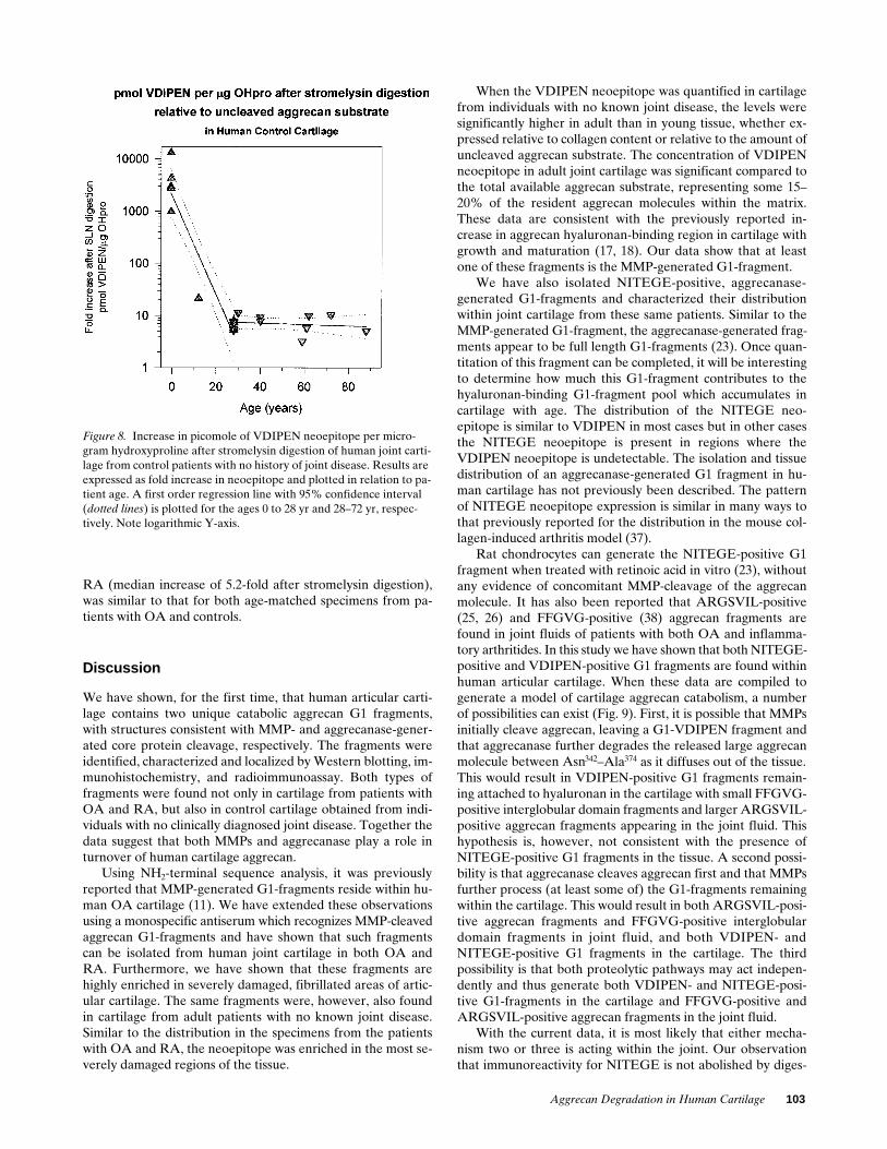
lines) is plotted for the ages 0 to 28 yr and . 28 yr, respectively. Note logarithmic Y-axis.
Figure 7. VDIPEN neoepitope (pmol) per microgram hydroxypro-line in extracts of human joint cartilage from control patients with no history of joint disease. Results are plotted in relation to patient age. A first order regression line with 95% confidence interval (dotted

Aggrecan Degradation in Human Cartilage 101
there were only z 20 mol (range 16–37) of available substrateper mole of resident VDIPEN-positive G1 fragment in thesamples from the 12-yr-old subject. When the remaining con-trol samples were examined as a group (age range 28–88),there were from 3- to 25-fold increases in signal after stromel-ysin digestion with a median increase of about eightfold (TableIII). Again, it appeared that in the matrix the ratio ofVDIPEN-positive G1 fragments to the available aggrecan sub-strate increased up to age 20–30, then remained at a steadystate with increasing age (Fig. 8).
Together, these data suggest that the VDIPEN-positive G1fragments are found at low levels in cartilage from very youngindividuals in relation to both collagen and available aggrecansubstrate, but that this G1 fragment accumulates with age upto about 25 yr.
Quantitation of VDIPEN neoepitope in human OA and RA
cartilage specimens. There was little difference in the averagelevels of FVDIPEN neoepitope relative to collagen in eitherthe adult control samples (0.20560.08 pmol/mg hypro [n 5 15median levels]), OA samples (0.27460.177 pmol/mg hypro [n 5
Table II. Quantitation of VDIPEN in Relation to Hydroxyproline Content in Human Knee Joint Cartilage Samples
Sample patient Age Median Range n Diagnosis
yr pmol VDIPEN/mg hypro
A 22-wk gestation 0.002 1 C
B 22-wk gestation 0.002 1 C
F 30 d 0.019 0.011–0.038 5 C
G1 12 0.035 0.029–0.056 8 C
G2 12 0.030 0.025–0.056 9 C
H1 28 0.110 0.070–0.276 8 C
H1 (femur) 28 0.168 0.030–0.257 8 C
H2 28 0.156 0.088–0.282 7 C
H2 (femur) 28 0.217 0.038–0.302 6 C
I 29 0.288 0.237–0.315 3 CT
J (right) 30 0.303 0.081–0.435 6 C
J (left) 30 0.329 0.212–0.593 7 C
K 40 0.107 0.094–0.119 2 CT
L 40 0.105 1 C
M 45 0.182 0.108–0.650 8 C
N 59 0.264 1 C
O 61 0.312 0.200–0.423 2 CT
P 62 0.109 0.035–0.325 11 C
Q 72 0.196 0.071–0.242 8 C
R 88 0.231 1 C
17 56 0.112 0.091–0.133 2 RA
4 59 0.163 1 OA
13 60 0.331 0.105–0.556 2 OA
12 64 0.419 0.303–0.534 2 OA
7 65 0.109 1 OA
9 65 0.157 0.125–0.187 2 OA
10 65 0.342 0.322–0.361 2 OA
1 66 0.022 1 RA
3 67 0.310 1 RA
16 69 0.203 0.093–0.214 3 OA
2 71 0.146 1 RA
11 76 0.687 0.213–1.154 3 OA
5 78 0.140 1 OA
6 78 0.211 0.077–0.215 3 RA
14 79 0.310 1 RA
18 82 0.191 1 OA
19 83 0.129 1 OA
8 84 0.408 0.324–0.433 3 OA
15 84 0.087 0.078–0.095 2 OA
Extracts of human knee joint cartilage (Diagnosis: C, control; CT, Control from tumor case) were prepared and the VDIPEN neoepitope quantified
by RIA. The collagen content in the collagen-rich pellet was then determined using a hydroxyproline assay and the data reported as picomoles
VDIPEN/microgram hydroxyproline. n denotes the number of samples examined from each joint. Unless noted, all samples were from tibial
condyles. Control samples were numbered A–R, for numbering and further information on OA and RA patient samples, see Table I.

102 Lark et al.
10 median levels]) or RA samples (0.18560.114 pmol/mghypro [n 5 6 median levels]). Furthermore, the range of levelsof the VDIPEN neoepitope relative to collagen in both theOA and RA samples was similar to those seen in the age-matched control specimens (Table II). When the extracts ofthe OA samples were examined after stromelysin digestion,
there were z 8 mol of available aggrecan substrate for everymole of resident VDIPEN-positive G1 fragment (Table III).These levels were similar to those in the age-matched controlcartilages.
The relative increase in the levels of VDIPEN neoepitopeafter stromelysin digestion in the specimens from patients with
Table III. VDIPEN Neoepitope in Human Knee Joint Cartilage Relative to Uncleaved Aggrecan Substrate (Expressed as Fold Increase after Stromelysin Digestion)
Sample patient Age Fold increase after stromelysin digestion Range n Diagnosis
yr
A 22-wk gestation . 13172 1 C
B 22-wk gestation 979 1 C
C 19-wk gestation . 2953 2175–3730 2 C
D 20-wk gestation . 2732 2705–4221 3 C
E 23-wk gestation . 4280 3282–4465 4 C
F 30 d ND C
G1 12 21.43 16.11–37.05 9 C
G2 12 20.97 12.04–30.45 8 C
H1 28 7.87 5.63–9.58 8 C
H1 (femur) 28 5.74 5.31–10.68 7 C
H2 28 5.30 4.37–7.23 7 C
H2 (femur) 28 7.27 5.93–13.45 6 C
I 29 11.26 8.99–12.79 3 CT
J (right) 30 ND C
J (left) 30 ND C
K 40 9.30 8.80–9.80 2 CT
L 40 7.90 1 C
M 45 ND C
N 59 3.22 1 C
O 61 5.76 3.26–8.26 2 CT
P 62 9.99 5.48–24.91 6 C
Q 72 10.18 3.79–20.46 7 C
R 88 4.98 1 C
17 56 6.31 5.54–7.08 2 RA
4 59 2.14 1 OA
13 60 18.03 4.35–31.70 2 OA
12 64 7.18 5.90–8.45 2 OA
7 65 25.33 1 OA
9 65 9.38 8.95–9.81 2 OA
10 65 7.47 5.82–9.11 2 OA
1 66 3.01 1 RA
3 67 3.68 1 RA
16 69 23.82 10.40–40.50 3 OA
2 71 4.05 1 RA
11 76 7.14 5.43–8.33 3 OA
5 78 14.04 1 OA
6 78 8.57 5.18–18.00 3 RA
14 79 7.16 1 RA
18 82 3.14 1 OA
19 83 7.39 1 OA
8 84 4.57 4.31–7.28 3 OA
15 84 11.40 11.11–11.65 2 OA
Extracts of human knee joint cartilage (Diagnosis: C, control; CT, Control from tumor case) were prepared and the FVDIPEN neoepitope quantified
by RIA. The samples were then exhaustively digested with stromelysin-1 to cleave all uncleaved, available aggrecan substrate and the FVDIPEN con-
tent again quantified using the RIA. Data are reported as fold increase after stromelysin digestion. For numbering and further information, see Table I.
Aggrecan Degradation in Human Cartilage 103
RA (median increase of 5.2-fold after stromelysin digestion),was similar to that for both age-matched specimens from pa-tients with OA and controls.
Discussion
We have shown, for the first time, that human articular carti-lage contains two unique catabolic aggrecan G1 fragments,with structures consistent with MMP- and aggrecanase-gener-ated core protein cleavage, respectively. The fragments wereidentified, characterized and localized by Western blotting, im-munohistochemistry, and radioimmunoassay. Both types offragments were found not only in cartilage from patients withOA and RA, but also in control cartilage obtained from indi-viduals with no clinically diagnosed joint disease. Together thedata suggest that both MMPs and aggrecanase play a role inturnover of human cartilage aggrecan.
Using NH2-terminal sequence analysis, it was previouslyreported that MMP-generated G1-fragments reside within hu-man OA cartilage (11). We have extended these observationsusing a monospecific antiserum which recognizes MMP-cleavedaggrecan G1-fragments and have shown that such fragmentscan be isolated from human joint cartilage in both OA andRA. Furthermore, we have shown that these fragments arehighly enriched in severely damaged, fibrillated areas of artic-ular cartilage. The same fragments were, however, also foundin cartilage from adult patients with no known joint disease.Similar to the distribution in the specimens from the patientswith OA and RA, the neoepitope was enriched in the most se-verely damaged regions of the tissue.
When the VDIPEN neoepitope was quantified in cartilagefrom individuals with no known joint disease, the levels weresignificantly higher in adult than in young tissue, whether ex-pressed relative to collagen content or relative to the amount ofuncleaved aggrecan substrate. The concentration of VDIPENneoepitope in adult joint cartilage was significant compared tothe total available aggrecan substrate, representing some 15–20% of the resident aggrecan molecules within the matrix.These data are consistent with the previously reported in-crease in aggrecan hyaluronan-binding region in cartilage withgrowth and maturation (17, 18). Our data show that at leastone of these fragments is the MMP-generated G1-fragment.
We have also isolated NITEGE-positive, aggrecanase-generated G1-fragments and characterized their distributionwithin joint cartilage from these same patients. Similar to theMMP-generated G1-fragment, the aggrecanase-generated frag-ments appear to be full length G1-fragments (23). Once quan-titation of this fragment can be completed, it will be interestingto determine how much this G1-fragment contributes to thehyaluronan-binding G1-fragment pool which accumulates incartilage with age. The distribution of the NITEGE neo-epitope is similar to VDIPEN in most cases but in other casesthe NITEGE neoepitope is present in regions where theVDIPEN neoepitope is undetectable. The isolation and tissuedistribution of an aggrecanase-generated G1 fragment in hu-man cartilage has not previously been described. The patternof NITEGE neoepitope expression is similar in many ways tothat previously reported for the distribution in the mouse col-lagen-induced arthritis model (37).
Rat chondrocytes can generate the NITEGE-positive G1fragment when treated with retinoic acid in vitro (23), withoutany evidence of concomitant MMP-cleavage of the aggrecanmolecule. It has also been reported that ARGSVIL-positive(25, 26) and FFGVG-positive (38) aggrecan fragments arefound in joint fluids of patients with both OA and inflamma-tory arthritides. In this study we have shown that both NITEGE-positive and VDIPEN-positive G1 fragments are found withinhuman articular cartilage. When these data are compiled togenerate a model of cartilage aggrecan catabolism, a numberof possibilities can exist (Fig. 9). First, it is possible that MMPsinitially cleave aggrecan, leaving a G1-VDIPEN fragment andthat aggrecanase further degrades the released large aggrecanmolecule between Asn342–Ala374 as it diffuses out of the tissue.This would result in VDIPEN-positive G1 fragments remain-ing attached to hyaluronan in the cartilage with small FFGVG-positive interglobular domain fragments and larger ARGSVIL-positive aggrecan fragments appearing in the joint fluid. Thishypothesis is, however, not consistent with the presence ofNITEGE-positive G1 fragments in the tissue. A second possi-bility is that aggrecanase cleaves aggrecan first and that MMPsfurther process (at least some of) the G1-fragments remainingwithin the cartilage. This would result in both ARGSVIL-posi-tive aggrecan fragments and FFGVG-positive interglobulardomain fragments in joint fluid, and both VDIPEN- andNITEGE-positive G1 fragments in the cartilage. The thirdpossibility is that both proteolytic pathways may act indepen-dently and thus generate both VDIPEN- and NITEGE-posi-tive G1-fragments in the cartilage and FFGVG-positive andARGSVIL-positive aggrecan fragments in the joint fluid.
With the current data, it is most likely that either mecha-nism two or three is acting within the joint. Our observationthat immunoreactivity for NITEGE is not abolished by diges-
Figure 8. Increase in picomole of VDIPEN neoepitope per micro-gram hydroxyproline after stromelysin digestion of human joint carti-lage from control patients with no history of joint disease. Results are expressed as fold increase in neoepitope and plotted in relation to pa-tient age. A first order regression line with 95% confidence interval (dotted lines) is plotted for the ages 0 to 28 yr and 28–72 yr, respec-tively. Note logarithmic Y-axis.

104 Lark et al.
tion of tissue sections with stromelysin-1, would perhaps sug-gest that the MMP- and aggrecanase-dependent pathways actindependently, and argue in favor of mechanism three (Fig. 9 C).
It has been shown by both in vivo and in vitro experimentsthat there are at least two pools of aggrecan surrounding chon-drocytes which have different rates of turnover (39–41). In thisstudy, we have shown that both the NITEGE and VDIPENneoepitopes are generated, and in some cases, enriched in theimmediate pericellular matrix. There is also neoepitope in re-gions far away from the chondrocyte. The intense stainingimmediately surrounding the cells suggests that both the ag-grecanase enzyme and the MMPs are likely generated and oractivated by the chondrocytes. The neoepitope-related reactiv-ity also present in matrix further removed from the cells, onthe other hand, suggests that the fragments may move awayfrom the cells once generated or that the enzymes responsiblefor the aggrecan cleavage may be active within the interterrito-rial matrix. Since aggrecan molecules in the pericellular matrixappear to turn over more rapidly than those in the surroundingmatrix (41), it would be interesting to examine the generationof these two different neoepitopes in vitro and determine ifthey turn over with different rates in these different matrixcompartments.
Very limited immunostaining for NITEGE, and no immu-nostaining for VDIPEN was detected in normal mouse articu-lar cartilage, but immunostaining for both epitopes was readilydetected in the cartilage of mice with type II collagen-inducedarthritis (37, 42). The data suggest, that in the normal articularcartilage of these animals there are few G1-fragments termi-nating in VDIPEN341 or NITEGE373, but that with increasingpathology there is a significant increase in both MMP- and ag-grecanase-generated aggrecan cleavage.
Our present observations indicate that the content ofMMP-generated aggrecan G1-fragments in human joint carti-lage increases up to about 20–30 yr of age, and that the propor-tion (relative to both collagen and available aggrecan sub-strate) then remains constant with further increase in age.Moreover, the proportion of G1-VDIPEN in OA and RAjoint cartilage, relative to collagen and to available aggrecansubstrate, was about the same as in adult joint cartilage.
The observations on the occurrence of these molecularfragments in normal and pathologic human and animal tissuesshould be interpreted in the light of the fact that we can, by im-munohistochemistry or by extraction of these human cartilagesamples, detect only those fragments which remain in the tis-sue after being cleaved by MMP or aggrecanase. Aggrecan G1fragments which diffuse into the joint fluid or are taken up bythe chondrocytes escape our detection by these means. The ag-grecan fragments detected here are thus the accrued result of aseries of synthetic and catabolic events: aggrecan synthesis andsecretion, proteolytic cleavage of the interglobular domain,and removal of the G1 fragment by chondrocyte uptake or dif-fusion (Fig. 10). The balance of these processes may differ be-
Figure 9. Hypothetical models for pathways of proteolytic break-down of aggrecan in cartilage. (A) Primary cleavage of aggrecan in-terglobular domain (IGD) by a classical matrix metalloprotease (MMPx), followed by aggrecanase (Agnase) cleavage of a 30-amino acid peptide from the liberated G2-containing fragment. Only VDIPEN neoepitope would be detected in matrix, while fragments with ARGSV or FFGVG amino termini would be present in joint fluid. (B) Primary cleavage of IGD by aggrecanase, followed by sec-ondary cleavage by metalloproteinase activity. Depending on effi-ciency of second cleavage step, both NITEGE and VDIPEN neoepitopes may be detected in cartilage matrix, while large frag-ments with an amino terminus of ARGSV and a small 30-amino acid peptide with FFGVG- and -NITEGE termini will be detected in joint fluid. (C) Aggrecanase and a classic matrix metalloproteinase activity
independently cleave aggrecan IGD. The G1-domains may remain bound to hyaluronan in matrix, while the remainder of the aggrecan molecules (including the G2-domain) are free to diffuse into joint fluid. Cleavage epitopes -NITEGE and -VDIPEN may be detected in tissue, while ARGSV- and FFGVG- may be detected in synovial fluid.

Aggrecan Degradation in Human Cartilage 105
tween tissues at different growth rates or ages, and with differ-ent degrees of pathology, and be influenced by changes inother matrix molecules which may interact with aggrecan. Anexample of such change is the increasing damage to type II col-lagen which has been demonstrated in aging and OA cartilage(43). It appears that these processes may reach a steady statein adult articular cartilage matrix, at least with regard to G1-VDIPEN. Recently, VDIPEN fragments have been isolatedfrom human OA joint fluids (Lark, M.W., and L.S. Lohmander,unpublished observations), suggesting that these fragments areindeed released from human articular cartilage. In support, itwas shown that within 1 h after injection of stromelysin-1 intorabbit joints, z 30% of the VDIPEN-positive G1 fragmentsare released into the joint fluid, suggesting that there is furtherprocessing of the HA-G1 complex resulting in release from thearticular cartilage (44). In this context, it is interesting to notethat the presence of aggrecan G1-fragments has been shown injoint fluids from patients with RA or OA (45, 46). Moreover,in vitro work on bovine joint cartilage has indicated that hyal-uronan and aggrecan are catabolized at similar rates and it wassuggested that at least part of the degradation involved inter-nalization by the chondrocytes (47, 48). Examination of theseevents in a closed experimental system, such as cartilage ex-plants or chondrocytes grown in alginate in vitro, may help toclarify some of these questions.
The results of our study indicate that both MMPs and ag-grecanase play roles in the degradation of human cartilage ag-grecan in normal cartilage as well as in osteoarthritic and rheu-matoid cartilage. We find it interesting that similar aggrecandegradation mechanisms may be active in osteoarthritis andrheumatoid arthritis, in spite of the contrasting pathologicaland clinical features of these two conditions. It will be impor-tant to compare the distribution and turnover of the aggrecanG1 fragments carrying the VDIPEN and NITEGE neoepi-topes in samples from patients with various joint diseases, totry to unravel the seemingly complex controls regulating ag-grecan metabolism within the joint. Such information may behelpful in efforts to intervene with cartilage destruction in jointdisease.
Acknowledgments
L.S. Lohmander was supported by the Swedish Medical ResearchCouncil, the King Gustaf V 80-year Birthday Fund, the Kock and theZoega Foundations, the Medical Faculty of Lund and Lund Univer-sity Hospital, and Merck Research Laboratories.
References
1. Dean, D.D., J. Martel-Pelletier, J.-P. Pelletier, D.S. Howell, and J.F.J.Woessner. 1989. Evidence for metalloproteinase and metalloproteinase inhibi-tor imbalance in human osteoarthritic cartilage. J. Clin. Invest. 84:678–685.
2. Gravallese, E.M, J.M. Darling, A.L. Ladd, J.N. Katz, and L.H. Glimcher.1991. In situ hybridization studies of stromelysin and collagenase expression inrheumatoid synovium. Arthritis Rheum. 34:1076–1084.
3. McCachren, S.S. 1991. Expression of metalloproteinases and metallopro-teinase inhibitor in human arthritic synovium. Arthritis Rheum. 34:1085–1093.
4. Firestein, G.S., M.M. Paine, and B.H. Littman. 1991. Gene expression(collagenase, tissue inhibitor of metalloproteinases, complement, and HLA-DR) in rheumatoid arthritis and osteoarthritis synovium. Quantitative analysisand effect of intraarticular corticosteroids. Arthritis Rheum. 34:1094–1105.
5. Walakovits, L.A., V.L. Moore, N. Bhardwaj, G.S. Gallick, and M.W.Lark. 1991. Detection of high levels of stromelysin and collagenase in synovialfluid from patients with rheumatoid arthritis and post-traumatic knee injury.Arthritis Rheum. 35:35–42.
6. Nguyen, Q., J.S. Mort, and P.J. Roughley. 1992. Preferential mRNA ex-pression of prostromelysin relative to procollagenase and in situ localization inhuman articular cartilage. J. Clin. Invest. 89:1189–1197.
7. Lohmander, L.S., L.A. Hoerrner, and M.W. Lark. 1993. Metalloprotein-ases, tissue inhibitor and proteoglycan fragments in knee synovial fluid in hu-man osteoarthritis. Arthritis Rheum. 36:181–189.
8. Wolfe, G.C., K.L. MacNaul, F.F. Buechel, J. McDonnell, L.A. Hoerrner,M.W. Lark, V.L. Moore, and N.I. Hutchinson. 1993. Differential in vivo expres-sion of collagenase messenger RNA in synovium and cartilage. Quantitativecomparison with stromelysin messenger RNA levels in human rheumatoid ar-thritis and osteoarthritis patients and in two animal models of acute inflamma-tory arthritis. Arthritis Rheum. 36:1540–1547.
9. Lohmander, L.S., L.A. Hoerrner, and M.W. Lark. 1995. Increased con-centrations of collagenase (MMP-1) in joint fluid after knee injury and in os-teoarthritis. Trans. Orthop. Res. Soc. 20:9. (Abstr.)
10. Nguyen, Q., G. Murphy, P.J. Roughley, and J.S. Mort. 1989. Degrada-tion of proteoglycan aggregate by a cartilage metalloproteinase. Evidence forthe involvement of stromelysin in the generation of link protein heterogeneityin situ. Biochem. J. 259:61–67.
11. Flannery, C.R., M.W. Lark, and J.D. Sandy. 1992. Identification of astromelysin cleavage site within the interglobular domain of human aggrecan:evidence for proteolysis at this site in vivo in human articular cartilage. J. Biol.
Chem. 267:1008–1014.12. Fosang, A.J., K. Last, V. Knäuper, P.J. Neame, G. Murphy, T.E. Har-
Figure 10. Schematic diagram of metabolic pathways of G1-containing fragments of joint cartilage aggre-can. The cartilage matrix content of aggrecanG1 fragments containing the carboxy termini of VDIPEN341 or NITEGE373 is the result of (a) the rate of production of aggrecan by the chondrocyte, (b) the rates of aggrecan IGD cleavage by MMP or aggre-canase, (c) the rate of chondrocyte uptake of the ter-nary complex of hyaluronan-link-G1 or free G1, and (d) the rate of diffusion out of the matrix of the ter-nary complex of hyaluronan-link-G1 or free G1. This assumes that there is no further COOH-terminal pro-cessing of either fragment. AGN, aggrecan; HA, hy-aluronan; LP, link protein. Small arrows, proteolytic cleavage of aggrecan core protein. Large arrows, pathways of addition or elimination of aggrecan or its cleavage products from matrix.
106 Lark et al.
dingham, H. Tschesche, and J.A. Hamilton. 1993. Fibroblast and neutrophilcollagenases cleave at two sites in the cartilage aggrecan interglobular domain.Biochem. J. 295:273–276.
13. Fosang, A.J., P.J. Neame, T.E. Hardingham, G. Murphy, and J.A.Hamilton. 1991. Cleavage of cartilage proteoglycan between G1 and G2 do-mains by stromelysin. J. Biol. Chem. 266:15579–15582.
14. Wu, J.-J., M.W. Lark, L.E. Chun, and D.R. Eyre. 1991. Sites of stromel-ysin cleavage in collagen types II, IX, X, and XI of cartilage. J. Biol. Chem. 266:5625–5628.
15. Liu, J.J., D. Cassidy, A. Allan, P.J. Neame, J.S. Mort, and P.J. Roughley.1992. Link protein shows species variation in its susceptibility to proteolysis. J.
Orthop Res. 10:621–630.16. Hollander, A.P., T.F. Heathfield, C. Webber, Y. Iwata, R. Bourne, C.
Rorabeck, and A.R. Poole. 1994. Increased damage to type II collagen in os-teoarthritic cartilage detected by a new immunoassay. J. Clin. Invest. 93:1722–1732.
17. Roughley, P.J., R.J. White, and A.R. Poole. 1985. Identification of a hy-aluronic acid-binding protein that interferes with the preparation of high-buoy-ant density proteoglycan aggregates from adult human articular cartilage. Bio-
chem. J. 231:129–138.18. Bayliss, M.T., M.W.A. Holmes, and H. Muir. 1989. Age-related changes
in the stoichiometry of binding region, link protein and hyaluronic acid in hu-man articular cartilage. Trans. Orthop. Res. Soc. 14:32. (Abstr.)
19. Sandy, J.D., P.J. Neame, R.E. Boynton, and C.R. Flannery. 1991. Catab-olism of aggrecan in cartilage explants. Identification of a major cleavage sitewithin the interglobular domain. J. Biol. Chem. 266:8683–8695.
20. Ilic, M.Z., C.H. Handley, H.C. Robinson, and M.T. Mok. 1992. Mecha-nism of catabolism of aggrecan by articular cartilage. Arch. Biochem. Biophys.
294:115–122.21. Loulakis, P., A. Shrikande, G. Davis, and C.A. Maniglia. 1992. N-termi-
nal sequence of proteoglycan fragments isolated from medium of interleukin-1-treated articular-cartilage cultures. Biochem. J. 284:589–593.
22. Plaas, A.H., and J.D. Sandy. 1993. A cartilage explant system for studieson aggrecan structure, biosynthesis and catabolism in discrete zones of themammalian growth plate. Matrix. 13:135–147.
23. Lark, M.W., J.T. Gordy, J.R. Weidner, J. Ayala, J.H. Kimura, H.R. Wil-liams, R.A. Mumford, C.R. Flannery, S.S. Carlson, M. Iwata, and J.D. Sandy.1995. Cell-mediated catabolism of aggrecan. Evidence that cleavage at the “ag-grecanase” site (Glu373-Ala374) is a primary event in proteolysis of the interglob-ular domain. J. Biol. Chem. 270:2550–2556.
24. Hughes, C.E., B. Caterson, A.J. Fosang, P.J. Roughley, and J.S. Mort.1995. Monoclonal antibodies that specifically recognize neoepitope sequencesgenerated by ‘aggrecanase’ and matrix metalloproteinase cleavage of aggrecan:application to catabolism in situ and in vitro. Biochem. J. 305:799–804.
25. Lohmander, L.S., P. Neame, and J.D. Sandy. 1993. The structure of ag-grecan fragments in human synovial fluid: Evidence that aggrecanase mediatescartilage degradation in inflammatory joint disease, joint injury and osteoar-thritis. Arthritis Rheum. 36:1214–1222.
26. Sandy, J.D., C.R. Flannery, P.J. Neame, and L.S. Lohmander. 1992. Thestructure of aggrecan fragments in human synovial fluid: Evidence for the in-volvement in osteoarthritis of a novel proteinase which cleaves the glu 373-ala374 bond of the interglobular domain. J. Clin. Invest. 89:1512–1516.
27. Fosang, A.J., K. Last, P.J. Neame, G. Murphy, V. Knäuper, H.Tschesche, C. Hughes, B. Caterson, and T.E. Hardingham. 1994. Neutrophilcollagenase (MMP-8) cleaves at the aggrecanase site E373-A-374 in the inter-globular domain of cartilage aggrecan. Biochem. J. 304:347–351.
28. Lark, M.W., H. Williams, L.A. Hoerrner, J.R. Weidner, J.M. Ayala,C.F. Harper, A. Christen, J. Olszewski, Z. Konteatis, R. Webber, et al. 1995.Quantification of a matrix metalloproteinase-generated aggrecan G1 fragmentusing monospecific anti-peptide serum. Biochem. J. 307:245–252.
29. Doege, K.J., M. Sasaki, T. Kimura, and Y. Yamada. 1991. Completecoding sequence and deduced primary structure of the human cartilage largeaggregating proteoglycan, aggrecan. J. Biol. Chem. 266:894–902.
30. Hascall, V.C., and J.H. Kimura. 1982. Proteoglycans: isolation and char-acterization. Methods Enzymol. 82:769–800.
31. Woessner, J.F., Jr. 1961. The determination of hydroxyproline in tissueand protein samples containing small proportions of this imino acid. Arch. Bio-
chem. Biophys. 93:440–447.32. MacLean, I.W., and P.K. Nakane. 1974. Periodate-lysine-paraformalde-
hyde fixative. A new fixation for immunoelectron microscopy. J. Histochem.
Cytochem. 35:647–655.33. Shu, S., G. Ju, and L. Fan. 1988. The glucose oxidase-DAB nickel
method in peroxidase histochemistry of the nervous system. Neurosci. Lett. 85:169–171.
34. Mankin, H.J., H. Dorfman, L. Lippiello, and A. Zarins. 1971. Biochemi-cal and metabolic abnormalities in articular cartilage from osteo-arthritic hu-man hips. II. Correlation of morphology with biochemical and metabolic data.J. Bone Jt. Surg. (Am.) Vol. 53:523–537.
35. Lark, M.W., L.A. Walakovits, T.K. Shah, J. Vanmiddlesworth, P.M.Cameron, and T.-Y. Lin. 1990. Production and purification of prostromelysinand procollagenase from IL-1 beta-stimulated human gingival fibroblasts. Con-
nect. Tissue Res. 25:49–65.36. Teahan, J., R. Harrison, M. Izquierdo, and R.L. Stein. 1989. Substrate
specificity of human fibroblast stromelysin. Hydrolysis of substance P and itsanalogues. Biochemistry. 28:8497–8501.
37. Singer, I.I., S. Scott, D.W. Kawka, E.K. Bayne, S.A. Donatelli, J.R.Weidner, H.R. Williams, R.A. Mumford, M.W. Lark, J. McDonnell, et al. 1995.Aggrecanase and metalloproteinase-specific aggrecan neoepitopes are inducedin the articular cartilage of mice with collagen II arthritis. Trans. Orthop. Res.
Soc. 20:330.38. Fosang, A.J., K. Last, and R.A. Maciewicz. 1996. Aggrecan is degraded
by matrix metalloproteinases in human arthritis. Evidence that matrix metallo-proteinases and aggrecanase activities can be independent. J. Clin. Invest. 98:2292–2299.
39. Lohmander, S. 1977. Turnover of proteoglycans in guinea pig costal car-tilage. Arch. Biochem. Biophys. 180:93–101.
40. Sandy, J.D. 1992. Extracellular metabolism of aggrecan. In ArticularCartilage and Osteoarthritis. K.E. Kuettner, R. Schleyerbach, J.G. Peyron, andV.C. Hascall, editors. Raven Press, Ltd., New York. 21–33.
41. Mok, S.S., K. Masuda, H.J. Häuselmann, M.B. Aydelotte and E.J.-M.A.Thonar. 1994. Aggrecan synthesized by mature chondrocytes suspended in algi-nate. Identification of two distinct metabolic pools. J. Biol. Chem. 269:33021–33027.
42. Singer, I.I., D.W. Kawka, E.K. Bayne, S.A. Donatelli, J.R. Weidner,H.R. Williams, J.M. Ayala, R.A. Mumford, M.W. Lark, T.T. Glant, et al. 1995.VDIPEN, a metalloproteinase-generated neoepitope, is induced and immu-nolocalized in articular cartilage during inflammatory arthritis. J. Clin. Invest.
95:2178–2186.43. Hollander, A.P., I. Pidoux, A. Reiner, C. Rorabeck, R. Bourne, and
A.R. Poole. 1995. Damage to type II collagen in aging and osteoarthritis startsat the articular surface, originates around chondrocytes, and extends into thecartilage with progressive degeneration. J. Clin. Invest. 96:2859–2869.
44. Bayne, E.K, K.L. MacNaul, S.A. Donatelli, A. Christen, P.R. Griffin,L.A. Hoerrner, J.R. Calaycay, J.M. Ayala, K. Chapman, W. Hagmann, et al.1995. Use of an antibody against the matrix metalloproteinase-generated aggre-can neoepitope FVDIPEN-COOH to assess the effects of stromelysin in a rab-bit model of cartilage degradation. Arthritis Rheum. 38:1400–1409.
45. Saxne, T., and D. Heinegård. 1992. Synovial fluid analysis of two groupsof proteoglycan epitopes distinguishes early and late cartilage lesions. Arthritis
Rheum. 35:385–390.46. Sandy, J.D., A.H.K. Plaas, and T.J. Koob. 1995. Pathways of aggrecan
processing in joint tissues—implications for disease mechanism and monitoring.Acta Orthop. Scand. 66 (Suppl. 266):26–32.
47. Morales, T.I., and V.C. Hascall. 1988. Correlated metabolism of pro-teoglycans and hyaluronic acid in bovine articular cartilage organ cultures. J.
Biol. Chem. 263:3632–3638.48. Morales, T.I., and V.C. Hascall. 1989. Effects of interleukins and li-
popolysaccharides on protein and carbohydrate metabolism in bovine articularcartilage organ cultures. Connect. Tissue Res. 19:255–275.