the insect bite reaction - connecting repositories · insect bite reaction 407 area three times to...
TRANSCRIPT

THE INSECT BITE REACTION
I. THE MECHANISM*
LEON GOLDMAN, M.D., PATRICIA JOHNSON, B.S., AND JAMES RAMSEY, M.S.
"In addition to the clinical problems sometimes presented by the severe local,or more rarely, generalized reactions of sensitized persons to insect-bites, thefeeding mechanisms of blood sucking insects, the nature of the lesions producedby their bites, and the subsequent reactions of the human host are of more thanacademic interest because of their bearing on the transmission and developmentof the parasites causing insect borne diseases. Thus the course of an insect-bornedisease may be influenced by these factors at any one of the three natural phases—that is, at the time when the parasite is taken up by the vector, at the timewhen it is deposited in the new host, and finally during the early stages of itssubsequent development." These remarks by Prof. R. M. Gordon and W. Crewe(1) serve to introduce this review, primarily for the dermatologist, of the studyof the mechanism of the insect bite. We felt that no detailed study of sensitivityto the bites of various arthropods or clinical investigations on human skin of re-peflents and toxicants could be done without some prior attempts to study thisinsect bite reaction in man.
From a study of the anatomy of the biting parts of some of the arthropodsincluded in the report, it seems to us that in general for the dermatologist, thebiting mechanism in sequence can be classified as to: 1. whether or not the ar-thropod fastens itself securely to the surface of the skin, a condition peculiar toslow feeders as it were; 2. which portion of the apparatus is used to puncture theskin; 3. whether the actual puncture is a direct stab into or a biting of the skin;4. what mechanisms may puncture or lacerate the blood vessel wall. This classifi-cation is shown in Table 1.
It is not the purpose to review in detail the anatomy of the biting parts (2) ofthe various insects under study. These are depicted in a diagrammatic fashionin Figure 2. It is indicated by Gordon and Crewe that "since observations duringthe actual process of biting are scanty and incomplete, it must be concluded thattheir (various authors on the mechanism and effect of insect bites) interpreta-tions and functions of the mouth-parts, although probably correct, are mainlyconjectural" (1). The mechanism, in brief, is thought to consist first in cutting
* From the Department of Dermatology and Syphilology of the College of Medicine ofthe University of Cincinnati.
This investigation was supported by a research grant from the Division of ResearchGrants and Fellowships of the National Institutes of Health, United States Public HealthService.
Parts of the study were done through Dr. W. V. King, in Charge, United States Depart-ment of Agriculture, Agricultural Research Administration, Bureau of Entomology andPlant Quarantine, Orlando, Florida.
Read before The Meeting of The Twelfth Annual Meeting of The Society for Investiga-tive Dermatology, Inc., Atlantic City, N. J., June 7 and 8, 1952.
403

404 THE JOURNAL OF INVESTIGATIVE DERMATOLOGY
or laceration or puncture of the skin by the insect, then the insertion of the pro-boscis through the wound, the injection of saliva, the probing for a blood vessel,the blood sucking either directly from the blood vessel or from a pool of blood(pool feeding), and finally the withdrawal of the proboscis.
For the purpose of our investigation it was decided to attempt to limit the work to hu-man skin even though we realized the inherent and obvious difficulties of such a study.Various insects were therefore observed on normal skin especially on the extremities, back,and ears; also on various types of skin lesions. Because of the vast amount of details neces-sary to evaluate the allergic reaction, this particular portion was made a separate studyunder the direction of Evelyn Rockwell.
The instrumentation in the study of the mechanism of the bite reaction included briefly:1. Microscopes
A. With the stereo microscope of Zeiss observation of the insects on skin andthe bite reactions.
TABLE 1General outline of the steges of some insect bites in human skin
AEDE5 CISEEX PEDICULU5 XENOPSYLLA AMBLYOMMA KUTROMBICULA(MOSQUITO) (sEnsUo) (LOUSE) (FLEA) (TICK) (nnooEa)
A. 5km attachment.. None None By meaos of Only soperfi- By means of Chelieerae im-short, cia!. Mazil- hypostoms bedded intoothed, lary lobes with rever- epidermisevertible hold steady sible barbs.haostellom position
B. Skin ponetore.... Serrate tip- Serrate tip- Slender, sty- Blade-like Sharp, pincer- Epidermisped maail- ped mandi- let-like lab- maxillary like, artieo- only; bylas bles him laeiniae lated rheli- ehelieerae
eerasC. Blood vessel punc- Stylet-like Stylet.like Slender stylet Blade-like ? None: histo-
ture mandible, maxillae formed by maxillary lytie seere-with finely apposition laeiniae tion di,-serrated tips of maxillae solves tis-
soc bits
Orderofabovesteps B-C B-'C A—'B -÷C A?-B C BA-c? A-BorB-A
B. Portable skin microscopes (3, 4) chiefly for studying the development of the actualbite produced.
C. Infra-red microscopes (5)—for observing bite reaction and for photomicrographyof structure.
2. Skin membrane studies—attempts were made to use human skin as a membrane forstudying the technics of puncture and feeding.
3. Tracer technics—to attempt to study the progress of the bite after addition of a tracersubstance to the feeding material for mosquitoes and after the direct injection intolarger arthropods as the Cimex and the Amblyomma americanum.A. FluoresceinB. Isotopes
4. Fixed tissue sections—serial section technics of bite reactions in human and animalskin, early and late reactions in sensitive and non-sensitive skin, biting parts of in-sects in situ and in some instances the entire insect on the skin of animals.
5. Materials applied or injected locally to affect the bite reaction already produced.Insects were obtained from the U. S. Department of Agriculture, Agricultural Research
Administration, Bureau of Entomology and Plant Quarantine, Orlando, Florida, through

B
—"I -tuaa 't
INSECT BITE REACTION 405
the direction of Dr. W. V. King. The arthropods included mosquitoes (Aedes aegypti) whichwere raised in the Department of Dermatology, ticks (Amblyomma americanuin) orientalrat fleas (Xenopsylla cheopis) bedbugs (Cimex lectularius) and the chiggers (Eutrombiculaalfreddugesii). Several trips were taken to Orlando in conjunction with the study for criti-cal review of the findings. Conferences were held also at Research Department, Christ
Fin. 1. Insects on the surface of the skin photographed with skin microscopes; Black andwhite prints from Kodachromes. A. Mosquito—A edes aegypti—4OX; B. Rat flea—Xenop-sylla cheopis—40X ; C. Tick—nymph lone star—Anthlpemma ernericanum—40X ; D. Tick—adult female lone star—Arnblyomme americanum—100X; E. Louse—Pediculus humanuscorporis on a patch of psoriasis—40X; F. Chigger—Eutrombicula elf reddugesii—JOOX.
Hospital, Cincinnati, with Dr. Leon Schmidt and Dr. Fred Coulston. Anopheles quedri-maculetus used in these investigations were obtained from the Christ Hospital unit. Theclinical phases were studied in the wards and clinics at the Cincinnati Ceneral Hospital,Children's Hospital, and in practice.
One of the first portions of the study was the attempt to determine whetherthe insect has selective localizations for biting on the surface of human skin.
406 THE JOURNAL OF INVESTIGATIVE DERMATOLOGY
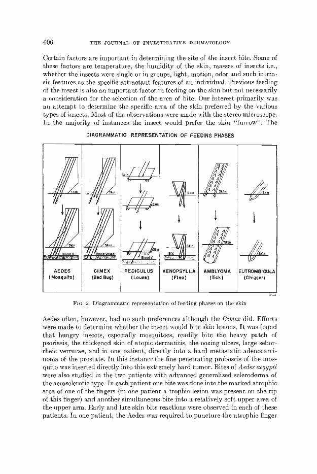
Certain factors are important in determining the site of the insect bite. Some ofthese factors are temperature, the humidity of the skin, masses of insects i.e.,whether the insects were single or in groups, light, motion, odor and such intrin-.sic features as the specific attractant features of an individual. Previous feedingof the insect is also an important factor in feeding on the skin but not necessarilya consideration for the selection of the area of bite. Our interest primarily wasan attempt to determine the specific area of the skin preferred by the varioustypes of insects. Most of the observations were made with the stereo microscope.In the majority of instances the insect would prefer the skin "furrow". The
DIAGRAMMATIC REPRESENTATION OF FEEDING PHASES
Aedes often, however, had no such preferences although the Cimex did. Effortswere made to determine whether the insect would bite skin lesions. It was foundthat hungry insects, especially mosquitoes, readily bite the heavy patch ofpsoriasis, the thickened skin of atopic dermatitis, the oozing ulcers, large sebor-rheic verrucae, and in one patient, directly into a hard metastatic adenocarci-noma of the prostate. In this instance the fine penetrating proboscis of the mos-quito was inserted directly into this extremely hard tumor. Bites of Aedes aegyptiwere also studied in the two patients with advanced generalized scleroderma ofthe acroscierotic type. In each patient one bite was done into the marked atrophicarea of one of the fingers (in one patient a trophic lesion was present on the tipof this finger) and another simultaneous bite into a relatively soft upper area ofthe upper arm. Early and late skin bite reactions were observed in each of thesepatients. In one patient, the Aedes was required to puncture the atrophic finger
FIG. 2. Diagrammatic representation of feeding phases on the skin

z,r__ 4n r- .- t is.
D '?'
r
a
INSECT BITE REACTION 407
area three times to secure an adequate blood meal. Each of these areas producedan immediate bite reaction. In this area the pruritus associated with the bite wasmore severe than the pruritus of the bite reaction on the upper arm. The bitereactions were repeated in one of the patients during a prolonged course ofACTH which produced some improvement of the scleroderma. Under ACTHtherapy, no pruritus or immediate or late reactions were observed.
On the hairy areas, of course, the Pediculus humanus corporis moved aroundin an attempt to search for smooth skin. In previous studies Phthirus pubis was
FIG. 3. Infra-redphotomicrographsto show biting parts: A. Mouth parts of tick (Ambly-omma arnericanuni)—75X; B. Mosquito (Aedes aegypti) showing mandible, maxilla andIabium—75X; C. Head of bedbug (Cirnex lectularius) with maxillae separated out—75X;D. Dog flea (Ctenocephalides canis) showing maxillary paips; piercing structures partiallyobscured—75X.
observed to bite the scalp of children. Attempts to have the Phthirus pubis bitethe scalp of adults were unsuccessful. In one experiment, a Pediculus humanuscorporis bit the scalp of a young child. Aedes aegypti also bite through the thickskin on the palms of the hand. In observations under the microscope, it wasusually not apparent why the insect in response to its reflex responses, probesaround before it finds a favorable site. Often the hungry mosquito, especially theAedes, did not probe but bit immediately. The reason may be, of course, in theavailability of the blood supply in that area rather than any factors in or on thesurface of the skin. In the artificial skin membranes with the human skin, the
'ø
B
408 THE JOURNAL OF INVESTIGATIVE DERMATOLOGY
few mosquitoes that did feed, did so after prolonged probing. So, in general withthese preliminary experiments, there did not seem to be any selective skin sitefor the hungry insect. Photographs in color at 40X and 100X of the insect onthe surface of the skin were taken with the technics reported previously (4).Moving pictures at 20X and 40X of the progress of the insect across the surfaceof the skin have not been done but are being planned.
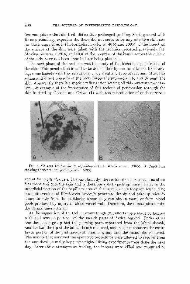
The next phase of the problem was the study of the technic of penetration ofthe skin. This penetration is said to be done either by means of lancet-like stick-ing, some lancets with tiny serrations, or by a cutting type of reaction. Muscularaction and direct pressure of the body forces the proboscis into and through theskin. Apparently there is a specific reflex action setting off this puncture mechan-ism. An example of the importance of this technic of penetration through theskin is cited by Gordon and Crewe (1) with the microfilariae of onchocerciasis
Fin. 4. Chigger (Eutrombicula alfrcddugesii): A. Whole mount—240X; B. Capitulumshowing chelicerae for piercing skin—510X.
and of Bancrofti filariasis. The simulium fly, the vector of onchocerciasis as otherflies rasps and cuts the skin and is therefore able to pick up microfilariae in thesuperficial portion of the papillary area of the dermis where they are found. Themosquito vectors of Wnchereria bancrofti penetrate deeply and take up microfi-lariae directly from the capillaries where they can obtain more, or from bloodpools produced by injury to blood vessel wall. Therefore, these mosquitoes missthe dermal microfilariae.
At the suggestion of Lt. Col. Jaswant Singh (6), efforts were made to tamperwith and remove portions of the mouth parts of Aedes aegypti. Under etheranesthesia one group had the piercing parts separat.ed from the labial sheath,anot.her had the tip of the labial sheath removed, and in some instances the entirelower portion of the proboscis, still another group had the mandibles removed.The insects that survived the operative procedures were allowed to recover fromthe anesthesia, usually kept over night. Biting experiments were done the nextday. After these attempts at feeding, the insects were killed and mounted to

INSECT BITE REACTION 409
study the details of the remaining mouth parts. In general, those insects that didsurvive, probed or rather attempted to probe. The most active group, of course,was the group with the piercing parts completely separated from the labialsheath. For successful penetration of the skin, both the piercing parts and foodchannel must be present.
The obvious difficulty of observation under the microscope was that, for themost part, the view through the microscope was vertically above the biting partsof the insect. It was not possible then to see the actual beginning penetration ofthe skin save in a few instances when this was done with Cimex. If the insectswere placed on the skin and covered with small transparent plastic boxes (7)which could be placed in the field of the microscope, observations on movementsover the surface of the skin and into the skin could be done earlier than if theinsects were placed on the skin with a test tube. Attempts were made to study theimage transmitted through prisms placed adjacent to the biting areas. Theseexperiments were not successful. Efforts were made to select areas—whose sur-face could be shifted in different planes such as forearm, finger, etc. In observa-tions on biting on the forearm, it was often not possible to keep the insect in viewwhile it was looking for a biting area with the forearm placed in varying positions.By means of the lucite rod attached to microscopic lamps, as we had describedin previous work with cutaneous microscopy (3), efforts were made to try tobring this "cold" light close to the biting area of the insect and on the undersurface of the skin of the web of the fingers for interdigital biting experiments inman. This technic would give clear lighting, translucent and silhouette lighting,but it was not of value for studying actual penetration through the skin. Withthis lucite rod attachment the biting parts of the bedbug were made more trans-parent. It was possible with the Cimex to see the internal mouth part structuresthat slide through the labium at the initial piercing and continued piercing of theskin and during feeding. Observation on feeding on the tip of the ear and the webof the fingers still did not make the actual technic of epidermal and dermal pene-tration clearer. Clearing agents on the skin interfered with the biting and oft.enserved as repellents.
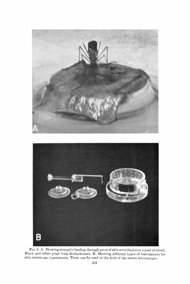
To provide one type of artificial membrane through which the insects couldbite, human skin was used. These membranes included fresh skin from the op-erating room and from post-mortem material. Small plastic chambers weredevised by Lyons (7) to provide both a reservoir for the blood pooi under theskin and an attachment for the skin on top. Later, humidity and temperaturecontrol were provided by a special additional box arrangement. As the illustra-tions indicate, these transparent appliances were small so they could be manipu-lated readily under the microscope. In some instances, the skin before use wasscraped to make it thinner, warmed gently and covered with glucose solution.The details of the successful animal skin membrane technic of Bishop and Gil-christ (8, 9) were not followed, for our primary purpose was to study the technicof penetration rather than that of feeding. With various technics utilizing humanskin, the mosquitoes Aedes and Anopheles did much less preliminary probingthan the Cimex indicating perhaps that the latter is a more shallow feeder. How-
, 4
Ii
FIG. 5. A. Showing mosquito feeding through piece of skin stretched over a pooi of blood.Black and white print from Kodachromes. B. Showing different types of instruments forskin membrane experiments. These can be used in the field of the stereo microscopes.
410

INSECT BITE REACTION 411
ever, the percentages of insects probing or feeding with our human skin mem-brane technics were not high enough to warrant continuing this. However,efforts are being continued to study membrane technics especially for purposes offeeding. A few experiments were done on the rabbit ear and attempts \vere madeto use a flap of excised skin with the skin made translucent by lucite rod trans-mitting the light. Recently Griffiths and Gordon (14) have reported on an ap-paratus for the direct viewing of living skin.
The next step in the actual progress of the bite is the study of the penetrationof the blood vessel. In the transparent frog web, Gordon and Lummsden observedthe proboscis during feeding. They believed that there were two types of feeding,a direct intravascular and a pool feeding from a previously lacerated vessel. Instudies on anesthetized guinea pigs with 14edcs aegypti, Gordon and Crewe foundlocalized hemorrhages one hour after biting and in other instances no abnor-malities (serial section technic). These negative findings they interpreted pre-sumably as evidence of intracapillary feedings. In sections 24 hours after bitingthey observed localized hemorrhage and edema with separation of collagenfibers. The flexibility of the proboscis during feeding has been shown for themosquito and also for the Glossina and Gimex (1). As indicated previously, at-tempts to study the relationship of the biting parts to the superficial blood vesselswere not successful in the human and with the rabbit ear, even under infra-redmicroscopy. Therefore, with our direct microscopic technics during the actualbiting process in human skin we could not tell whether there was any penetrationof the blood vessel. During feeding we could observe in certain individuals red-ness about the bite area and edema. This redness was studied in detail with clear-ing agents on the skin. This reaction was found chiefly to be due to markeddilatation of vessels presumably in the subpapillary plexus area. This was studiedchiefly with the mosquitoes and Cimex. However, the fact that the Cimcx takeslonger time and more effort than the mosquito in instituting a flow of blood intoits body could be an indication of an initial laceration of a vessel for pool produc-tion. We believe without direct proof, that longer feeding insects such as theAmblyomma americanum and Cimcx use pool feeding. The Eutrombicula is re-ported after penetration to produce lysis (partial digestion?) of tissue and toaspirate this digested material without any direct or indirect vascular feeding.As yet, we have not examined any Eutrombicula after feeding, nor do we have,as yet, any serial section studies on human skin with Eutrombicula. It is believedthat at times bits of chitinous materials of the arthropod may be left in tissue.
Fluorescein was utilized as a tracer substance for experiments preliminaryto the use of isotopes in insects. Fluorescein was added to the sugar water forAedes aegypti. There was no detection of fluoroscein 3, 6, and 24 hours afterfeeding. A normal type of whitish fluorescence appears in the white bands of thehind tarsi of Acdcs. No fluorescence was visible in the bite area of some of thesemosquitoes on the skin. Ten Cimcx were injected with a minute drop of fluores-cein. The patterns of injection were studied and appeared concentrated in thelocal area of injection and were more pronounced in 23 hours than in 3—4 hours.No fluorescence was visible in the skin area of the bites of 3 of these injected
B
412 THE JOURNAL OF INVESTiGATIVE DERMATOLOGY
Cimex.. These few experiments then demonstrated that it was possible to injecta tracer substance in the larger insects.
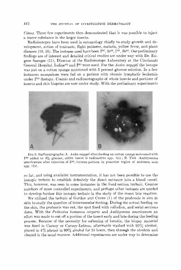
Radioisotopes have been used in entomology chiefly to study growth and de-velopment, action of toxicants, flight patterns, malaria, yellow fever, and plantdiseases (15, 16). The isotopes used have been p22, Sr89, JG, Br82. Our preliminaryfindings are of interest and detailed critical studies are under way with Dr. Eu-gene Saenger (11), Director of the Radioisotope Laboratory at the CincinnatiGeneral Hospital. Iodine'31 and 32 were used. For the Aedes aegypti the isotopewas put on a cotton sponge moistened with 5 percent glucose solution. In a fewinstances mosquitoes were fed on a patient with chronic lymphatic leukemiaunder 32 therapy. Counts and radioautographs of whole insects and portions ofinsects and skin biopsies are now under study. With the preliminary experiments
FIG, 6. Radioautographs: A. Aedes aegypti afterfeeding on cotton sponge moistened withI" added to 5% glucose; entire insect is radioactive app. 12X; B. Tick—Amblyommaamericanum after injection of 1131; festoon pattern in posterior region of abdomen seenapp. 12X.
so far, and using available instrumentation, it has not been possible to use theisotopic technic to establish definitely the direct entrance into a blood vessel.This, however, was seen in some instances in the fixed section technic. Greaternumbers of more controlled experiments, and perhaps other isotopes are neededto develop further this isotopic technic in the study of the insect bite reaction.
We utilized the technic of Gordon and Crewe (1) of the proboscis in situ inskin to study the question of intravascular feeding. During the actual feeding onthe skin, the proboscis was cut, the spot fixed with collodion, and serial sectionsdone. With the Pediculus humanus cor poris and Amblyomma americanum aneffort was made to cut off a portion of the insect early and late during the feedingprocess. Because of the necessity for softening of keratin, the biopsy materialwas fixed in Carnoy or Carnoy-Lebrun, afterwards washed with 95% alcohol,placed in 4% phenol in 80% alcohol for 24 hours, then through the alcohols andcleared in the usual manner. Additional experiments are under way to determine

H
1k
A
INSECT BITE REACTION 413
the preferred type of fixation technic both for the insect and the skin. A fewexperiments were done also with rabbit and guinea pig skin.
The proboscis in situ also serves as an indicator for studying the intensity ofthe bite reaction in that particular area of the skin. It is emphasized again thatunless one does careful serial sections a critical evaluation of the intensity of thetissue response to the irritation of the bite cannot he given. It is felt that thereis needed a detailed description of the histopathology of the early arthropodbite reaction qualified in terms of known sensitivity of the individual to the bite
Fie. 7. Biopsies with proboscis of mosquito Anopheles quadrimaculatus in situ HE.stain: A. Showing fasicle adjacent to blood vessel in mid dermis. Note absence of bleeding—25X; B. Showing depth of fasicle in skin. Note absence of any inflammatory infiltrate oredcma—25X.
of that particular arthropod. There have been excellent studies of the late reac-tions but relatively few studies of the early reactions. A brief review of our histo-pathologic studies nw indicates, of course, that the intensity of the tissue re-sponse depends upon the sensitivity of the individual, the type of arthropod, andthe duration of feeding. First, actually little is seen save edema, then there is aperivascular polymorphonuclear and lymphocytic response localized to the areaof the bite. The early reaction may show very little even in those biopsies withthe proboscis in situ. Later, the edema is less intense and perivascular collars ofinflammatory cells, especially lymphocytes, are present, often surprisingly deepin tissue. Later, the eosinophilic, plasma cellular, and histiocytic responses maydevelop. This is but a brief summary of the progressive stages of development.Further details of the early and late reactions will be presented in the report on

414 THE JOURNAL OF INVESTIGATIVE DERMATOLOGY
Fie. S. The arthropod bite reaction as affected both by its location on lower leg and insome instances by the sensitivity of the patient; Black and white prints from Kodachromes.A. Mosquito—with eczematoid and petechial response; B. Chigger—4 weeks duration;with ulceration, hackground of stasis syndrome; C. Mosquito—persistent eczematoid anddiffuse hemosideric response; no local medications; ID. Mosquito—simultaneous bites onhand and ankle—on the hand a mild papular reaction, on the ankle a bullous reaction.

:2
INSECT BITE REACTION 415
the study of the allergic phases of the insect bite reaction. The detailed histo-pathologic studies will also be presented in a separate report.
There is still considerable confusion as to whether there is a constant flow ofsalivary secretion into the bite area or just initial "lubrication" of the puncturearea. Gordon and Lummsden (10), in the frog web experiments, described theinjection of a fluid presumably salivary secretion, in the tissues at variousstages of penetration of the proboscis. Secretion from the salivary gland doesenter the tissue of the host. Many studies have been done of the salivary glandsof various insects and such materials as heparin-like substances, agglutinins,hemolysins, etc., have been found. For example, the salivary secretion of Andesaegypti is said not to contain any agglutinins or hemolysins or anti-coagulins,that of Ann pheles maculipennis, agglutinins. Gimex are supposed to have a hemo-lytic factor in the salivary secretion (12). However, the saliva of the various
Fin. 9. Some types of nodular responses following arthropod bites. Black and white printsfrom Kodachromes: A. Localized neurodermatitis, thigh—6 months duration—mosquito;B. Petechial inflammatory—finger—3 weeks—mosquito; C. "Spontaneous" nodule—lowerleg—years—mosquito (no biopsy).
insects do differ from each other (13). In many cases the longer the insect feeds,the more pronounced will be the reaction. For example, the adult Cimex maycause more reaction than the smaller nymphs. In addition, the insect may con-tain bacteria and fungi. However, these do not produce the picture of the insectbite reaction.
The persistence of the insect bite reaction depends to some extent as indicatedpreviously upon the amount of tissue response. However, certain obvious exter-nal factors can influence the persistency of the insect bite reaction. Scratchingand rubbing will aggravate the insect bite reaction by increasing the tissuedamage. Varied pictures of secondary localized, so-called, neurodermatitis mayensue. Gordon and Crewe (1) indicate that in regard to the bites of lice and redu-viid bugs, the irritation from scratching may facilitate the entry of parasitesand increase infection with typhus or South American trypanosomiasis. Second-ary infection may develop of course in an insect bite and varied types of pyo-
416 THE JOURNAL OF INVESTIGATiVE DERMATOLOGY
derma result. Granuloma pyogenidum may also follow an insect bite. Withsimultaneous insect bites on the upper and lower extremities, those in the latterarea show greater persistency and may even be more severe with vesiculo-bullousreactions and petechiae. In individuals with stasis syndrome of the lower extremi-ties, insect bites may cause severe reactions including extensive ulceration. Thereaction of the insect bite to an external factor will produce persistent infiltratednodules in the skin. Such nodules have been seen not only after scabies, but alsoafter the bites of other arthropods.
Independent of external irritant factors, the insect bite reaction may of itselfpersist for some time. As Allen (19) and others (20) have indicated, these chroniclesions may sometimes simulate lymphomas. Allen believes that some agent ofthe arthropod may persist actively in the tissue for some time. We have observedbits of "cuticular" material in the upper dermis after tick bite.
Preliminary experiments (17, 18) with the injection of saline, procaine, andcortisone and Compound F into the insect bite reaction by means of the syringeand by the Hypospray jet injection apparatus showed, in a few instances, someincreased healing with cortisone. This was not marked. Additional experimentsare being done utilizing much smaller amounts of cortisone. Local Compound Fhas been found to cause definite inhibition of the skin reaction to bites of mos-quitoes. The pruritus of chigger bites may be lessened by direct intra-lesionalinjection with procaine and Compound F by the Hypospray apparatus. Morecontrolled experiments of this type are needed.
The attempted study of the insect bite mechanism directly and dynamicallyin the human skin illustrates many of the difficulties in such an investigation.This report details important phases with only preliminary experiments done formany of these phases. Instrumentation with various types of microscopes has itslimitations. Attempts in the direction of skin flap and skin fold and human skinmembrane could be continued. The study of the infra-red microscope was disap-pointing especially for direct use on the skin. Animal membranes will have to betried. Perhaps the detailed and controlled study of prepared fixed tissue wouldbe of greater value. Because of the delicacy of the biting parts and the physicalproperties of light on and through human skin, it may not be possible to studythe exact progress of this mechanism dynamically in human skin. The clinicaldermatologist must be interested in the mechanism of the insect bite reactionand is urged to contribute his critical findings.
REFERENCES
1. GORDON, R. M. AND CREWE, W.: The mechanisms by which mosquitoes and tsetse fliesobtain their blood meal, the histology of the lesions produced, and the subsequentreactions of the Kammalian host; together with some observations on the feedings ofchrysops and cimex, Ann. Trop. Med. and Parasitology, 42: 334, December 1948.
2. MAT1IESON, ROBT.: Medical Entomology, 24th Ed., Comstock Publishing Co., Ithaca,New York, 1950.
3. GOLDMAN, LEON AND YOUNKER, WALDO: Studies in microscopy of the surface of theskin, J. Invest. Dermat., 9: 11, 1947.
4. GOLDMAN, LEON: Investigative studies with pigmented nevi, J. Invest. Dermat. (inpress).

INSECT BITE REACTION 417
5. Kj,ncE, JAS.: Personal communication to the authors.6. SINCH, LT. COL. JA5WANT: Personal communication to the authors.7. LYONS, WILFRED: Personal communication to the authors.8. Bisuop, ANN AND CILCHRIST, BARBARA M.: A method for collecting sporozoites of plas-
modium gallinaceum by feeding of Aedes aegypti through animal membranes, Nature,153: 713, 1944.
9. Bisuor', ANN AN0 CILcI-IRIST, BARBARA M.: Experiments upon the feeding of Aedesaegypli through animal membranes with a view to applying this method to the chemo-therapy of malaria, Parasitology, 37: 85, January, 1946.
10. Gostoo, R. M. AND LLTMMSDEN, W. H. R.: A study of the behavior of the mouth-partsof mosquitoes when taking up blood from living tissue; together with some observa-tions on the ingestion of microfilariae, Ann. Trop. Med. and Parasitology, 33: 259,1939.
11. SAENORR, EUGENE: Personal communication to the authors.12. SANGI0RGI, C. AND FRowNs, D.: Di un pricipio emolitico ("cimicina") nella saliva del
"Cimex lectularius", Pathologica 32: (583) 189—191, May, 1940. Abstracted by Bettini,S.: Biol. Abstr. 21: 10529, 1947.
13. BARER, ARTHUR C.: Personal communication to the authors.14. GRIFFITIIS, E. B. AND CORDON, R. M.: A simple apparatus designed in order to observe
insects feeding on living tissue or penetration of helminth larvae, Trans. RoyalSoc. Trop. Med. and Hyg., 44: 336, Feb., 1951.
15. JENKINS, DALE W. AND BASSETT, CRARLES C.: Radioisotopes in Entomology, Nu-cleonics, 6: 5, March, 1950.
16. BUGHER, JOHN C. AND TAYLOR, MARJORIE: Radio-phosphorus and radio-strontium inmosquitoes, Science, 110: 146, August 5, 1949.
17. PRESTON, ROBERT B., GOLDMAN, LEON, AND THOMPSON, LT. COL. ROBERT C.: The useof the Rypospray in dermatology, Arch. Dermat. and Syph. 64: 327 (Sept.) 1951.
18. COLOMAN, LEON, THoMPsoN, LT. CDL. ROBERT C., AND TRICK, CAPT. RANDOLPH E.:Cortisone acetate in skin disease. Local effect on the skin from topical applicationand local injection A. M. A. Arch Dermat. and Syph. 65: 177 (Feb.) 1952.
19. ALLEN, ARTHUR C.: Persistent "insect bites" (dermal eosinophilic granulomas) simu-lating lymphoblastomas, histiocytosis and squamous cell carcinoma, Amer. J. Path.,24: 367, 1948.
20. WINER, L. B. AND STRAKO5cH, E. A.: Tick bites—Dermacentor Variahilis (Say), J.Invest. Dermat., 4: 249, June, 1941.
DISCUSSION
DR. PECK, New York: During the var, the mechanism of insect bites astypified by the louse bite was of practical importance because of the role of licein spreading typhus fever. My associates and I investigated the louse bite (Cu-taneous Reactions Due to the Body Louse (Pediculus humanus), Peck, S. M.,Wright, W. H. and Cant, J. Q.; J. A. M. A., 123: 821—825, Nov. 27, 1943). Weraised colonies of lice on ourselves and in this way were able to observe the proc-ess very carefully. The first evidence of a reaction was always a purpuric spotbecause of the bite. As allergy developed, the typical louse bite reaction was seenwhich was papular and urticarial. We were able to demonstrate that the allergywas due to an antigen in the feces of the louse as well as in the salivary secre-tions.
Du. BEERMAN, Philadelphia: I am certain that in his rush for time, Dr. Gold-man did not mean to imply that eight-legged arthropods \vere insects but, rather

418 THE JOURNAL OF INVESTIGATIVE DERMATOLOGY
arachnids (mites, etc.). These are to be sure, arthropods, but not of the insectfamily.
DR. GOLDMAN: The pediculi were adapted to feeding on rabbits through thetechnic of Culpepper. This is a significant step, by the way, in the study ofinsect-borne diseases. He has also succeeded in getting small colonies to grow onwhite rats.
Now that point brought up about the feces is extremely important in regard toscratching the area of an insect bite. For example, South American trypanosomia-sis or Chagas disease is perhaps actually acquired that way, i.e., by rubbing inthe bite area the infected feces of the arthropod. Typhus, as Dr. Peck mentioned,is another example. We have seen also eczematization from bites of pediculi.
Also, we did not mention the technic of studying the bite reaction in situ underthe microscope in determining the vascular patterns, which, incidentally, incases of mosquitoes may sometimes persist for an appreciable length of time.
The point Dr. Beerman makes is a very excellent one. Many of the insect bitereactions were done in entomologists. This gave us an unusual opportunity tostudy bite reactions in people who knew exactly what species bit them. And theywere always very interested in correcting our terminology. Arthropod is of coursethe general term which should be used.