the innervatio onf the gut of the brown trout (salmo...
TRANSCRIPT

199
The Innervation of the Gut of the Brown Trout(Salmo trutta)
By G. BURNSTOCK
(From the Department of Zoology, King's College, London; present address,Pharmacology Department, University of Oxford)
With two plates (figs. 5 and 7)
SUMMARY
1. The extrinsic and intrinsic innervation of the trout gut has been examined byusing methylene blue, osmium tetroxide, and silver staining techniques.
2. Left and right vagus nerves penetrate the oesophageal wall and run between thelongitudinal and circular muscle-coats of the stomach, but do not reach the intestine.Branches of these nerves anastomose with each other and become incorporated intoAuerbach's plexus. Pericellular endings of vagal fibres are found about enteric neuronesin the stomach, while some fibres end directly on the striated muscle-cells of theoesophagus.
3. A single, fine, anterior splanchnic nerve anastomoses along the major branchesof the coeliaco-mesenteric artery to supply both the stomach and intestine. Most ofthe splanchnic fibres pass directly into the smooth muscle-coats, but some fibres runinto the subserous plexus.
4. Two posterior autonomic nerves provide separate innervation of the rectum.5. Three main types of nerve-cells have been distinguished in the gut by their
distribution as well as structure.(a) Small uni-, bi-, and multi-polar cells (10-15 ju. m diameter) form the main body
of Auerbach's plexus, lying between the muscle-coats. Throughout most of the gutthey are not arranged in true ganglia, since only from 1 to 3 neurones occur at thenodes and many cells lie singly along the internodal connectives.
(b) A smaller number of large multipolar neurones (30-60ft in diameter) lie half-embedded in the longitudinal muscle along the whole length of the gut, theirprincipal axons passing down to lie between the muscle-coats in Auerbach's plexus.
(c) A third cell type consisting mainly of large mono- or bi-polar cells (30-70 fx indiameter) contributes to Auerbach's plexus in the stomach only and is associated withthe endings of vagus nerve-fibres.
6. Bundles of nerve-fibres pass down through the circular muscle-coat fromAuerbach's plexus to join a loosely arranged plexus of nerve-fibres, the submucousplexus; but unlike Meissner's plexus of mammals, which occupies a similar region, nonerve-cells are present.
7. Nerve-endings, some of which may be sensory, are located at the bases of themucous epithelial cells and are continuous with fibres forming a prominent sub-epithelial plexus from which fibres pass to both submucous and Auerbach's plexuses.
8. A subserous plexus and 'interstitial cell network' are also present in the fish gut.9. A quantitative study of nerve-cell densities in different regions of the trout gut
revealed density peaks of 93 cells per square mm and 170 cells per square mm in theposterior cardiac stomach and anterior duodenum respectively. The ratio of smallto large neurone types in Auerbach's plexus is about 20 to 1 in all regions. Thetotal number of nerve-cells in the whole gut was calculated to be about half a million.
[Quarterly Journal of Microscopical Science, Vol. 100, part 2, pp. 199-220, June 1959.]421.2 P
Burnstock—Innervation of Gut of Trout
INTRODUCTION
ANUMBER of workers have described the autonomic system of teleosts(Stannius, 1849; Chevrel, 1887; Herrick, 1898; Cole and Johnstone,
1901; Miiller and Liljestrand, 1918; Young, 1931; Hirt, 1934; Kirtisinghe,1940) but few of them traced out the exact course of the nerves supplyingthe gut. Young (1931), however, made a detailed investigation of the extrinsicinnervation of the gut of Uranoscopus and Lophius. In this work no vaguselements were found to extend beyond the stomach into the intestine, while asingle anterior splanchnic nerve supplied both the stomach and intestine. Hefailed to discover any separate innervation of the hind part of the gut andfound nothing corresponding to the sacral parasympathetic outflow which ispresent in all tetrapods.
The intrinsic innervation of the teleostean gut was first described by Monti(1895) in the tench [Tinea tinea). A complicated plexus between the muscle-coats was observed, with small ganglia at the nodal points, formed by multi-polar nerve-cells of a single type. Fibres from the plexus were traced throughthe circular muscle-coat to form a submucosal mesh of fibres, some finebranches ending in boutons on muscle-fibres of the muscularis mucosae. Inaddition a subepithelial plexus was described, composed of delicate nerve-endings at the bases of glandular cells. Polyaxonal cells present in this regionwere said to form the fibres of the plexus. The tench gut, however, cannot betaken as typical of the teleostean condition since it contains striated muscle-coats throughout its entire length.
The enteric nervous system of the perch was described by Sakussef (1897),who found nerve-cells of two types in Auerbach's plexus, which correspondedto Dogiel's types I and II, but only type I cells were said to be present inthe intestine. Fibres were traced from the enteric plexus to the mucosa wherethey ramified to form a subepithelial plexus, from which fine varicose threadscame off to form networks about individual epithelial cells. Both Sakussef andMonti described nerve-endings on smooth muscle-cells.
Kirtisinghe (1940) studied the myenteric nervous system of a number oflower vertebrates. His main teleost example was Saccobranchus (Indian cat-fish), but he included in his paper some study of Motella and Ophiocephalus.For the most part he seemed content to refer back to the descriptions of Montiand Sakussef, but there were a number of additional features included in thepaper. He found that the nerve-cells were usually to be seen placed singly inthe meshes of the plexus, rather than aggregated at the nodal points to formganglia. An 'interstitial cell' system lying between the muscle-coats of the gutof Ophiocephalus was described in his paper. The cells were reported to be ofvariable shape with three to four branched processes, arranged syncytially butmaintaining no connexion with the enteric neurones. Those processes notuniting with neighbouring cells penetrated between the muscles. Pericellulararborizations of vagal fibres were found on nerve-cells of type II, as well as

Burnstock—Innervation of Gut of Trout 201
large boutonic nerve-endings on cells of the same type, which he suggestedbelonged to the axons of type I cells.
The object of the present paper is to examine the innervation of the gut of atypical teleost for comparison with those described in other vertebrates. Inaddition this work formed the anatomical basis of studies made on thephysiology and pharmacology of the trout gut (Burnstock, 1957, 1958 a, b).
The brown trout (Salmo trutta) was chosen because it belongs to a teleosteanfamily that retains many primitive and generalized features and because it hasnot adopted a specialized feeding habit. The musculature and general morpho-logy of the trout gut has been described previously (Burnstock, 1959).
TECHNIQUES
Extrinsic nerves
Tracing the paths of the vagus and splanchnic nerves was largely a matterof delicate dissection under a simple lens or binocular microscope. Trout thathad been freshly killed or were preserved in formalin or alcohol were used forthis purpose.
Fish kept in formalin for about three months provided the best workingmaterial for the examination of the vagus nerves; but for the finer brancheslying between the muscle-coats of the stomach wall, whole mounts of freshstomach stained with methylene blue were used. These preparations were bestexamined with a lens while stretched about a glass tube and strongly illumi-nated from within.
The splanchnic nerve is particularly delicate in the trout and is closelyapplied to blood-vessels, so that it was extremely difficult to trace by ordinarydissection methods. It was found necessary to remove lengths of carefullycleaned blood-vessels (and nerve) from fresh material and place them in asolution of o-oi% osmium tetroxide overnight. These preparations, afterbeing well washed, were mounted in glycerine or Apathy's syrup for closerexamination. This technique was also used to trace out fine nerves issuing fromthe posterior trunk region to the rectum.
Enteric plexus
For a comprehensive study of the enteric plexus, both whole mounts andserial sections were required.
Whole mounts. For whole-mount preparations the methylene blue method,modified from Kirtisinghe (1940), proved to be by far the most successfultechnique. The reason for its particular success with trout probably lies in thenature of the gut morphology, namely in the extremely thin outer longi-tudinal muscle-coat in the stomach and intestine. This meant that the dyesolution, which normally has a poor penetration, could reach the plexuswithout disturbance of the nerve arrangement by dissection. Another advantageis the presence of the stratum compactum, which forms a strong stable basewhen manipulating the preparations.

202 Burnstock—Innervation of Gut of Trout
During the development of the technique, control experiments werearranged so that the optimum conditions for each of the following factors werediscovered: fish size; composition of the dye itself and the strength of thesolution used; acidity; salinity; time of immersion of preparations in the dyesolutions; temperature; fixative and fixation time; clearing agent; use of suchadditional substances as hyalase, glucose, sodium acetate.
The final technique used was as follows:1. Young fish, about 10 cm long, were pithed and the gut dissected out.
The gut was then cleaned out thoroughly with o-8% saline solution.2. The preparations were stretched both evenly and maximally in both a
circular and longitudinal direction. The most efficient method of achievingthis end was by extending large cylindrical pieces of the gut over tapered,constricted glass tubes, a whole size-range of which was made. Preparationsextended in this way were very easy to handle with the minimum of dis-turbance. For some regions of the gut it was more convenient to stretch thepreparations, muscle-coat uppermost, on a thin wax plate by means of entomo-logical pins. The plates were then placed face down in a dish of dye solution.In yet other cases the gut was pumped up with dye solution and sutured.
3. The most effective staining was obtained by placing the preparations inthe dye solution immediately after killing the fish. After experiments with 6refined methylene blue dye types, crude dye (made by Williams and Hopkin in1913) proved to be the most effective. The dye was made up in a solution ofsalinity o-8%. At greater concentration the penetration improved but thenerves disintegrated; when the salinity was less than o-8%, the nerve-cellsonly were stained and the contrast was poor. A weak dye solution (o-oi%),taking 2 or 3 h to penetrate, produced the most delicate results with thegreatest contrast. The acidity of the solution proved to be an important factor.The best results were obtained at pH about 5-5 and 6-5 for the stomach andintestine respectively. The solution was maintained at about 12° C duringstaining.
4. On completion of staining, the preparations were exposed to the air forabout 4 min in order to oxidize and remove any excess dye, before transferringthem to the fixative, 8% ammonium molybdate being used. In two experi-ments the fixative containing osmium tetroxide recommended by Alexan-drowicz (1951) was used, and gave a better contrast between the nucleus andcytoplasm of the nerve-cells. Kirtisinghe (1940) suggested a duration of 30min for fixation, but it was found that a fixation time of less than 10 h led torapid fading of the nerve-fibres.
5. After fixation the gut was cut off the glass tube by a longitudinal incisionand laid out flat. At that stage, the mucous coat could easily be scraped offwith a blunt knife down to the stratum compactum. The preparation was thenwashed with distilled water for 2 to 4 h.
6. The pieces of gut were then gently flattened between two slides and putin a dish of 95% alcohol for about 1 h, transferred to absolute alcohol for afurther hour, and then cleared in cedarwood oil. At that stage the stratum

Burnstock—Innervation of Gut of Trout 203
compactum could be separated off from the muscle-coat so that a thin, flatmuscle preparation could be mounted on large slides, longitudinal layeruppermost.
In most regions the longitudinal muscle-coat is thin, and this, together withthe fact that the nerve-arrangement between the two muscle-layers is verycomplex, makes further dissection a disadvantage. In the rectum, however,it was sometimes an advantage to dissect off the longitudinal coat after thepreparation had been in the stain solution for about 1 h. Otherwise the staindid not penetrate to the nerves.
A number of further refinements in methylene blue technology have beensuggested in the literature, but the following recommendations have notproved to be particularly successful with trout gut staining. The use ofhyaluronidase as a pretreatment 'spreading factor' (Weddell and Pallie, 1954)removed the staining contrast, the muscle staining blue as well as the nerves.The recommendations of Schabadasch (1934) to increase penetration of thedye by means of glucose or pyruvic acid and to improve the staining of finestructure with sodium acetate and magnesium bromide proved to be of littleadvantage with the trout gut. The method of heating the saline solution to500 C before adding the dye, when making up the staining solution, suggestedby Hillarp (1946), produced no specially good results.
Injections of the dye solution during life have not given results of a standardcomparable to those obtained by the supravital technique outlined above.
Both the gold chloride (Gairns, 1930) and silver methods (Gunn, 1951;Hill, 1927) of preparing whole mounts of the enteric plexus yielded poorresults, owing mainly to heavy staining of the muscle as well as the nerve.
Serial sections. A number of very careful attempts to stain sections of thetrout gut with silver were made but with no outstanding success, despitecontrolled variations of fixatives, pH, temperature, and times of staining.This supports the view that fresh-water teleosts are particularly refractory tosilver staining (Young, 1931).
Both the Bodian (1937) technique using protargol and Holmes's technique(Holmes, 1947) produced some staining of the subepithelial nerves of thestomach and intestine, as well as blackening of the sub-serous region.
The silver technique recommended by Peters (1955) produced by far themost consistent results. In general the nerve-fibres, especially those ofthe submucous coat, stained very well but the nerve-cells usually only tookthe stain peripherally.
The osmium tetroxide technique for staining fixed sections recommendedby Kiss (1932) produced an even stain of muscle and nerve.
Permanent records were made of characteristic features of the entericsystem by means of photomicrographs. The contrast of methylene blue pre-parations was found to be optimal for photographic purposes at about threemonths, after which time there was a slow fading of the stained parts.
A study was made of the density of nerve-cells present in Auerbach'splexus in the oesophagus and in different regions of the stomach and intestine.

204 Burnstock—Innervation of Gut of Trout
Sections of the gut-wall showed that the nerve-cells of Auerbach's plexuslie roughly in one plane, so that counts could be made from the surface. Thecounts were based on the number of cells contained in sample mm squares.Only selected parts of the best stained preparations were used. Stomach countswere made on preparations taken from 7 fish and intestinal counts from 5.About 5 separate counts were made on each preparation of one region fromeach fish.
RESULTS
Extrinsic nerves
Charts showing the paths of the nerves supplying the gut of the trout havebeen mapped out from dissections of over 30 fish.
The paths of the vagus nerves are shown in fig. 1. The main vagus trunkspenetrate the gut-wall at the oesophagus and run down the stomach betweenthe circular and longitudinal muscle-coats. Two or three branches come offeach of the left and right vagus nerves before they enter the gut-wall. Thesebranches contain fibres which innervate the striated muscles of the oesophagusdirectly. Both vagus nerves run up towards the dorsal surface of the stomachsoon after penetrating the gut, the left trunk giving off four major branchesventrally, while three ventral branches come off the right vagus. Thesebranches follow a fairly constant pattern in the first part of their passagebetween the muscle-coats but the more posterior innervation and minorbranch connectives are by no means constant in their arrangement. Thereare frequent anastomoses between left and right vagal elements, both dorsallyand ventrally, although the density of vagus branches is greater on the ventral,inner curvature of the stomach than dorsally. The vagus fibres beyond theantrum are collected in very fine bundles. No fibre bundles have been tracedas far as the pyloric sphincter, and it seems most unlikely that any vaguselements extend into the intestine.
Fig. 2 shows the paths of the branches of the coeliaco-mesenteric artery. Thesplanchnic nerve is very fine and anastomoses during its route along themajor branches of this blood-vessel.
Fine left and right branches from the first abdominal sympathetic gangliajoin at the junction of the gastric and hepato-intestinal arteries to form a smallsplanchnic ganglion just anterior to the opening of the pneumatic duct intothe right dorsal surface of the oesophagus. There are three main branchesfrom the coeliaco-mesenteric artery at about this level; the gastric and hepato-intestinal arteries and a smaller vessel which supplies the region of the'duodenal-ileal'junction. Three main splanchnic nerves come off the ganglionto follow these blood-vessels.
The gastric artery and accompanying nerve split up almost immediately inthe oesophageal region into left and right branches, which closely adhere tothe gut-wall and are embedded in adipose tissue. The left branch supplies theleft side of the cardiac stomach and the spleen, while the right artery branches

Burnstock—Innervation of Gut of Trout 205
<rynx
pylorus
FIG. I. Dorsolateral views of the innervation of the stomach of a trout 228 mm long by (A) leftvagus nerve, (B) right vagus nerve.
again to supply the right side of the cardiac stomach and the pyloric stomach.Fine branches of these nerves penetrate the gut-wall at frequent intervalsalong the stomach, accompanying minor blood-vessels. Elements from theright splanchnic gastric nerve join with fibres from both the left and rightvagus nerves to innervate the pneumatic duct and swim bladder. Thesplanchnic nerve following the hepato-intestinal artery divides into twobranches, one supplying the pyloric caeca and 'duodenum', the other runningdown the length of the intestine. A number of the fine splanchnic branchesterminate in the walls of the arteries and do not reach the gut.

206 Burnstock—Innervation of Gut of Trout
splanchnic /„/•,.gastric artery. -£_„/,_„ , t e f t
. \ 9pnglion vagus nerv
rightvagus'nerve
Fie. 2. Path of splanchnic nerve to the trout gut. Fine splanchnic nerves follow all the majorbranches of the coeliaco-mesenteric artery. The stomach is shown from the dorsal surface.
spinal nerve,rib
left sympatheticchain
right sympatheticchain
kidney
body wall*
FIG. 3. Innervation of the trout rectum by posterior autonomic nerves.
A posterior 'sympathetic' nerve innervates the rectum as well as the gonads(fig. 3). The nerve originates in the last right abdominal sympathetic ganglion,but an extremely fine nerve joins it close to this ganglion from the leftsympathetic chain. Two segments farther back another nerve penetrates thekidney and swim bladder to supply the gonads, urinary bladder, and rectum.It was not possible to see whether this nerve originated from the sympatheticchain or from spinal nerves.

Burnstock—Innervation of Gut of Trout 207
Enteric plexus
General plexus structure. The distribution of nervous elements in the gut-wall is shown diagrammatically in fig. 4.
FIG. 4. Diagrammatic transverse section through the wall of the trout stomach, to show thegeneral arrangement of nervous elements. Compiled from both silver and methylene bluestained preparations, cm, circular muscle; Im, longitudinal muscle; me, mucosal epithelium;mm, muscularis mucosa; NI, NIII, types I and III nerve-cells; s, serosa; sc, stratum com-
pactum; sm, sub-mucous coat.
Auerbach's plexus lies prominently between the longitudinal and circularmuscle-coats. Throughout most of the gut there are no true ganglia in theplexus since only from 1 to 3 nerve-cells occur at the nodes and many cells liesingly along the internodal connectives (fig. 5, A). A smaller number of largenerve-cells is embedded in the longitudinal muscle, their axons passing downto join the plexus (fig. 5, B). A third cell type, which is clearly associated withvagal fibres, is found only in the oesophagus and stomach.

208 Burnstock—Innervation of Gut of Trout
Bundles of fibres pass down through the circular muscle at irregularintervals from Auerbach's plexus to join a prominent and loosely arrangedplexus of nerve-fibres which lies between the stratum compactum and thecircular muscle-coat. This plexus is present in the intestine as well as in thestomach, despite the absence of a sub-mucous coat and muscularis mucosaein the former. It occupies a similar position to Meissner's plexus of mammals,but no nerve-cells are present. A complex subepithelial plexus is present. Itis very prominent in the stomach, some of the fibres ending about glandularcells while others pass at frequent intervals through the stratum compactumto join the 'sub-mucous' plexus. In the intestine considerably fewer fibres areto be seen subepithelially and the stratum compactum is only rarely penetratedby nerves. Structures embedded in the epithelium, especially at the bases ofthe villi, stain strongly with silver and appear to be connected to the sub-epithelial plexus. A few small, poly-axonal nerve-cells have been seen in thesubepithelium.
A complex arrangement of cells staining with methylene blue lies betweenthe muscle-coats, slightly embedded in the longitudinal muscle; it is showndiagrammatically in fig. 6, A. Processes appear to penetrate between themuscle-fibres. This arrangement bears a close similarity to descriptions ofthe 'interstitial cell network of Cajal' seen in other vertebrate enteric systems.
A sub-serous nerve plexus, which is particularly prominent in the oesophagusand rectum, is formed of a meshwork of extremely fine fibres, connected withroughly spherical masses, 1-4 fj. in diameter, embedded in the serous coat.Some of the fibres run down through the longitudinal coat to j oin the myentericplexus.
The main branches of the splanchnic nerve run superficially along the gut.Minor branches, following the course of the smaller blood-vessels, penetratethe serous coat and longitudinal muscle-layer along the whole length of thegut to join Auerbach's plexus (fig. 6, B). Sometimes fine fibres in the splanchnicbranches pass to the sub-serous plexus. Other fibres appear to pass directly tothe smooth muscle-coats.
The main branches of the vagus nerves run down the stomach between themuscle-coats. From these trunks minor branches are given off which, besidesanastomosing with each other, soon merge intimately with the enteric plexusproper (fig. 6, c). Pericellular endings of vagal fibres are found about entericneurones.
Nerve-cells. Three main types of nerve-cells have been distinguished inAuerbach's plexus, both by their structure and distribution (fig. 7).
Large (30-60 p, diameter), multipolar cells in the outer region of Auerbach's
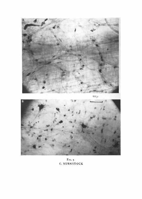
FIG. S (plate), A, photomicrograph of a methylene blue 'whole mount' preparation of theposterior cardiac stomach. Auerbach's plexus can be seen lying between the muscle-coats.Type III nerve-cells lie at the nodes and along the internodal connexions.
B, photomicrograph of a methylene blue preparation of the intestine. It is focused so thatthe type I nerve-cells embedded in the longitudinal muscle can be seen clearly, while theplexus proper formed from type III cells is just visible in the background.
100/1
4*
FIG. S
G. BURNSTOCK

210 Burnstock—Innervation of Gut of Trout
into the oesophagus. These cells are nearly always pear-shaped and mono-polar. Many more cells lie along the vagus branches and a number join cellsof type III to form large diffuse ganglia in the oesophagus and anterior part ofthe stomach. These are often elongate, bipolar cells, or monopolar cells inwhich the process divides into two soon after leaving the cell. Towards theposterior part of the cardiac stomach, and even more frequently in the pyloricstomach, the cells have two or three long dendrites whose branches end indelicate boutons. The axon possesses collaterals. These posteriorly placedtype II cells are sometimes difficult to distinguish from type I cells. It is onnerve-cells of type II that pericellular endings of vagal fibres are found.
Type III neurones are small cells (10-15^1 in diameter) with very largenuclei which do not take the stain (fig. 7, c). They form the main body ofAuerbach's plexus between the muscle-coats, where they lie both at the nodalpoints and along the internodal connectives. Cells of type III may be uni-,bi-, or multi-polar. Uni- and bi-polar cells are found alone, mainly at minorjunctions in the plexus, their axons passing down into the circular muscle,while multipolar cells with up to 5 short branching dendrites are found atthe larger nodal junctions, where other cells are present. In some preparationsthe dendrites appear to lie on the cell bodies of adjacent cells.
Nerve endings. Fine, smooth vagal fibres run alongside individual striatedmuscle-cells in the oesophagus. They give off from 3 to 6 branches, whichdivide further to end in fine clumps on the surface of the muscle, over a distanceof 100 to 350 ft. This arrangement is shown diagrammatically in fig. 8, A. Itwas not possible to see the relation of the nerve-fibres to the sarcoplasm.
Nerve-cells of type II in the cardiac stomach are commonly found to beenveloped by from 1 to 4 fine, intertwining, varicose fibres, which in anumber of cases have been traced back to vagal trunks (figs. 8, B, a ) . Not allthe varicose fibres terminate on the cell surface. Some pass on to rejoin theplexus and end on other type II cells. Some of the cells are enveloped by acapsule on which the intertwining fibres lie, rather than on the cell membrane.
Most of the axons of all the enteric neurones and the dendrites of type IIand III cells run into the plexus and cannot be traced very far. Fine fibresseparate from Auerbach's plexus and run down to end in the circular muscle-coat. They terminate, after branching, in small, heavily stained swellings orboutons. However, like most previous workers who have used silver andmethylene blue staining techniques, I have not been able to see the precisenature of the relation between the nerves and individual smooth muscle-cells.
Nerve-endings are present in the mucosa. In the stomach, fibres separateoff from the subepithelial plexus and surround the gastric gland cells. Heavily
FIG. 7 (plate). Photomicrographs of nerve-cells.A, type I nerve-cell embedded in the longitudinal muscle-coat. Note the short dendrites and
the collateral branches coming off the axon.B, type II nerve-cell, associated with the vagal nerve. Note the long dendrites.C, type III nerve-cells, forming the plexus proper. Note the relatively small size and the
prominent, lightly staining nucleus.

950>/
FIG. 7G. BURNSTOCK

Burnstock—Innervation of Gut of Trout
striatedoesophagealmuscle
W
.subepithelialplexus
columnar^epithelium
10y/
FIG. 8. A, diagram showing the innervation of a striated oesophageal muscle by a vagal fibre.B, c, pericellular endings of vagal fibres about type II nerve-cells (NII). A capsule envelopsthe neurone shown in B. D, diagram showing structures lying between the bases of the columnarepithelial cells, from which fibres run to the subepithelial plexus. E, diagram showing the formof the sub-serous plexus under high magnification, and its connexion with structures embedded
in the serous coat.
staining structures lie towards the bases of the epithelial cells of the intestine,especially at the proximal ends of the villi. They appear to be connected withfibres of the subepithelial plexus (fig. 8, D). In addition, fibres are often seen torun along the bases of the epithelial cells. Whether they form part of local

2i2 Burnstock—Innervation of Gut of Trout
reflex arcs or whether they pass directly to higher centres by way of theextrinsic nerves cannot be decided from the preparations.
Fine fibres of the subserous plexus divide arboreally to end in very delicateboutons usually on the surface of small (3-10 /u, diameter) ovoid structures(fig. 8, E). These endings seem to be confined to parts of the rectum and to theanterior oesophagus.
Regional analysis. The plexus is very diffuse and heterogeneous in theoesophagus. The nodal points sometimes contain up to 8 or 9 nerve-cells(types II and III) while the interganglionic connectives are long and containonly a few fibres (fig. 9, A).
The vagus nerves enter the gut in this region. Aggregations of type II nerve-cells are found along the vagus trunks, both before and after their entry intothe gut. A number of fine side branches of the vagus nerves are given offin the oesophagus. These become incorporated in the beginnings of theenteric plexus.
Fine branches of the splanchnic nerve enter the oesophageal wall, very closeto the right vagus nerve, and contribute directly to the plexus. No nerve-cellaggregations are connected with the entry of the splanchnic branches. Notype I nerve-cells have been seen in the oesophagus, perhaps because nosmooth muscles are present. The subserous plexus is particularly prominentin this region. The plexus in the anterior cardiac stomach is similar to that inthe oesophagus, with nodes containing up to 8 nerve-cells, from which 5 to 8irregular inter-ganglionic connectives radiate (fig. 9, B). From the mid-cardiacstomach to the antrum, however, the plexus becomes progressively morehomogeneous and more densely packed. The nodal points become muchsmaller with only from 1 to 4 nerve-cells involved (fig. 9, c). The internodalfibres are much shorter and only from 3 to 5 radiate from each nodal point.Where nerve-cells lie along internodal connectives, one or two fibres come offat this point and pass down into the circular muscle. Nerve-cells of type IIIalmost exclusively form the plexus proper towards the posterior part, but typeII cells are seen in the more anterior nodes and at points where the vagusbranches merge with the plexus. Further type II cells bearing pericellularnerve-endings are present in the internodal spaces. Type I cells are found inthis region, embedded in the longitudinal coat.
The subepithelial plexus is very prominent. Fibres from this plexusfrequently end round the glandular cells. In the pyloric stomach the plexusitself does not appear to be so dense as that found in the posterior cardiacstomach and the antrum. It is formed largely of type III nerve-cells at thenodal points. Type I cells are fairly prominent in this region and type II cellswith long branching dendrites are frequently present. Branches of the vagusnerves, although now fine and intimately related to the plexus proper, passdown the pyloric stomach, but no vagus elements have been traced as far asthe pyloric sphincter. Splanchnic fibres to the pyloric stomach are suppliedmainly from branches coming off the right gastric nerve.
Auerbach's plexus is very dense in the intestine. One to three nerve-cells of

Burnstock—Innervation of Gut of Trout 2 1 3
FIG. 9. Form of Auerbach's plexus in different regions of the gut. Same scale throughout.A, diagram of the diffuse arrangement of the myenteric plexus seen in the oesophagus, withisolated nerve-cells of types II and III (NII and N III), B, typical large ganglion-type nodecontaining both type II and III nerve-cells, as found in the anterior cardiac stomach. C,typical small node containing about 3 nerve-cells of type III (N III), as found in middle andposterior cardiac stomach and pyloric stomach. A single nerve-cell can be seen lying along aninternodal connective. D, diagram of the characteristic arrangement of type III nerve-cells(N III) found in the myenteric plexus of the intestine. The axon of a type I nerve-cell can be
seen joining the plexus proper.
type III occupy the nodal points and some lie along the short internodalconnectives (fig. 9, D). Nerve-cells of type I are present. The plexus lyingbetween the mucosa and the circular muscle is prominent in the intestine and

214 Burnstock—Innervation of Gut of Trout
many fibres run through the circular muscle to join the myenteric plexus.The subepithelial plexus, however, is not prominent. Extrinsic control ismediated entirely by branches of the splanchnic nerve. This region, especiallymore posteriorly, is highly vascular and many blood-vessels follow the complexpattern of the nerve-plexus. The pyloric caeca contain a continuation of theintestinal nerve-plexus between the muscle-coats.
Preparations of the rectum were poor. They did show, however, that theplexus is more evenly arranged than that seen in the more anterior regions,with fairly regularly shaped inter-plexus spaces. The cells at the nodal pointsand along the connectives are all of type III. Cells of type I are present. Thesubserous nerve-plexus is prominent in the rectum.
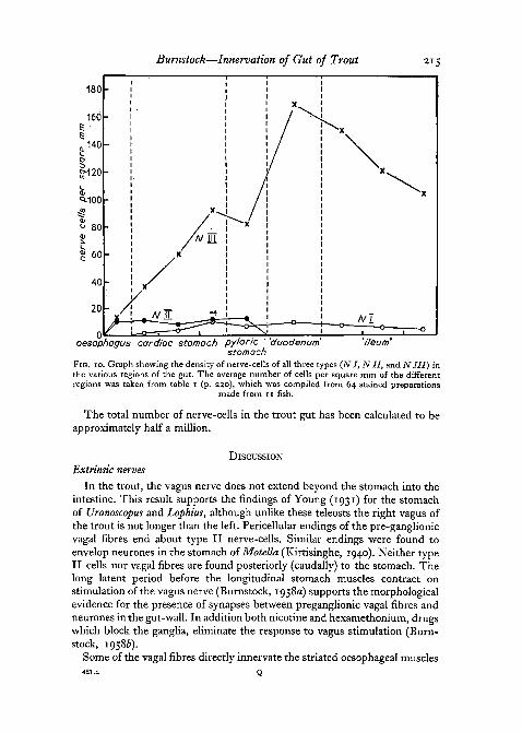
Table i summarizes the results of regional nerve-cell counts. The figuresshowing the number of nerve-cells per square mm for each fish represent theaverage taken from 2 to 5 counts made on each regional preparation. Separatecounts of nerve-cells of all 3 types were included in the calculations. Prepara-tions from the following regions were used: oesophagus, anterior, middle andposterior cardiac stomach, pyloric stomach, 'duodenum' (defined for con-venience as the anterior part of the intestine from which the pyloric caecaarise), anterior, middle, and posterior 'ileum' (intestinal region between'duodenum' and rectum). At the foot of the table the average number ofnerve-cells of types I, II, and III per square mm found in each region of allthe fish used is given. These figures are represented graphically in fig. 10.
Type I cells first appear in the anterior cardiac stomach and rise to adensity of 11 cells per square mm at the posterior cardiac stomach, fromwhich there is a gradual decline towards the rectum. There is a definitecorrelation of cell I density with that of cell III, i.e. there is one type I cell toapproximately 20 type III cells.
Type II cells show a fairly constant density of about 10 cells per square mmthroughout the oesophagus and stomach. They are not present in the intestine.The distribution of these cells is not even, however, especially in the oesopha-gus, where the figures reveal a variation of from 1 to 31 cells per square mmin areas taken at random. The explanation is that most of the cells are aggre-gated about the vagal trunks, so that areas counted of the plexus propercontain very few type II cells.
Type III cells are present in much greater numbers than those of the othertwo types. Furthermore, they show a definite density gradient. There is agradual increase from 14 to 93 cells per square mm from the anterior toposterior cardiac stomach. This density drops very slightly in the pyloricstomach before rising to 170 cells per square mm in the duodenum. From theduodenum there is a gradual density fall to 105 cells per square mm at theposterior ileum. There are two nerve-cell density peaks, one at the antrum andone in the duodenum. It is interesting to note that in the living system, manymovements of the stomach first appear at the antrum. In the intestine, peri-staltic waves of contraction move down the gut from a point about one-thirdof the way down the duodenum.
Burnstock—Innervation of Gut of Trout 215
180
160
.140
§-120
^100
80
60
40
20
oesophagus cardiac stomoch pyloric 'duodenum 'ileurri1
stomachFIG. IO. Graph showing the density of nerve-cells of all three types (NI, Nil, and NIII) inthe various regions of the gut. The average number of cells per square mm of the differentregions was taken from table i (p. 230), which was compiled from 64 stained preparations
made from 11 fish.
The total number of nerve-cells in the trout gut has been calculated to beapproximately half a million.
DISCUSSIONExtrinsic nerves
In the trout, the vagus nerve does not extend beyond the stomach into theintestine. This result supports the findings of Young (1931) for the stomachof Uranoscopus and Lophius, although unlike these teleosts the right vagus ofthe trout is not longer than the left. Pericellular endings of the pre-ganglionicvagal fibres end about type II nerve-cells. Similar endings were found toenvelop neurones in the stomach of Motella (Kirtisinghe, 1940). Neither typeII cells nor vagal fibres are found posteriorly (caudally) to the stomach. Thelong latent period before the longitudinal stomach muscles contract onstimulation of the vagus nerve (Burnstock, 1958a) supports the morphologicalevidence for the presence of synapses between preganglionic vagal fibres andneurones in the gut-wall. In addition both nicotine and hexamethonium, drugswhich block the ganglia, eliminate the response to vagus stimulation (Burn-stock, 19586).
Some of the vagal fibres directly innervate the striated oesophageal muscles

216 Burnstock—Innervation of Gut of Trout
of the trout and on stimulation of the vagus nerves these muscles contractrapidly some seconds before the smooth stomach-muscle responds. It isinteresting, however, that the oesophageal muscles are capable of sustainedcontraction if the stomach is extended with food, even after the vagus nerveshave been severed.
Chevrel (1887) and Young (1931) showed that the 'sympathetic' innerva-tion of the teleost gut consists of a single anterior splanchnic nerve, branchesof which closely follow the main arteries supplying the gut. In the trout, too, avery fine splanchnic nerve supplies the stomach and intestine. The response ofthe stomach to stimulation of the splanchnic nerve is rapid, in contrast to thedelayed reaction on vagus stimulation (Burnstock, 19586). This fact supportsthe morphological evidence that most of the splanchnic fibres reaching thegut are postganglionic and innervate the muscles directly. Thus vagal and'sympathetic' innervation of the teleost gut corresponds to that described inmammals (Dogiel, 1899; Kuntz, 1922; Lawrentjew, 1931).
Separate autonomic innervation of the hind gut is present in the trout,contrary to the findings of Chevrel (1887) and Young (1931) working onother teleosts. This discovery has been given physiological support in aprevious paper, where the result of electrical stimulation of these nerves isdescribed (Burnstock, 19586). The anterior of the two nerves is clearly'sympathetic', originating in the last abdominal ganglia, but whether the othernerve contains 'sympathetic' or 'parasympathetic' fibres is not clear fromthe dissections. It is conceivable that posterior autonomic control has evolvedin teleost fish in relation to the specialized development of separate urinaryand reproductive systems. Alternatively it may have developed in relation tothe complicated rectal septum apparatus described elsewhere (Burnstock,1959), the function of which is not yet clear.
Enteric plexuses
The nerve-cells present in the trout gut lie diffusely in the meshes ofAuerbach's plexus throughout most of its length and are not aggregated intoganglia, as is the case in mammals (Kuntz, 1953; Hill, 1927). In this respectthe form of the trout plexus resembles that of other teleosts described bySakussef (1897) and Kirtisinghe (1940), as well as those found in the amphi-bians (Gunn, 1951) and elasmobranchs (Kirtisinghe, 1940).
In this paper, nerve-cells forming the trout myenteric plexus have beenclassified into three distinct types by their distribution as well as by theirstructure. Despite the general lack of agreement about enteric nerve-cellclassification, it is clear from descriptions and pictures that these cells are verysimilar to those described in other teleosts (Kirtisinghe, 1940) and amphibians(Gunn, 1951). Type III cells, however, which form the major part of theplexus proper in the trout, do not appear to be represented in the mammaliangut.
Embryological studies have shown that a number of enteric neuronesmigrate down the vagal trunks at a late stage in gut development (Kuntz,

Burnstock—Innervation of Gut of Trout 217
1920; Van Campenhout, 1941). The pear-shaped type II cells seen along thevagal nerves are less complex than those cells of the same type with branchingdendrites found in the meshes of the plexus in the pyloric stomach. Type IIcells are clearly concerned with vagal control. Pericellular endings of pre-ganglionic vagal fibres are found to envelop a large number of type II cells.These cells may be the vagal motor neurones, or they may represent a thirdneurone in the peripheral 'parasympathetic' pathway, their axons each supply-ing a large number of motor neurones, a suggestion originally put forwardtheoretically by Langley (1922).
Type III cells probably form the basic connective, motor, and inhibitoryneurones of the enteric system. That elements with these functions may existin the trout gut has been demonstrated (Burnstock, 19586). Because typeII cells are clearly associated with the vagus nerves, and type I cells are fewin number with their dendrites ending in longitudinal muscle only, cells oftype III are the neurones most likely to be concerned with this function. Inaddition axons of type III cells have been seen to innervate the muscles.
Type I cells may be sensory. Their dendrites are embedded in the longi-tudinal muscle and might be concerned with recording stretch. The existenceof sensory neurones involved in the peristaltic reflex arc seen in mammalianguthasbeen clearly demonstrated recently (Biilbring, Lin, and Schofield, 1958).Alternatively they may represent 'pacemaker' cells, one type I cell controlling20 type III cells. Keith (1915) put forward the suggestion that the gut maypossess intestinal 'pacemakers' by analogy with the sinu-atrial node of theheart.
The existence of a sub-mucous plexus has been reported previously in theteleost gut (Monti, 1895; Sakussef, 1897), but unlike Meissner's plexus, socharacteristic of the mammalian sub-mucous coat (Stohr, 1932), no nerve-cellsare present. A similar condition has been described for Amphibia (Gunn,
The sub-epithelial plexus, also found in mammals (Schabadasch, 1930), isa particularly prominent feature of the trout gut, and small polyaxonal cellssimilar to those described in the tench by Monti (1895) are present. No suchcells have been reported in any other vertebrate class.
As in other vertebrate classes (Dogiel, 1896; Hill, 1927; Gunn, 1951),sensory endings in the mucosal epithelium have been identified in the troutgut. Capparelli (1891) described sensory endings in the mucosal epitheliumof Amphibia, which are very similar to those observed in the trout. Sensoryendings of a rather different nature have been described in the epithelium ofthe perch by Sakussef (1897). Whether they form part of local peristalticreflex arcs in the manner suggested by Biilbring, Lin, and Schofield (1958) orwhether they connect directly to higher centres by way of extrinsic nervescannot be decided from the preparations.
A subserous nerve plexus has been described in the gut of mammals(Worobiew, 1913; Kondratjew, 1928; Schabadasch, 1930). Although notpreviously reported in fish, this plexus exists in the trout gut, where it is

218 Burnstock—Innervation of Gut of Trout
prominent in the oesophagus and rectum. The tiny clumps embedded in theserous coat and connected to the plexus bear a very close resemblance to theencapsulated tactile nerve-endings in teleost skin described by Weddell andPallie (1954). If indeed these structures are sensory, further investigation oftheir precise function would be interesting.
A structure resembling descriptions of the 'interstitial network of Cajal'is also present in the trout gut. This is a feature which appears to be commonto all vertebrate enteric systems (Hillarp, 1946; Meyling, 1953). In the troutgut, however, it seems to be connected with the longitudinal muscle-coatonly.
Studies of the numbers of nerve-cells present in the enteric plexus of thetrout reveal density gradients with peaks in the antrum and anterior 'duo-denum'. No quantitative studies of this kind have been done on any other lowervertebrates. Studies of the mammalian gut, however, show that there aredensity peaks at the pylorus and anus of the guinea-pig (Irwin, 1931). Theactual density of nerve-cells present in the myenteric plexus of the trout(100 to 200 cells per square mm) is about the same as that reported in mammals(Irwin, 1931; Ohkubo, 1936). However, the total number of cells calculatedto be present in the trout gut (about half a million) is considerably less thanthe figure given for mammals (5 million in the small intestine of the cat(Sauer and Rumble, 1946)). Prominent peristaltic waves are initiated wherethere are nerve-cell density peaks, i.e. at the antrum and one-third of the waydown the duodenum. This result supports Alverez's 'gradient' theory (Alverez,1927) for the mechanism of peristalsis, but a reflex action also seems to beinvolved in the trout. Experiments with short segments of the stomachsuspended under tension to record circular muscle-contractions, still exhibitregular waves of constriction moving away from the area of greatest nervedensity. Thus it seems that while peristalsis is basically a reflex action, thefirst circular contraction occurs at the point of greatest cell density in any onesegment, while the direction of the wave is normally dictated by the directionof the neurone density gradient. Thus although the cardia, the antrum, andthe region one-third of the way down the 'duodenum' may be called the'pacemakers' of the peristaltic waves seen in the trout gut, they need notpossess any special feature other than the peak nerve density values of thegradient.
Fig. 1 has been reproduced from the Journal of Physiology by kind per-mission of the Editorial Board.
REFERENCES
ALEXANDROWICZ, J. S., 1951. Quart. J. micr. Sci., 92, 163.ALVEREZ, W. C, 1927. Amer. J. Physiol., 80, 493.BODIAN, D., 1937. Anat. Rec, 69, 153.BOLBRING, E., LIN, R. C. Y., and SCHOFIELD, D. G., 1958. Quart. J. exp. Physiol. 43, 26.BURNSTOCK, G., 1957. Nature, 180, 1491.

Burnstock—Innervation of Gut of Trout 219
BURNSTOCK, G., 1958a. J. Physiol., 141, 35.19586. Brit. J. Pharmacol., 13, 216.1959. Quart. J. micr. Sci., 100, 183.
CAPPARELLI, A., 1891. In Oppel, 1896, Lehrb. vergl. mikr. Anat, 1, 119.CHEVREL, R., 1887. Arch. Zool. exp. gen., 5 (suppl.), 1.COLE, F. J., and JOHNSTONE, J., 1901. L.M.B.C. Memoir 8. London (Williams & Norgate).DOGIEL, A. S., 1896. Anat. Anz., 11, 679.
1899. Arch. Anat. Physiol., Physiol. Abt., S> I3°-GAIRNS, F. W., 1930. Quart. J. micr. Sci., 74, 151.GUNN, M., 1951. Ibid., 93, 55.HERRICK, C. J., 1898. J. comp. Neurol., 8, 162.HILL, C. J., 1927. Phil. Trans., 215, 355.HILLARP, N. A., 1946. Acta Anat., 2 (suppl. 4), 153.HIRT, A., 1934., In Bolke's Handb. vergl. Anat. Wirbelt., 2. Berlin (Urban & Schwarzenberg).HOLMES, W., 1947. In Recent Advances in Clinical Pathology. London (Churchill).IRWIN, D. A., 1931. Amer. J. Anat., 49, 141.KEITH, A., 1915. Lancet, 2, 371.KIRTISINGHE, P., 1940. Quart. J. micr. Sci., 81, 521.Kiss, F., 1932. J. Anat., 66,488.KONDRATJEW, N., 1928. Z. Anat. Entw. Gesch., 86, 320.KUNTZ, A., 1920. J. comp. Neurol., 32, 173.
1922. Anat. Rec, 24, 193.1953- The Autonomic Nervous System. 4th ed. Philadelphia (Lea & Febiger).
LANGLEY, J. N., 1922. J. Physiol., 56, 39.LAWRENTJEW, B. I., 1931. Z. mikr.-anat. Forsch, 23, 527.MEYLING, H. A., 1953. J. comp. Neurol., 99, 495.MONTI, R., 1895. Arch. ital. biol., 24, 188.MCLLER, E., and LILJESTRAND, G., 1918. Arch. Anat. Physiol., 137.OHKUBO, K., 1936. Jap. J. med. Sci., I. Anat, 6, 1, 21, 219.PETERS, A., 1955. Quart. J. micr. Sci., 96, 323.SAKUSSEF, S., 1897. Trav. Soc. Nat. St. Petersbourg, 27, 29.SAUER, M. E., and RUMBLE, C. T., 1946. Anat. Rec, 96, 373.SCHABADASCH, A., 1930. Z. Zellforsch, 10, 320.
1934- Ibid., 21, 213.STANNIUS, H., 1849. Das peripherische Nervensystem der Fische. Rostock (Stiller).STOHR, P., 1932. Z. Zellforsch, 16, 123.VAN CAMPENHOUT, E., 1941. Arch. Biol. Paris, 52, 472.WEDDELL, G., and PALLIE, W., 1954. Quart. J. micr. Sci., 95, 389.WOROBIEW, W. P., 1913. Die Nerven des Magens des Hundes. Kharkow.YOUNG, J. Z., 1931. Quart. J. micr. Sci., 74, 492.

to
TABL
E I.
T
he t
able
sho
ws
the
num
ber
of n
erve
-cel
ls o
f eac
h ty
pe (
I, I
I an
d II
I) p
er s
quar
e m
m in
eac
h pa
rt o
f th
e al
imen
tary
can
al
&
Fis
h , 2 3 4 5 6 7 8 9
Me,
No.
prep
ara
ns
7 8 4 5 8 6 3
lion
sO
esop
hagu
sI
II
III
Tot
al
0 2
O
I
0 I
O
I I
6 8
13
' 4
8 9
14
25
A I I 2 2
77 6 9
I S
III
Tota
l
58
68
34
SI
7 4 2
Stc
Mid
. ca
rdia
cII
H
I T
otal
5 13 6
69 88 67 57
83 98 84 6s
mac
h
Pos
t, c
ardi
acI
II
III
Tot
al
8
16 9 21
1
9 7 7
87
95
97
130
105
143
Pyl
I g 8
77 9 9
III
T
84
I
78 91
Ich ital
05 95 08
7
10 8
'Duo
denu
II
III
0 0
18
2
14
4
Tot
al
19
2
15
2
I 8 R 7
77 0
7
777
«3
7
140
151
ntes
lin
n To
tal
'47
159
e
I 6 5 6 6
Mid
. il
eum
II
III
To
tal
0 73
0 12
7
14
2 78 133
128
Post
, il
eum
I II
II
I T
ota
l
6
0
106
11
2
5
0
105
n
o
3 s