the impact of structured plaque control for patients with...
TRANSCRIPT

This work is licensed under a Creative Commons Attribution-NonCommercial 3.0 Unported License
Newcastle University ePrints - eprint.ncl.ac.uk
Stone SJ, Heasman PA, Staines KS, McCracken GI. The impact of structured
plaque control for patients with gingival manifestations of oral lichen planus:
a randomized controlled study. Journal of Clinical Periodontology 2015, 42(4),
356-362.
Copyright:
This is the peer reviewed version of the following article: Stone SJ, Heasman PA, Staines KS, McCracken GI.
The impact of structured plaque control for patients with gingival manifestations of oral lichen planus: a
randomized controlled study. Journal of Clinical Periodontology 2015, 42(4), 356-362., which has been
published in final form at http://dx.doi.org/10.1111/jcpe.12385. This article may be used for non-
commercial purposes in accordance with Wiley Terms and Conditions for Self-Archiving.
DOI link to article:
http://dx.doi.org/10.1111/jcpe.12385
Date deposited:
24/09/2015
Embargo release date:
27 February 2016

Version 3.0
The impact of structured plaque control for patients with gingival manifestations of oral lichen planus: a randomized controlled study
Simon J Stone1
Peter A Heasman1 Konrad S Staines2
Giles I McCracken1 1School of Dental Sciences, Newcastle University, Framlington Place, Newcastle upon Tyne. NE2 4BW 2School of Oral and Dental Sciences, Unversity of Bristol, Lower Maudlin Street, Bristol. BS1 2LY
Keywords: Oral lichen planus, desquamative gingivitis, oral health impact profile, plaque control.
Address for correspondence: Dr S J Stone Room 5.014 Centre for Oral Health Research School of Dental Sciences Newcastle University Framlington Place Newcastle upon Tyne NE2 4BW
A paper submitted for consideration for publication in The Journal of Clinical Periodontology

Version 3.0
Acknowledgements: None Conflict of interest and sources of funding: This was an investigators own study. The powered toothbrushes and interdental brushes were provided by Philips Consumer Lifestyle, WA, USA and Molar Ltd., Kent, UK. Clinical relevance Clinical guidelines suggest that patients with oral lichen planus should improve their oral hygiene. Systematic reviews have suggested that patient-centered outcome measures be used in oral lichen planus outcome studies. Principal findings A structured plaque control intervention was more effective than control in improving the oral health related quality of life for subjects with gingival oral lichen planus. Practical implications This study provides support to the existing clinical guidelines that recommend optimizing oral hygiene for patients with oral lichen planus. Structured plaque control should form part of the first line treatment for patients presenting with gingival lesions.
Version 3.0
The impact of structured plaque control for patients with gingival manifestations of oral lichen planus: a randomized controlled study
Abstract
Aim: To evaluate the impact of a structured plaque control intervention on
clinical and patient-centered outcomes for patients with gingival manifestations
of oral lichen planus.
Materials and methods: Eighty-two patients were recruited into a 20-week
randomised controlled trial. The intervention was structured plaque control
comprising powered tooth brushing and inter-dental cleaning advice. Control
subjects continued with their normal dental plaque control regimen. The
primary outcome measure was the oral health impact profile (OHIP) with
secondary outcomes of pain, plaque index, mucosal disease score and cost-
effectiveness.
Results: Overall the intervention patients showed statistically significant
improvements in OHIP sum ordinal and dichotomous scores compared with
control. There were improvements in the functional limitation, psychological
discomfort and physical disability domains at 4- and 20-weeks and in the
psychological disability domain at 20-weeks. The intervention was successful in
reducing plaque compared to control (p<0.001) and improvements were
observed using the mucosal disease indices at the 4- and 20-week follow ups
(p<0.001).
Conclusion
A structured plaque control intervention was effective in improving the oral
health related quality of life and clinically observed gingival lesions. This study
provides evidence to include intensive plaque control within patients’ initial and
on-going management.

Version 3.0
Introduction
Gingival manifestations are commonly seen in the erosive, ulcerative and
atrophic forms of oral lichen planus (OLP) (Jadinski and Shklar, 1976, Scully and
Porter, 1997, Leao et al., 2008, Lo Russo et al., 2009). The lesions vary in extent
and severity from mild localized patches to widespread intense erythema and
ulceration with areas of spontaneous haemorrhage (Scully and Porter, 1997).
The symptomatology of milder presentations may include sensitivity to spicy or
acidic foods or discomfort with particular dentifrices. The more severe
presentations of the disease are likely to be symptomatic, painful and have
significant impact on patients’ lives (Cheng et al., 2012). Interventions are
generally initiated based on controlling symptoms; the current recommended
clinical pathway suggests that following diagnosis, initial treatment should focus
upon controlling oral hygiene, avoid precipitating factors (e.g. drugs, foods,
chemicals) and provide reassurance (Lodi et al., 2005, Thongprasom et al., 2003,
Thongprasom et al., 2011). Improving plaque control as a conservative strategy
has been the subject of increasing interest, but there remains limited evidence
for the effectiveness of this intervention and, as a result, topical corticosteroids
remain the first line treatment (BSOM, 2010, Holmstrup et al., 1990, Lo Russo et
al., 2008, Guiglia et al., 2007, Lopez-Jornet and Camacho-Alonso, 2010, Salgado et
al., 2013).
It is important for clinicians to understand the disease from the patient’s
perspective so that treatments are proportionate to the reported symptoms.
Pain scores are easy to administer and may provide some insight, but they do not
capture other symptoms, which might be more pertinent. These might include
challenges with eating, in particular avoiding particular foods that may
exacerbate symptoms, chronic soreness, frequency of ulcers, aching, general
discomfort or even changes in mood. Over the last 20 years there has been a
shift to understand the effect that diseases impact upon patients’ lives. The
rationale being that newly developed treatment strategies should address the
issues that are important to patients. Patient-based outcome measures have
been developed to measure the consequences of impaired oral health from the
patient’s perspective, frequently referred to as oral health related quality of life.
Version 3.0
These instruments have been subsequently evaluated for reliability and
consistency and are frequently used in clinical studies as primary or secondary
outcome measures. Rarely have patient-based outcome measures been used to
evaluate interventions for oro-mucosal diseases including OLP, and those that
do, generally use it as a secondary outcome measure (Hegarty et al., 2002,
McGrath et al., 2003, Gorouhi et al., 2007, Lopez-Jornet et al., 2009, Riordain and
McCreary, 2010, Salgado et al., 2013). It has been recommended that future
evaluations should include pain symptoms, report adverse effects of treatment
and include cost-effectiveness with lengthy follow-up periods to evaluate long-
term benefit (Lodi et al., 2012). In this paper we report outcomes from a 20-
week randomized controlled trial evaluating the impact of plaque control on
gingival oral lichen planus from the patient’s perspective.
Aim To evaluate the impact of a structured plaque control intervention on clinical and
patient-centered outcomes for patients with gingival manifestations of oral
lichen planus.
Methods Study design The study was conducted at Newcastle Dental Hospital, UK in accordance with
ICH good clinical practice (GCP) between February 2011 and May 2012, a
favourable ethical opinion was provided by Sunderland Research Ethics
Committee, UK (Ref. 10/H0904/48). The design, (study overview, sample size,
recruitment, randomisation and examiner calibration) has previously been
described (Stone et al., 2013).
Briefly: a parallel group, longitudinal randomized controlled trial (RCT) was
conducted to evaluate the effectiveness of a structured plaque control
programme. Inclusion criteria were: adult patients aged 18 years and above;
willing and able to complete questionnaires; able to provide consent, newly
referred or under review at Newcastle Dental Hospital with a provisional
diagnosis of OLP with clinical signs of gingival involvement. Exclusion criteria

Version 3.0
were: unable to attend for the additional appointments prior to biopsy; unable to
complete questionnaires (large print format were made available for those with
visual impairment, alternatively questionnaires would be read by a researcher);
involved in a research study within the previous 28 days. All subjects whose
diagnosis of OLP was not previously confirmed by biopsy and histopathological
analysis had this performed along with direct immunofluorescence and blood
tests where appropriate (BSOM, 2010). The intervention group received
structured oral hygiene instruction using a powered toothbrush, Sonicare
FlexCare+ HX6942/20 (Philips Oral Healthcare Inc. Bothell, WA, USA) with
interdental cleaning aids, either appropriately sized TePe® extra soft interdental
brushes (TePe Munhygienprodukter, Sweden) ranging from ISO size 1-6 or Oral-
B dental floss (Procter & Gamble, UK). All products were provided for the
complete duration of the study along with standardised toothpaste for all
subjects (Pronamel®, GlaxoSmithKline, Brentford, Middlesex, UK). The control
group was asked to continue with their normal plaque control regimen and did
not receive this additional intervention or advice. Follow up was carried out at 4
and 20 weeks, compliance was not formally recorded. Clinical record forms were
coded and anonymised, each participant was assigned a study number,
participant information was kept confidential and individual subject records
contained only sufficient data to allow identification of the participants
throughout the study.
Oral health-related quality of life was the primary outcome measure in this
study. It was measured using the 49-item version of the Oral Health Impact
Profile (OHIP-49), which has been shown to be reliable, valid and responsive to
clinical change (Locker and Allen, 2002). Participants were asked to rate each of
the responses on a 5-point Likert scale. Responses were coded 0 (never), 1
(hardly ever), 2 (occasionally), 3 (fairly often), and 4 (very often). The OHIP was
self-administered but checked for completeness to ensure no missing data points
(Guyatt et al., 1993). The questionnaire items provide information about the
frequency (burden) by which participants had experienced specific impacts in
the month preceding completion of the questionnaire; OHIP does not aim to
measure severity. Short reference periods have been used with OHIP
successfully in the past (Sutinen et al., 2007) and in clinical trials (Allen et al.,

Version 3.0
2001), therefore a 4-week reference period was used at all appointments to
allow evaluation of change over the course of the study. Secondary outcomes
included visual analogue scales for pain, global transition scores and validated
clinical indices for mucosal disease and plaque control along with evaluation of
cost-effectiveness (Silness and Löe, 1964, Slade and Spencer, 1994, Stone et al.,
2013, Escudier et al., 2007). The data for the secondary outcomes have been
previously reported with the exception of global change scores (Stone et al.,
2013). These were included at each follow-up visit to provide some overall
context to the subjects’ symptoms and also to provide validity to the OHIP data.
Analytic plan
The primary outcome measure for this study was overall sum OHIP score and by
domain. No weightings were to be applied to the OHIP data, sum OHIP data were
used (Allen and Locker, 1997). Overall sum OHIP (ordinal) data were used in
statistical analysis to examine both intervention and control groups, ordinal data
were used to examine each OHIP domain. Data was also dichotomised by grouping
subjects reporting items frequently often or very often (coded 1), and those
reporting items occasionally, hardly ever and never (coded 0). These data were
summarised and analysed to further differentiate negative impacts on quality of life
for intervention and control subjects.
Descriptive statistics were produced for each of the primary and secondary
outcomes according to treatment received. Statistical analysis of the data
including parameter and 95% confidence interval (CI) estimation was
accomplished using SAS software. For age and gender, continuous variables p-
values were calculated by t-test and chi-squared for categorical variables.
Comparisons for the primary outcome measure, OHIP and clinical parameters
between treatment groups were performed using ANOVA for baseline and
ANCOVA for the 4-week and 20-week follow-ups. The p-values are based on a
mixed model F-test (H0=both intervention and control treatments equal). The
population to be analysed comprised all randomised subjects with a baseline and at
least one post-baseline OHIP evaluation, those without were excluded from the
analysis. Minor differences to those previously reported may exist based on the way
in which those with missing data points were handled. Global change scores were
Version 3.0
calculated and analysed using Mann Whitney U statistic, assuming an abnormal
distribution.
Results
Recruitment for the study ran from February 2011 to June 2012. 120 patients
were invited to participate, 82 accepted, were enrolled and randomised, (39
intervention and 43 control subjects). 3 intervention and 2 control subjects
were lost to follow up. One subject failed to attend visit 2 but was retained
through to visit 3. 79 of the 82 participants data were analysed.
The mean age for all randomised subjects was 61.4 years at the time of
enrolment; the gender balance of 18.3% males to 81.7% females reflected the
greater number of females with oral lichen planus in the wider population. The
numbers of subjects taking concomitant topical steroid medication was evenly
distributed in both control and intervention groups (n=23). Three participants
were taking concomitant systemic steroids (prednisolone); two in the
intervention group and one in the control group. Recruitment selection bias was
not considered to be significant.
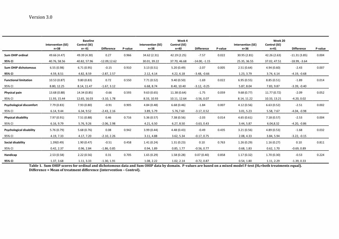
A summary of the baseline and follow-up OHIP data is presented in Table 1.
There were no significant differences between the groups in baseline
demographics or for any other parameter at baseline (p>0.05). The groups had
similar baseline mean OHIP sum scores 49.66 (ordinal) and 6.55 (dichotomous)
for the intervention group and 49.39 (ordinal) and 6.71 (dichotomous) for the
control group. At week 4 and week 20 the distributions shift with both groups
showed a reduction in OHIP ordinal scores overall. The shift for the intervention
group was more than the control group. Both groups contained subjects who
experienced net negative change (post-baseline minus baseline) in OHIP ordinal
and dichotomous scores. The negative net change in ordinal scores indicated
subjects experiencing an overall improvement in given domain, taking into
account both the improvement and deterioration to individual statements.
Negative net change in dichotomous scores indicates an overall decrease in the
impacts that happened “fairly often” or “very often” from baseline, taking into
account some impacts happened more frequently, and some happened less
frequently. The intervention group experienced greater negative net change in
Version 3.0
both ordinal and dichotomous scores indicating improvements in oral health
related quality of life these differences were statistically significant at the 4-week
(p=0.022) and 20-week (p=0.004) follow-up.
Evaluation of the individual domain scores provides further insight into the
range of impacts that oral lichen planus has on a subject’s quality of life.
Statistical output for each domain was undertaken using ordinal data and
presented in Table 1.
Those domains with significant differences between the intervention and control
groups at 4 and 20 weeks respectively were functional limitation (p=0.022;
p=0.014), psychological discomfort (p=0.007; p=0.002) and physical disability
(p=0.014; p=0.004). The psychological disability domain showed significant
differences at the 20-week (p=0.003) but not at the 4-week follow up (p=0.435).
There were no significant correlations in the social disability (p=0.763; p=0.811)
and handicap domains (p=0.858; p=0.224) at either time point and a constant
although not statistically significant effect was observed in the physical pain
domain (p=0.059, p=0.052).
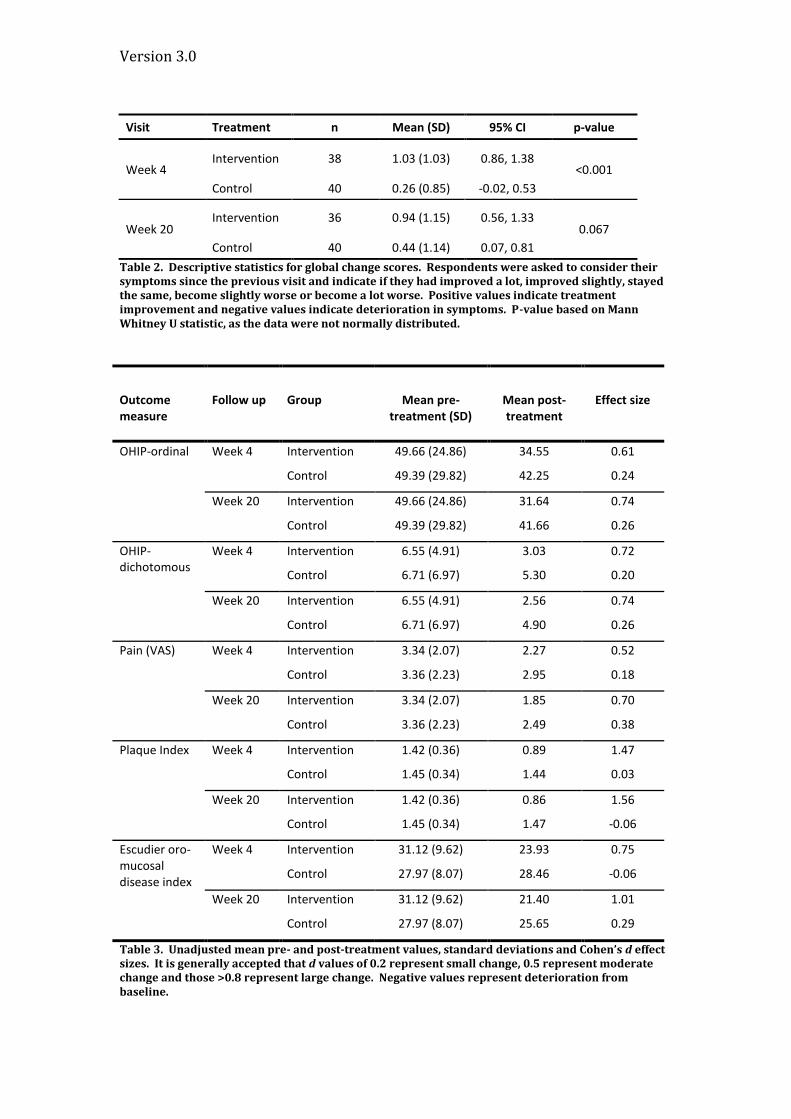
Global change
Global change scores, recorded on a 5-point Likert scale, were coded positively
for improvements and negatively for deteriorations in symptoms: Improved a
lot (2), improved slightly (1), stayed the same (0), become slightly worse (-1),
become a lot worse (-2). Results are displayed in Table 2. At week 4 the mean
global change with 95% CI was 1.03 (0.86, 1.38) for the intervention group
indicating that the subjects in that group felt that their symptoms improved
slightly. The control group still showed a positive mean global score [0.26 (-0.02,
0.53)] and there were statistically significant differences between the two groups
(p<0.001) at the 4-week follow-up. At the 20-week follow-up the difference in
improvements between the groups from the 4-week time point was not
statistically significant (p=0.067) suggesting that the greatest effect came in the
intervention group in the time initially following the intervention.
Effect sizes
The effect of treatment was examined using Cohen’s d (Table 3). Interpretation
of effect sizes differs but it is generally agreed that those values above 0.2 are
Version 3.0
seen to be having a small treatment effect, above 0.5 to have a moderate effect
and above 0.8 to have a large effect.
Moderate treatment effects were seen in the intervention group for OHIP ordinal
scores at week 4 (0.61) and week 20 (0.74). Moderate treatment effects were
also observed in the intervention group for pain (VAS) both at the 4-week (0.52)
and 20 weeks (0.70). Large effect sizes were observed in the interventions group
for PI at week 4 (1.47) and week 20 (1.56). A moderate effect was seen in
Escuder’s oro-mucosal disease index at week 4 (0.75) and a large effect at week
20 (1.01). Small changes were observed for the control group in OHIP scores and
VAS scores at 4 and 20 weeks. No effect was observed in PI for the control group
at any time point. These can be used alongside the surrogate measures of health
(clinical indices) and subjective measures of health (OHIP) in an attempt to
provide comprehensive assessment of the effect of the intervention.
Version 3.0
Discussion
The evaluation of the effectiveness of a structured plaque control programme
used tools that were both objective and subjective measures of health and
disease. For context, clinical evaluation of the intervention was based on Plaque
Index (Silness and Löe, 1964) and the Escudier index (Escudier et al., 2007). In
this study the mean Plaque Index scores reduced for the intervention group by
39.5% at the 20-week follow up compared to a marginal improvement of 4.1%
control group. The intervention was, therefore successful in reducing plaque
compared to control and was sustained to the end of the study at 20 weeks
(p<0.001). Clinical improvements were observed using the Escudier indices at
the 4- and 20-week follow up (p<0.001).
The mean OHIP ordinal scores at baseline were 49.7 for the intervention and
49.4 for the control group. In the original validation studies for OHIP involving
older adults, the mean sum OHIP ordinal score was 31.5 (Slade, 1998). The
subjects with gingival manifestations of oral lichen planus in this study reported
more frequent impacts at baseline indicating poorer oral health related quality of
life. It is important though to examine the individual domains to determine the
social impact of the disease. The physical pain domain in OHIP contains
statements that relate to the frequency that subjects experienced painful gums,
sore spots and discomfort when eating. These symptoms are commonly
reported in outpatient clinical settings therefore, it is reasonable to assume that
this domain would have the potential for change. Although improvements in the
domain score were observed, they were not statistically significant from the
control group (p>0.05). The VAS scores for pain were also not statistically
different at the 4- and 20- week follow up (p>0.05). Pain might therefore not be
significantly affected by the intervention or perhaps the symptoms that have the
greatest effect on quality of life do not include pain. In the functional limitation
domain there were significant differences between the control and intervention
groups (p=0.012, p=0.014). This domain contains statements relating to
appearance, difficulty chewing, taste and digestion. It may be that improvements
in clinical signs of inflammation then bring about these secondary outcomes
measured in this and other domains. The largest differences between groups
Version 3.0
were observed with the psychological discomfort and physical disability
domains. The psychological discomfort domain relates to being worried, self-
conscious, miserable, concerned about appearance and tension. Perhaps the
intervention is, by resolving inflammation, reducing symptoms, so that subjects
are consequently being less concerned about their oral health. There may also
be some positive effect by which participating in the study affects this domain;
particularly a study that monitors subjects more frequently than through their
conventional clinical pathway. This may be particularly important when
examining a cohort of patients with a potentially pre-malignant diagnosis
(Holmstrup, 2010). Within the physical disability OHIP domain, which contains
statements relating to being unable to brush teeth, avoidance of eating and
unsatisfactory diet, there were statistical differences between the groups in
favour of the intervention group at week 20 (p=0.004). It is impossible to tell,
without further adjunctive qualitative interviewing, which part of the
intervention was the most important: the advice and reassurance, or the
provision of appropriate aids that facilitate the perceived improvements in this
domain. Comparatively few impacts were observed in the final three domains:
psychological disability, social disability and handicap. This suggests that oral
lichen planus does not have large disabling effects but carries significant
psychological impact associated with the diagnosis and chronic discomfort.
Anxiety has previously been strongly associated with the initiation of oral lichen
planus and frequent observation and monitoring during a clinical study may go
some way to alleviating this anxiety (Vallejo et al., 2001)
Studies have suggested that painful atrophic mucosa may discourage patients
from brushing effectively, additionally it has been reported that powered tooth
brushing can cause minor gingival abrasions (Erpenstein, 1985, Robinson et al.,
2005). The intervention therefore had the potential to exacerbate the lesions
particularly with the friable, atrophic nature of the gingival tissue. It has been
suggested that plaque removal would potentiate new lesions resulting from
mechanical trauma, however this hypothesis lacked evidence (Hermann, 1963).
Contrary to the thoughts of Erpenstein, (1985) the results of this study showed
that the structured advice and products provided to the subjects facilitated
Version 3.0
improvements in plaque control, brought about improvements in the severity of
the OLP lesions and improved subjects’ oral health related quality of life; the
intervention did not result in any adverse outcomes and analysis of its cost-
effectiveness has previously been reported (Stone et al., 2013).
Conclusions
A structured plaque control intervention was effective in improving the oral
health related quality of life and clinically observed gingival manifestations of
oral lichen planus. This study provides evidence to include intensive plaque
control within patients’ initial and on-going management. Intensive plaque
control should, therefore, become an important initial phase of treatment, which
can be delivered pre-referral by general dentists and dental hygienists.
Version 3.0
Baseline Week 4 Week 20 Intervention (SE)
n=38 Control (SE)
n= 41 Difference P-value Intervention (SE)
n=38 Control (SE)
n=40 Difference P-value Intervention (SE)
n=36 Control (SE)
n=41 Difference P-value
Sum OHIP ordinal 49.66 (4.47) 49.39 (4.30) 0.27 0.966 34.62 (2.31) 42.19 (2.25) -7.57 0.022 30.95 (2.81) 42.26 (2.63) -11.31 (3.85) 0.004
95% CI 40.76, 58.56 40.82, 57.96 -12.09,12.62 30.01, 39.22 37.70, 46.68 -14.00, -1.15 25.35, 36.55 37.02, 47.51 -18.99, -3.64
Sum OHIP dichotomous 6.55 (0.98) 6.71 (0.95) -0.15 0.910 3.13 (0.51) 5.20 (0.49) -2.07 0.005 2.51 (0.64) 4.94 (0.60) -2.43 0.007
95% CI 4.59, 8.51 4.82, 8.59 -2.87, 2.57 2.12, 4.14 4.22, 6.18 -3.48, -0.66 1.23, 3.79 3.74, 6.14 -4.19, -0.68
Functional limitation 10.53 (0.87) 9.80 (0.83) 0.72 0.550 7.71 (0.52) 9.40 (0.50) -1.69 0.022 6.95 (0.55) 8.85 (0.51) -1.89 0.014
95% CI 8.80, 12.25 8.14, 11.47 -1.67, 3.12 6.68, 8.74 8.40, 10.40 -3.12, -0.25 5.87, 8.04 7.83, 9.87 -3.39, -0.40
Physical pain 13.68 (0.88) 14.34 (0.85) -0.66 0.593 9.63 (0.65) 11.38 (0.64) -1.75 0.059 9.68 (0.77) 11.77 (0.72) -2.09 0.052
95% CI 11.93, 15.44 12.65, 16.03 -3.10, 1.78 8.33, 10.93 10.11, 12.64 -3.56, 0.07 8.14, 11.22 10.33, 13.21 -4.20, 0.02
Psychological discomfort 7.79 (0.83) 7.93 (0.80) -0.91 0.905 4.84 (0.48) 6.68 (0.46) -1.84 0.007 4.12 (0.56) 6.63 (0.52) -2.51 0.002
95% CI 6.14, 9.44 6.34, 9.52 -2.43, 2.16 3.89, 5.79 5.76,7.60 -3.17, 0.52 3.01, 5.23 5.58, 7.67 -4.04, -0.98
Physical disability 7.97 (0.91) 7.51 (0.88) 0.46 0.716 5.36 (0.57) 7.38 (0.56) -2.03 0.014 4.65 (0.61) 7.18 (0.57) -2.53 0.004
95% CI 6.16, 9.79 5.76, 9.26 -2.06, 2.98 4.21, 6.50 6.27, 8.50 -3.63, 0.43 3.44, 5.87 6.04,8.32 -4.20, -0.86
Psychological disability 5.76 (0.79) 5.68 (0.76) 0.08 0.942 3.99 (0.44) 4.48 (0.43) -0.49 0.435 3.21 (0.56) 4.89 (0.53) -1.68 0.032
95% CI 4.19, 7.33 4.17, 7.20 -2.10, 2.26 3.11, 4.88 3.62, 5.34 -0.17, 0.75 2.08, 4.33 3.84, 5.94 -3.22, -0.15
Social disability 1.39(0.49) 1.90 (0.47) -0.51 0.458 1.41 (0.24) 1.31 (0.23) 0.10 0.763 1.26 (0.29) 1.16 (0.27) 0.10 0.811
95% CI 0.42, 2.37 0.96, 2.84 -1.86, 0.85 0.94, 1.89 0.85, 1.77 -0.56, 0.77 0.68, 1.83 0.62, 1.70 -0.69, 0.89
Handicap 2.53 (0.58) 2.22 (0.56) 0.31 0.705 1.65 (0.29) 1.58 (0.28) 0.07 (0.40) 0.858 1.17 (0.32) 1.70 (0.30) -0.53 0.224
95% CI 1.37, 3.68 1.11, 3.33 -1.30, 1.91 1.08, 2.22 1.02, 2.14 -0.72, 0.87 0.54, 1.80 1.11, 2.29 -1.39, 0.33
Table 1. Sum OHIP scores for ordinal and dichotomous data and Sum OHIP data by domain. P-values are based on a mixed model F-test (H0=both treatments equal). Difference = Mean of treatment difference (intervention – Control).
Version 3.0
Visit Treatment n Mean (SD) 95% CI p-value
Week 4 Intervention 38 1.03 (1.03) 0.86, 1.38
<0.001
Control 40 0.26 (0.85) -0.02, 0.53
Week 20 Intervention 36 0.94 (1.15) 0.56, 1.33
0.067
Control 40 0.44 (1.14) 0.07, 0.81
Table 2. Descriptive statistics for global change scores. Respondents were asked to consider their symptoms since the previous visit and indicate if they had improved a lot, improved slightly, stayed the same, become slightly worse or become a lot worse. Positive values indicate treatment improvement and negative values indicate deterioration in symptoms. P-value based on Mann Whitney U statistic, as the data were not normally distributed.
Outcome measure
Follow up Group Mean pre-treatment (SD)
Mean post-treatment
Effect size
OHIP-ordinal Week 4 Intervention 49.66 (24.86) 34.55 0.61
Control 49.39 (29.82) 42.25 0.24
Week 20 Intervention 49.66 (24.86) 31.64 0.74
Control 49.39 (29.82) 41.66 0.26
OHIP-dichotomous
Week 4 Intervention 6.55 (4.91) 3.03 0.72
Control 6.71 (6.97) 5.30 0.20
Week 20 Intervention 6.55 (4.91) 2.56 0.74
Control 6.71 (6.97) 4.90 0.26
Pain (VAS) Week 4 Intervention 3.34 (2.07) 2.27 0.52
Control 3.36 (2.23) 2.95 0.18
Week 20 Intervention 3.34 (2.07) 1.85 0.70
Control 3.36 (2.23) 2.49 0.38
Plaque Index Week 4 Intervention 1.42 (0.36) 0.89 1.47
Control 1.45 (0.34) 1.44 0.03
Week 20 Intervention 1.42 (0.36) 0.86 1.56
Control 1.45 (0.34) 1.47 -0.06
Escudier oro-mucosal disease index
Week 4 Intervention 31.12 (9.62) 23.93 0.75
Control 27.97 (8.07) 28.46 -0.06
Week 20 Intervention 31.12 (9.62) 21.40 1.01
Control 27.97 (8.07) 25.65 0.29
Table 3. Unadjusted mean pre- and post-treatment values, standard deviations and Cohen’s d effect sizes. It is generally accepted that d values of 0.2 represent small change, 0.5 represent moderate change and those >0.8 represent large change. Negative values represent deterioration from baseline.
Version 3.0
References Allen, P. F. & Locker, D. (1997) Do item weights matter? An assessment using the
oral health impact profile. Community Dental Health 14, 133-138. Allen, P. F., McMillan, A. S. & Locker, D. (2001) An assessment of sensitivity to
change of the Oral Health Impact Profile in a clinical trial. Community Dent Oral Epidemiol 29, 175-182.
BSOM (2010) Guidelines for the management of oral lichen planus in secondary care [WWW document]. URL http://www.bsom.org.uk/LP_guidelines_-_BSOM.pdf [accessed on 09/01/2013]
Cheng, S., Kirtschig, G., Cooper, S., Thornhill, M., Leonardi-Bee, J. & Murphy, R. (2012) Interventions for erosive lichen planus affecting mucosal sites. Cochrane Database of Systematic Reviews 2, CD008092. doi:10.1002/14651858.CD008092.pub2.
Erpenstein, H. (1985) Periodontal and prosthetic treatment in patients with oral lichen planus. Journal of Clinical Periodontology 12, 104-112.
Escudier, M., Ahmed, N., Shirlaw, P., Setterfield, J., Tappuni, A., Black, M. M. & Challacombe, S. J. (2007) A scoring system for mucosal disease severity with special reference to oral lichen planus. The British Journal of Dermatology 157, 765-770. doi:BJD8106 [pii]
10.1111/j.1365-2133.2007.08106.x. Gorouhi, F., Solhpour, A., Beitollahi, J. M., Afshar, S., Davari, P., Hashemi, P., Nassiri
Kashani, M. & Firooz, A. (2007) Randomized trial of pimecrolimus cream versus triamcinolone acetonide paste in the treatment of oral lichen planus. Journal of the American Academy of Dermatology 57, 806-813. doi:S0190-9622(07)01040-7 [pii]
10.1016/j.jaad.2007.06.022. Guiglia, R., Di Liberto, C., Pizzo, G., Picone, L., Lo Muzio, L., Gallo, P. D., Campisi, G.
& D'Angelo, M. (2007) A combined treatment regimen for desquamative gingivitis in patients with oral lichen planus. Journal of Oral Pathology and Medicine 36, 110-116. doi:10.1111/j.1600-0714.2007.00478.x.
Guyatt, G. H., Feeny, D. H. & Patrick, D. L. (1993) Measuring health-related quality of life. Annals of Internal Medicine 118, 622-629.
Hegarty, A. M., McGrath, C., Hodgson, T. A. & Porter, S. R. (2002) Patient-centred outcome measures in oral medicine: Are they valid and reliable? International Journal of Oral and Maxillofacial Surgery 31, 670-674.
Hermann, D. (1963) Lichen ruber planus der Mundschleimhaut, Untersuchungen uber Klinik, Histologie und Therapie in 60 Fallen. Deutsche Zahnarztliche Zeitschrift 18, 346-363.
Holmstrup, P. (2010) The malignant potential of oral lichen planus. Oral Diseases 16, 509-510. doi:10.1111/j.1601-0825.2010.01750.x.
Holmstrup, P., Schiotz, A. W. & Westergaard, J. (1990) Effect of dental plaque control on gingival lichen planus. Oral Surgery, Oral Medicine, and Oral Pathology 69, 585-590.
Jadinski, J. J. & Shklar, G. (1976) Lichen planus of the gingiva. Journal of Periodontology 47, 723-733.
Leao, J. C., Ingafou, M., Khan, A., Scully, C. & Porter, S. (2008) Desquamative gingivitis: retrospective analysis of disease associations of a large cohort. Oral Diseases 14, 556-560. doi:ODI1420 [pii]
10.1111/j.1601-0825.2007.01420.x.

Version 3.0
Lo Russo, L., Fedele, S., Guiglia, R., Ciavarella, D., Lo Muzio, L., Gallo, P., Di Liberto, C. & Campisi, G. (2008) Diagnostic pathways and clinical significance of desquamative gingivitis. Journal of Periodontology 79, 4-24. doi:10.1902/jop.2008.070231.
Lo Russo, L., Fierro, G., Guiglia, R., Compilato, D., Testa, N. F., Lo Muzio, L. & Campisi, G. (2009) Epidemiology of desquamative gingivitis: evaluation of 125 patients and review of the literature. International Journal of Dermatology 48, 1049-1052. doi:IJD4142 [pii]
10.1111/j.1365-4632.2009.04142.x. Locker, D. & Allen, P. F. (2002) Developing short-form measures of oral health-
related quality of life. J Public Health Dent 62, 13-20. Lodi, G., Carrozzo, M., Furness, S. & Thongprasom, K. (2012) Interventions for
treating oral lichen planus: a systematic review. The British Journal of Dermatology 166, 938-947. doi:10.1111/j.1365-2133.2012.10821.x.
Lodi, G., Scully, C., Carrozzo, M., Griffiths, M., Sugerman, P. B. & Thongprasom, K. (2005) Current controversies in oral lichen planus: report of an international consensus meeting. Part 2. Clinical management and malignant transformation. Oral Surgery Oral Medicine Oral Pathology Oral Radiology and Endodontics 100, 164-178. doi:10.1016/j.tripleo.2004.06.076.
Lopez-Jornet, P. & Camacho-Alonso, F. (2010) Application of a motivation-behavioral skills protocol in gingival lichen planus: a short-term study. Journal of Periodontology 81, 1449-1454. doi:10.1902/jop.2010.100245.
Lopez-Jornet, P., Camacho-Alonso, F. & Lucero Berdugo, M. (2009) Measuring the impact of oral mucosa disease on quality of life. European Journal of Dermatology 19, 603-606. doi:ejd.2009.0762 [pii]
10.1684/ejd.2009.0762. McGrath, C., Hegarty, A. M., Hodgson, T. A. & Porter, S. R. (2003) Patient-centred
outcome measures for oral mucosal disease are sensitive to treatment. International Journal of Oral and Maxillofacial Surgery 32, 334-336. doi:S0901-5027(02)90377-2 [pii]
10.1054/ijom.2002.0377. Riordain, R. N. & McCreary, C. (2010) The use of quality of life measures in oral
medicine: a review of the literature. Oral Dis 16, 419-430. doi:10.1111/j.1601-0825.2009.01647.x.
Robinson, P. G., Deacon, S. A., Deery, C., Heanue, M., Walmsley, A. D., Worthington, H. V., Glenny, A. M. & Shaw, W. C. (2005) Manual versus powered toothbrushing for oral health. Cochrane Database of Systematic Reviews, CD002281. doi:10.1002/14651858.CD002281.pub2.
Salgado, D. S., Jeremias, F., Capela, M. V., Onofre, M. A., Massucato, E. M. & Orrico, S. R. (2013) Plaque control improves the painful symptoms of oral lichen planus gingival lesions. A short-term study. J Oral Pathol Med 42, 728-732. doi:10.1111/jop.12093.
Scully, C. & Porter, S. R. (1997) The clinical spectrum of desquamative gingivitis. Seminars in Cutaneous Medicine and Surgery 16, 308-313.
Silness, J. & Löe, H. (1964) Periodontal Disease in Pregnancy II. Correlation between Oral Hygiene and Periodontal Condition. Acta Odontologica Scandinavica 22, 112-135.

Version 3.0
Slade, G. D. (1998) Assessing change in quality of life using the Oral Health Impact Profile. Community Dentistry and Oral Epidemiology 26, 52-61.
Slade, G. D. & Spencer, A. J. (1994) Development and evaluation of the Oral Health Impact Profile. Community Dental Health 11, 3-11.
Stone, S. J., McCracken, G. I., Heasman, P. A., Staines, K. S. & Pennington, M. (2013) Cost-effectiveness of personalized plaque control for managing the gingival manifestations of oral lichen planus: a randomized controlled study. J Clin Periodontol 40, 859-867. doi:10.1111/jcpe.12126.
Sutinen, S., Lahti, S., Nuttall, N. M., Sanders, A. E., Steele, J. G., Allen, P. F. & Slade, G. D. (2007) Effect of a 1-month vs. a 12-month reference period on responses to the 14-item Oral Health Impact Profile. European Journal of Oral Sciences 115, 246-249. doi:10.1111/j.1600-0722.2007.00442.x.
Thongprasom, K., Carrozzo, M., Furness, S. & Lodi, G. (2011) Interventions for treating oral lichen planus. Cochrane Database of Systematic Reviews, CD001168. doi:10.1002/14651858.CD001168.pub2.
Thongprasom, K., Luengvisut, P., Wongwatanakij, A. & Boonjatturus, C. (2003) Clinical evaluation in treatment of oral lichen planus with topical fluocinolone acetonide: a 2-year follow-up. Journal of Oral Pathology and Medicine 32, 315-322. doi:130 [pii].
Vallejo, M. J., Huerta, G., Cerero, R. & Seoane, J. M. (2001) Anxiety and depression as risk factors for oral lichen planus. Dermatology 203, 303-307.