the immune proteins of bovine colostrum and plasma* · the immune proteins of bovine colostrum and...
TRANSCRIPT

THE IMMUNE PROTEINS OF BOVINE COLOSTRUM AND PLASMA*
BY EMIL L. SMITH+
(From the Biological Laboratories, E. R. Squibb and Sons, New Brunswick)
(Received for publication, March 29, 1946)
The important r61e of the colostrum in the transmission of antibodies from mother to offspring, and particularly in the protection of the new-born in ruminants to infectious disease, has been shown by the work of many investigators (1). Howe has demonstrated by salt fractionation studies that the serum of t)he new-born calf taken prior to suckling is deficient in a globulin fraction, and that this globulin, generally associated with anti- bodies, aapears after the calf has ingested colostrum (2). More recent studies b.y means of the electrophoretic technique of Tiselius have shown t,hat, the serum of the new-born calf contains little or no r-globulin, but that the slow moving globulin component appears after the feeding of colos- triim (3).
Ea,rlv work on the composition of colostrum demonstrated that the pro- tein content was far higher than that of milk, and that the colostrum was narticularly rich in a globulin precipitable at low concentrations of salts such as ammonium sulfate. Crowther and Raistrick (4) separated colos- t,rum into the three fractions which have usually been prepared from milk; namely, casein, lactoglobulin, and lactalbumin. Their studies of the nitro- gen distribution by the Van Slyke method showed that these fractions could readily be differentiated from one another.
In the present study, colostrum and the protein fractions derived from it have been studied electrophoretically. It was found that an electrophoreti- tally homogeneous globulin could be readily isolated in high yield by the conventional precipitation with ammonium sulfate. This lactoglobulin possesses all of the immune properties of colostrum and has been studied with a view to its characterization by physical and chemical methods. The immune fractions of bovine plasma have also been isolated in order to com- pare the properties of immune proteins found in different body fluids. It is convenient to refer to the globulins which are associated with immunity and found in colostrum and plasma as “immune globulins.” It is fully realized that the immune properties probably account for only a very small part of the total protein.
*A preliminary report of this work was presented before the Thirty-seventh annual meeting of the American Society of Biological Chemists at Atlantic City, 1946 (Federation Proc., 6, 154 (1946)).
t Present address, School of Medicine, University of Utah, Salt Lake City.
345
by guest on July 4, 2018http://w
ww
.jbc.org/D
ownloaded from

346 BOVINE IMMUNE PROTEIRS
EXPERIMEI’ZTAL
Electrophoretic studies were performed at 1” in a Tiselius apparatus equipped with the Longsmort,h scanning device. Unless otherwise specified, the solutions were equilibrated by dialysis for at least 48 hours with a vero- nal (dicthyl barbiturate) buffer at pH 8.4 to 8.6 and at an ionic strength of 0.1.
Fractionation of Colostrum
In order to study the properties and quantities of the various proteins, the colostrum was arbitrarily separated into several fractions. 2 liters of colostrum collected 1 hour post partum were centrifuged thoroughly and the orange-colored fat layer was discarded. The colostrum (1700 cc.) was then diluted 4-fold with distilled water and slowly adjusted with 0.5 M HCl to pH 4.5. The casein precipitate (Fraction A) was removed by filtration on coarse fluted paper and the solution was clarified by filtration through a thick layer of paper pulp. The filtrate was brought to pH 6.0 with 0.5 M NaOH and successive fractions were removed at 0.3 (Fraction B), 0.5 (Fraction C), and 0.9 (Fraction D) saturation with ammonium sulfate. Fractions B, C, and D were redissolved in water at about 5 per cent concen- tration and reprecipitated within the same limits of salt concentration after any turbidity present at the lower salt concentration was first removed by filtration. Each of these fractions was dialyzed at 2” until salt-free and then dried by lyophilizing. Fraction A was redissolved with the minimum quantity of 0.1 M NaOH, filtered clear, and then reprecipitated at pH 4.5. This precipitate was washed twice with distilled wat.er, redissolved with the aid of dilute alkali, and then lyophilized.
Fig. 1 shows the electrophoretic patterns obtained Jvit.h each of the four fractions and with the original colostrum. Table I presents the data for the yield of each fraction and its composition as determined from the electro- phoretic pattern. The components included in the vertical columns of Table I are not necessarily to be regarded as the same in all cases but are listed in that manner for tabular convenience. The separate fractions have been designated by the name of the principal protein which lvas found to be present in each fraction. Fractions A (casein) and D (p-lactoglobulin) will be discussed only briefly and are therefore presented first.
Casein (Fraction .4)-It is apparent that the crude casein (Fraction A) is complex in nature, like that of milk, and contains at least txvo components (5). The nature of the third component has not been invest,igated further but, it may be a globulin which precipitated because of the alt,eration in salt concentration on dilution of the colostrum and adjustment of the PH. The boundary migrating at -4.2 X 10e5 sq. cm. per volt per second in the whole colostrum is obviously composite in nature and includes casein as Tvell as
by guest on July 4, 2018http://w
ww
.jbc.org/D
ownloaded from
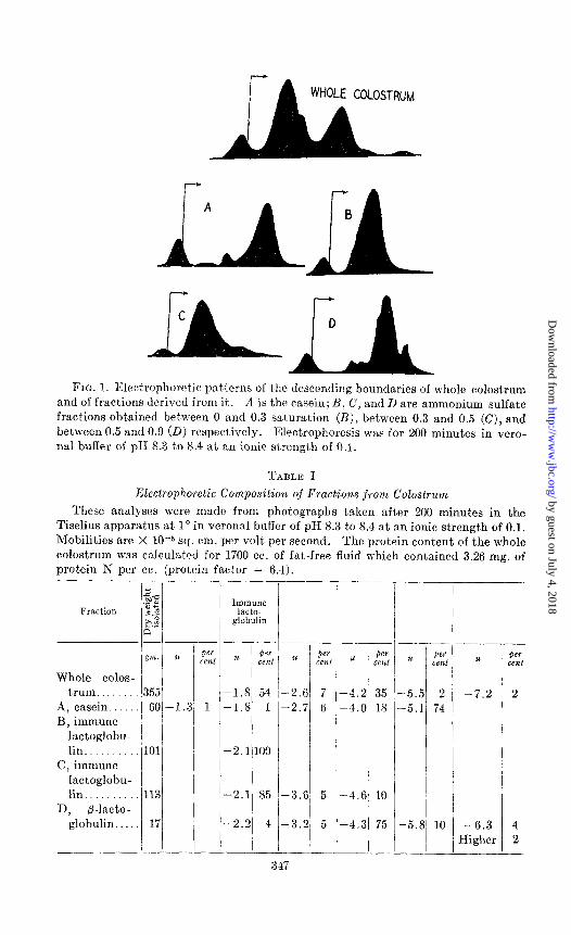
FIG. 1. Electrophoretic pal terns of the descending boundaries of whole colostrum and of fractions derived from it. n is the casein; B, C, and D are ammonium sulfate fractions obtained between 0 and 0.3 saturation (B), between 0.3 and 0.5 (C), and between 0.5 and 0.0 (D) respectively. Electrophoresis was for 200 minutes in vero- nal buffer of pII 5.3 to 8.4 at an ionic strength of 0.1.
TABLE I
Electrophoretic Composition of Fractions from Colostrum
These analyses were made from photographs taken after 200 minutes in the Tiselius apparatus at 1’ in vcronal buffer of pH 8.3 to 8.4 at an ionic strength of 0.1. Mobilities are X 10-j sq. cm. per volt per second, The protein content of the whole colostrum was calculated for 1700 cc. of fat-free fluid which contained 3.26 my. of protein N per cc. (protein factor = 6.4).
Whole colos- trum.
A, casein. B, immune
lactoglobu- lin.
C, immune lactoglobu- lin
n, b-lacto- globulin.
Per cent
355 60-1.3 1
113
17
-
IlllmUne lacto-
globulin
-1.8 54 -1.8 1
-2.1 100
-2.1 85
-2.2 4
-2.6 7 -2.7 6
-3.6 5
-3.2 5
u Per G&
-4.2 35 -4.0 18
-4.6 10
-4.3 75
u I 2%
-5.5 2 -5.1 74
-5.8 10
u
-7.2
-6.3 Higher
-
t
-
Per X?if
2
4 2
- 347
by guest on July 4, 2018http://w
ww
.jbc.org/D
ownloaded from
348 BOVINE IMMUNE PROTEINS
other components. It should be noted that electrophoresis of whole co- lostrum or milk has always shown differences in mobility for casein and p-lactoglobulin compared with the mobility for these same components in a more homogeneous state.
fi-Lactoglobulin (Fraction D)-This is a major component of the fraction which is called “lactalbumin” in the older literature. As is shown in Table I, only a small part of the protein in colostrum was isolated by precipitation at high concentrations of ammonium sulfate; this is in agreement with the data of Crowther and Raistrick (4) and Engel and Schlag (6). We have found that 75 per cent of the protein in this fraction, as estimated from the electrophoretic pattern, is the p-lactoglobulin originally isolated from milk by Palmer (7). When Fraction D was thoroughly dialyzed and adjusted to pH 5.2, the globulin crystallized in high yield. It possessed the crystalline form and other properties described by Palmer and others for this protein. A recrystallized sample was homogeneous on electrophoresis in Verona1 buffer at pH 8.4 and migrated with a mobility of -4.9 X lop5 sq. cm. per volt per second. Other preparations of crystalline fl-lactoglobulin from milk migrated with mobilities of -4.9 to -5.2 X low5 sq. cm. per volt per second at pH 8.4 to 8.5.
Immune Lactoglobulin (Fractions B and C)-The animals from which this and other samples of colostrum were obtained had been hyperimmunized. While the immunity of these animals is the object of a separate study by Dr. August Holm of these laboratories and will be described by him else- where, it may be stated that the total immune activity of colostrum is definitely associat,ed with the protein of low electrophoretic mobility (- 1.8 to -2.2 X 10m5 sq. cm. per volt per second at pH 8.4). Immune activity has not been found in fractions free from this protein, and conversely the isolated protein accounts completely for the immune properties of colostrum. It is convenient to refer to this protein from colostrum or milk as immune lactoglobulin; this will serve to distinguish it from p-lactoglobulin.
The immune protein is definitely globulin in character. This is partly indicated by its precipitation at low concentrations of ammonium sulfate, but is more clearly demonstrated by its low solubility in the neighborhood of the isoelectric point, about pH 5.8 to 6.2, and by the marked increase in solubility in the presence of neutral salts. Moreover, as will be discussed below, prolonged dialysis at the isoelectric point causes a precipitation of a water-insoluble portion of the immune lactoglobulin.
It is apparent from Table I that the immune lactoglobulin is the main protein constituent of colostrum as determined both from the electro- phoretic analysis of the whole colostrum and from the composition of Fractions B and C. Adding the total yield of Fraction B (101 gm.) and 85 per cent of the yield of Fraction C (96 pm.) shows that 55 per cent of
by guest on July 4, 2018http://w
ww
.jbc.org/D
ownloaded from
E. L. SMITH 349
the original protein in the whole colostrum was isolated as the immune lactoglobulin, as compared with the 54 per cent found by the electrophoretic analysis of the whole colostrum. This surprisingly good recovery shows the great preponderance of this protein compared with any other present in colostrum.
With other samples of colostrum obtained from different animals, it was found that, after removal of the casein as described above, the immune lactoglobulin could be obtained quantitatively from the colostrum whey free of other proteins determined electrophoretically. This was accom- plished by precipitation at 0.4 saturation with ammonium sulfate at pH 6.0, solution in water followed by filtration, and reprecipitat,ion at 0.4 saturation. This operation was carried out three times in all; the final precipitate was filtered, pressed free of excess sulfate between thick layers of filter paper, dialyzed thoroughly at 2”, and then lyophilized. From 2 liters of colostrum obtained 1 hour post partum, there were obtained 1295 cc. of fat-free colostrum which contained 40.0 mg. of protein N per cc. or 332 gm. of protein, with the factor 6.4. Since 184 gm. of homo- geneous immune lactoglobulin were isolated, the yield was 55 per cent of the total. From this same cow, another 2 liters of colostrum obtained 10 hours post part,um gave 1620 cc. of fat-free fluid and from t,his there were obtained 230 gm. of globulin or 55 per cent of the total protein as before. Although this second milking contained considerably less fat than the first sample, the total protein content and the composition of the aqueous phase remained the same. However, the colostra of two other COWS gave con- siderably smaller quantities of globulin for the second milking, the yields for which were 12 and 30 per cent of those obtained for the first milking. By the 2nd day, the composition of the colostrum begins to approach that of milk and the immune lactoglobulin fraction can no longer be obtained free of other proteins by the simple method described above. The change in composition from colostrum to milk has been described in detail by Crowther and Raistrick (4) and Engel and Schlag (6).
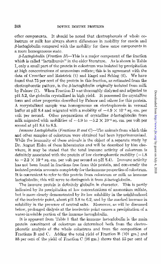
Exhaustive dialysis of the immune lactoglobulin fraction may result in the separation of water-insoluble and water-soluble or euglobulin and pseudoglobulin fractions. In only one instance was this done; for the other specimens the total immune globulin was used for characterization of these proteins. The euglobulin was separated by centrifuging; it was washed twice in the centrifuge wit’h distilled water at 2” to remove traces of pseudoglobulin and then suspended in water and lyophilized. The pseudoglobulin was diluted 4-fold with distilled water at 2” and then filtered through a sterilizing pad and lyophilized. The ratio of pseudo- globulin to euglobulin was about, 3:4. In Fig. 2 the electrophoretic patterns for the euglobulin and pseudoglobulin are shown. These runs
by guest on July 4, 2018http://w
ww
.jbc.org/D
ownloaded from
350 BOVINE IMMUNE PROTEIXS
were carried out in Verona1 buffer at pH 8.5. The pseudoglobulin migrated at a slightly higher mobility than the euglobulin, -2.2 compared with - 1.9 X 10M5 sq. cm. per volt per second. Immune activity was present in both the pseudoglobulin and euglobulin fractions.
The above isolations have clearly demonstrated that the initial colos- trum is extraordinarily rich in protein and that the principal protein is the immune lactoglobulin. For the three animals which me have studied, the aqueous phases contained 25.6, 20.9, and 15.4 per cent protein respectively and the immune lactoglobulin accounted for 55, 55, and 41 per cent of the total protein.
C L FIG. 2. Electrophoretic patterns of immune globulin from colostrum. R and B!
euglobulin after 100 and 200 minuks; C and D, pseudoglobulin after 100 and 200 minutes. The runs were performed in Verona1 buffer of pH 8.5 and at an ionic strength of 0.1.
Isolation of Plasma. Globulins
It has long been recognized that the colostrum or milk globulin is related to a serum globulin. The immunological studies of Wells and Osborne (8) had clearly shown this, and Crowther and Raist,rick had pointed out earlier that lactoglobulin and serum globulin were chemically indistinguishable by the methods available to them. It must be recognized that the proteins studied by these investigators were not homogeneous by present day standards. While there is no doubt of the relationship of the globulins of blood and milk, one cannot assume identity without further study. The plasma of hyperimmunized cows was therefore fract,ionated to obtain t.he components with which the immune activity is associated. It was soon recognized that, as in the plasma of the horse (9), the immune activity of bovine plasma is present in two components, y and T. Both of these
by guest on July 4, 2018http://w
ww
.jbc.org/D
ownloaded from
E. L. SMITH 351
components could be readily isolated by the methods elaborated by Cohn and his collaborators (10, 11) for the fractionation of plasma.
Fraction I (fibrinogen) was precipitated at 8 per cent ethanol and pH 7.3 at -2” from the plasma. The Fraction II + III, which contained most of the T and y components, was then removed at 25 per cent ethanol and pH 6.9 at -5”. From 24 liters of plasma there were obtained 2900 gm. of wet Fraction II + III paste. This paste was suspended in water and then fractionated by Method 3C (I 1). The yields of lyophilized powders were 115 gm. of Fraction 111-l and 100 gm. of Fraction II.
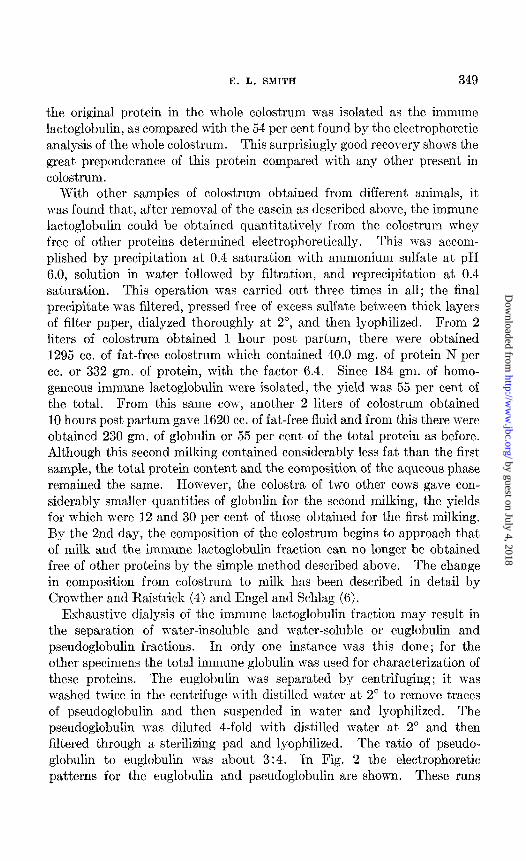
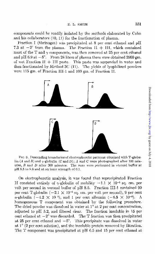
FIG. 3. Descending boundaries of electrophoretic patterns obtained with T-globu- lin (A and B) and y-globulin (C and D); A and C were photographed after 100 min- utes, B and L> after 200 minutes. The runs were performed in Verona1 buffer at pH 8.5 to 8.6 and at an ionic strength of 0.1.
On electrophoretic analysis, it n-as found that reprecipitated Fraction II consisted entirely of -y-globulin of mobility -1.1 X 10h5 sq. cm. per volt per second in Verona1 buffer of pH 8.5. Fraction III-1 contained 90 per cent T-globulin (-2.1 X 1O-5 sq. cm. per volt per second), 9 per cent a-globulin (-4.2 X 10⁻⁵), and 1 per cent albumin (-6.5 X 10P5). A homogeneous T component was obtained by the following procedure. The dried powder was dissolved in water at 1” at 2 per cent concentration, adjusted to pH 5.2, and filtered clear. The fraction insoluble in 15 per cent ethanol at -2” was discarded. The T fraction was then precipitat,ed at 25 per cent ethanol and -5”. This precipitate was dissolved in water at 1” (2 per cent solution), and the insoluble protein removed by filtration. The T component was precipitated at pH 6.5 and 15 per cent ethanol at
by guest on July 4, 2018http://w
ww
.jbc.org/D
ownloaded from

352 BOVINE IMMUNE PROTEINS
-2”, and was lyophilized. It proved to be homogeneous on electrophoresis at pH 8.6, and had a mobility of -2.4 X lop5 sq. cm. per volt per second. Fig. 3 shows the electrophoretic patterns obtained with the homogeneous y and T fractions. Other properties of Obese proteins will be described below in comparison with the colostrum globulin.
Isoelectric Points
The purified globulins were studied electrophoretically in the Tiselius apparatus with univalent buffers at an ionic strength of 0.1 and at lo.
4
0 Colostrum Globulin
-3 3 4 5 6 7 8 9
FIG. 4. Elect.rophoretic mobility as a function of pH for colostrum globulin, T- globulin, and r-globulin. All of the measurements were calculated from descending migrations in univalent, buffers at 1”. The mobility is in sq. cm. per volt per second.
At all pH values studied these proteins migrated as single components, although t.he patterns showed greater symmetrical spreading than would normally be expected for homogeneous proteins. The electrophoretic mobilities calculated from the data are shown in Fig. 4 as a function of the pH. The apparent isoelectric point of the r-globulin is at pH 7.2, that of the T component at pH 6.15, and of the total immune globulin of colostrum at pH 5.85. For the protein from the colostrum of another animal, the euglobulin was found to have a higher isoelectric point, about pH 6.2, than the pseudoglobulin, which was at pH 6.0. It is apparent from these data
by guest on July 4, 2018http://w
ww
.jbc.org/D
ownloaded from
E. L. SMITH 353
that the ‘r-globulin and colostrum globulin are similar although not neces- sarily ident,ical in their variation of mobility with pH, while the r-globulin is distinctly different from either of these. Since the homogeneity of the plasma proteins with respect to sedimentation and solubility is still un- known and the colostrum immune globulin is known to be a mixture, rigid comparisons are not possible at present. The T component, does, however, resemble the pseudoglobulin more than it does the euglobulin or total colostrum globulin.
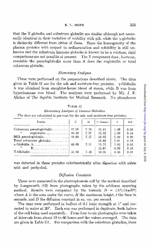
Elementary Analyses
These were performed on the preparations described above. The data given in Table II are for the ash and moisture-free proteins. r-Globulin A was obtained from slaughter-house blood of steers, while B was from hyperimmune cow blood. The analyses were performed by Mr. J. F. -4licino of The Squibb Institute for Medical Research. No phosphorus
TABLE II
Elementary Analyses of Immune Globulins
The data are calculated in per cent for the ash- and moisture-free proteins.
Protein C
Colostrum pseudoglobulin “ euglobulin.. . .
Milk pseudoglobulin.. Total colostrum globulin.. T-Globulin A.. .
“ B . . .
T-Globulin .
.
. .
47.G8 48.50 48.09
51.93
H
7.18
7.27 7.17
7.11
7.15 !
S (Dumas)
15.44 15.53 15.63 15.63 15.75 15.67 16.01
S
1.08
1.09
1.09
1.01
1.05
0.99
0.95
Ash
0.05 0.14 0.89 0.18 0.01 0.18
0.31
was detected in these proteins calorimetrically after digestion with nitric acid and perhydrol.
Dij’usion Constants
These were measured in the electrophoresis cell by the met,hod described by Longsworth (12) from photographs taken by the schlieren scanning method. Results were computed by the formula D = (A2)/(4atH2) where A is tlhe area under the curve, H the maximum height, t the time in seconds, and D the diffusion constant in sq. cm. per second.
The runs were performed in buffers of 0.1 ionic strength at 1” and cor- rected to water at 20”. Each run was performed in duplicate, both halves of the cell being used separately. From four to six photographs were taken at intervals from about 12 to 60 hours and the values averaged. The data are given in Table III. For comparison with the colostrum globulins, there
by guest on July 4, 2018http://w
ww
.jbc.org/D
ownloaded from

354 13OVISE IMMUNE PROTEINS
are included mea.surements on the T- and r-globulins of the cow and the r-globulin of the horse. The horse globulin was derived from antitetanus serum and was electrophoretically homogeneous.
It is apparent that, the diffusion constants found for the colostrum globulins are of the same ma,gnitude as those for the T- and y .globulins and are in good agreement w&h the results of Peclersen (13) for bovine r-globu- lin who found DZDw = 3.‘i4, 3.58, and 3.81. Our measurements suggest a small difference in DzOw for the euglobulin and pseudoglobulin, which may be significant. Nevertheless, all three bovine immune proteins are of approximately t’he same size, and, by analogy with similar proteins, indicate molecular weights in the region of 160,000 to 190,000.
For the horse r-globulin, Neurath, Cooper, and Erickson (14) found for their GI pseudoglobulin Dzou = 4.1 X lo-’ when t,heir result is corrected for the viscosity difference in water betI\-een 25” and 20”. Cohn el al.
Colost~rum A., ‘L “ “
pseudoglobulin 13. Bovine -pglobulin..
‘< T-globuliw Ilorsc ~-globulin.
‘i “ .,
*w cen1 0.4 I .i 1.2 0.8 1.8 1.5 0.6 1.6
BllfftY
Verona1 Cncodylat,e Verona1
“ ‘L
“
<‘
-- -
PH
8.45 6.62 8.45 8.46 8.37 8.61 8.43 8.54
Dzow x 107
- sq. ‘tn. per se‘.
3.50 f 0.06 3.69 f 0.11 3.34 f 0.00 3.86 f 0.19 3.53 f 0.09 3.60 f 0.23 4.08 f 0.23 3.78 f 0.04
(15) also cite for horse r-globulin, D20w = 4.1 X 10e7. Pappenheimer, Lundgren, and Williams (.16) ol)tained 4.4 X lo-’ for a purified diphtheria antit,osic globulin.
dnaphylactic Tests in Guinea Pigs
Wells and Osborne (8) carefully reviewed the older data on the immuno- logical relat,ions of the milk and plasma proteins and extended this work, using the milk prot,cin preparations prepared by Osborne and his collabo- rators. They clearly differentiated by anaphylaxis in guinea pigs the lactoglobulin from casein, lactalbumin, and the alcohol-soluble protein of milk, and showed that only the globulin sensitizes to beef serum or causes reactions in animals sensitized to beef serum. These observations have been confirn:ed with the homogeneous r-globulin and colostrum globulin.
Guinea pigs weighing 300 f 10 gm. Tl-ere sensit’ized by the intraperitoneal
by guest on July 4, 2018http://w
ww
.jbc.org/D
ownloaded from
E. L. SXITH 355
injection of 10.8 mg. of colostrum imn-une globulin in a volume of 1 cc. These animals mere tested 30 days later by intravenous inject’ion of y- globulin and colostrum globulin. For both of these proteins, severe re- actions with convulsions were produced at levels of 0.2 to 0.4 rrg., and fatal reactions invariably resulted from t,he inject.ion of 0.5 to 0.6 mg. At the time of inject,ion, t,he guinea pigs weighed 390 -I 20 gm. Xo differences in time or quality of response could be observed and the two proteins were quantitatively equivalent.
DISClTSIOS L_ ,, 6
It has been amply demonstrated in the past that l-he principal proteins of milk, casein, and &lactoglobulin (or lactalbnmin), are present only in the mammary secretion, and that these proteins arc distinct from any knot-n plasma proteins. However, the presence of immlme properties in milk and colostrum raises many questions regarding the relationship of the proteins possessing this funct’ion in these secretions compared Ivith Ihe proteins circulating in the blood stream. Although this question has been partially answered by the anaphylactic studies of many investigators, particularly Osborne and Wells, the problem remained unsolvctl.
In the cow, as in t’he horse, the immune activity of the plasma is present in at least two well defined components which c!ifYcr in their electrophoretic mobility and isoelectric points. Althocgh the immune globulin which has now been isolated from colostrum is similar to the T component of plasma in t,hese properties, the two proteins are not identical, as is indicated by differences in amino acid composition and ultraviolet absorption spectra (17, 18). Undoubt.edly, all of the proteins concerned with immunity in the same species are closely related (as is partly shown by the anaphylactic studies) but differ somewhat from each other. However, the molecules may vary in composition in portions of th.e molecule not concerned with either the immune properties or species specificity. That, part of the horse antitoxins is not concerned wit,h immune activity is suggested by the studies which have shown that approximately half of the molecule may be digested without impairment of the antitoxic functions of the remaining smaller molecules (19).
The absorption of immune globulin from colostrum into the blood st,ream of the new-born calf has been shown to produce a new electrophoretic component of plasma (3), which has been called r-globulin. The studies reported here have demonstrated that the colostrum globulin is readily differentiated electrophoret’ically from the r-globulin. It has been found that the absorbed globulin in the serum of the new-born calf possesses the mobility of the colostrum globulin and not’ t’hat of r-globulin.’
1 Smith, E. I,., and Helm, A., to he published.
by guest on July 4, 2018http://w
ww
.jbc.org/D
ownloaded from

356 BOVINE IMMUNE PROTEINS
From the work reported in this paper on colostrum, it is clear that the older concept of a lactalbumin fraction is not justified. Palmer showed that more than half of the protein in the lactalbumin fraction of milk whey could be obtained as a crystalline globulin. Unpublished data on the electrophoresis of milk whey have shown that about 60 per cent of the total protein is Palmer’s ,&lactoglobulin. It is not implied that proteins possessing the properties of albumins are not present in milk or colostrum, but it must be emphasized that what has hitherto been called “lactalbumin” is not primarily an albumin at all. The name has been used both for the total coagulable protein of milk whey, which is a complex mixture contain- ing predominantly two or more globulins in addition to a large number of other proteins, or it has been used in referring to a “lactalbumin fraction”
TABLE IV
Some Properties o.f Two Globulins o.f Milk and Colostrum
Isoelectric point, PH.. Mol. wt.. Dzo,“........................ Carbohydrate.. . Sulfur, %. Leucine, %. Valine, %. . Tryptophane, ‘%. Phenylalanine, %. Dialysis at isoelectric point.
Precipitation with (SH,)nSOI.. Immune activity..
-
_ @-Lactoglobulin Immune lactoglobulin
5.2 5.85 42,000 Ca. 160,000-190,000 7.3 x IO-’ 3.6 X lo-’ None Present 1.60 1.07 15.6 8.9 5.83 10.2 1.94 2.74 3.54 3.6 Crystals Insoluble euglobulin
and soluble pseudo- globulin
Ca. 0.4-0.7 saturated Cu. 0.25-0.4 saturated None Present
which contains a wide variety of proteins in small amount in addition to ,&lactoglobulin. It is only necessary to recall some of the proteins isolated in recent years from bovine whey, i.e. a flavoprotein (xanthine oxidase), a copper-protein, a peroxidase, as well as others.
In order to show clearly the distinctive properties of the globulin whose isolation has been described above, some of the properties of p-lactoglobulin and the immune protein are summarized in Table IV. These substances are readily differentiated by their size, carbohydrate content, solubility, and analytical composition with respect to sulfur content and the few amino acids for which data have been obtained on the total immune protein of colostrum (17). The analytical data for p-lactoglobulin have been taken from the paper by Brand and his collaborators (20).
by guest on July 4, 2018http://w
ww
.jbc.org/D
ownloaded from

E. L. SMITH 357
It is a pleasure to acknowledge the indebtedness of the author to Dr. August Holm for mak.ing available the colostrum and bovine plasma used in this study and for his cooperation. Gratitude is also due to Leo Zucker- man and Douglas M. Brown for their technical assistance, and to T. D. Gerlough for his help and cooperation.
SUMMARY
1. Electrophoretic analysis and isolation have shown that the immune lact,oglobulin is the predominant protein in bovine colostrum. This protein has been isolated in electrophoretically homogeneous form.
2. The immune activity of bovine plasma is present, in both T and y components. Both of these have been isolated and characterized in com- parison with the colostrum globulin by their elementary composition, isoelectric points, and diffusion constants.
3. The colostrum immune globulin and the plasma y-globulin have been shown to be quantitatively equivalent in producing anaphylaxis in guinea pigs.
4. The relationship of the various immune proteins has been discussed and it has been pointed out that, while the colostrum globulin is closely related to the y- and T-globulins, they are not identical. The immune lactoglobulin of colostrum has also been shown to be easily distinguished from p-lactoglobulin.
BIBLIOGRAPHY
1. Ehrlich, I’., Z. Hyg. u. Infektionskrankh., 12, 183 (1892). Famulener, L. W., J. Infect. nis., 10, 332 (1912). Smith, T., and Little, R. R., .I. Exp. Med.,36,181 (1922). Mason, J. S., Dalling, T., and Gordon, W. S., J. Path. and Bact., 33, 783 (1930).
2. Howe, P. E., Physiol. Rev., 6, 439 (1925). 3. Jameson, E., Alvarez-Tostado, C., and Sortor, H. H., Proc. Xoc. Ezp. Biol. and
iMed., 61, 163 (1942). San Clemente, C. L., and Huddleson, I. F., Michigan Agr. Exp. Sta., Bull. 182, 3 (1943).
4. Crowther, C., and Raistrick, H., B&hem. ,I., 10,434 (1916). 5. Mellander, O., B&hem. Z., 300, 240 (1939). Warner, R. C., J. Am. Chem. Sot.,
66, 1725 (1944). 6. Engel, H., and Schlag, H., Milchwirtschuft. Forsch., 2,1 (1924). 7. Palmer, A. H., J. Biol. Chem., 104, 359 (1934). 8. Wells, H. G., and Osborne, T. B., J. Infect. Dis., 29,200 (1921). 9. van der Scheer, J., Wyckoff, R. W. G., and Clarke, .F. H., J. Immunol., 39, 65
(1940). 10. Cohn, E. J., Strong, 1,. E., Hughes, W. I,., Jr., Xulford, 1). J., Ashworth, J. S.,
Melin, M., and Taylor, H. L., J. Am. Chem. SW., 68, 459 (1946). 11. Oncley, J. L., Melin, M., Richert, D. A., Cameron, J. W., and Gross, I’. M., J.
Am. Chem. Sot., 68, in press (1946). 12. Longsworth, I,. G., Ann. New York Acad. SC., 41, 267 (1941).
by guest on July 4, 2018http://w
ww
.jbc.org/D
ownloaded from
358 BOVIR’E IMMUNE PROTEINS
13. Pedersen, K. O., Ultracent,rifugal studies on serum and serum fractions, Upsala (1945).
14. Neurath, H., Cooper, G. IL., and Erickson, J. O., J. Biol. Chem., 138,411 (1941). 15. Cohn, E. J., McMeekin, T. L., Oncley, J. I,., Newell, J. M., and Hughes, W. L.,
6. Am. Chem. Sot., 62, 3386 (1940). 16. l’appenheimer, ii. M., Jr., Lundgren, II. I’., and Williams, J. W., J. Ezp. Med.,
71, 247 (1940). 17. Smith, E. L., Greene, R. D., and Bartner, E., J. Biol. Chem., 164,359 (1946). 18. Smith, E. L., and Coy, N. H., J. Biol. Chem., 164, 367 (1946). 19. Petermann, M. L., and Pappenheimer, A. M., Jr., J. Phus. Chem., 46, 1 (1941).
Rothen, A., J. Gen. Physiol., 26,487 (1942). 20. Brand, E., Saidel, L. J., Goldwater, W. H., Kassell, B., and Ryan, F. J., J. Am.
Chem. SOL, 67, 1524 (1945).
by guest on July 4, 2018http://w
ww
.jbc.org/D
ownloaded from

Emil L. SmithCOLOSTRUM AND PLASMA
THE IMMUNE PROTEINS OF BOVINE
1946, 164:345-358.J. Biol. Chem.
http://www.jbc.org/content/164/1/345.citation
Access the most updated version of this article at
Alerts:
When a correction for this article is posted•
When this article is cited•
alerts to choose from all of JBC's e-mailClick here
tml#ref-list-1
http://www.jbc.org/content/164/1/345.citation.full.haccessed free atThis article cites 0 references, 0 of which can be
by guest on July 4, 2018http://w
ww
.jbc.org/D
ownloaded from