the gingival oral lichen planus: a periodontal-oral...
TRANSCRIPT
Case ReportThe Gingival Oral Lichen Planus: A Periodontal-OralMedicine Approach
Abdulhameed Alsarraf,1 Kunj Mehta,2 and Nabil Khzam 3
1Ministry of Health, Kuwait2Private Practice, Perth 6000 Western Australia, Australia3NK Perio & Implants, Perth 6172 Western Australia, Australia
Correspondence should be addressed to Nabil Khzam; [email protected]
Received 5 August 2018; Revised 12 November 2018; Accepted 10 December 2018; Published 6 January 2019
Academic Editor: Jose López-López
Copyright © 2019 Abdulhameed Alsarraf et al. This is an open access article distributed under the Creative Commons AttributionLicense, which permits unrestricted use, distribution, and reproduction in any medium, provided the original work isproperly cited.
We present a case of a 77-year-old female who suffered from oral lichen planus (OLP) involving her gingiva and bilateral buccalmucosa for over 6 months. We showed that oral hygiene measures and conventional periodontal treatment and strictmaintenance were sufficient to control the gingival involvement of OLP. The mechanism of OLP is complex and not yet fullyunderstood. The focus of discussion in our case was that knowledge and understanding of gingival pathology are fundamentalfor a determined management approach. Our case was managed according to the suggested protocols in previous case studies. Amultidisciplinary approach allowed for accurate diagnosis and treatment tailored to the presented case.
1. Introduction
Lichen planus is a chronic immune-mediated inflamma-tory disease that affects the skin and mucous membranes.Oral lichen planus (OLP) is the mucosal counterpart ofcutaneous lichen planus and it was first described in1866 as white papular eruptions in the oral cavity [1].According to the American Academy of Periodontology,OLP is classified as nonplaque-induced gingival lesions[2]. Classically, OLP appears in a roughly symmetrical dis-tribution of well-defined white striations on a backgroundof mild erythema, commonly involving the buccal mucosaand tongue [3]. Other clinical patterns reported in theliterature include atrophic, erosive, papular, plaque-like,and bullous-type lesions [4]. To ensure accurate diagnosisof OLP, key histopathological features from biopsy speci-mens should be reported by an oral pathologist to allow clin-icopathological correlation and exclusion of oral epithelialdysplasia. The pathogenesis of the disease is characterizedby cytotoxic CD8+ T lymphocytes migration to the epithe-lium inducing apoptosis of basal keratinocytes [5]. A numberof etiological factors in OLP have been proposed such as
local and systemic inducers of cell-mediated hypersensitivity,drugs, dental materials, infectious agents, and stress [6]. OLPmanagement is directed at controlling active inflammationand the reduction of associated symptoms; therefore, patienteducation and awareness is important [7]. In addition,maintaining good oral hygiene, absence of oral candidosis,periodontal disease, and salivary gland hypofunction are allfactors to successful management outcomes [8]. Varioustherapeutic agents are described for controlling OLP includ-ing topical, intralesional, and systemic corticosteroids,immunosuppressive agents, retinoids, and immunomodula-tors. These treatment regimens may be used sequentiallyuntil effective symptomatic control is reached due to varia-tions in patient responses [9–11]. Of note, the malignantpotential of OLP is reported in the literature with an overallmalignant transformation of 1%. This is due to cases of oralcancer arising from patients with OLP demonstratingabsence of epithelial dysplasia [12, 13]. OLP has thereforebeen designated as an oral potentially malignant disorder. Itmust also be noted that similar clinical presentations ofOLP are induced by oral lichenoid reactions; however, it isnot always simple to distinguish between the two different
HindawiCase Reports in DentistryVolume 2019, Article ID 4659134, 3 pageshttps://doi.org/10.1155/2019/4659134

diseases clinically as well as histopathologically [9]. With thatbeing said, most oral lichenoid lesions are directly presentedadjacent to direct restorative materials such as amalgam andcomposite [9]. Further understanding and clinical experienceaids in implementing a long-term management plan forOLP cases.
2. Case Report
A 77-year-old female patient presented to her general dentistdue to bleeding gums. The dentist referred the patient to aspecialist periodontist for a consultation regarding the assess-ment and treatment of generalized chronic periodontitis. Afull comprehensive periodontal and radiographic examina-tion revealed a periodontal diagnosis of generalized moderateto advanced chronic periodontitis. Clinical signs of gingivalinflammation and periodontal pockets of 5mm and morewith calculus and bleeding upon probing were present ontwo or more aspects of each tooth. The radiographic exami-nation revealed a generalized horizontal bone loss of 40 to50% around most of the dentition. The patient was thenreferred to the Oral Medicine Clinic for diagnosis and furthermanagement of OLP-like lesions. Incisional biopsies wereperformed from the left buccal mucosa and 13/14 labial gin-giva (Figures 1 and 2). Histopathological assessment showedhyperkeratosis and band-like lymphocytic infiltrate in thelamina propria (Figure 3). No epithelial dysplasia was noted.These features are consistent with the diagnosis of OLP.Patient education and awareness was delivered in the contextof diagnosis, potential triggering factors, and disease malig-nant potential. Long-term observation is necessary, and thepatient will be followed up regularly to monitor diseasebehavior and progression.
3. Discussion
The diagnosis of OLP requires both clinical manifestationsand characteristic histopathological features. The practiceof good oral hygiene and achieving optimum periodontalhealth contribute to successful management outcomes. From
a clinical standpoint, the proposed periodontal treatmentplan was patient education, in the form of information onperiodontitis in terms of pathogenesis, treatment, and pre-vention. Detailed oral hygiene instructions include recom-mendations for the use of electric ultrasoft tooth brush,spongy dental floss, and soft interdental brushes. The patientwas encouraged to maintain high level of oral hygiene andattend regular periodontal review appointments in order tomaintain periodontal stability. The patient underwent twosessions of nonsurgical periodontal therapy on the samemonth, followed by reevaluation of periodontal tissue condi-tions 12 weeks after completion of debridement. This wasfollowed by supportive periodontal treatment (3 monthlyintervals). Overall, the patient had an excellent response tothe nonsurgical periodontal therapy with good oral hygiene.In view of the involved OLP, additional attention is requiredin the overall management of the case. OLP is frequentlyasymptomatic or associated withminor discomfort only. Thisis especially true for papular, reticular, and plaque-typelesions which are often unnoticed by the patient and thegeneral dentist. In contrast, atrophic and ulcerative lesionsmay give rise to severe pain, particularly related to food intakeand aggressive oral hygiene procedures. In the presented case,the OLP lesion involving the gingiva of teeth 13 and 14 wassymptomatic; therefore, the patient was instructed to use a
Figure 1: Gingival OLP: a mixed red and white lesion in thebackground of white striations involving the gingiva betweenteeth 13 and 14. No to minimal plaque is noted and the involvementof the attached gingiva distinguishes OLP from plaque-inducedgingivitis.
Figure 2: Left buccal mucosa: predominantly white plaque-typelesion and white striations noted in the background of milderythema.
Figure 3: Histopathological assessment from an incisional biopsyperformed from the left buccal mucosa showed hyperkeratosis andband-like lymphocytic infiltrate in the lamina propria.
2 Case Reports in Dentistry
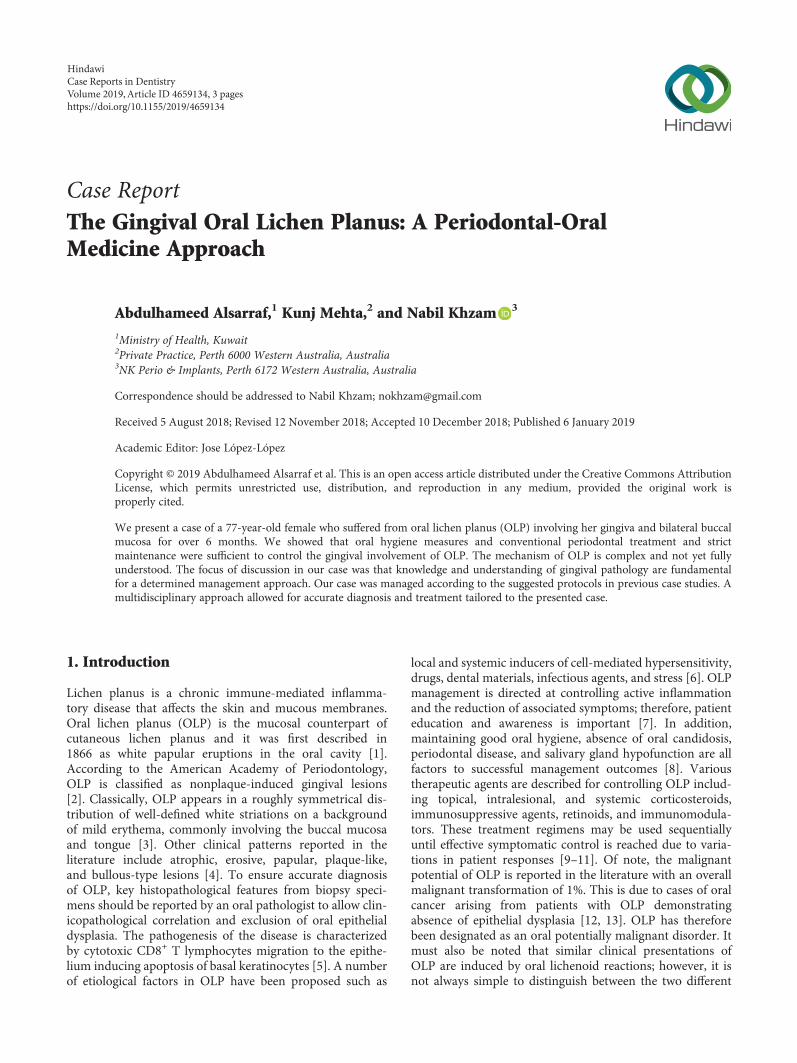
topical corticosteroid ointment three times daily for twoweeks applied directly to the lesion. Specifically, betametha-sone valerate 0.05% was used as a topical application to theaffected areas. Successful resolution of background erythemaand active gingival inflammation was noted in the reviewappointment (Figure 4). Other areas involving the oral cavitysuch as the left buccal mucosa improved significantly. From aperiodontal perspective, the most important part of thetherapeutic regimen is treatment of periodontal disease(conservative nonsurgical approach), an atraumatic oralhygiene in terms of gentle tooth brushing and flossing usingspongy type floss. Evidence of the literature supports thisstatement in the context of personalized plaque control forOLP gingival manifestations [14]. This will in turn result insignificant improvement in a large number of patients. Effec-tive plaque removal without traumatic influence on the gingi-val tissues must be established. In case of persistent paintypically associated with ulcerative and atrophic types, anti-fungal treatment may be necessary if the lesion contains can-dida species [8]. In chronic and symptomatic OLP cases, theresponse to the above treatment regimens is usually poor;therefore, topical corticosteroids are considered the first-linemedications to control patients’ symptoms. However, relapsein such cases is very common and intermittent episodes oftreatment may be required over an extended period of time.
4. Conclusion
This case has demonstrated the periodontal management of apatient with oral lichen planus. While oral hygiene measuresand conventional periodontal treatment were sufficient tocontrol the gingival involvement of OLP, patient educationand awareness of triggering factors were important to controlOLP activity with the aid of topical corticosteroids. It is how-ever important to consider the side effects related tolong-term use of topical corticosteroids in the oral cavity. Itis therefore important that both knowledge and understand-ing of gingival pathology are fundamental for a determinedmanagement approach in such cases.
Conflicts of Interest
The authors declare that they have no conflicts of interest.
References
[1] E. Wilson, “On lichen planus: the lichen ruber of Hebra,” BMJ,vol. 2, no. 302, pp. 399–402, 1866.
[2] G. C. Armitage, “Development of a classification system forperiodontal diseases and conditions,” Annals of Periodontol-ogy, vol. 4, no. 1, pp. 1–6, 1999.
[3] F. Gorouhi, P. Davari, and N. Fazel, “Cutaneous and mucosallichen planus: a comprehensive review of clinical subtypes,risk factors, diagnosis, and prognosis,” The Scientific WorldJournal, vol. 2014, Article ID 742826, 22 pages, 2014.
[4] Y.-S. L. Cheng, A. Gould, Z. Kurago, J. Fantasia, and S. Muller,“Diagnosis of oral lichen planus: a position paper of theAmerican Academy of Oral andMaxillofacial Pathology,”OralSurgery, Oral Medicine, Oral Pathology and Oral Radiology,vol. 122, no. 3, pp. 332–354, 2016.
[5] Z. B. Kurago, “Etiology and pathogenesis of oral lichen planus:an overview,” Oral Surgery, Oral Medicine, Oral Pathology andOral Radiology, vol. 122, no. 1, pp. 72–80, 2016.
[6] D. Farhi and N. Dupin, “Pathophysiology, etiologic factors,and clinical management of oral lichen planus, part I: factsand controversies,” Clinics in Dermatology, vol. 28, no. 1,pp. 100–108, 2010.
[7] V. Crincoli, M. B. Di Bisceglie, M. Scivetti, A. Lucchese,S. Tecco, and F. Festa, “Oral lichen planus: update onetiopathogenesis, diagnosis and treatment,” Immunopharma-cology and Immunotoxicology, vol. 33, no. 1, pp. 11–20, 2011.
[8] I. M. C. Lundström, G. B. Anneroth, and K. Holmberg,“Candida in patients with oral lichen planus,” InternationalJournal of Oral Surgery, vol. 13, no. 3, pp. 226–238, 1984.
[9] I. Al-Hashimi, M. Schifter, P. B. Lockhart et al., “Orallichen planus and oral lichenoid lesions: diagnostic andtherapeutic considerations,” Oral Surgery, Oral Medicine,Oral Pathology, Oral Radiology, and Endodontology, vol. 103,pp. S25.e1–S25.e12, 2007.
[10] F. Agha-Hosseini, N. Sheykhbahaei, and M.-S. SadrZadeh-Afshar, “Evaluation of potential risk factors that contributeto malignant transformation of oral lichen planus: a literaturereview,” The Journal of Contemporary Dental Practice, vol. 17,no. 8, pp. 692–701, 2016.
[11] E. M. Otero-Rey, F. Suarez-Alen, M. Penamaria-Mallon,J. Lopez-Lopez, and A. Blanco-Carrion, “Malignant transfor-mation of oral lichen planus by a chronic inflammatoryprocess. Use of topical corticosteroids to prevent thisprogression?,” Acta Odontologica Scandinavica, vol. 72,no. 8, pp. 570–577, 2014.
[12] G. Lodi, C. Scully, M. Carrozzo, M. Griffiths, P. B. Sugerman,and K. Thongprasom, “Current controversies in oral lichenplanus: report of an international consensus meeting. Part 1.Viral infections and etiopathogenesis,” Oral Surgery, OralMedicine, Oral Pathology, Oral Radiology, and Endodontology,vol. 100, no. 1, pp. 40–51, 2005.
[13] S. Warnakulasuriya, N. W. Johnson, and I. van der Waal,“Nomenclature and classification of potentially malignantdisorders of the oral mucosa,” Journal of Oral Pathology &Medicine, vol. 36, no. 10, pp. 575–580, 2007.
[14] S. J. Stone, G. I. McCracken, P. A. Heasman, K. S. Staines, andM. Pennington, “Cost-effectiveness of personalized plaquecontrol for managing the gingival manifestations of oral lichenplanus: a randomized controlled study,” Journal of ClinicalPeriodontology, vol. 40, no. 9, pp. 859–867, 2013.
Figure 4: Resolution of erythema and active gingival inflammationdue to OLP lesion involving the gingiva between teeth 13 and 14following use of topical corticosteroid ointment.
3Case Reports in Dentistry

DentistryInternational Journal of
Hindawiwww.hindawi.com Volume 2018
Environmental and Public Health
Journal of
Hindawiwww.hindawi.com Volume 2018
Hindawi Publishing Corporation http://www.hindawi.com Volume 2013Hindawiwww.hindawi.com
The Scientific World Journal
Volume 2018Hindawiwww.hindawi.com Volume 2018
Public Health Advances in
Hindawiwww.hindawi.com Volume 2018
Case Reports in Medicine
Hindawiwww.hindawi.com Volume 2018
International Journal of
Biomaterials
Scienti�caHindawiwww.hindawi.com Volume 2018
PainResearch and TreatmentHindawiwww.hindawi.com Volume 2018
Preventive MedicineAdvances in
Hindawiwww.hindawi.com Volume 2018
Hindawiwww.hindawi.com Volume 2018
Case Reports in Dentistry
Hindawiwww.hindawi.com Volume 2018
Surgery Research and Practice
Hindawiwww.hindawi.com Volume 2018
BioMed Research International Medicine
Advances in
Hindawiwww.hindawi.com Volume 2018
Hindawiwww.hindawi.com Volume 2018
Anesthesiology Research and Practice
Hindawiwww.hindawi.com Volume 2018
Radiology Research and Practice
Hindawiwww.hindawi.com Volume 2018
Computational and Mathematical Methods in Medicine
EndocrinologyInternational Journal of
Hindawiwww.hindawi.com Volume 2018
Hindawiwww.hindawi.com Volume 2018
OrthopedicsAdvances in
Drug DeliveryJournal of
Hindawiwww.hindawi.com Volume 2018
Submit your manuscripts atwww.hindawi.com