the eye 一. layers of the eye corneoscleral coat : fibrous layer, include the sclera, the white...
TRANSCRIPT
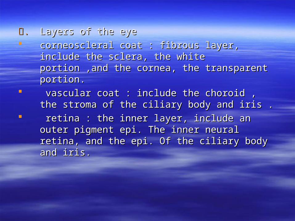
.壹.壹 Layers of the eyeLayers of the eye corneoscleral coat : fibrous layer, include the corneoscleral coat : fibrous layer, include the
sclera, the white portion ,and the cornea, the sclera, the white portion ,and the cornea, the transparent portion.transparent portion.
vascular coat : include the choroid , the vascular coat : include the choroid , the stroma of the ciliary body and iris .stroma of the ciliary body and iris .
retina : the inner layer, include an outer retina : the inner layer, include an outer pigment epi. The inner neural retina, and the pigment epi. The inner neural retina, and the epi. Of the ciliary body and iris.epi. Of the ciliary body and iris.
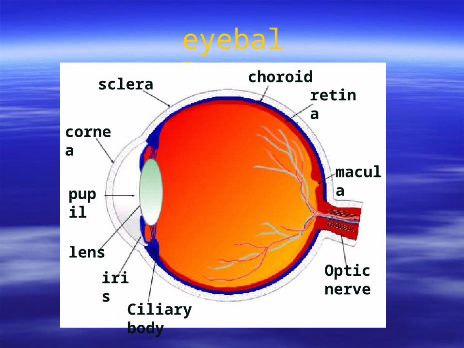
eyeball
pupil
lens
iris
Ciliary body
Optic nerve
macula
cornea
scleraretina
choroid
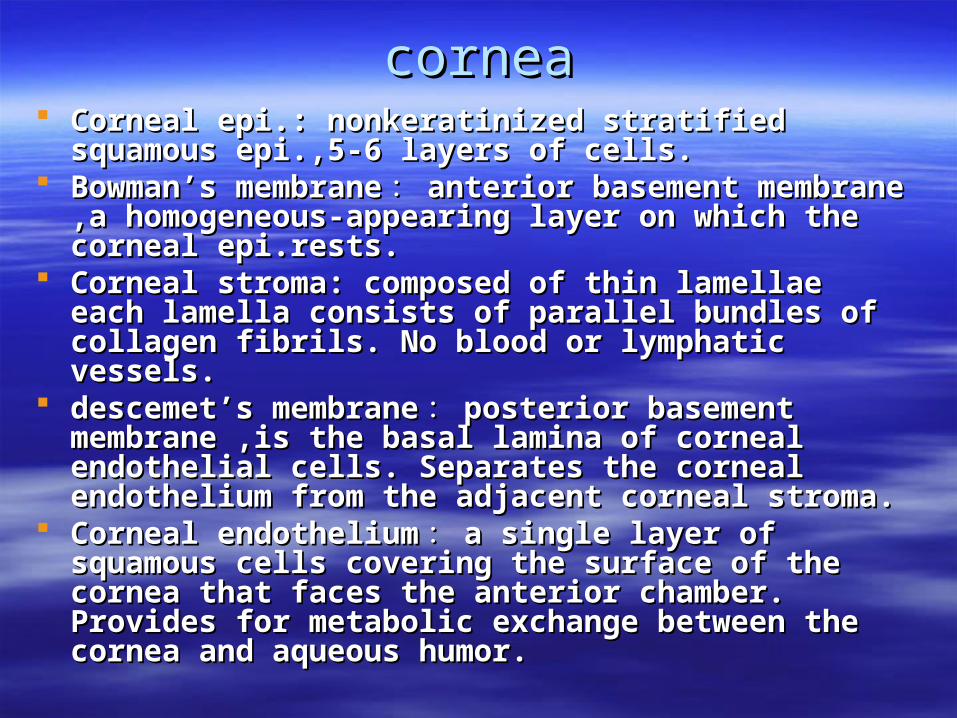
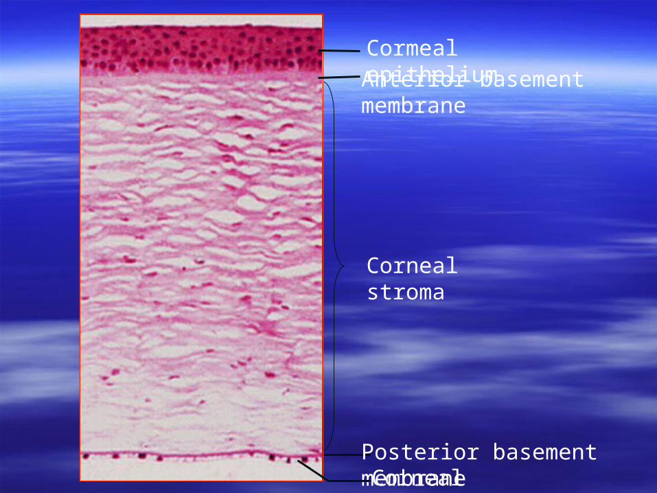
corneacornea Corneal epi.: nonkeratinized stratified Corneal epi.: nonkeratinized stratified squamous epi.,5-6 layers of cells.squamous epi.,5-6 layers of cells.
Bowman’s membraneBowman’s membrane :: anterior basement anterior basement membrane ,a homogeneous-appearing layer on membrane ,a homogeneous-appearing layer on which the corneal epi.rests.which the corneal epi.rests.
Corneal stroma: composed of thin lamellae Corneal stroma: composed of thin lamellae each lamella consists of parallel bundles of each lamella consists of parallel bundles of collagen fibrils. No blood or lymphatic collagen fibrils. No blood or lymphatic vessels.vessels.
descemet’s membranedescemet’s membrane :: posterior basement posterior basement membrane ,is the basal lamina of corneal membrane ,is the basal lamina of corneal endothelial cells. Separates the corneal endothelial cells. Separates the corneal endothelium from the adjacent corneal stroma. endothelium from the adjacent corneal stroma.
Corneal endotheliumCorneal endothelium :: a single layer of a single layer of squamous cells covering the surface of the squamous cells covering the surface of the cornea that faces the anterior chamber. cornea that faces the anterior chamber. Provides for metabolic exchange between the Provides for metabolic exchange between the cornea and aqueous humor. cornea and aqueous humor.
Cormeal epitheliumAnterior basement membrane
Corneal stroma
Posterior basement membraneCorneal endothelium
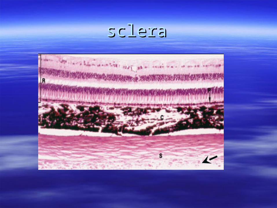
sclerasclera Is an opaque layer that consists Is an opaque layer that consists
predominantly of dense CT.predominantly of dense CT. The corneoscleral limbus : is the transitional The corneoscleral limbus : is the transitional
zone between the cornea and sclera . An zone between the cornea and sclera . An abrupt transition from the avascular cornea to abrupt transition from the avascular cornea to the well-vascularized sclera occurs here. the well-vascularized sclera occurs here.
Sclera venous sinus: in the irisocorneal angle Sclera venous sinus: in the irisocorneal angle contains the apparatus for the outflow of contains the apparatus for the outflow of aqueous humor which is produced by the aqueous humor which is produced by the ciliary processes .ciliary processes .
sclerasclera
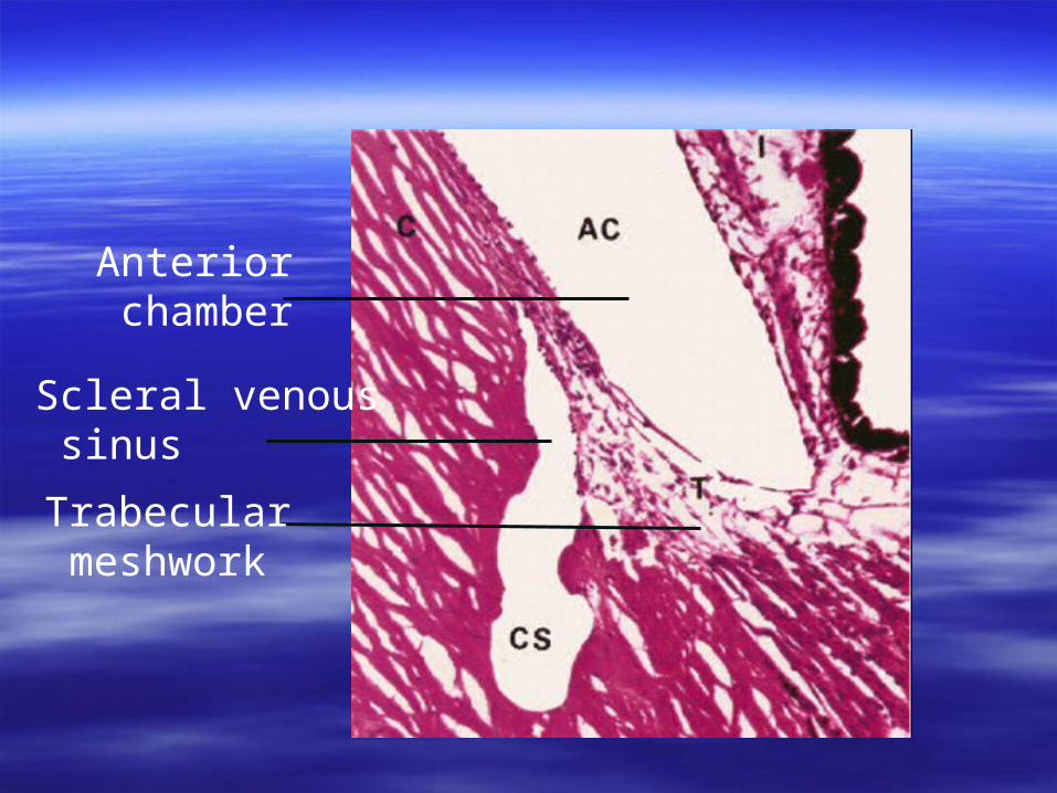
Anterior chamber
Scleral venous sinus
Trabecular meshwork
iris
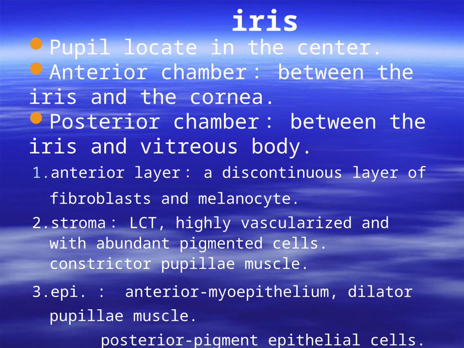
1. anterior layer : a discontinuous layer of fibroblasts and
melanocyte.
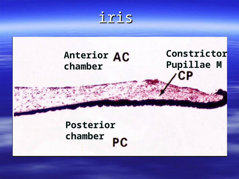
2. stroma : LCT, highly vascularized and with abundant pigmented cells. constrictor pupillae muscle.
3. epi. : anterior-myoepithelium, dilator pupillae muscle.
posterior-pigment epithelial cells.
Pupil locate in the center.Anterior chamber : between the iris and the cornea.Posterior chamber : between the iris and vitreous body.
irisiris
前房
Posteriorchamber
ConstrictorPupillae M
Anteriorchamber
Ciliary bodyCiliary body
Is the thickened anterior portion of Is the thickened anterior portion of the vascular coat and is located the vascular coat and is located between the iris and choroid.between the iris and choroid.
Ciliary muscle
The stroma
Epithelial layer
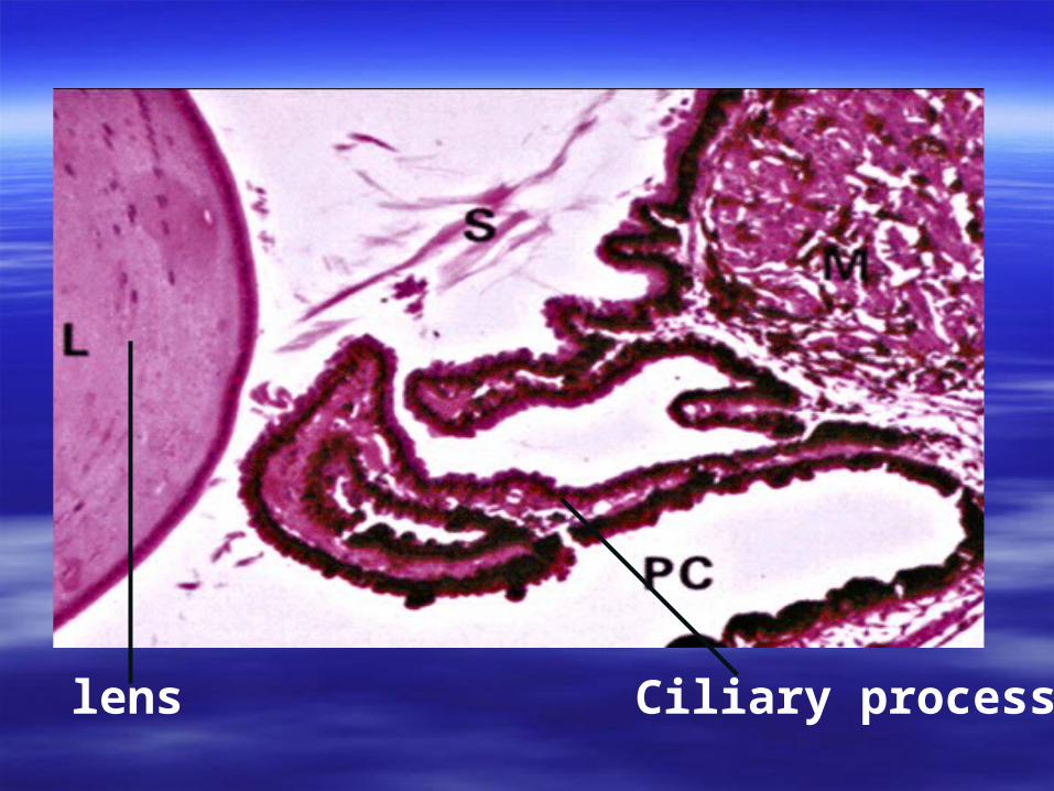
Ciliary processlens
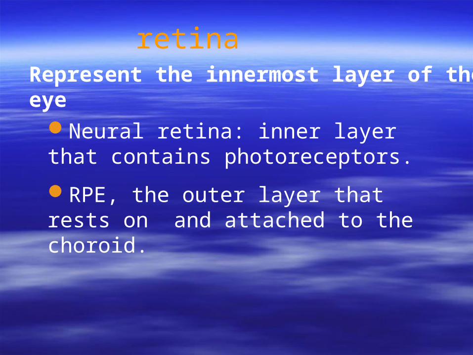
retinaRepresent the innermost layer of the eye
Neural retina: inner layer that contains photoreceptors.
RPE, the outer layer that rests on and attached to the choroid.
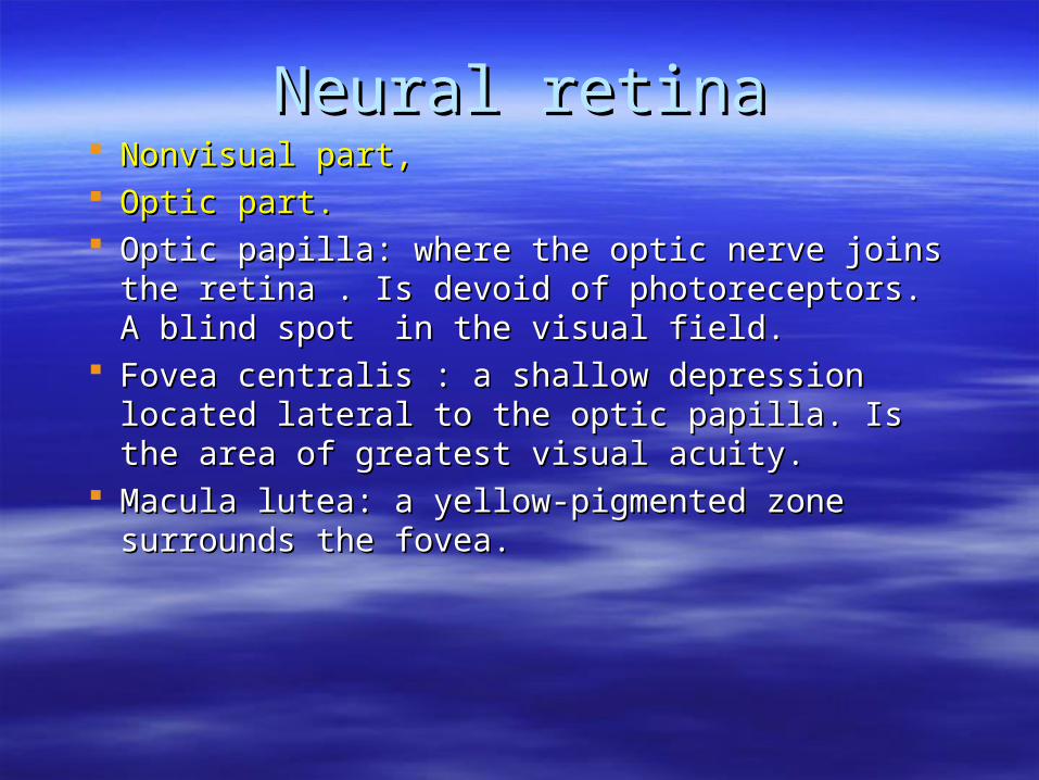
Neural retinaNeural retina Nonvisual part, Nonvisual part, Optic part.Optic part. Optic papilla: where the optic nerve joins the Optic papilla: where the optic nerve joins the
retina . Is devoid of photoreceptors. A blind spot in retina . Is devoid of photoreceptors. A blind spot in the visual field. the visual field.
Fovea centralis : a shallow depression located Fovea centralis : a shallow depression located lateral to the optic papilla. Is the area of greatest lateral to the optic papilla. Is the area of greatest visual acuity.visual acuity.
Macula lutea: a yellow-pigmented zone surrounds Macula lutea: a yellow-pigmented zone surrounds the fovea.the fovea.
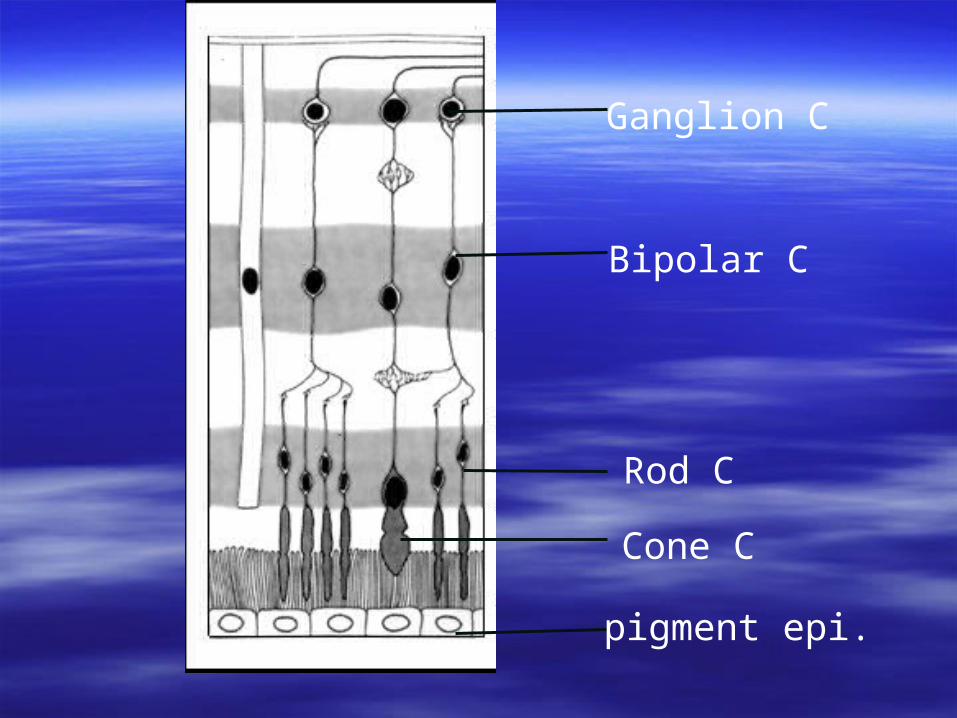
pigment epi.
Ganglion C
Bipolar C
Cone C
Rod C
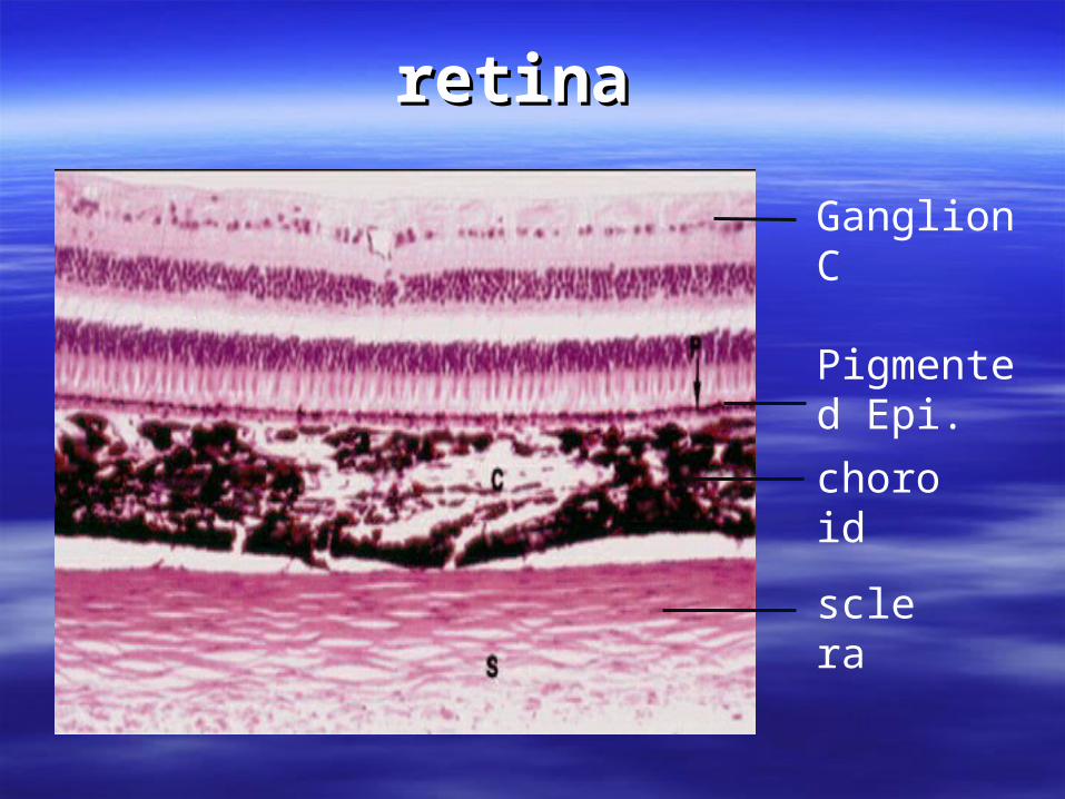
sclera
choroid
Pigmented Epi.
Ganglion C
retinaretina
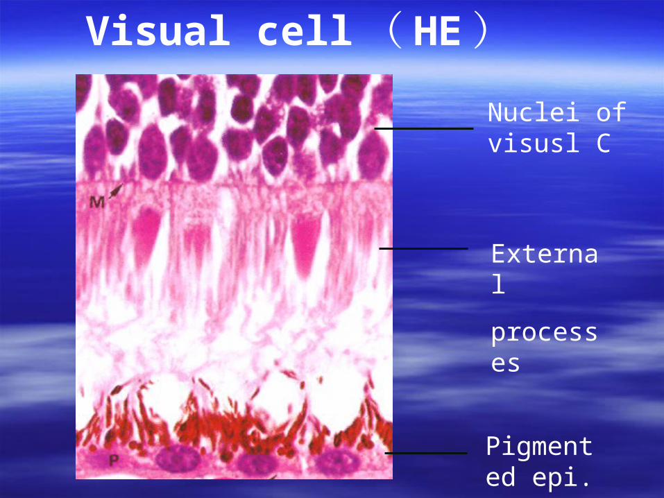
Nuclei of visusl C
External
processes
Pigmented epi.
Visual cell ( HE)
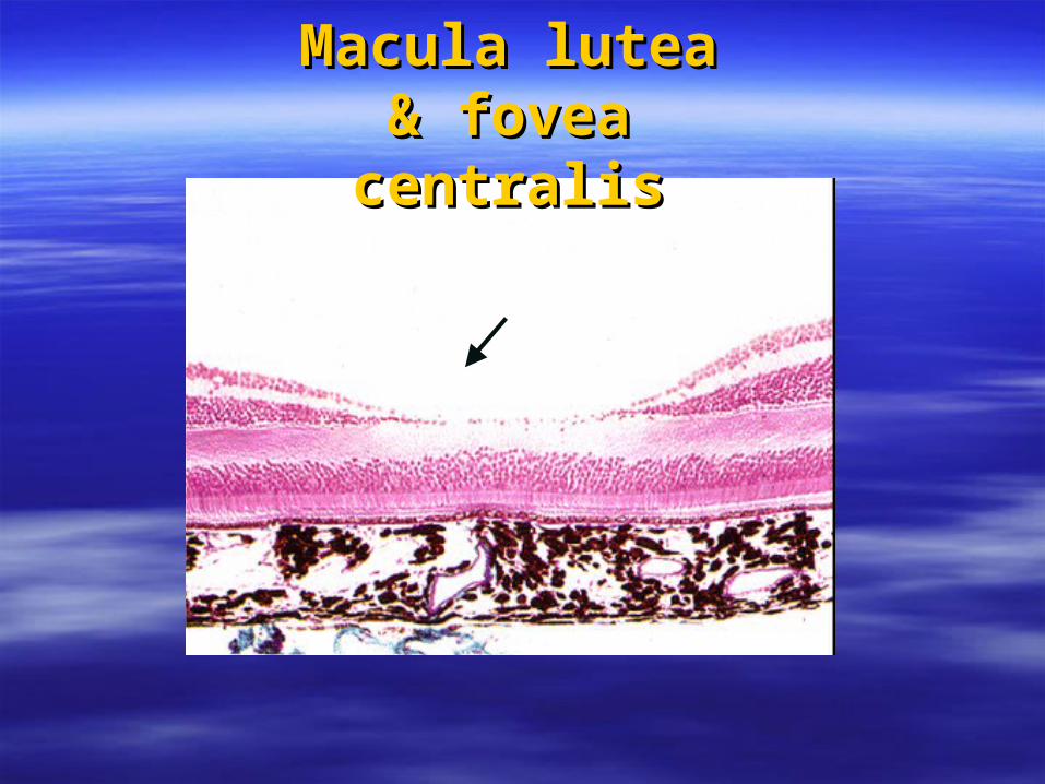
Macula lutea Macula lutea & fovea & fovea centraliscentralis
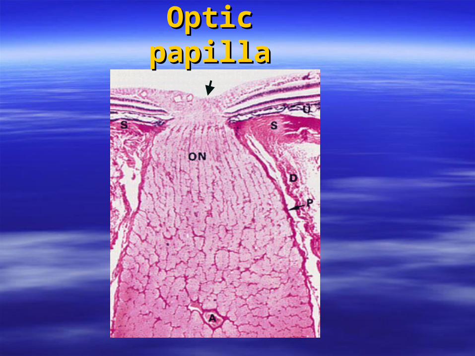
Optic Optic papillapapilla
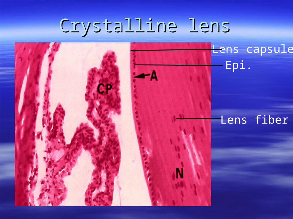
Lens capsuleEpi.
Lens fiber
Crystalline lensCrystalline lens
Crystalline lensCrystalline lens Is a transparent, avascular, biconvex Is a transparent, avascular, biconvex
structure.structure. The components of the lens:The components of the lens:
①① lens capsule, a thick basal laminalens capsule, a thick basal lamina
②② subcapsular epi., a cuboidal layer subcapsular epi., a cuboidal layer of of
cells. on the anterior face of the cells. on the anterior face of the lenslens
①① lens fibers, derived from lens fibers, derived from subcapsular epi.subcapsular epi.
Vitreous bodyVitreous body
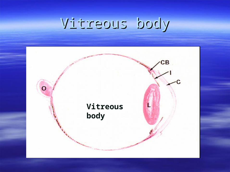
Vitreous body
Vitreous body Vitreous body
Is the transparent jelly-Is the transparent jelly-like substance that fill like substance that fill the vitreous chamber in the vitreous chamber in the posterior segment of the posterior segment of the eye.the eye.