the effects of organic and harsh cleaners on anolis ... · the effects of organic and harsh...
TRANSCRIPT

THE EFFECTS OF ORGANIC AND HARSH CLEANERS ON ANOLIS
CAROLINENSIS INTEGUMENT
A Report of a Senior Study
by
Kristen Rolston
Major: Biology
Maryville College
Spring, 2017
Date approved , by
Faculty Supervisor
Date approved , by
Division Chair
Formatted: Font:Italic
Formatted: Font:Italic

2
ABSTRACT
Household bleach has been used in the home for cleaning hard surfaces since the 18th century
and has caused a number of injuries over time. This study further investigates how
detrimental this caustic substance can be to the epidermis of Anolis carolinensis in
comparison to organically branded products claiming to be safer. Bleach cause significantly
reduced cell width (p=0.001) and number (p=0.009) on stratum corneum , whereas the
organic cleaner showed no difference. This study illustrates that Anolis carolinensis are an
appropriate model organism to examine the effects of certain substances on the
integumentary system and how the skin recovers could lead to valuable insight in the fields
of dermatology and stem cell research.
PAGE NUMBERS ARE INCORRECT THROUGHOUT
Formatted: Font:Italic
Deleted: It was determined that there was a significant effect
Deleted: cell width (p=2.59x10⁻¹⁴) and number (p=0.009)
Deleted: between the bleach and control groups
Deleted: By understanding

3
ACKNOWLEDGEMENTS
ADD ACKNOWLEDGEMENTS HERE
Deleted: [This section is not required. If included, it has a 2” top margin.]

4
TABLE OF CONTENTS
Page List of Tables vi List of Figures vii Chapter I Introduction 1 Chapter II Title of Chapter 2 Chapter III Title of Chapter 3 Chapter IV Title of Chapter 4 Appendix (or Appendices, as appropriate) 5 Works Cited 7
FIX THIS
Deleted: [This section has a 2” top margin.]

5
LIST OF TABLES
Table Page 1. ???????????????? 9 2. Full first sentence of table description 19 3. Full first sentence of table description 24
Deleted: Full first sentence of table description
Deleted: [If no more than two tables in document, you may omit this page. If included, it has a 1” top margin.]

6
LIST OF FIGURES
Figure Page 1. ???????????????????? 2 2. Full first sentence of figure description 7 3. Full first sentence of figure description 15 [If no more than two figures in document, you may omit this page. If included, it has a 1” top margin
Deleted: Full first sentence of figure description
7
CHAPTER I
INTRODUCTION
EVOLUTION OF AMNIOTE INTEGUMENT
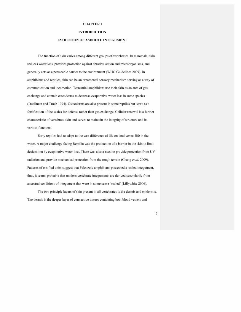
The function of skin varies among different groups of vertebrates. In mammals, skin
reduces water loss, provides protection against abrasive action and microorganisms, and
generally acts as a permeable barrier to the environment (WHO Guidelines 2009). In
amphibians and reptiles, skin can be an ornamental sensory mechanism serving as a way of
communication and locomotion. Terrestrial amphibians use their skin as an area of gas
exchange and contain osteoderms to decrease evaporative water loss in some species
(Duellman and Trueb 1994). Osteoderms are also present in some reptiles but serve as a
fortification of the scales for defense rather than gas exchange. Cellular renewal is a further
characteristic of vertebrate skin and serves to maintain the integrity of structure and its
various functions.
Early reptiles had to adapt to the vast difference of life on land versus life in the
water. A major challenge facing Reptilia was the production of a barrier in the skin to limit
desiccation by evaporative water loss. There was also a need to provide protection from UV
radiation and provide mechanical protection from the rough terrain (Chang et al. 2009).
Patterns of ossified units suggest that Paleozoic amphibians possessed a scaled integument,
thus, it seems probable that modern vertebrate integuments are derived secondarily from
ancestral conditions of integument that were in some sense ‘scaled’ (Lillywhite 2006).
The two principle layers of skin present in all vertebrates is the dermis and epidermis.
The dermis is the deeper layer of connective tissues containing both blood vessels and
8
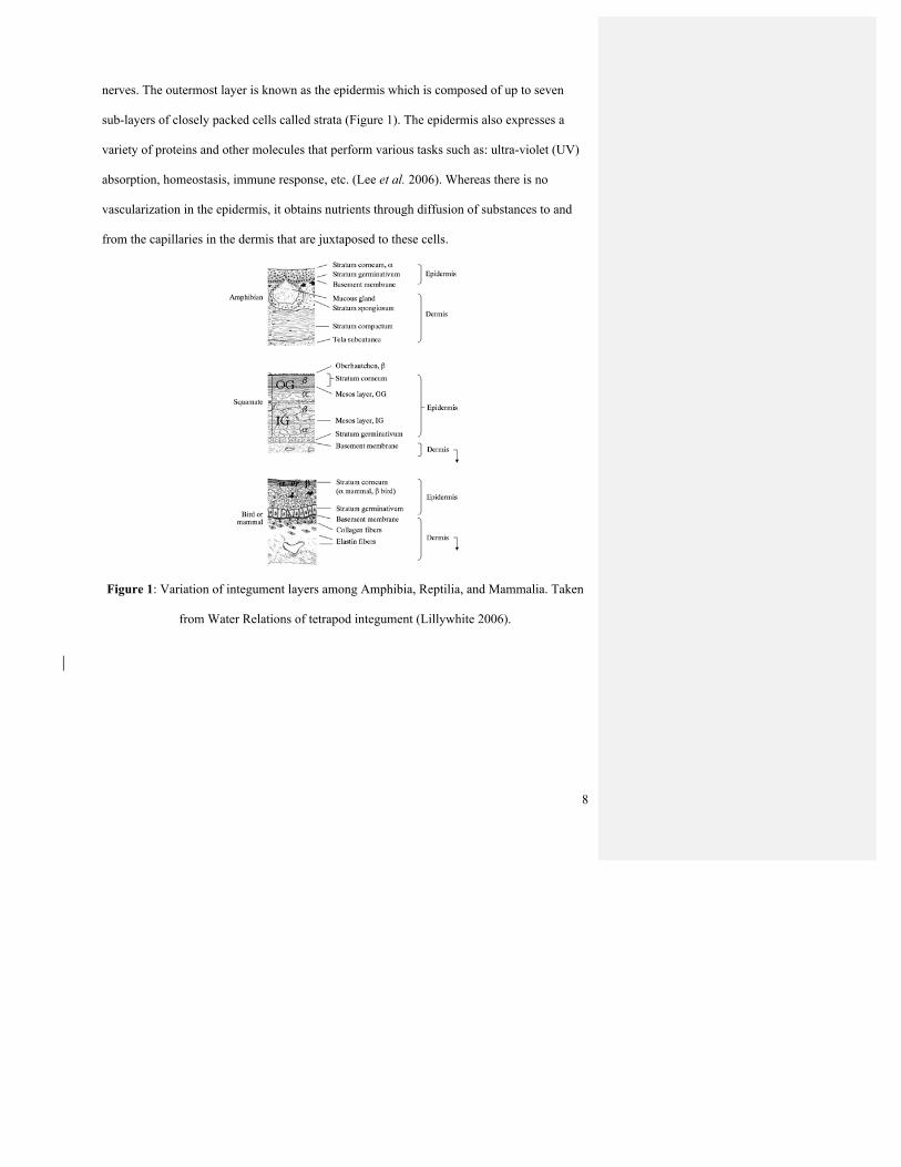
nerves. The outermost layer is known as the epidermis which is composed of up to seven
sub-layers of closely packed cells called strata (Figure 1). The epidermis also expresses a
variety of proteins and other molecules that perform various tasks such as: ultra-violet (UV)
absorption, homeostasis, immune response, etc. (Lee et al. 2006). Whereas there is no
vascularization in the epidermis, it obtains nutrients through diffusion of substances to and
from the capillaries in the dermis that are juxtaposed to these cells.
Figure 1: Variation of integument layers among Amphibia, Reptilia, and Mammalia. Taken
from Water Relations of tetrapod integument (Lillywhite 2006).
9
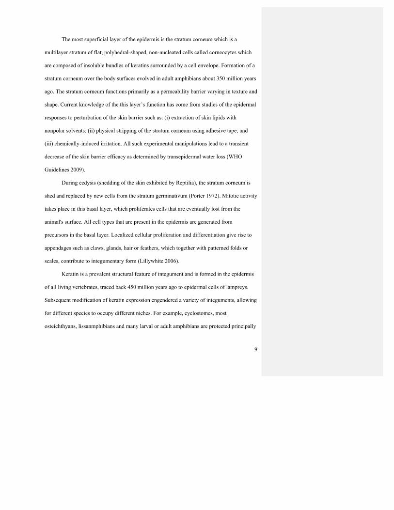
The most superficial layer of the epidermis is the stratum corneum which is a
multilayer stratum of flat, polyhedral-shaped, non-nucleated cells called corneocytes which
are composed of insoluble bundles of keratins surrounded by a cell envelope. Formation of a
stratum corneum over the body surfaces evolved in adult amphibians about 350 million years
ago. The stratum corneum functions primarily as a permeability barrier varying in texture and
shape. Current knowledge of the this layer’s function has come from studies of the epidermal
responses to perturbation of the skin barrier such as: (i) extraction of skin lipids with
nonpolar solvents; (ii) physical stripping of the stratum corneum using adhesive tape; and
(iii) chemically-induced irritation. All such experimental manipulations lead to a transient
decrease of the skin barrier efficacy as determined by transepidermal water loss (WHO
Guidelines 2009).
During ecdysis (shedding of the skin exhibited by Reptilia), the stratum corneum is
shed and replaced by new cells from the stratum germinativum (Porter 1972). Mitotic activity
takes place in this basal layer, which proliferates cells that are eventually lost from the
animal's surface. All cell types that are present in the epidermis are generated from
precursors in the basal layer. Localized cellular proliferation and differentiation give rise to
appendages such as claws, glands, hair or feathers, which together with patterned folds or
scales, contribute to integumentary form (Lillywhite 2006).
Keratin is a prevalent structural feature of integument and is formed in the epidermis
of all living vertebrates, traced back 450 million years ago to epidermal cells of lampreys.
Subsequent modification of keratin expression engendered a variety of integuments, allowing
for different species to occupy different niches. For example, cyclostomes, most
osteichthyans, lissanmphibians and many larval or adult amphibians are protected principally

10
by mucus, and thus the body is covered largely by non-keratinizing epidermis that suffices
for aquatic life (Maderson and Alibardi, 2000). Additional mechanical support is provided by
a cytoskeleton and a terminal web of keratin filaments in superficial cells.
Amphibians are exceptional among tetrapods in having very little keratin and a thin
stratum corneum (Lillywhite 2006). Although keratin was presumably present in basal
amphibians, neither extensive keratinization nor synthesis of β-type keratins characterizes the
skin of modern amphibian lineages. Therefore, amphibians prevent desiccation through
secretion of a superficial aqueous film, which must be replenished, or by shielding the
stratum corneum with superficial lipids (Lillywhite 2006).
The skin of amphibians is glandular and produces three principal categories of
secretions: mucus, various toxins, and lipids. Mucus plays various roles in the biology of
integument and is especially effective in relation to lubrication and keeping the skin hydrated
and moist. Those amphibians without a mucus film either avoid drying conditions or absorb
water from the substrate around them. Therefore, the importance of mucus secretions in
protecting exposed epidermal surfaces from dehydration suggests a fundamental dichotomy
of skin organization and water balance (Lillywhite and Maderson, 1988).

11
Figure 2: Electron Micrograph showing details of stratum corneum and permeability barrier
of terrestrial vertebrates. (A) Section through a portion of cocoon of a burrowing hylid frog,
Pternohyla fodiens. (B) Section through mesos layer of snake epidermis (Natrix natrix).
Laminated lipids occur between darker bands of keratin layers. (C) Section through stratum
corneum of human skin. Lipids occur between distinct layers of keratin. (D) Section through
epidermis of canary, showing nucleated layers as well as stratum corneum (top). As
presented by Lillywhite 2006.

12
In contrast, reptiles have a thick scale layer made up of β-keratins and other complex
lipids. Three typical reptile scales exist today: overlapping, tuberculate and elongated (frill),
with overlapping scales being the most common. In some species, the scales are modified
into sharp spines to dissuade predators and some are fortified with internal bony plates called
osteoderms (Halliday and Adler 2000). But scales do not necessarily provide strength of
structure, nor, as commonly misunderstood, do they provide an effective waterproofing
function (Roberts and Lillywhite, 1980).
In Sauropsid amniotes (the ancestors of reptiles and birds), a β-keratinized layer
formed above the α-keratinized layer and became the major constituents of scales and
feathers, providing mechanical support as well as a way to uptake solar energy (Porter
1972). β-keratins have a small molecular weight of about 10-25 kD and exhibit unique
arrangements of pleated sheets (Shames et al. 1989). Molecular studies suggest that β-
keratins in reptiles and birds occupy a functional role analogous to that of mammalian keratin
associated proteins (mKAPs, Rogers, 2004). Furthermore, the proteins so far indicated as β-
keratins seem to represent the reptilian equivalent of the keratin-associated or matrix proteins
present in mammalian hairs, claws, and horns.
The main components of mammalian hair are cysteine-rich type I and type II keratins
also known as hard α-keratins or “hair keratins”. Eckhard et al. 2008 determined that the
genome of chickens contained one type II hair keratin-like gene and the Anolis carolinensis
(green anole lizard) contained two type I and four type II hair keratin-like genes (expressed
most strongly in the digits). This study also suggests that cysteine-rich α-keratins are not
restricted to mammals and the evolution of mammalian hair involved the co-option of pre-
existing structural proteins.

13
A comparison of the half-cysteine and glycine content of vertebrate α and Ø keratins
suggests that the α and Ø proteins of reptiles may be related to the soft α-keratins of
mammals and amphibians (Jean Wyld and Alan Brush 1978). The hard keratin (claws, scales,
feathers, etc.) probably represents a uniquely derived group of proteins dissimilar to that of
vertebrate keratins. The X-ray diffraction pattern of reptiles is very similar to that obtained
from the avian hard keratins, leading to the conclusion that the framework of the filaments
was also composed of β-sheets, same as the avian keratins (Fraser and Parry 1996). It has
been proposed that the feather β-keratin subfamily (claws, feathers, feather-like, and scale)
evolved from the scale β-keratin subfamily through a deletion event followed by gene
duplication, whereas others suggest that the feather genes are basal to the avian scale genes
(Greenwold and Sawyer 2010). Total or near absence of scales is a derived character in many
amphibians, mammals and birds (Maderson and Alibardi, 2000).
In Therapsid amniotes, the ancestors of mammals, scales were lost and the α-
keratinized layer was strengthened by mammalian-type HRP (histidine-rich protein) rather
than by β-keratins (Wu et al. 2004). α-keratin molecules show a helical arrangement and
form polymers. They exist in the epidermis of all vertebrates and have a molecular weight of
about 40-70 kDa. It is the presence of HRPs in the stratum corneum of mammals that
provides a barrier to water loss. HRP is also known as filaggrin which functions as a keratin
matrix protein within the epidermis. It is a highly polar protein while being poor in nonpolar
amino acids (Lynley and Dale 1983).
14
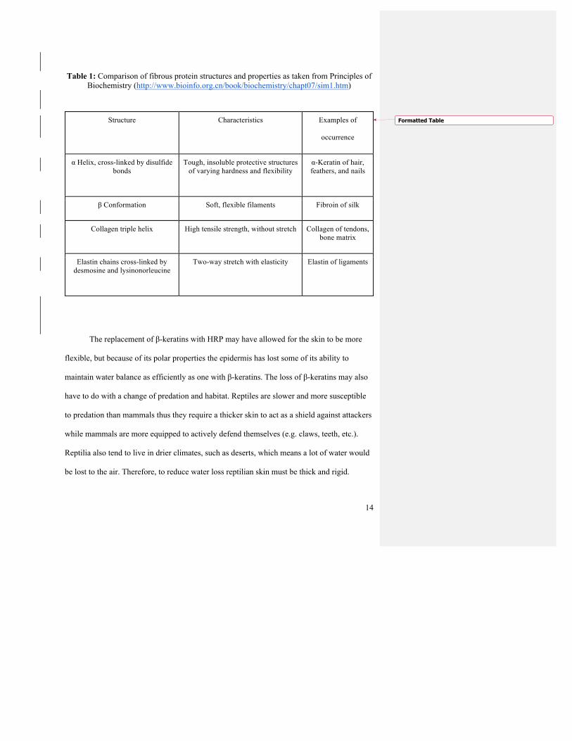
Table 1: Comparison of fibrous protein structures and properties as taken from Principles of Biochemistry (http://www.bioinfo.org.cn/book/biochemistry/chapt07/sim1.htm)
The replacement of β-keratins with HRP may have allowed for the skin to be more
flexible, but because of its polar properties the epidermis has lost some of its ability to
maintain water balance as efficiently as one with β-keratins. The loss of β-keratins may also
have to do with a change of predation and habitat. Reptiles are slower and more susceptible
to predation than mammals thus they require a thicker skin to act as a shield against attackers
while mammals are more equipped to actively defend themselves (e.g. claws, teeth, etc.).
Reptilia also tend to live in drier climates, such as deserts, which means a lot of water would
be lost to the air. Therefore, to reduce water loss reptilian skin must be thick and rigid.
Structure Characteristics Examples of
occurrence
α Helix, cross-linked by disulfide bonds
Tough, insoluble protective structures of varying hardness and flexibility
α-Keratin of hair, feathers, and nails
β Conformation Soft, flexible filaments Fibroin of silk
Collagen triple helix High tensile strength, without stretch Collagen of tendons, bone matrix
Elastin chains cross-linked by desmosine and lysinonorleucine
Two-way stretch with elasticity Elastin of ligaments
Formatted Table

15
Using reptiles as the model of human skin, this study hopes to determine that reptile
integument behaves similarly if not the same when exposed to external pollutants or irritants.
Anolis carolinensis (Green Anole) was chosen as a model for understanding tail anatomy and
regeneration in lizards for three main reasons. First, it is a suitable taxon for developing and
maintaining captive breeding populations, and colonies of healthy animals can be developed
and maintained relatively easily (Lovern et al., 2004). A. carolinensis is a small, arboreal
iguanid lizard commonly found on some Caribbean islands and in the Southeastern United
States, with northern most extent being North Carolina and Tennessee. Green anoles form
hierarchies in which the subordinates become browner in coloration and the dominant
becomes a bright green (Greenburg et al. 1984). While the body and claws share similar
amino acids with bird claws and feathers, the head and tail contain similar amino acids also
contained in mammalian hair, making them a good candidate for human skin cell
comparison.

16
Figure 3: Transverse section of A. carolinensis tail as shown in Kristian W. et al 2012
(http://journals.plos.org/plosone/article?id=10.1371/journal.pone.0051803)
A daily tasks of janitorial staff is to clean schools, churches, large industries using
typically bleach-based cleaners due to the high success of killing microbes. Bleaches based
on sodium hypochlorite (NaOCl) are most widely used even in households to disinfect hard
surfaces and bleach laundry (Racioppi et al. 2002). Bleach is cheap and easily obtainable,
making it a better option for most when cleaning. It is known to be effective against a broad
range of pathogens: gram-positive and gram-negative bacteria, fungi, spores and viruses
including human immunodeficiency virus (HIV), however it causes acute inflammation
followed by necrosis when it comes in contact with all tissue except heavily keratinized
epithelium (Mehdipour et al. 2007).
17
In recent years, there has been a growing demand for safe alternatives to these harsh
cleaners, especially in the home setting. This has brought up brands like Honest Company
and Simple Green that promise to make their products without potentially health-
compromising chemicals or compounds. One of the ingredients in the Honest Company’s All
Surface Cleaner is Phenoxyethanol which was found as the cause of contact dermatitis in
Lovell et al. 1984. However, it also contain Citrus grandis oils that have benefits of free
radical scavenging (Tsai et al. 2007). While these cleaners may be promoted by
environmentally savvy industries, this study hopes to determine that these cleaners are in fact
safer than bleach by studying the composition of the skin after longer-term exposure.

18
CHAPTER II
METHODOLGY
TREATMENT AND COLLECTION OF A. CAROLINENSIS INTEGUMENT
Animals and Exposure
Fourteen small, unsexed Green Anoles (Anolis carolinensis) were purchased from
Carolina Biological Supply Company (Item# 147232). Each anole was put into a separate
container with holes drilled into the sides and tops. They were randomly split into three
groups: control, organic, and bleach. Five anoles served as the control, five as the organic
and four as bleach.
The Anoles were kept in a light controlled room on a cycle of twelve hours of light
and twelve hours of darkness. Each tank was kept on a tall shelf in order to reduce animal
stress and each tank was equipped with plastic plants and a water bowl. Each Anole was fed
a diet of 2-3 crickets every two to three days and water was sprayed into each tank daily for
consumption and humidity control. Two heat lamps were fixed a safe distance away (12in.)
from each enclosure to ensure that one side was well warmed while leaving the other side
cool to thermoregulation. These lamps were turned on in the morning and turned off in the
evening.
Carefully handling the Anoles by holding them firmly by the torso, the five controls
were treated by dipping the tails into a 100 mL beaker of water for 30 seconds to simulate the
same stress the organic and bleach might experience. Five Anoles were treated with Honest
Co. MultiSurface Cleaner, 26 fl. oz. (Walmart Model#1607027) by dipping their tails into a
beaker of 100mL of cleaner for 30 seconds (see Table 2 for active ingredients). The last four
were treated with bleach using this dipping method, filling a beaker with 20 mL of bleach
Deleted:
Deleted: more

19
(Great Value Cleaning Bleach Walmart # 553565758, see Table 2 for ingredients) and filling
the rest with water to 100 mL. All applications were done daily for three weeks. At the end of
week three each Anole was subjected to a tail clip of 2cm from the tip of the tail, pressing
firmly against wounds that bled, and placed immediately into Bouin’s fixative in a Falcon
tube enough to cover all the tissue. Any premature deaths were immediately subjected to tail
clips and fixed in Bouin’s before being disposed of properly. Animal husbandry and
treatments were approved by Maryville College IACUC approval number 201716 (see
appendix 1).
INSERT TABLE 2
Endpoints Measured
The clipped tail was placed in Bouin’s fixative to prevent decay of the tissue. After
one week, the tissue was transferred to 70-75% alcohol to clear out excess fixative. Then the
tissue was dehydrated using the procedures listed in Humasin (2016). After dehydration,
tissues were infiltrated with paraffin wax under a vacuum for several hours in varying waxes
before being embedded in a wax block. Once the wax was dry, excess wax was cut away
from the preserved tissue and then the tissue was placed on a microtome to be sectioned as
desired. These ribbons were then floated on a warm water bath with a pinch of gelatin in the
bath and then they were mounted on slides. These slides were allowed to dry before staining.
The staining process is a hematoxylin and eosin staining process discussed in Humasin
(2016).
Data Analysis
Once the sections were stained, the prepared slides were viewed under a microscope.
The thickness, width, size and shape of the cells were examined among the control, the
Deleted: clean
Deleted: attempting to stain them
Deleted: cells
Deleted: between

20
organic treatment and the bleach treatment. These slides were then also compared to a known
slide of human epidermis provided by the Maryville College Animal Physiology and
Anatomy lab. Any notable changes were marked and photographed.
A t-test assuming equal variance was performed to show whether or not there was
significance between the control group vs. organic group, control group vs. bleach group, and
organic group vs. bleach group in terms of number of cells in the width of the stratum
corneum.

21
CHAPTER III
RESULTS
QUALITATIVE AND QUANTITATIVE REVIEW OF A. CAROLINENSIS TAIL
HISTOLOGY
Quantitative
Measurements of the stratum corneum, which appeared as a dark purple band near the
outer edge of the cell, determined that the control, organic and bleach groups had average
cell number of three, two and one cells across, respectively (see Figure 1). Cell number was
significantly lower in the bleach group compared to the controls (p=0.009). However, there
was no significant change between the organic group and the bleach group (p=0.175).
Likewise, there was no statistically significant difference between the control and the organic
group (p=0.067).
The average cell width of the stratum corneum was also measured with control
groups averaging at 0.55µm, organic groups at 0.57µm, and bleach groups at 0.23µm (see
Figure 2). Cell width was significantly less in the bleach tails compared to controls
(p<0.001), while there was no significant variation between control and organic (p=0.356).
Qualitative
There were two Anoles, a control and an organic, that died during the
experimentation process due to stress and did not receive the full three week exposure. For
this reason the results from these two individuals were removed from the rest of the data.
There does appear to be a slight thinning of the Oberhauchen layer, shown in light
pink outside of the stratum corneum that may be a result of increased shedding to relieve skin
Deleted: s
Deleted: and 2
Deleted: A t-test did show significant difference between the control and
Deleted: with
Deleted: a
Deleted: -value of
Deleted: There
Deleted: a
Deleted: difference between the cell width of the control and the cell width of the
Deleted: groups
Deleted: =2.59x10⁻¹⁴

22
cell damage or agitation. Furthermore, there is the appearance of a thin black layer beneath
the stratum corneum of the organic and bleach groups--worse so in the bleach groups. The
composition of this mass is unknown and would require further investigation to determine its
composition and its formation.
While the organic group varied slightly from the control in thickness and
composition, there is an abundance of damage to the dermis in the bleach groups where there
are obvious signs of thinning to 1 cell across (see Figure 3C). There is also a more prominent
Oberhauchen layer in some areas and areas where it is nearly nonexistent. There also appears
to be less structural integrity within the tail itself. Most individuals in the bleach samples had
blackened tails which confirms the atrophy of the cells within the tail.
0
0.5
1
1.5
2
2.5
3
Averagenu
mbe
rofcells
Bleach Organic Control
(4)
(5) (5)
Deleted: , bleach

23
Figure 4 (FIX IN TEXT): Comparison of average cell number+1 SE in stratum corneum after exposure to generic bleach, Honest Company Multipurpose cleaner (organic), and water
(control).
Figure 5 (FIX IN TEXT): Comparison of average cell width after exposure to generic
bleach, Honest Company Multipurpose Cleaner (organic), and water (control) for 3 weeks.
0
0.1
0.2
0.3
0.4
0.5
0.6
0.7Ce
llwidth(µ
m)
Control Organic Bleach
(4) (5)
(5)
Deleted: 1
Deleted: 2

24
Figure 6 (FIX IN TEXT): Anolis carolinensis tail sample stratum corneum after three weeks
exposure to water (control, A), Honest Company MultiPurpose Cleaner (organic, B), and Bleach (C) under 400x magnification
A
B
C
Deleted: 3

25
CHAPTER IV
DISCUSSION
ANALYSIS OF A. CAROLINENSIS AS A HUMAN INTEGUMENT MODEL
The results of this study show that household bleach does have an effect on the
structural integrity of the epidermis, specifically the stratum corneum, while there is little
effect of organic cleaners on these cells. Thus, buying less caustic cleaning products would
save the consumer the potential of doing one’s self harm or potential others during disposal
of said products.
Without a doubt, the integumentary system remains a vital aspect of all creatures in
which it plays roles from immune defense and physical protection to locomotion and sensory
communication. When exposed to certain substances this structure and its functions begin to
diminish, but in many cases the skin can heal and regenerate itself.
BETTER TRANSITION NEEDED…Studies done on skin regeneration have begun
to focus on the use of human umbilical cord Wharton’s jelly–derived mesenchymal stem
cells (hUC-MSCs) in the recovery of deep skin injury. In this study a middorsal, full
thickness incisional wound was performed on mice and new regeneration of Langerhans
cells, sebaceous glands, sweat glands and epidermis could be seen (Zhang et al. 2012).
Similarly, Kishi et al. 2012 found that fetal regeneration may be caused by reduced
expression of TGF-β1 and higher levels of hyaluronan in the extracellular matrix, allowing
for wounds to heal without inflammatory responses, granulation proliferation, and scar
Deleted: With certainty it can be said that
Deleted: It is safe to say that

26
formation as is observed in adults. Therefore skin injuries passed the gestation period remain
a growing concern especially in the area of fully body burns.
The exposure of the skin to even minor levels of bleach has shown just how sensitive
the skin truly is. Hypochlorite, a component of bleach, was determined to exert profound
cytotoxic effects on fibroblasts--which play a role in wound healing--as low as 0.05% of
concentration. After four hours of exposure and a concentration less than 0.01% the
mitochondria’s survival diminished from 71% to 10% (Racioppi et al. 1994). Sodium
hypochlorite (NaOCl) has a pH of 11-12.5 which causes injury primarily by oxidation of
proteins (Mehdipour et al. 2007). It is likely that since bleach is intended to kill bacteria that
it could compromise the healthy bacteria living on the skin upon contact and perhaps in
serious cases it could affect the Langerhans cells of the epidermis that alert the immune
system to invading viruses and bacteria.
The stratum corneum provides one of the key factors to regulate cutaneous
sensitization and by understanding this barrier is important for understanding self-defense
mechanisms because of its close functional link to skin-associated innate an adaptive
immunity. Accumulating evidence also shows that enhanced cutaneous sensitization is one of
the major causes of many allergic disorders including atopic dermatitis, asthma, food allergy
and anaphylaxis (Matsui et al. 2015). While reptile stratum corneum possesses a highly
impermeable barrier based on stiff epidermis, mammalian stratum corneum has evolved to be
highly moistened, resulting in the acquisition of a soft epidermis and thus leaving our skin
susceptible to infections and/or allergy with a complicated crosstalk between innate and
adaptive immune systems.

27
The Langerhans cells are located in the stratum spinosum beneath the stratum
granulosum which rests under the stratum corneum. These cells are thought to be involved in
induction of antigen-specific TH2 responses as well as maintenance of peripheral tolerance.
In the resting state the tips of the Langerhans cell dendrites are aimed at the apical side of the
stratum germinativum. Once activated, they extend their dendrites through the tight junction
beneath the stratum corneum where external antigens can be absorbed (see Figure ???).
Furthermore, the function and dysfunction of the stratum corneum determines the nature and
quality of cutaneous sensitization.
Figure ????: Structure of mammalian epidermis and its three barrier elements as
taken from Matsui et. al 2015.
Although recent research has told a great deal about the regenerative abilities and
factors of lizards’ tails, there has been little discussion about the importance of their
integument system as a model for human integument. The present study confirms it is
possible for Anolis carolinensis to serve as a model for humans due to the similarities of the
Deleted: 1
Deleted: 1
Formatted: Font:Italic

28
stratum corneum; however, it is still uncertain what the exact mechanism of skin regeneration
is and to what extent it can be modified. A future study of the anole tails is warranted to
analyze the recovery process after the exposure to environmental irritants.
What is already known about anole skin regeneration on the tail after autotomy (tail
loss) is that remnants of the torn original integument collapse over the site of loss and, within
days, a wound epithelium begins to form. There is also an alignment or chain of cell nuclei
that localize in the dermis creating a cellular zipper that may facilitate controlled rupturing of
the dermis during autotomy (Gilbert et al. 2015). Some lizards are also capable of what is
known as regional integumentary loss. Regional integumentary loss seems to be associated
with a discrete zone of structural weakness within the stratum compactum of the dermis that
are not located in the fractural plane (the area of breakage between scales that forms during
autotomy). Not much is truly known about the mechanisms.
There also appears to be little histology analysis of reptilian tail tissue specifically
investigating skin and its layers, and many questions remain. What are the mechanisms of
skin regeneration in lizards and is the mechanism similar if not the same in humans? If they
are different, can these mechanisms be mimicked in humans? With a focus on burn recovery,
could lizards be used to study the effects of burns and how the regenerative process proceeds
or is inhibited? Understanding these principles of the integument would not only provide
insight into the anatomy and physiology of lizards but also serve as a valuable complement to
the fields of dermatology and potential regenerative medicine.
Deleted: ,
Deleted: cells

29
APPENDICES
[Included only if there is more than one appendix.]

30
APPENDIX 1: TITLE OF APPENDIX IACUC?????

31
WORKS CITED MISSING REFS: HUMASON, ETC. CHECK EACH ONE.
Chang, C., Wu, P., Baker, R. E., Maini, P. K., Alibardi, L., & Chuong, C. (2009). Reptile
scale paradigm: Evo-Devo, pattern formation and regeneration. The International
Journal of Developmental Biology,53(5-6), 813-826. doi:10.1387/ijdb.072556cc
Duellman, W. E., & Trueb, L. (1994). Biology of amphibians. Baltimore: The Johns
Hopkins University Press.
Eckhart, L., Valle, L. D., Jaeger, K., Ballaun, C., Szabo, S., Nardi, A., . . . Tschachler, E.
(2008). Identification of reptilian genes encoding hair keratin-like proteins
suggests a new scenario for the evolutionary origin of hair. Proceedings of the
National Academy of Sciences,105(47), 18419-18423.
doi:10.1073/pnas.0805154105
Fraser, R., & Parry, D. (1996). The molecular structure of reptilian keratin. International
Journal of Biological Macromolecules,19(3), 207-211. doi:10.1016/0141-
8130(96)01129-4
Greenberg, N., Chen, T., & Crews, D. (1984). Social status, gonadal state, and the
adrenal stress response in the lizard, Anolis carolinensis. Hormones and
Behavior,18(1), 1-11. doi:10.1016/0018-506x(84)90045-x
Greenwold, M. J., & Sawyer, R. H. (2010). Genomic organization and molecular
phylogenies of the beta (β) keratin multigene family in the chicken (Gallus gallus)
and zebra finch (Taeniopygia guttata): implications for feather evolution. BMC
Evolutionary Biology,10(1), 148. doi:10.1186/1471-2148-10-148
Halliday, T., & Adler, K. (2000). The Encyclopedia of reptiles and amphibians. New
York: Facts on File.
Formatted: Font color: R,G,B (50,50,50)Formatted: Normal, Indent: Left: 0.25", Hanging: 0.5", Nobullets or numberingFormatted: Font color: R,G,B (50,50,50)
Formatted: Font color: R,G,B (50,50,50)
Formatted: Font color: R,G,B (50,50,50)
Formatted: Font color: R,G,B (50,50,50)
Formatted: Font color: R,G,B (50,50,50)
Formatted: Font color: R,G,B (50,50,50)

32
Lee, S. H., Jeong, S. K., & Ahn, S. K. (2006, July 30). An Update of the Defensive
Barrier Function of Skin. Retrieved March 05, 2017, from
https://synapse.koreamed.org/DOIx.php?id=10.3349%2Fymj.2006.47.3.293
Lillywhite, H. B. (2006). Water relations of tetrapod integument. Journal of Experimental
Biology,209(2), 202-226. doi:10.1242/jeb.02007
Lillywhite, H. B. and Maderson, P. F. A. (1988). The structure and permeability of
integument. Am. Zool. 28,945 -962.
Lynley, A. M., & Dale, B. A. (1983). The characterization of human epidermal filaggrin
[Abstract]. Biochimica et Biophysica Acta (BBA) - Protein Structure and
Molecular Enzymology,744(1), 28-35. doi:10.1016/0167-4838(83)90336-9
Maderson, P. F. A. and Alibardi, L. (2000). the development of the sauropsid integument:
A contribution to the problem of the origin and evolution of feathers. Am.
Zool. 40,513-529.
Porter, K. R. (1972). Herpetology. Philadelphia: W. B. Saunders.
Roberts, J. B. and Lillywhite, H. B. (1980). Lipid barrier to water exchange in reptile
epidermis. Science 207,1077 -1079.
Rogers G. Hair follicle differentiation and regulation. Int. J. Dev. Biol. 2004;48:163–
170. [PubMed]
Shames RB, Knapp LW, Carver WE, Washington LD, Sawyer RH. Keratinization of the
outer surface of the avian scutate scale: Interrelationship of alpha and beta keratin
filaments in cornifying tissue. Cell and Tissue Research. 1989;257:85–92.
WHO Guidelines on Hand Hygiene in Health Care: First Global Patient Safety Challenge
Clean Care Is Safer Care. Geneva: World Health Organization; 2009. 6,
Formatted: Font color: R,G,B (50,50,50)
Formatted: Font color: R,G,B (50,50,50)
Formatted: Font color: R,G,B (50,50,50)
Formatted: Font color: R,G,B (50,50,50)
Formatted: Font color: R,G,B (50,50,50)

33
Physiology of normal skin. Available from:
https://www.ncbi.nlm.nih.gov/books/NBK144027/
Wu, P., Hou, L., Plikus, M., Hughes, M., Scehnet, J., Suksaweang, S.(2004). Evo-Devo
of amniote integuments and appendages. The International Journal of
Developmental Biology,48(2-3), 249-270. doi:10.1387/ijdb.15272390
Wyld, J.A. & Brush, A.H. J Mol Evol (1979) 12: 331. doi:10.1007/BF01732028