the effect of two different bracket types on the salivary levels of s mutans and s sobrinus in the...
TRANSCRIPT
Original Article
The effect of two different bracket types on the salivary levels of S mutans
and S sobrinus in the early phase of orthodontic treatment
Antonija Jurelaa; Dario Repicb; Slavica Pejdab; Hrvoje Juricc; Renata Vidakovica; Igor Maticd;Andrija Bosnjake
ABSTRACTObjective: To determine the difference in the levels of Streptococcus mutans and S sobrinus instimulated saliva in orthodontic patients with different bracket types (stainless steel and estheticbrackets) using polymerase chain reaction and cultivation method.Materials and Methods: Thirty-two patients, aged 13 to 30 years, were selected following thesecriteria: 1) orthodontic treatment indication, 2) systemic health, and 3) no tobacco and antibioticconsummation for three months prior to the commencement of the study. Patients were dividedinto two groups according to the bracket type; 16 patients formed the conventional bracket group(stainless steel brackets), and 16 patients formed the esthetic bracket group (plastic brackets). Thelevels of S mutans and S sobrinus in stimulated whole saliva samples were collected prior to fixedorthodontic appliance placement (T1) and 12 weeks after placement (T2), as were the Decayed,Missing, and Filled Surface Index (DMFS) and Oral Hygiene Index-Simplified (OHI-S). Mann-Whitney, Wilcoxon, and chi-square tests were used for statistical analysis.Results: Statistical analysis (chi-square test) showed no difference in S mutans and S sobrinuscounts among patients with different brackets at either T1 or T2. There was no difference in totalbacteria counts after fixed orthodontic appliance placement.Conclusion: The number of colony-forming units of S mutans and S sobrinus in stimulated salivasamples does not seem to be significantly different between patients with stainless steel bracketsand patients with plastic brackets. (Angle Orthod. 2013;83:140–145.)
KEY WORDS: S Mutans; S sobrinus; Polymerase chain reaction; Plastic brackets; Stainless steelbrackets
INTRODUCTION
Complex design of fixed orthodontic appliancespromotes development and retention of supragingivalplaque due to reduced efficiency in self-cleaning and
oral hygiene measures.1 Supragingival plaque presentsa reservoir of complex cariogenic bacterial strains anda risk factor for enamel demineralization and dentalcaries.2 Microbial adhesion and plaque maturation inorthodontic patients depends on different variables,such as bracket design and material, proximity of thegingival sulcus to the bracket, surface area of the labialenamel relative to the bracket, position of the teeth in thedental arch, material of ligation and, mainly, individualoral hygiene habits.3 Numerous studies4,5 have provedthat placement of fixed orthodontic appliance leads to anincrease in the number of mutans Streptococci. Amongthem, Streptococcus mutans and S sobrinus have beenrecognized as prime causative organisms of dentalcaries.2 Since the popularity of plastic brackets hasgrown during the last few years due to increaseddemand for superior esthetics during orthodontic treat-ment, it is crucial to identify possible variations in theadhesion pattern of S mutans and S sobrinus on dif-ferent bracket material in order to decrease the risk ofpossible side effects of such therapy.
a Private practice, Zagreb, Croatia.b Resident, School of Medicine, University of Split, Split,
Croatia.c Professor, School of Dental Medicine, University of Zagreb,
Zagreb, Croatia.d Research assistant, Faculty of Science, University of
Zagreb, Zagreb, Croatia.e Professor, School of Medicine, University of Split, Split,
Croatia.Corresponding author: Dr Slavica Pejda, Resident, Depart-
ment of Orthodontics, Medical University of Split, Soltanska 2,Split 21000, Croatia(e-mail: [email protected])
Accepted: May 2012. Submitted: March 2012.Published Online: July 5, 2012G 2013 by The EH Angle Education and Research Foundation,Inc.
DOI: 10.2319/030612-187.1140Angle Orthodontist, Vol 83, No 1, 2013

In the literature there are diverse results regardingplaque and bacterial adhesion on different bracketmaterials that can be attributed to various methodol-ogies used in published research. The aim of thisclinical study was to detect the effect of two differenttypes of brackets (esthetic and stainless steel) oncaries-related factors such as a) Oral Hygiene Index-Simplified (OHI-S), b) paraffin-stimulated whole saliva,and c) levels of S mutans and S sobrinus in theparaffin-stimulated whole saliva in patients undergoingorthodontic treatment.
MATERIALS AND METHODS
Thirty-two patients of both sexes were included inthis prospective clinical study. The Ethical Committeeof the School of Dental Medicine approved the protocolof the research. Patients were recruited in a privateorthodontic office in Zagreb, Croatia, and had to satisfythe following inclusion criteria: 1) indication for fixedorthodontic treatment, 2) healthy systemic condition, 3)healthy periodontium, 4) nonsmoker, and 5) withoutantibiotic therapy in the last three months. The aimsand the methods of research were explained to allpatients, and they or their parents/guardians wereasked to sign informed consent.
Patients were divided into two groups according tobracket type: 16 patients formed the conventional bracketgroup (Discovery; Dentaurum, Ispringen, Germany), and16 patients formed the esthetic (plastic) bracket group(Spirit MB; Ormco/A Company, Orange, Calif). All bracketswere bonded with the same adhesive (Transbond XT; 3MUnitek, Monrovia, Calif) and ligated using elastomereligature (Latex-free Unicyles a Chain; Masel, Carlsbad,Calif). All patients received oral hygiene instructions atbaseline and at each regular check-up and were asked torefrain from any chlorhexidine use during the study.
From Pandis et al.,5 Forsberg et al.,6 and Attin et al.,7
it is known that a sample size of 16 patients per group,at a 5 .05, yields a statistical power close to 0.8 for thiskind of study.5–7
All clinical measurements were performed by thesame investigator at two time points: 1) prior toplacement of fixed orthodontic appliance (T1) and 2)12 weeks after placement of the appliance (T2). Timepoint T2 has been determined as a period with highestprevalence of oral microbiota according to Ristic et al.8
Further clinical measurements were performed atthese two time points in the following sequence: 1)paraffin-stimulated whole saliva (SS), 2) OHI-S, 3)Decayed, Missing, and Filled Surface Index (DMFS).
Paraffin-Stimulated Whole Saliva Collection
All patients were asked to refrain from eating,drinking, and brushing 2 hours prior to all clinical
examination. Collection of stimulated saliva and otherclinical parameters took place in a dental chair between0900 and 1100 hours. Patients were asked to chew 1 gof paraffin wax for 1 minute, followed by collection ofsaliva into measured, dry, and sterilized plastic tubesfor 5 minutes. After sampling time, the tubes weremeasured and the whole amount of collected stimulatedsaliva was calculated by subtracting the weight of theempty tube. Total amount of saliva was measured ing/min, which is equal to ml/min.9
Decayed, Missing, and Filled Surface Index (DMFS)
Decayed, Missing, and Filled Surface Index wasrecorded using criteria of the World Health Organiza-tion for permanent dentition.10
Oral Hygiene Index Simplified
In order to quantify supragingival plaque, we usedthe OHI-S of Greene and Vermillion,11 which is dividedinto debris index and calculus index, the sum of whichpresents the value of OHI-S. Fluorescein dye (Plak-Check, Butler, Germany) was applied with cotton tipsand illuminated with a halogen light (Blue phase G2;Ivoclar-Vivadent, Schaan, Liechtenstein) in order todetect yellow efflorescences. Supragingival plaqueand calculus were recorded on the buccal aspects ofthe permanent right and left maxillary first molars,permanent right maxillary central incisor, and perma-nent left mandibular central incisor, as well as on thelingual aspects of the permanent right and left man-dibular first molars. The OHI-S score was computed asthe mean of the plaque scores for the observed andscored (index) teeth. The criteria for classification ofthe OHI-S are good (from 0.0 to 0.6), regular (from 0.7to 1.8), and poor (from 1.9 to 3.0).
Bacterial Markers
Saliva samples were serially diluted 10-fold withpotassium phosphate-buffered saline (pH 7), and100 mL of diluted samples were plated, as previouslydescribed,12 on Mitis-Salivarius agar containing baci-tracin and sucrose (MBS agar). Another 100 mL ofdiluted saliva was plated on nonselective agar (Co-lumbia agar, BBL). Samples were incubated in ahumified chamber at 37uC for 2 days. The numberof colony-forming units (CFU) of S mutans and Ssobrinus per milliliter of saliva was determined fromtypical colonial morphology. All colonies with typicalmacro- and micromorphology were further subculti-vated overnight in thioglycollate broth in a shakerincubator (37uC). Pure culture strains were biochem-ically identified by API Strep test (BioMerieux Inc,Durham, NC).
SALIVARY STREPTOCOCCI IN ORTHODONTIC PATIENTS 141
Angle Orthodontist, Vol 83, No 1, 2013
Saliva (1 mL) was centrifuged for 15 minutes at12,000 g, and the pellet was resuspended in 200 mL ofcell lysis buffer (10 mM Tris-HCl, 1 mM Ethylenedi-aminetetraacetic acid, 1% Triton X-100; pH 8.0). Thesuspension was boiled for 10 minutes and centrifuged.Supernatant was used for DNA extraction.13,14
The sequences of the primers used for nestedpolymerase chain reaction (PCR) detection of Smutans and S sobrinus are shown in Table 1. GenomicDNA was analyzed by nested PCR for the glucosyl-transferase genes (gtfB of S mutans and gtfI of Ssobrinus). First sets of primers GTFB-F,-R and GTFI-F,-R were used for the first run of PCR. The secondround of PCR was performed using second sets ofprimers GTFB-FIN,-RIN, GTFI-FIN,-RIN, and 0.1 mL ofthe first run mixture as a template. Cycling conditionsfor the first and second PCR run were 6 minutesof denaturation at 95uC followed by 30 cycles of30 seconds of denaturation at 95uC, 30 seconds ofannealing at 59uC, and 1 minute of extension at 72uC,with a final extension of 7 minutes at 72uC. Aspreviously described, using this method we are ableto identify both S mutans and S sobrinus at a level of$104 CFU/ml saliva.12 PCR products were stained withethidium-bromide and analyzed on 1.8% agarose gels.
Complete microbiological treatment was performedat the Department of Clinical and Molecular Microbi-ology, University Hospital Centre Zagreb, Croatia.
Data analysis was performed by SPSS version10.0 (SPSS Inc, Chicago, Ill). Differences in age,OHI-S, SS, and DMFS between bracket groupswere analyzed by Mann-Whitney test, and differ-ences within the group between two time periodswere analyzed by Wilcoxon test. Chi-square testwas applied for difference assessment in bacteriacount, with the cutoff point at 104 CFU/ml forcultivation and PCR method. Cohen Kappa wasused for agreement assessment of bacteria detec-tion between cultivation and PCR method, with acutoff point 104 CFU/ml.
RESULTS
Gender distribution did not show a statisticallysignificant difference in the two bracket groups; therewere 10 female (62.5%) and 6 male patients (37.5%)in each group. Age distribution showed statisticallysignificant differences in the two bracket groups;patients with plastic brackets were older (25.8 6 4.9)than patients with stainless steel brackets (18.7 6 5.3;P , .001). There were no statistically significant dif-ferences between the two bracket groups regardingoral hygiene variables (OHI-S and DMFS) at T1(Table 2). Results indicate statistically significantimprovement in OHI-S in both groups (plastic brackets:P 5 .019, stainless steel brackets: P 5 .038; Table 2).SS flow did not show statistically significant differencesamong different brackets groups at either T1 or T2. Astatistically significant increase in stimulated salivaflow was determined in both groups between T1 andT2 (plastic brackets: P 5 .010, stainless steelbrackets: P 5 .030; Table 2). Cohen Kappa showedgood correlation between the two methods of detection(cultivation and PCR method) of S mutans and Ssobrinus with detection level at 104 CFU/ml (k 5 0.718for S mutans and 0.792 for S sobrinus, P , .001). Chi-square test revealed statistically insignificant differ-ences in the number of colony-forming units per
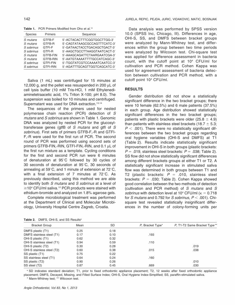
Table 1. PCR Primers Modified from Oho et al.14
Species Primers Sequence
S mutans GTFB-F 59-ACTACACTTTCGGTGGCTTGG-39
S mutans GTFB-R 59-CAGTATAAGCGCCAGTTTCATC-39
S sobrinus GTFI-F 59-GATAACTACCTGACAGCTGACT-39
S sobrinus GTFI-R 59-AAGCTGCCTTAAGGTAATCACT-39
S mutans GTFB-FIN 59-AAAGCAGATTCTAARGAATCGA-39
S mutans GTFB-RIN 59-AATGTAAAATTTTGCCATCAGC-39
S sobrinus GTFI-FIN 59-TGGTATCGTCCAAAATCAATCC-39
S sobrinus GTFI-RIN 59-AGATTTGCAGTTGGTCAGCATC-39
Table 2. DMFS, OHI-S, and SS Resultsa
Bracket Group Mean SD P, Bracket Type* P, T1-T2 Same Bracket Type**
DMFS plastic (T1) 0.25 0.18
DMFS stainless steel (T1) 0.17 0.10 .193
OHI-S plastic (T1) 0.62 0.32
OHI-S stainless steel (T1) 0.94 0.59 .110
OHI-S plastic (T2) 0.30 0.28 .019
OHI-S stainless steel (T2) 0.60 0.38 .015 .038
SS plastic (T1) 0.75 0.22
SS stainless steel (T1) 0.64 0.24 .160
SS plastic (T2) 0.90 0.26 .010
SS steel (T2) 0.87 0.45 .669 .030
a SD indicates standard deviation; T1, prior to fixed orthodontic appliance placement; T2, 12 weeks after fixed orthodontic appliance
placement; DMFS, Decayed, Missing, and Filled Surface Index; OHI-S, Oral Hygiene Index-Simplified; SS, paraffin-stimulated saliva.
* Mann-Whitney test; ** Wilcoxon test.
142 JURELA, REPIC, PEJDA, JURIC, VIDAKOVIC, MATIC, BOSNJAK
Angle Orthodontist, Vol 83, No 1, 2013

milliliter of S mutans and S sobrinus between the twobracket groups at either T1 or T2 (Tables 3 and 4).
DISCUSSION
Although some studies1,8,15 reported a negativeinfluence of fixed orthodontic appliances on quantita-tive and qualitative distribution of oral microbiota,an unequivocal and consistent conclusion regardingchanges of oral microbiota during orthodontic treat-ment is still lacking.
We were unable to detect any negative effect onmicrobial flora (S mutans and S sobrinus) 12 weeks
after fixed orthodontic appliance placement, which isconsistent with the results of Pandis et al.,5 Lara-Carrillo et al.,16 and Mota et al.17
In order to obtain a more uniform conclusionregarding the influence of fixed orthodontic applianceson oral microbiota, it is necessary to conduct clinicalstudies for a longer duration. Jordan and LeBlanc18
reported similar results in their four month longitudinalstudy. They linked the inconsistent pattern of mutansStreptococci prevalence observed among their pa-tients to the oral hygiene practices of individual pa-tients, something that may be attributable to thepatients in this study as well. However, at 12 weeks
Table 3. Summary Data for S mutans and S sobrinus Isolation Using the Cultivation Methoda
T1/T2 ,104 CFU/mL $104 CFU/mL P, Bracket Type*
P, T1-T2 Same
Bracket Type*
S mutans (plastic brackets) T1, N 16 0
% 100 0
T2, N 15 1
% 93.8 6.2 .312
S mutans (stainless steel brackets) T1, N 13 3
% 81.3 18.7 .069
T2, N 12 4
% 75 25 .143 .666
S sobrinus (plastic brackets) T1, N 16 0
% 100 0
T2, N 15 1 .312
% 93.8 6.2
S sobrinus (stainless steel brackets) T1, N 16 0
% 100 0
T2, N 15 1
% 93.8 6.2 1.0 .312
a T1 indicated baseline; T2, 12 weeks after fixed orthodontic appliance placement; CFU/mL, the number of colony-forming units per milliliter of
stimulated saliva of S mutans and S sobrinus based on morphology on MSB agar according to the method developed by Gold et al.12
* Chi-square test.
Table 4. Summary Data for S mutans and S sobrinus Isolation Using Nested PCRa
T1/T2 Nonconfirmed Confirmed P, Bracket Type*
P, T1-T2 Same
Bracket Type**
S mutans (plastic brackets) T1, N 15 1
% 93.8 6.2
T2, N 14 2
% 87.5 12.5 .541
S mutans (stainless steel brackets) T1, N 12 4
% 75 25 .333
T2, N 10 6
% 62.5 37.5 .220 .446
S sobrinus (plastic brackets) T1, N 16 0
% 100 0
T2, N 15 1
% 93.8 6.2 .312
S sobrinus (stainless steel brackets) T1, N 16 0
% 100 0
T2, N 14 2
% 87.5 12.5 1.0 .144
a T1 indicates baseline; T2, 12 weeks after fixed orthodontic appliance placement.
* PCR method detected S mutans and S sobrinus at $104 CFU/mL (number of colony-forming units per milliliter of stimulated saliva according
to the method developed by Oho et al.14).
** Chi-square test.
SALIVARY STREPTOCOCCI IN ORTHODONTIC PATIENTS 143
Angle Orthodontist, Vol 83, No 1, 2013

we were unable to detect any plaque on the teeth ofour subjects, attributable to the rigid and continuousinstructions that were performed at all checkups. Thepositive effects of oral hygiene instructions for patientswith fixed orthodontic appliances have been recog-nized,1 and significant improvement of the OHI-S indexwas observed in our study as well; this is inaccordance with the results by Al-Jewair et al.,19 whoreported good OHI compliance in 73% of patients.Improvement in OHI-S can be explained by the preciseinstructions in oral hygiene measures during eachcheckup and the resolving of crowding during the first12 weeks of orthodontic treatment, but it can also beattributable to the Hawthorne effect (patients’ aware-ness of being examined and evaluated).20
It is mainly in vitro studies that have been conductedto analyze the adhesion pattern of microbiota on dif-ferent orthodontic materials, but without uniformconclusions for implications in clinical practice.2,21–23
Since organic acids produced by investigated bacteriahave been recognized as the main pathological factorsin dental caries and enamel demineralization,24 wewanted to compare if there is any difference in theprevalence of these microbial species between pa-tients with different types of brackets.
Because of the difficulty in obtaining and controllingthe size of the plaque sample, we used stimulated salivaas the representative sample to quantitatively deter-mine levels of these microorganisms. Togelius et al.25
and Dasanayake et al.26 showed excellent correlationbetween stimulated saliva samples and plaque samplesfor quantitative assessment of mutans Streptococci.Results from our study indicate that there is no sta-tistically significant difference in levels of S mutans andS sobrinus from samples of paraffin-stimulated salivabetween patients with different bracket material, al-though Eliades et al.21 identified stainless steel as asurface material with increased potential for microbialattachment after measuring free surface energy andwork of adhesion of raw materials and compared it topolycarbonate and ceramic material. In contrast, resultsfrom Fournier et al.22 indicate weaker in vitro affinity ofS mutans for metallic brackets than for plastic brackets,which is in accordance with the results of a studyconducted by Ahn et al.,2 who made in vitro multiplecomparisons of cariogenic adhesion amounts onstainless steel, plastic, ceramic, and titanium brackets.Besides significant differences in adhesion pattern ofdifferent cariogenic strains, their results showed higheradherence of cariogenic streptococci on plastic brack-ets than on four other types of brackets. This wasexplained by the surface characteristics of plasticbrackets that were modified with filler, which conse-quently led to increased adhesion of cariogenicstreptococci.
Contrary to the results from aforementioned studies,microbiological data from our study did not indicate anystatistically significant difference in total cariogenicStreptococci in paraffin-stimulated whole saliva oforthodontic patients with different bracket material.Similar results were reported by Papaioannou et al.,3
who were unable to find statistically significant dif-ferences regarding adhesion patterns among stainlesssteel, plastic, and ceramic brackets. In their in vitro studythey underscored the important role of the salivarypellicle, which might negate any differences in cariogenicsurface characteristics (surface free energy), togetherwith the presence of histatins and lyzozymes in salivathat possess antibacterial activity and can contribute todecreased amounts of microbiota adhesion.
CONCLUSIONS
N The numbers of colony-forming units of S mutansand S sobrinus per milliliter of paraffin-stimulatedsaliva were not influenced at a statistically significantlevel by fixed orthodontic appliance placement duringthe first 12 weeks of orthodontic treatment.
N The numbers of colony forming units of S mutansand S sobrinus per milliliter of paraffin-stimulatedsaliva were not significantly altered by bracket type,either stainless steel or plastic.
REFERENCES
1. Smiech-Slomkowska G, Jablonska-Zrobek J. The effect oforal health education on dental plaque development and thelevel of caries-related Streptococcus mutans and Lactoba-cillus spp. Eur J Orthod. 2007;29:157–160.
2. Ahn SJ, Lee SJ, Lim BS, Nahm DS. Quantitative determi-nation of adhesion patterns of cariogenic streptococci tovarious orthodontic brackets. Am J Orthod DentofacialOrthop. 2007;132:815–821.
3. Papaioannou W, Gizani S, Nassika M, Kontou E, Nakou M.Adhesion of Streptococcus mutans to different types ofbrackets. Angle Orthod. 2007;77:1090–1095.
4. Sandham HJ, Nadeau L, Phillips HI. The effect of chlorhexidinevarnish treatment on salivary mutans streptococcal levels inchild orthodontic patients. J Dent Res. 1992;71:32–35.
5. Pandis N, Papaioannou W, Kontou E, Nakou M, Makou M,Eliades T. Salivary Streptococcus mutans levels in patientswith conventional and self-ligating brackets. Eur J Orthod.2010;32:94–99.
6. Forsberg CM, Brattstrom V, Malmberg E, Nord CE. Ligaturewires and elastomeric rings: two methods of ligation, andtheir association with microbial colonization of Streptococ-cus mutans and lactobacilli. Eur J Orthod. 1991;13:416–420.
7. Attin R, Thon C, Schlagenhauf U, Werner C, Wiegand A,Hannig C, Attin T. Recolonization of mutans steptococci onteeth with orthodontic appliances after antimicrobial therapy.Eur J Orthod. 2005;27:489–493.
8. Ristic M, Vlahovic Svabic M, Sasic M, Zelic O. Clinical andmicrobiological effects of fixed orthodontic appliances onperiodontal tissues in adolescents. Orthod Craniofac Res.2007;10:187–195.
144 JURELA, REPIC, PEJDA, JURIC, VIDAKOVIC, MATIC, BOSNJAK
Angle Orthodontist, Vol 83, No 1, 2013
9. Navazesh M. Methods for collecting saliva. Ann N Y AcadSci. 1993;694:72–77.
10. World Health Organization. Oral Health Surveys: BasicMethods. 4th ed. World Health Organization, Geneva; 1997.
11. Greene JC, Vermillion JR. The simplified oral hygiene index.J Am Dent Assoc. 1964;68:7–13.
12. Gold OG, Jordan HV, Van Houte J. A selective medium forStreptococcus mutans. Arch Oral Biol. 1973;18:1357–1364.
13. Watanabe K, Frommel TO. Detection of Porphyromonasgingivalis in oral plaque samples by use of the polymerasechain reaction. J Dent Res. 1993;72:1040–1044.
14. Oho T, Yamashita Y, Shimazaki Y, Kushiyama M, Koga T.Simple and rapid detection of Streptococcus mutans andStreptococcus sobrinus in human saliva by polymerasechain reaction. Oral Microbiol Immunol. 2000;15:258–262.
15. Topaloglu-Ak A, Ertugrul F, Eden E, Ates M, Bulut H. Effectof orthodontic appliances on oral microbiota—6 monthfollow-up. J Clin Pediatr Dent. 2011;35:433–436.
16. Lara-Carrillo E, Montiel-Bastida NM, Sanchez-Perez L,Alanıs-Tavira J. Effect of orthodontic treatment on saliva,plaque and the levels of Streptococcus mutans and Lacto-bacillus. Med Oral Patol Oral Cir Bucal. 2010;15:924–920.
17. Mota SM, Enoki C, Ito IY, Elias AM, Matsumoto MA.Streptococcus mutans counts in plaque adjacent to ortho-dontic brackets bonded with resin-modified glass ionomercement or resin-based composite. Braz Oral Res. 2008;22:55–60.
18. Jordan C, LeBlanc DJ. Influences of orthodontic applianceson oral populations of mutans streptococci. Oral MicrobiolImmunol. 2002;17:65–71.
19. Al-Jewair TS, Suri S, Tompson BD. Predictors of adolescentcompliance with oral hygiene instructions during two-archmultibracket fixed orthodontic treatment. Angle Orthod.2011;81:525–531.
20. Feil PH, Grauer JS, Gadbury-Amyot CC, Kula K, McCunniffMD. Intentional use of the Hawthorne effect to improve oralhygiene compliance in orthodontic patients. J Dent Educ.2002;66:1129–1135.
21. Eliades T, Eliades G, Brantley WA. Microbial attachment onorthodontic appliances. I. Wettability and early pellicleformation on bracket materials. Am J Orthod DentofacialOrthop. 1995;108:351–360.
22. Fournier A, Payant L, Bouclin R. Adherence of Streptococ-cus mutans to orthodontic brackets. Am J Orthod Dentofa-cial Orthop. 1998;114:414–417.
23. Anhoury P, Nathanson D, Hughes CV, Socransky S, FeresM, Chou LL. Microbial profile on metallic and ceramicbracket materials. Angle Orthod. 2002;72:338–343.
24. Hirose H, Hirose K, Isogai E, Miura H, Ueda I. Closeassociation between Streptococcus sobrinus in the saliva ofyoung children and smooth-surface caries increment. CariesRes. 1993;27:292–297.
25. Togelius J, Kristoffersson K, Anderson H, Bratthall D.Streptococcus mutans in saliva: intraindividual variationsand relation to the number of colonized sites. Acta OdontolScand. 1984;42:157–163.
26. Dasanayake AP, Caufield PW, Cutter GR, Roseman JM,Kohler B. Differences in the detection and enumeration ofmutans streptococci due to differences in methods. ArchOral Biol. 1995;40:345–351.
SALIVARY STREPTOCOCCI IN ORTHODONTIC PATIENTS 145
Angle Orthodontist, Vol 83, No 1, 2013