the effect of core stability exercises (cse) on trunk ...core.ac.uk/download/pdf/9633069.pdf · the...
TRANSCRIPT
The effect of Core Stability Exercises (CSE) on trunk sagittal acceleration
A thesis submitted for the degree of Doctor of Philosophy
By
Augustine Adetokunbo Aluko
School of Health Sciences and Social Care Brunel University
2011

ii
CONTENTS PAGE
Acknowledgements vi
Abstract viii
Tables x
Figures xi
Appendices xii
Chapter 1 Introduction 1
1.1 Summary 1
1.2 Study background 1
1.3 Aetiology of low back pain 4
1.4 Impact of low back pain 5
1.5 Causes of low back pain 5
1.6 Historical management of low back pain 6
1.7 Historical beliefs about low back pain 8
1.8 Diagnostic imaging 8
1.9 Differential diagnosis 11
1.10 Non-specific low back pain 12
1.11 Factors influencing the onset of non-specific low back pain 12
1.12 Assessment of non-specific low back pain 13
1.13 Functional anatomy of the lumbar spine 14
1.14 Facet joints 17
1.15 Intervertebral discs (IVD) 20
1.16 Finite-element modelling 22
1.17 Skeletal muscle physiology 24
1.18 The pain-spasm pain model 27
1.19 The pain-adaptation model 27
1.20 Biomechanical properties of the lumbar spine 28
1.21 Trunk stability 28
1.22 Effects of force on the spine 32
1.23 Effects of mechanical stress and strain on the spine 35
1.24 Deformation of tissue as a response to loading 36
1.25 Creep 37
1.26 Energy absorption 39
1.27 Conclusions 39
Chapter 2 Literature review 41
2.1 Summary 41
2.2 Historical factors that influence the management of low back pain 41

iii
2.3 Acute non-specific low back pain 42
2.3.1 Classification of low back pain 43
2.3.2 Management of acute low back pain 45
2.4 Characteristics of acute non-specific low back pain 50
2.5 Physical/general exercise and low back pain 52
2.6 Review of the literature 54
2.7 Trunk functional performance 68
2.8 Trunk velocity 73
2.9 Trunk stability 79
2.10 Stability or robustness 82
2.11 Inverted pendulums 86
2.12 Intra-abdominal pressure 91
2.13 Trunk acceleration 92
2.14 This thesis research questions 95
2.15 Conclusions 95
Chapter 3 Developing the method 97
3.1 Summary 97
3.2 Operational definition of non-acute low back pain 97
3.3 Recruitment of participants 98
3.3.1 Inclusion criteria 98
3.3.2 Exclusion criteria 98
3.3.3 The sample size 100
3.3.4 Challenges to recruitment 100
3.3.5 The randomisation process 103
3.4 Tools and outcome measures 103
3.4.1 Pain 104
3.4.2 Disability 104
3.4.3 Trunk performance 105
3.4.3a Calibration of the Lumbar Motion Monitor (LMM) 106
3.5 The lumbar Motion Monitor protocol 108
3.6 Reliability of the LMM using a single task method 109
3.6.1 Participants for the pilot study to evaluate its reliability 109
3.6.2 Pilot study procedure 110
3.6.3 Results of the pilot study 113
3.6.4 The Bland-Altman plot of repeated measures 114
3.6.5 Conclusions derived from the pilot study 115
3.7 Evaluation of trunk kinematics 116
3.7.1 Recruitment of participants 116

iv
3.7.2 Procedure to evaluate trunk kinematics using the LMM 118
3.7.3 Statistical analysis 118
3.7.4 Results 118
3.7.5 Conclusions derived from trunk kinematic evaluation 121
3.8 Development of the intervention 121
3.8.1 Core stability exercises 121
3.8.2 Exercise compliance 123
3.9 The effects of missing data 125
3.10 Conclusions 125
Chapter 4 Method 126
4.1 Summary 126
4.2 Introduction 127
4.3 The research design 127
4.4 The study sample size 128
4.5 Study recruitment process 129
4.6 Randomisation and allocation process 133
4.7 Study procedure 133
4.8 Ethical considerations 137
4.9 Potential sources of error 137
4.10 Data preparation 139
4.11 Procedure for testing the study hypotheses 140
4.12 Conclusions 141
Chapter 5 Results 142
5.1 Study participants and attrition 142
5.2 Missing data analysis 144
5.3 Description of the data 144
5.4 Data transformation 157
5.5 Range of movement 157
5.6 Trunk sagittal acceleration 157
5.7 Pain 159
5.8 Disability 159
5.9 Between variable relationships 159
5.10 Conclusions 161
Chapter 6 Discussion 162
6.1 Introduction 162
6.2 Originality of the study 163
6.3 Contribution to existing knowledge 165
6.4 Results 166

v
6.4.1 Trunk sagittal acceleration 167
6.4.2 Pain 174
6.4.3 Disability 179
6.4.4 Variable relationships 182
6.4.4.1 Start of the study to 3 weeks 182
6.4.4.2 3 weeks to 6 weeks 184
6.4.4.3 6 weeks to 12 weeks 185
6.5 Key findings 186
6.6 Mechanism by which CSE may work 187
6.7 Study limitations 190
6.8 Study rigour and suggestions for improvement 192
Chapter 7 Study conclusions and recommendations for future research 193
References 196
Appendices 217

vi
Acknowledgements
I would like to thank my wife Sally, my children Justin and Kaillie as well as
the rest of my immediate family for giving me the support and time to complete
this work. I would also like to sincerely thank my supervisors Professor Lorraine
De Souza, Head of School and Professor of Rehabilitation School of Health
Sciences and Social Care, Brunel University and Professor Janet Peacock,
Professor of Medical Statistics Department of Primary Care and Public Health
Sciences, King’s College London for their immense support and guidance
throughout the time spent on this research project.
I would like to give special thanks to Professor Ian Sutherland and Dr
Svetlana Ignatova of the Brunel Institute for Bioengineering who provided me
with immeasurable support and assistance in developing and conducting a
calibration process for the Lumbar Motion Monitor.
My thanks also go to all the staff of Musculoskeletal Physiotherapy
Services,
Hillingdon Community Health who not only took a genuine interest in my work
but also helped with the recruitment process and provided valuable assistance in
the retention of participants.

vii
Finally I would I would like to thank all the participants who gave up their
time voluntarily and who worked very hard to adhere to the exercise compliance
required to make this study a success.

viii
Abstract
Aims
The aim of this study was to investigate Core Stability Exercise (CSE)
induced changes in trunk sagittal acceleration as a measure of performance in
participants following an acute onset of non-specific low back pain (LBP).
Methodology
A Lumbar Motion Monitor (LMM) was used to measure trunk sagittal
acceleration. The LMM was demonstrated to be reliable [Intra-Class Correlation
(ICC) for average sagittal acceleration (0.96, 95% CI 0.90-0.98) and peak sagittal
acceleration (0.89, 95% CI 0.75-0.96) with a 95% limit of agreement for the
repeated measure of between -100.64 and +59.84 Deg/s2 ]. Pain was measured
using the Visual Analogue Scale (VAS) and disability was measured with the
Roland Morris Disability Questionnaire (RMDQ).
Results
Differences in mean trunk sagittal acceleration between control and
experimental groups at time points were assessed using a regression analysis
(ratio of geometric means [95%CI]) and demonstrated to be not statistically
significant (3 weeks (20%) 1.2 [0.9 to 1.6], p=0.2; 6 weeks (10%) 1.1 [0.8 to 1.5],
p=0.7; 3 months (20%) 1.2 [0.8 to 1.9], p=0.9). Similarly, differences in mean pain
score (3 weeks (30%) 1.3 [0.8-2.2], p= 0.3); 6 weeks (20%) 1.2 [0.7-2.0], p=0.6; 3
months (0%) 1.0 [0.5-1.9], p=1.0) and difference in mean disability score (6 weeks
(0%) 1.0 [0.7-1.5], p= 1.0, 3 months (30%) 1.3 [0.8-1.9], p= 0.3) between groups were
also not statistically significant.

ix
Conclusions
This work does not infer that CSE are definitively effective in reducing pain,
improving subjective disability and improving trunk performance after an onset
acute of non-specific LBP. However, there is a suggestion of clinical importance
and a possible mechanism by which they may work. Further investigation into this
mechanism may provide future effective management strategies for intervention
of acute non-specific low back pain with optimistic cost implications for
healthcare delivery in general and Physiotherapy in particular.

x
TABLES Table Description Page
1.1 Red flags 6
1.2 Yellow flags 7
1.3 Diagnostic tools 9
1.4 Low back pain differential diagnosis 12
1.5 Motor unit characteristics 26
2.1 Treatment based classification low back pain 44
2.2 Literature search strategy 56
2.3 Evaluation of methodological quality of reviewed studies using PEDro 57
2.4 Literature review core stability exercises 58
3.1 Minimal clinically significant difference rule for disability scores 105
3.2 Pilot study group descriptive 109
3.3 Pilot study group comparison (Acceleration) 113
3.4 Pilot study intra-class correlation coefficient (ICC) 114
3.5 Pilot study acceleration differences 114
3.6 Database participant descriptive 117
3.7 Group trunk performance comparison (Kinematic evaluation) 119
3.8 Mean sagittal acceleration by level of gender and the
presence of low back pain
120
3.9 Core stability exercises belief systems 123
4.1 Participant demographics 130
4.2 Study ethical considerations 137
4.3 Statistical tools to test the study hypotheses 140
5.1 Study group descriptive 142
5.2 Between group differences in range of movement (ROM) 157
5.3 Study outcome measures by group 158
5.4 Relationship between mean variables scores 160
6.1 Study PEDro score 192

xi
FIGURES Figure Description Page
1.1 Osteoprosis 9
1.2 Spondylolithesis 10
1.3 Description of the lumbar spine 15
1.4 Transversus Abdominis 15
1.5 Multifidus muscle 16
1.6 Facet joint orientation comparing thoracic and lumbar spine 18
1.7 The intervertebral disc (IVD) 20
1.8 Loss of IVD integrity 22
1.9 Finite element model of the loaded spine 24
1.10 Section of skeletal muscle 24
1.11 The motor unit 25
1.12 The pain-spasm-pain model 27
1.13 The pain adaptation model 28
1.14 Trunk stability sub-systems 30
1.15 Load-displacement curve 32
1.16 Stress-strain curve 37
1.17 Load-deformation curve 38
2.1 Trunk core stability model 68
2.2 The Lyapunov Exponent 80
2.3 Effect of pace on the stability of the spine 80
2.4 Posterior view of a lateral pivot and lateral view of a flexion pivot of a spinal
segment
82
2.5 Effect of perturbation on the movement of the spine 83
2.6 Feedback control system 84
2.7 Two-degree of freedom model for balance 87
2.8 One –degree of freedom model for balance 87

xii
2.9 Illustration of inverted pendulum movement of the trunk 88
2.10 Simple harmonic motion wave 89
2.11 Velocity changes of vertebrae caused by angular displacement 90
2.12 Phase plane of trunk movement 93
3.1 The Lumbar Motion Monitor Exoskeleton 112
3.2 Pilot study comparison of sagittal acceleration Test 1Vs Test2 113
3.3 Bland-Altman plot showing levels of agreement of repeated measures for the
pilot study
115
4.1 Study flow chart 128
5.1 Description of study sample and attrition 143
5.2 Mean trunk saggittal acceleration (Control group) 145
5.3 Mean trunk sagittal acceleration (Experimental group) 146
5.4 Mean pain scores (Control group) 147
5.5 Mean pain scores (Experimental group) 148
5.6 Mean disability scores (Control group) 149
5.7 Mean disability scores (Experimental group) 150
5.8 Mean trunk sagittal acceleration (Control group) without missing data 151
5.9 Mean trunk sagittal acceleration (Experimental group) without missing data 152
5.10 Mean pain scores (Control group) without missing data 154
5.11 Mean pain scores (Experimental group) without missing data 154
5.12 Mean disability scores (Control group) without missing data 155
5.13 Mean disability scores (Experimental group) without missing data 156

xiii
Appendices
Appendix Description Page 1 Department of Works and Pensions musculoskeletal injury data 2006 217
2 Laboratory calibration of the Lumbar Motion Monitor 218
3 Measurements used for the calibration of the Lumbar Motion Monitor 219
4 Published pilot study: Evaluation of trunk acceleration in healthy
Individuals and those with low back pain
223
5 Short non-standardised questionnaire 231
6 Study stability exercises 1 232
7 Study stability exercises 2 233
8 Exercise compliance sheet 234
9 Core stability exercise class exercise sheets used by Hillingdon
Community Health sheet level 1
235
10 Core stability exercise class exercise sheets used by Hillingdon
Community Health sheet level 2
239
11 Short history sheet used by recruiting Physiotherapists 243
12 Patient information sheet 244
13 Consent form 251
14 Study Roland Morris Disability Questionnaire 253
15 Study Visual Analogue Scale 255
16 Approved amendments to study 256
17 Exercise compliance for experimental group (n=16) [x-exercises completed
at least twice a day]
265
18 Submitted paper under review- Quantification of trunk performance in a
sample population
266

1
Chapter 1
Introduction
1.1 Summary
This chapter introduces the complexity of LBP as a condition in general and
acute non-specific low back pain specifically. Acute non-specific low back pain in this
instance is low back pain with a period of less than 6 weeks duration and not
attributable to any specific underlying pathology (NICE, 2009). It explores the
characteristics and behaviour of trunk movement in response to axial loading and
also explores current diagnostic tools, considering their advantages and
disadvantages.
1.2 Study Background
Low Back Pain (LBP) is a health burden (Koes et al., 2006) and a twentieth
century health care enigma (Sieben et al., 2005). The incidence and prevalence of
LBP has been widely documented (Croft et al., 1998; Campbell and Muncer 2005;
Carragee et al., 2006; Cayea et al., 2006; Carreon et al., 2007; BackCare, 2007) and
the reality of the impact is demonstrated by musculoskeletal data and the number
of referrals to both primary and secondary care. A number of studies have
investigated different aspects of LBP. These include the underlying belief systems
(Fullen et al., 2008), resultant muscle pain and its affect on muscle activity and
coordination during an onset of LBP (Graven-Nielson et al., 1997), the effects of
manipulation as a treatment for LBP (Assendelft et al., 2003; Bronfort et al. 2004;
Ernst, 2007), the effect of loading on the spine and the mechanism by which it
precedes an onset of LBP (Callaghan et al., 1998), the effect posture on spinal

2
loading (Cholewicki et al., 2000), the use of stabilisation exercises as treatment
(Ferriera et al., 2006) and the general management of the onset of LBP (Hagen et
al., 2002, Hagen et al., 2005; Gullick, 2008). However advances in knowledge have
not made an impact on its prevalence nor suggested a gold standard of
intervention.
Effective treatment is dependent upon the understanding of the
mechanisms of onset of LBP. An understanding of trunk behaviour during functional
tasks is therefore important within this process. Chapter 1 explores what is already
known of the anatomy of the lumbar spine and attempts to put that knowledge
into a functional context. The chapter also discusses current diagnostic tools and
their merits highlighting the major flaw within their design; the inability to provide
definitive causal relationships between what is reported and LBP experienced
during functional activity. The diagnostic tools are therefore arguably of limited use
to Physiotherapy because it is change in relative functional movement of the trunk
structure that effectiveness and efficiency of intervention is measured by. The
quality and quantity of lumbar movement is therefore and important part of a
Physiotherapy assessment (Petty, 2006).
Objective measures of function are less likely to be susceptible to bias and
trunk higher order kinematics (acceleration and velocity) are the most reliable
objective measures as outcomes for the quantification of LBP (Kroemer et al.,
1990). Chapter 2 therefore explores the current knowledge of trunk performance in
this context and discusses the underlying principles by which trunk performance
may be more beneficial to a clinician. The chapter also discusses the increasing
popularity of Core stability exercises (CSE) as a method of treatment and explores
3
the possible mechanism by which they may work, especially in the context of
improving trunk performance following and onset of LBP. Although CSE are
increasingly being used to improve and provide trunk stability (Willardson, 2007b;
Willardson, 2007a), the clinical value for this concept remains controversial (May,
2008). The ambiguity in the effectiveness of CSE is because it is difficult to clinically
demonstrate (Teyhen et al., 2007) even though the concept of instability (Panjabi,
2003) has in the main been accepted.
Finally the chapter describes the 3 hypotheses of this study and the method
by which they were tested.
Because the study is unique, the method needed to be developed. Chapter 3
sets out how this was done. A pilot study evaluated the primary outcome tool and a
second study explored trunk behaviour in order that an attempt to interpret the
results of the study could be made. The Lumbar Motion Monitor (LMM) (Marras
and Wongsam, 1986; Ferguson et al., 2003; Ferguson and Marras, 2004) was
identified as the most appropriate equipment to measure trunk performance. This
was because the LMM is arguably the most practical for use in the clinical
environment because of its portability with minimal setup/labour time and its ability
to provide valid and reliable measures for the quantification of LBP (Marras and
Wongsam, 1986; Marras et al., 1990; Marras, 1996). Previous studies have provided
evidence of the validity and reliability of the other outcomes of pain (Crossley et al.,
2004) and disability (Roland and Morris 1983). Further reliability and validity tests
were therefore not considered with respect to this study.
Chapter 4 describes the method derived from the previous chapter used to
test the hypotheses of the study and the results are presented in the penultimate

4
chapter 5. The last chapter discusses the results and attempts to make logical
interpretation of those results and considering the limitations of the study and
exploring possible future work which would enhance our existing knowledge of the
effects of CSE on acute LBP.
1.3. Aetiology of low back pain
The incidence and prevalence of LBP suggests that LBP may be “a twentieth
century health care enigma” (Sieben et al., 2005). In the United Kingdom 14,754
occurrences of musculoskeletal were reported during a one year period between
July 2005 and June 2006 accounting for 23.5% of all reported occurrences of injury
(Appendix 1). Incidence and prevalence are descriptive epidemiological terms with
the incident rate described as the total number of events within a population at risk
of that event over a specified period of time (Guillemin, 2005). Prevalence is
described as the state of the population as affected by the condition at a given
specified time (Guillemin, 2005). It is therefore perceivable that both the incidence
and prevalence of LBP may vary according to the population being studied and the
period of time for which the data is collected (Guillemin, 2005). It is suggested that
both the number of episodes recorded and the total population considered being at
risk should come from the same data source (Guillemin, 2005).
The aetiology of Low Back Pain (LBP) is usually referred to in terms of either
its incidence or prevalence with much variation. More recent informed opinion
suggests that there is an annual prevalence equating to a third of adults being
affected by the condition (Macfarlane et al., 2006), however, other previous studies
suggest 40% (Papageorgiou et al., 1996), 15-20% (Wong and Deyo 2001), 7% (Stanley
et al., 2000), 60% (Jackson, 2001) and 80% (Haas et al., 2004).

5
1.4 Impact of low back pain
Lumbar spine disorders have a negative effect on physical health,
functioning and bodily pain as measured by the SF-36 health survey (Pahl et al.,
2006) and a better understanding of LBP will provide a means of developing
strategies to manage the condition.
The total cost of Low Back Pain (LBP) to the United Kingdom is between 1
and 2% of gross domestic product (GDP) (BackCare, 2007) and it is the second
largest reason for long term sickness with an estimated 7% of acute episodes of LBP
becoming chronic (BackCare, 2007). Although it is not possible to suggest that an
incorrect diagnosis may have an impact on these figures a correct management
strategy for LBP remains important because there is still ambiguity about the
mechanism by which LBP develops and what and how intervention works.
1.5 Causes of low back pain
Low Back Pain is the most common musculoskeletal condition seen in
primary care (Wong and Deyo, 2001) and everyone will be affected by it at some
stage in their lives (Macfarlane et al., 2006). LBP can be caused by mechanical
dysfunction as a result of strains, sprains, spondylosis, herniated intervertebral discs
and stenosis of the spine (Jayson, 1996) and by non-mechanical problems
associated with conditions such as inflammatory disorders, neoplasms, and
metabolic bone disorders (Jayson, 1996). Sometimes LBP has no apparent cause
and is termed idiopathic (NHS, 2005; BackCare, 2007). One anecdotal suggestion is
that LBP is caused by the disruptive forces affecting the spinal column. Core stability
exercises (CSE) are thought to provide a resistance to these forces (Willardson,

6
2007b). However, it is not clear what effect CSE has on acute LBP. Acute LBP is
defined as pain within an initial 6 week period following onset (NHS, 2005;
BackCare, 2007).
1.6 Historical management of low back pain
CSE are fast becoming a preferred method of rehabilitation following the
onset of LBP (Willardson, 2007a). It is thought that they may reduce the effect of
debilitating force generated within the spine during functional movement (Hodges
and Richardson, 1996; Hodges and Richardson, 1997; Barr et al., 2005; , Barr et al.,
2007).
Traditionally LBP has been seen as a medical problem with a medical
approach to management. However, a biopsychosocial model of management may
be a more appropriate approach (Waddell et al., 1984). Effective management
however, is dependent upon identifying possible ‘red ‘or ‘yellow’ flags which can
suggest either serious underlying pathological problems or other factors that may
influence the outcome of treatment, respectively (Samanta et al., 2003). Red flags
are possible warning signs that the presentation of LBP may be a guise for
something more sinister (Table 1.1) (Moffett and McLean, 2006).
Table 1.1: Red flags (Moffett and McLean 2006) Age of onset <20 or >50 years Violent trauma Constant progressive, non-mechanical pain Thoracic pain Past medical history of malignant tumour Prolonged use of corticosteroids Drug abuse, immunosupression, HIV Systematically unwell Unexplained weight loss Widespread neurology, including cauda equine Structural deformity Fever

7
Yellow flags (Table 1.2) may influence the mechanism by which an acute
onset of LBP can become chronic (Krismer and van Tulder, 2007; Gullick, 2008) but
there is no current informed opinion to suggest that these factors may influence an
initial onset of LBP.
Table1.2: Yellow flags (Krismer and van Tulder, 2007; Gullick, 2008) Belief that pain and activity are harmful Exhibits sickness behaviour Negative moods Effective treatment does not meet best practice Claims and compensation Recurrent claims of low back pain and associated time off work Work issues such as low morale and poor work satisfaction Unsociable working hours and heavy work Overprotective family or lack of support
These factors are not considered within this thesis because they will be part
of the routine physical assessment used by Physiotherapists as part of the normal
treatment process and were part of the exclusion criteria used for recruitment to
this study. The red flags indicate that physiotherapy is a contraindication without
further investigation and yellow flags indicate caution in treatment.
Most episodes of LBP resolve within 3months in 90% of cases (Croft et al.,
1998) but persistent back pain will resolve by the 6th week following onset (Jayson,
1996; BackCare, 2007) suggesting that although patients are not in distress from the
pain for very long periods the effects may never the less be debilitating.
Furthermore, it may be that the advice to ‘keep mobile’ (NICE, 2009) plays an
important role in restoring trunk function. It also suggests that any study designed
to investigate the effects of an acute onset of non-specific LBP may be difficult
because of rapid changes in any intended outcome measure. However, early
exercise intervention may not be beneficial because specific back exercises have
been demonstrated to increase symptoms in the acute phase of LBP (Atlas and

8
Deyo, 2001). However, the definition of ‘specific exercise’ as used by Atlas (2001) is
ambiguous and the term ‘specific core stability exercise’ may be an anomaly
because any exercise affecting the ‘core’ is a core stability exercise (McGill et al.,
2003). The ‘core’ is described as the lumbopelvic region (Willardson, 2007a;
Willardson, 2007b).
1.7 Historical beliefs about low back pain
The historical belief is that LBP is caused predominately by structural failure
of the spinal column caused by the loading of the spinal column (MacNab and
McCulloch, 1990). Spinal stability is important for the prevention and reduction in
episodes of acute mechanical LBP (Morgan and King, 1957; Pope and Panjabi, 1985;
Panjabi, 1994). The stability mechanism described by Reeves et al. (2007) (section
2.10) may offer an explanation of how the trunk compensates for the effects of
axial loading of the spine during functional movement (Reeves et al., 2007). There
is increasing inquiry into the efficiency and effectiveness of stability exercises to
prevent instability (Koumantakis et al., 2005) in addition to the effects of instability
on the changes in kinematics of the spine (Marras and Wongsam, 1986; Kroemer et
al., 1990; Marras et al., 1990; Marras, 1996). Unambiguous consensus for the effect
of LBP on trunk kinematics is scarce within the current literature.
1.8 Diagnostic imaging
Diagnostic applications may be of use when they can compliment an
objective history. There are different diagnostic tools for LBP (Table 1.3) (Patel,
2004). Each tool has its advantages and disadvantages.

9
Table 1.3: Diagnostic tools for low back pain (from Patel, 2004) Test Advantages Disadvantages Indications for Use
X-Rays
Simple Economical Fast Efficient
Outcome measures less reliable
One dimensional Definitive diagnosis
elusive
Spodylolisthesis Compression fractures Spinal alignment and
curvature analysis
Computerised Tomography Scan (CT scan)
Can perform Soft tissue analysis
Can perform Fluid analysis (e.g Blood)
Poor value for the evaluation of post-operative complications
Differentiation of soft tissue planes not clear
Poor evaluation of early degenerative changes
Spinal canal deficiencies Neuroforaminal stenosis Lumbar disc protrusions,
extrusions and sequestration
Arthropathies (e.g facet joint dysfunction)
Magnetic Resonance Imaging (MRI)
• It provides Clear definition of structures
• Very low levels of radiation
• Intimidating environment
• Lumbar Intervertebral Disc deficiencies
• Evaluation of neural tissue • Soft tissue differentiation
(e.g epidural scar Vs recurrent or residual disc herniation)
• Spinal cord compression
Single-Photon Emission Computed Tomography (SPECT)
• Can be used effectively when bone abnormality is suspected
• High levels of accuracy
• Role within diagnostic imaging still controversial
• Spondylodiscitis • Metastic lesion • Fractures involving the
Vertebrae • Degenerative disease • Spodylolysis • Facet joint osteoarthritis
Discography • Localised • Can provide accurate
diagnosis
• Procedure is provocative • Only skilled Physicians
can perform test • Can have side effects
(Discitis)
• Intervertebral disc dysfunction
X-RAY
Historically the first tool of choice for investigating an onset of LBP is the X-
Ray, which can identify problems associated with changes in bone density such as
osteoporosis (Fig. 1.1) (Yu, 2001).
Fig. 1.1: Osteoporosis (from Yu, 2001)

10
Changes in structural positional relationships may also be investigated (Yu,
2001), for example spondylolithesis when a vertebral body has migrated forwards in
relation to an adjacent vertebral body (Fig. 1.2).
Fig. 1.2: Spondylolithesis (from Yu, 2001)
The usefulness of this diagnostic tool is unclear because of the relationship
between the subjective psychological wellbeing of the patient and any subsequent
request for imaging (Lurie, 2005); the greater the discomfort the greater the
possibility for an X-ray request. But although there is evidence to suggest high levels
of satisfaction within this group of patients there is prolonged care and greater
reported disability three months after the initial onset (Lurie, 2005). Inappropriate
X-ray requests may also result in increased risk radiation induced side effects (van
den Bosch et al., 2004). 66% of the over 55 year olds will demonstrate degenerative
change, the use of X-rays is therefore often unjustified (van den Bosch et al., 2004).
The use of X-ray imaging for acute non-specific LBP as described later in section 1.9
may therefore be debatable.
Computerised Tomography Scan (CT scan)
CT scans provide much more detail than X-rays by analysing soft tissue as
well as bone mass (Semelka et al., 2007). It is however the largest contributor of
man-made radiation doses (Semelka et al., 2007) but its use to identify the

11
effectiveness of posterolateral fusion of the spine demonstrated its effectiveness in
predicting the presence of any non-union of this aspect of the spine (Carreon et al.,
2007). The CT scan can therefore be advantageous when an episode of LBP may be
suspected to involve an inflammatory process.
Magnetic Resonance Imaging (MRI)
The MRI is considered to be the gold standard diagnostic tool because of the low
levels of radiation emitted and the ability to distinguish both soft tissue and bone
mass (Patel, 2004). However the MRI has limitations (Carragee et al., 2006). The
MRI cannot provide evidence to suggest that observed changes are responsible for
reported symptoms and a scan before 12 weeks post onset is unreliable because no
structural changes can be observed before that time frame (Carragee et al., 2006).
1.9 Differential diagnosis
Effective clinical diagnosis is dependent upon an understanding of the 24
hour pattern of pain as part 0f the assessment process (Petty, 2006). Different
underlying problems may generate similar symptoms (Lurie, 2005). The common
various differential diagnoses are illustrated below (Table 1.4),

12
Table1.4: Low back pain differential diagnosis (from Lurie, 2005)
Regional Mechanical LBP
Non- Specific (Sprain, strain etc) Degenerative changes (Intervertabral discs, facet joints) Fractures (Osteoporosis, trauma) Deformity (Scoliosis, Kyphosis) Symptomatic Spondylolisthesis
Mechanical LBP with neurogenic leg pain
Prolapsed Intervertebral disc Spinal stenosis Spinal stenosis resulting from spondylolisthesis
Non-mechanical LBP
Neoplasia (Metastases, lymphoid tumours, spinal cord tumours Infection (Infective spondylitis, epidural abcess, endocarditis, herpes zoster, Lyme disease) Seronegative spondyloarthritides (Ankylosing Spondylitis, psoariatic arthritis, reactive arthritis, Reiter's Syndrome, inflammatory bowel disease)
Visceral disease Pelvic problems (Prostatitis, endometriosis, pelvic inflammatory disease) Renal problems (Nephrolistiasis, pyelonephritis, renal papillary necrosis) Aortic aneurysm Gastrointestinal problems (Pancreatitis, cholecystitis, peptic ulcer disease)
Miscellaneous Parathyroid disease Hemoglobinopathies
1.10 Non-specific low back pain
Response to LBP differs according to ethnicity (Campbell and Muncer, 2005)
and patients who are unyielding for the need to ‘cure’ their pain present with
numerous secondary issues associated with acute LBP (Campbell and Muncer,
2005). Similarly anecdotal evidence suggests that this subgroup along with
colleagues in the medical professions with an episode of LBP make the worst
patients as they appear to be sceptical of intervention and the outcome.
Low Back Pain can be classified in terms of its association with a specific
disorder or not, that is, whether it is organic or inorganic (Waddell et al., 1984;
Waddell, 1987). In the absence of underlying organic abnormality LBP is referred to
as being non-specific (Waddell et al., 1984; Waddell, 1987).
1.11 Factors influencing the onset of non-specific low back pain
Non-specific LBP is the most common type of LBP seen in primary care,
accounting for almost 95% of all reported cases (Gullick, 2008).

13
The prognosis for non-specific LBP is poor because of a lack of consistent
outcome measures for its treatment (Gullick, 2008). Previous work evaluating trunk
movement suggests that this problem may be overcome by using trunk kinematics
rather than strength as an outcome measure (Koumantakis et al., 2005; Marras and
Wongsam, 1986).
Logic suggests that there is a causal relationship between trunk structural
failure and the onset of pain. The direct relationship between movement and force
production (Ogrodnik, 1997c) suggests that specific exercises designed to improve
the integrity of trunk movement will reduce the effects of the force produced.
Furthermore reoccurrence may be due to a failure to restore this capability
following an episode of LBP. This is relevant because it is not possible to predict the
reoccurrence of onset of LBP from an earlier prognosis (Kent and Keating, 2004).
1.12 Assessment of non-specific low back pain
Management of LBP requires a good clinical assessment however this
process may vary according to the experience of the clinician (Doody and McAteer,
2002). The reliability of some of the tests used for objective assessment, for
example, the straight leg raise (SLR) in testing nerve root compromise is not reliable
(Gullick, 2008). However both the subjective and objective history as part of the
assessment is inextricably linked (van den Hoogen et al. 1995; Gullick, 2008; van
Tulder et al., 2008). But it is suggested that tests that do not provide reproducible
diagnostic value ought to be abandoned (Lurie, 2005). Poor outcome measures for
intervention however may be a net result of poor definition of subgroups for LBP
(Borkan et al., 1998).

14
1.13 Functional anatomy of the lumbar spine
The smallest functional part of the spine is the movement segment (Alam,
2002). Each movement segment consists of two adjacent vertebra connected by
two facet joints posteriorly and the intervertebral discs (IVD) anteriorly (Alam,
2002). The body of the vertebra is designed to carry load and in adults has an
epiphysial ring (a layer of cortical bone) that acts as a growth zone in the young but
becomes a point of attachment for the intervertebral disc in adulthood (MacNab
and McCulloch, 1990). A layer of hyaline cartilage lies within this epiphysial ring and
together these two structures form the end plate (MacNab and McCulloch, 1990).
The spine of each vertebra provides a location for the attachment of the
interspinous ligament and the articulating surfaces of each vertebra are found at
the end of the articular pillar, the end of which forms the facet joints. Transverse
processes protrude from the articular pillar to provide a location for muscular
attachment (Fig 1.3) (Martini et al., 1995).
Each end plate (Upper or lower) has a characteristic curvature which is
concave in humans and has an important role in load distribution (Langrana et al.,
2006). The location for the maximum warp of the curvature is dependent upon the
stress distribution on the vertebrae (Langrana et al., 2006).
The Transversus Abdominis and Multifidus muscles play an active role in
maintaining with trunk stability (Hodges and Richardson 1996; Hodges and
Richardson, 1997) (Figs. 1.4 and 1.5)

15
Fig. 1.3: Description of the lumbar spine (from Martini et al., 1995)
Fig. 1.4: Transversus Abdominis ( From Basmajian, 1976)

16
Fig. 1.5 Multifidus muscles (From Basmajian, 1976)
A study of vertebral segment movement demonstrated that posterior-
anterior pressure (PA), the ‘Maitland’ concept (Grieve, 1984; Petty, 2006), on the
spine of vertebrae in the lumbar region produces most movement at L1/2 in
asymptomatic individuals in contrast with symptomatic individuals who
demonstrated most movement at L2/3 (Kulig et al., 2007). The least amount of
movement occurred in both asymptomatic and symptomatic subjects at L4/5 (Kulig
et al., 2007). Active repetitive movements designed to produce centralisation of
pain based upon the ‘McKenzie’ concept (Moffett and McLean, 2006) produces
most movement at L5/S1 and L4/5 in the asymptomatic and symptomatic subjects
respectively with the least movement occurring at L1/2 in both groups of subjects
(Kulig et al., 2007). The ‘McKenzie’ concept may therefore be more relevant and
effective when dealing with LBP associated with hypo-mobility involving L5/S1,
however, the overall success of the technique may be limited by symptoms (Kulig et

17
al., 2007). There is paucity of literature to adequately demonstrate vertebral
segment movement during trunk flexion activity.
1.14 Facet joints
Facet joints are part of the stabilising structures during flexion movements
of the spine (Alam, 2002). This stability is achieved by using a ‘hook’ mechanism
derived from the angle at which the adjacent surfaces of the joint lie (Alam, 2002).
Each joint within the trunk inclines at 90 degrees above the horizontal plane and
deviates 45 degrees behind the frontal plane (Whiting and Zernicke, 1998b). The
joint surface allows a gliding/sliding movement with the outermost fibres of the
annulus fibrosus of the intervertebral discs playing a part in the overall control of
the amount of movement produced (Whiting and Zernicke, 1998b). Faults within
this mechanism predispose the lumbar spine to instability during functional
movement (MacNab and McCulloch 1990). The distinct orientation of the surfaces
of the facet joints limit rotation about a vertical axis (Watkins, 1999) and an
understanding of the implications of this alignment is useful when evaluating
episodes of LBP associated with facet joint dysfunction. However, the relationship
between objective findings suggesting facet joint dysfunction and reported
symptoms remains unclear (Atlas and Deyo 2001).
The intervertebral discs (IVD) are protected from strain by reducing
excessive functional rotation through the ability of the upper lumbar facet joints to
cope with axial displacements (Boyling and Jull, 2004). However, the lower down
the spine the facet joint is located within the lumbar spine, its orientation alters
making those lower facets more susceptible to damage compared to the facet

18
joints located higher up (Fig 1.6) (Boyling and Jull, 2004). This change in orientation
may follow the change in the natural curvatures of the spine in an erect posture.
Fig. 1.6: Facet joint orientation comparing Thoracic and Lumbar spine (from Boyling and Jull, 2004)
Rotational movements of the lumbar spine (Fig. 1.6 B) produce less facet
joint displacement compared to the thoracic spine (Fig. 1.6 A). The upper regions of
the lumbar vertebrae (L1-4), (Fig 1.6 C) demonstrate less compression and
separation when compared to L4/5 segment (Fig 1.6 D). The variation in joint
orientation may therefore influence effectiveness of physiotherapy treatment when
objective signs suggest a problem in either the upper or lower parts of the lumbar
spine.
Flexion and extension can either reduce or increase compression forces on
the facet joints, respectively, with corresponding reduction or increases in the load
on the articular pillar (Watkins, 1999). Long periods of standing can produce low
back pain because of trunk lordotic position with L4 and L5 making an angle of 15
and 25 degrees respectively, to the horizontal (Watkins, 1999).
Repeated micro trauma such as an impingement associated with excessive
loading and consequential stress on the articular pillar can result in spondylosis (Yu,

19
2001). Spondylosis increases with age and commonly affects L4 and L5 in 5% of the
general population (Yu, 2001). Anecdotal evidence suggests that this is the main
cause of LBP of insidious onset in an age group within which natural degenerative
change is occurring.
During trunk flexion movements in a cadaveric spine caudal facet joints
experience the most moment force and horizontal strain caused by displacement
and this strain is more than the vertical strain at the same levels (Ianuzzi et al.,
2004). During extension however, there is more vertical strain (mainly at L5/S1) than
horizontal strain (Ianuzzi et al., 2004). The intervertebral angle (IVA) is greatest at
L5/S1 during flexion/extension but it is greatest at L3/4 during side bending (Ianuzzi
et al., 2004). Such research evidence is informative, but limited, as it has been
obtained from experiments on cadavers. These movement characteristics however
differ from the left to the right side (Ianuzzi et al., 2004). There is paucity of
literature demonstrating similar trunk movement characteristics in vivo. However, it
has been demonstrated that during flexion, deformation of the lower intervertebral
discs occurs before that of the upper discs but during extension from flexion there
is little evidence to suggest that deformation occurs at all (Kanayama et al., 1995).
However, during extension from the neutral position deformation is mainly at L5/S1
(Kanayama et al., 1995).
Facet joints are important for both the quality and quantity of trunk
movement because removal of the posterior elements of a movement segment
decreases the resistance to rotation by 40-60% (Boyling and Jull, 2004).
15-40% of chronic LBP is associated with facet joint pathology but the routine
method of extension-rotation to test facet joint integrity has only 12% specificity

20
even though it does have a 100% sensitivity rating (Laslett et al., 2006). The reliability
of the traditional method of palpation is also poor because the reliability between
examiners of locating facet joints is low (Najm et al., 2003).
Each facet joint derives its nerve innervation from the medial branches the
dorsal ramus of the adjacent spinal nerve (Boyling and Jull 2004). Some localised
acute back pain may originate because the intra-articular synovial folds and joint
capsule share the same innervation and may offer an explanation as to why these
types of back pain respond to manipulation (Boyling and Jull, 2004).
1.15 Intervertebral discs (IVD)
Lumbar intervertebral discs (IVD) (Fig. 1.7) are between 7-10mm thick with an
anterior –posterior diameter of approximately 4cm (Urban and Roberts, 2003). Each
IVD has an outer annulus fibrosus (AF) and an inner nucleus pulposus (NP); the
boundaries of which are distinct in the young but degeneration occurs specifically
compared with other soft tissue of the musculoskeletal system (Urban and Roberts,
2003). Although the degenerative process can be asymptomatic, it is linked to the
onset of LBP (Urban and Roberts, 2003).
Fig.1.7: The Intervertebral disc (IVD) (from Holm, 1996)
The nucleus pulposus is a proteoglycan matrix with high water content (Giles
and Singer, 1997; Boyling and Jull, 2004) but collagen content increases with

21
maturity (Giles and Singer, 1997). The NP occupies approximately 75% of the disc
space and is surrounded by the AF consisting of collagen fibres or lamellae,
arranged at an angle of approximately 650 to the vertical and arranged in alternate
directions (Boyling and Jull, 2004). This arrangement provides an ideal mechanism
to resist rotational forces (Boyling and Jull, 2004). The thickness of the lamellae vary
depending upon their position with the thicker fibres found in the anterior and
lateral aspects (Boyling and Jull, 2004). Posterior fibres are more closely packed
together than other areas of the disc with 50% in the posterolateral aspect
appearing as incomplete rings (Boyling and Jull 2004).
There are two layers of lamellae, each with a specific role in the overall
biomechanics of the IVD; an outer layer links adjacent vertebrae, limiting movement
between them and an inner layer that links adjacent end-plates, providing a capsule
for the NP (Boyling and Jull, 2004).
The integrity of the IVD can become compromised by either of two ways
(Fig. 1.8).

22
Fig. 1.8: Loss of IVD integrity (from Watkins, 1999)
The IVD can be compromised because of the relationship between loss of
structural integrity of the IVD and crystal deposits found within the disc (Gruber et
al., 2007). The resulting loss of disc height may alter trunk movement characteristics
and influence an onset of symptoms of LBP. This is because as the disc loses height,
the articular surfaces experience greater compression and stress as discussed
earlier within this chapter (Gruber et al., 2007).
1.16 Finite-element modelling
Finite-element modelling developed by and for engineers in the mid 1950’s as
a method of simulating structural behaviour allows the investigation of tissue
response to external forces (Whiting and Zernicke, 1998a). Complex mathematical
AP-Annulus Fibrosus NP- Nucleus Pulposus IVD- Intervertebral Disc IVF-Intervertebral Foramen

23
calculations can be used to predict structural deformation caused by stress and
strains as a result of loading on the spine but finite element modelling can also
provide volumetric representation of the spine under stress/strain (Liebschner et
al., 2003). The technique has been used to demonstrate the response of facet joints
and the behaviour of the motion segment to compression forces (Gardner-Morse
and Stokes, 2004). It was shown that the load-displacement behaviour of facet
joints depends upon the axial compressive pre-load to maintain movement segment
stiffness during antero-posterior shear (Gardner-Morse and Stokes, 2004). The
behaviour of the movement segment to compression does not change when the
posterior articulating parts (including the facet joints) were removed suggesting
that the response is solely due to the intervertebral disc (Gardner-Morse and
Stokes, 2004).
Finite-element modelling has been used to provide evidence for how facet
joints play a role in the stability of the spine (Panjabi, 1994) demonstrating that the
centre of rotation of these joints migrate in response to forces applied to the
segment (Schmidt et al., 2008). The centre of rotation migrates outside the
intervertebral disc when the force is at its maximum (Schmidt et al., 2008).
During axial loading of the spine a vertebral body is damaged before an
intervertebral disc will be damaged (MacNab and McCulloch, 1990). Finite-element
modelling has been used to demonstrate this response in a healthy spine (Tabor et
al., 2005) (Fig 1.8). The resilience of the spine during activities such as lifting or
carrying a heavy load is demonstrated below with the accompanying scale
suggesting that the greatest loading occurs at the lower region of the spine (L5)
(Fig 1.9) (Tabor et al., 2005).

24
Fig. 1.9: Finite-element imaging of the loaded spine (from Tabor et al., 2005)
1.17 Skeletal muscle physiology
Actin and myosin are the main two proteins responsible for the contractile
properties of skeletal muscle and is approximately 80% of all protein found within it
(Jones and Round, 1990). A longitudinal section of the smallest contractile unit (the
myofibril) demonstrates an arrangement of light and dark bands (Fig 1.10).
Fig. 1.10: Section of skeletal muscle (from Jones and Round, 1990)
During muscle contraction polymerisation of the actin gives it a double helix
appearance. During polymerisation adenosine tri-phospahate (ATP) splits and
binding with adenosine bi-phosphate (ADP) occurs (Jones and Round, 1990).
High loading
Low loading
25
Tropomysin which is also a double helix structure is found across every 7
subunits of actin and has a main function of preventing contact between the actin
and myosin until movement brings them into close proximity when calcium binds
with troponin C a constituent of three smaller proteins (troponin I, C and T) also
found within muscle (Jones and Round, 1990).
Actin filaments join together to form the z- line and the distance between z-
lines is the sarcomere, the length of which changes during muscle contraction
(Jones and Round, 1990). The process of actin and myosin binding involves the actin
filaments sliding between the myosin filaments. This mechanism is known as the
‘sliding theory’ of muscle contraction (Jones and Round, 1990).
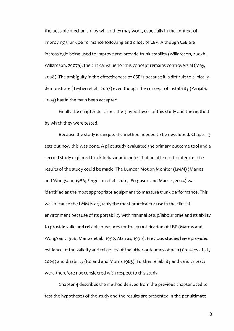
The functional unit of skeletal muscle is the motor unit (Jones and Round,
1990; Watkins, 1999). Each motor unit is made up of a motor neurone, its axon and
all its branches and the muscle fibres that are attached to them (Fig 1.11). The
branches may be from either an Aα or an Aβ motor neuron. A contraction is
precipitated when an action potential (AP) elicited by the motor neuron is
transmitted along the axon and its branches to reach the neuromuscular junction or
end plate to produce a muscle response (Jones and Round, 1990; Watkins 1999).
Fig. 1.11: The motor unit (from Watkins, 1999)
Motor Neuron
Axon Muscle fibres

26
If the AP is large enough and/or prolonged enough individual twitches
combine to form a contraction (Watkins, 1999). There are three types of motor
units each with unique properties that can be activated by stimuli (Table 1.5)
(Watkins, 1999).
Table 1.5: Motor unit characteristics adapted from Watkins (1999) Slow twitch Fast twitch
(Fatigue Resistant) Fast twitch (Fatigable)
Activation threshold Low Moderate High
Contraction time (ms) 100-120 40-45 40-45
Innervation ratio of motor units Low Moderate High
Type of muscle fibres 1 2a 2b
Type of Axon Aβ Aα Aα
Speed (m/s) 40-80 65-120 65-120
Duration and size of force Prolonged Low force
Prolonged Relatively high force
Intermittent High force
There are equal proportions of both slow and fast twitch motor units in each
muscle however the ratio may vary according to the action of the muscle (Jones
and Round, 1990; Watkins, 1999). Muscles used for fast responses possess a greater
number of fast twitch than slow twitch motor units and in muscles responsible for
posture and prolonged activity there are greater number of slow twitch motor units
(Jones and Round, 1990; Watkins, 1999).
The innervation ratio of motor units is the ratio between the numbers of
muscle fibres per axon and the greater the ratio the greater the force produced by
its activation (Watkins, 1999). Trunk muscle group activation and the sequence of
activation is ambiguous (Barr et al., 2007). It is suggested that the Transversus
Abdominis (TA) and Multifidus (MF) are activated first to maintain stability of the
spine during movement of the extremities but the TA is activated before the MF
(Barr et al., 2005). However, trunk stability is achieved through a tripartite
arrangement involving three sub-systems; the passive (bones, IVD and ligaments),

27
the active (muscles) and the neural (sensory receptors, cortical and sub-cortical
controls) (Panjabi, 1994). A 20lb load is sufficient to cause the collapse of the spine
if the muscles are removed (Crisco, 1989). Muscle response to pain is by either a
‘pain-spasm-pain’ or ‘pain adaptation’ model (van Dieen et al., 2003).
1.18 The pain-spasm-pain model
Pain causes a muscular response which creates further pain and discomfort
(van Dieen et al., 2003). This model is elicited by either of two ways (Fig. 1.12).
Fig. 1.12: The pain-spasm-pain model (from van Dieen et al., 2003)
Nociceptor activity (N) via the posterior horn influences the brain through
the neural pathway simultaneously increasing muscle activity by causing excitation
(E) of the α-neurons at the level of the segment of the spine which they supply
(Johansson and Sojka, 1991). Alternatively the nociceptors (N) increase muscle
spindle (S) activity via γ- afferents. This hyperactivity proceeds to excite the α-
neurons (Johansson and Sojka, 1991).
1.19 The pain-adaptation model
The excitation of both inhibitory (I) and excitatory (E) interneurons results in
a process where pain reduces agonist muscle group activity simultaneously
increasing antagonist muscle group activity (van Dieen et al., 2003). This process is
controlled by the central nervous system through motor command (Fig. 1.13) (van
Dieen et al., 2003).

28
Fig. 1.13: The Pain adaptation model (from van Dieen et al., 2003)
It is not clear as to how this model may fit in with the complex nature of the
synergy demonstrated by trunk muscles because studies suggesting this model
have only used large muscles such as the gastrocnemius (Graven-Nielson et al.,
1997). However this model may result in a reduction in movement velocity in the
spine (van Tulder et al., 2000).
1.20 Biomechanical properties of the lumbar spine
The restoration of trunk stability using core stability exercises (CSE) is
increasing in popularity (Willardson, 2007a; Willardson, 2007b). The effectiveness of
CSE is uncertain (Standaert and Herring, 2007; Standaert et al., 2008). However
trunk biomechanical characteristics are influenced by non-specific LBP (Barr et al.
2005; Barr et al., 2007).
1.21 Trunk stability
There is no definitive definition of trunk stability. However several studies
have described ‘Instability’ as being a phenomenon resulting from applied loads
causing abnormal movement of the movement segment (Panjabi, 1994; Fritz et al.,
1998; O'Sullivan, 2000; Alam, 2002). Mechanisms for stability have also been

29
suggested as the tripartite arrangement described earlier (section 1.15) (Panjabi,
1994; Barr et al., 2005; Barr et al., 2007).
Radiological tests demonstrate that traction spurs, IVD space narrowing,
asymmetric collapse of the IVD, mal-alignment of associated vertebrae and
abnormal glide and rotational movements of the spine during flexion and extension
can be evidence of instability (Alam, 2002). It has been proposed that specific
stabilisation exercises were not effective in reducing pain and disability in acute LBP
but may be effective in reducing reoccurrence after an episode (Ferriera et al.,
2006) however they are more effective than other forms of active intervention for
the management of chronic LBP (May and Johnson, 2008).
Trunk stability is dependent upon 3 sub-systems- passive, active and neural
control systems (Panjabi, 1994; Fritz et al., 1998) (Fig 1.14).
The passive subsystem consists of the IVD, ligaments and facets (Fig 1.14).
The annulus fibrosus of the IVD portrays a unique arrangement of fibres, an
orientation of +30 to -30 degrees in adjacent laminae which provides stability to
counter axial torque and excessive lateral flexion (Panjabi, 1994). Injuries to the
annulus may cause pain and discomfort in a single direction whereas the injury to
the nucleus pulposus may be identified by pain and discomfort during
multidirectional movements (Panjabi, 1994).

30
Fig. 1.14: Trunk stability sub-systems
Ligaments provide trunk stability but their efficiency is dependent upon the
size of the ligament in terms of its length and cross sectional area, its location from
the centre of movement of the segment and the direction of movement it is
supposed to regulate (Panjabi, 1994). Facet joint hypertrophy as the spine
degenerates through a natural ageing process is also considered to be a cause of
LBP (Panjabi, 1994) and it has been demonstrated that the removal of just one facet
joint within a segment exposes the segment to significant to multidirectional
instability (Abumi et al., 1990).
Muscles are important for trunk stability (Fig 1.15) (Panjabi, 1994). The ratio
of tolerable physiological load to critical load that will result in the spinal column
collapse is approximately 17:1 when in the flexed position of 20 degrees
(Nachemson and Morris, 1964; Crisco, 1989). This is prevented by muscles exhibiting
similar characteristics to guy wires (Panjabi, 1994). These characteristics are assisted
by the large cross sectional area of the muscles around the low back region and
their large lever arms (Panjabi, 1994). Trunk muscles either produce movement or
inhibit it, a process that provides spinal control (Norris, 1995). Muscles are grouped
as either global muscles responsible for gross movement of the spine or deep local
Instability
The Passive Subsystem (Spinal column)
•Position •Movement
The Active Subsystem •Muscles T d
The Neural Control Subsystem •Monitors and corrects position of spine
•Monitors response to load •Regulates movement in response to load

31
muscles responsible for segmental adjustments required to maintain local stability
(Barr et al., 2005). However, specific exercise is not beneficial as a treatment for LBP
in the absence of any clinical signs suggesting instability (Koumantakis et al., 2005;
Ferriera et al., 2006).
Exercise for the management of LBP using the large muscles with large lever
arms, such as back extension exercises in the prone position can cause an
exacerbation of symptoms in some patients (Callaghan et al., 1998). They may
therefore not be appropriate if the source of the LBP involves a nucleus pulposus
which does not tolerate multidirectional movement (Kanayama et al., 1995). The
aforementioned exercise also increases the stress on the facet joints (Callaghan et
al., 1998).
Tendons are an integral part of the muscle system operating across the
lumbar spine and are inaccessible by non-invasive techniques. They contain similar
structural composition as other soft tissue and as such exhibit similar viscoelastic
properties of stress, strain and fatigue etc. (Watkins, 1999).
The neural control subsystem is responsible for the control and regulation of
the other two sub-systems (Fritz et al., 1998). Neural input is provided by both the
lower centre of the spinal cord through reflex loops and from the brain that has the
capacity to override instruction from the spinal cord (Fritz et al., 1998). Poor
neuromuscular control can result in recurrent episodes of LBP but there is no
current evidence to suggest that poor neuromuscular control pre-empts a first
episode of LBP (Fritz et al., 1998). However, it is suggested that inadequacies in this
subsystem can reduce the ability of the spine to anticipate the effect of applied load
as demonstrated by Multifidus and Transversus Abdominis activity preceding active

32
limb movements in the absence of LBP (Hodges and Richardson, 1996; Hodges and
Richardson, 1997) but alterations to this sequence of events are evident in chronic
LBP (Barr et al., 2007). It is therefore possible to differentiate pain and disability
associated with either psychosomatic pain or somatopsychic pain because the latter
will demonstrate alterations in activation of either or both the Transversus
Abdominis and Multifidus muscle groups (Hodges and Richardson, 1996; Hodges
and Richardson, 1997).
A load-displacement curve for trunk flexion-extension movements (Fig 1.15)
demonstrates trunk biomechanical characteristics in response to load.
Fig. 1.15: Load-displacement curve (from Panjabi, 1994)
The spine is flexible enough to low loads but stiffens to high load (Panjabi,
1994). This is demonstrated by a non-linear curve which suggests two distinct areas
of a neutral zone where the segment is providing very little resistance and an actual
range of movement zone (Fig. 1.15). If the neutral zone is greater than the actual
range of movement zone instability results with the likelihood of accompanying
pain (Panjabi, 1994).
1.22 Effects of force on the spine
When the spine is subjected to loading the activity of all the components of
the spinal column compliment each other (Cripton et al., 2000). Their response
33
however, varies according to tissue type and the amount of force applied (Cripton
et al., 2000). The stability of the spinal movement segment is dependent upon the
IVD (Cripton et al., 2000) and the nucleus pulposus has a major role within this
mechanism (Nachemson, 1981).
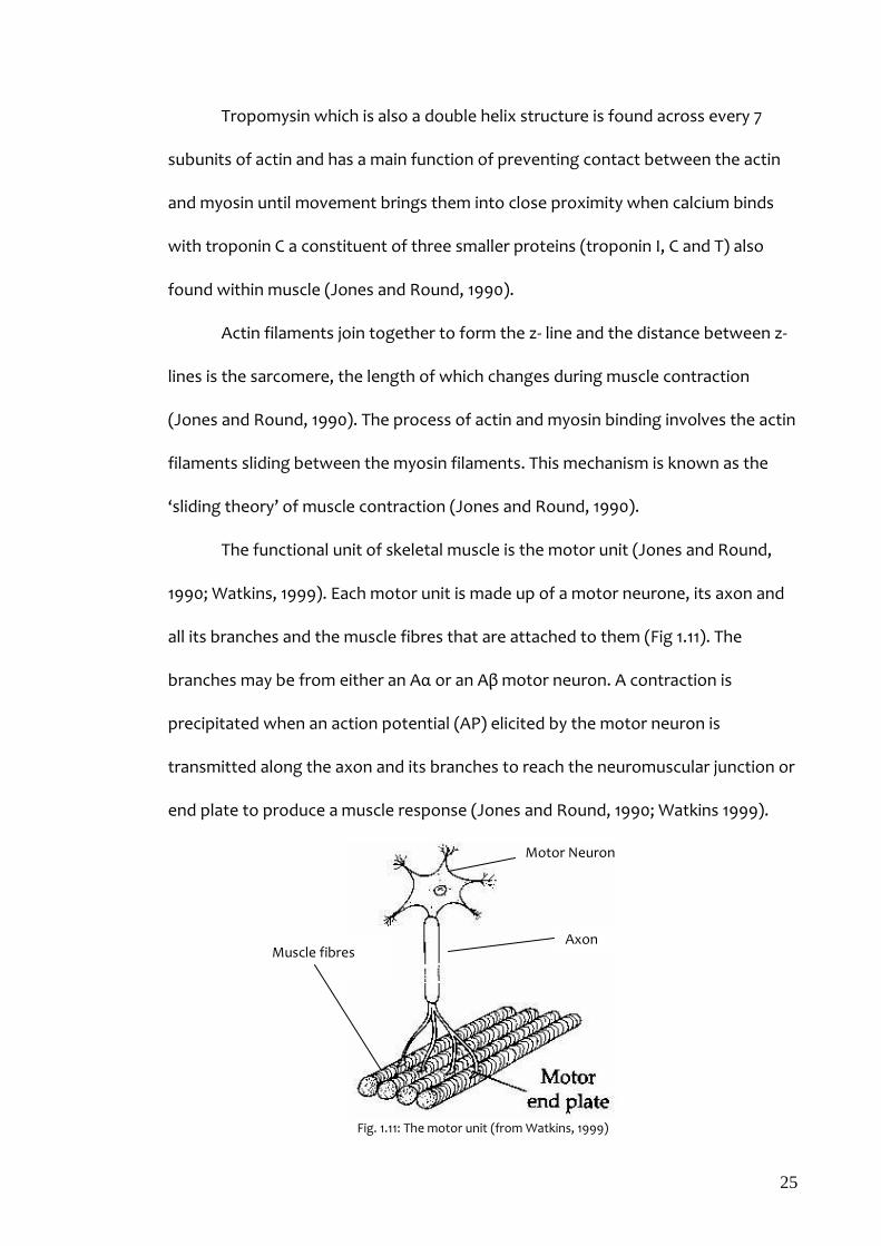
The pressure within the nucleus pulposus is expressed in mathematical
terms as;
P (nucleus pulposus) = K (F (disc) /A (disc)) (Nachemson, 1981)
(F (disc) - vertical force applied to the IVD, A (disc) -surface area and K- coefficient [between 1.3 and 1.6])
The intradiscal pressure is greatest during trunk flexion when compared to
side flexion or extension (Cripton et al., 2000). This response creates more pain in
the early morning compared to the latter parts of the day because of the
hydrophilic nature of the disc material (Giles and Singer 1997; Boyling and Jull, 2004)
and the gradual loss of fluid caused by the effects of gravity when in an erect
position.
Anecdotal evidence suggests that lumbar traction has been used for many
years to treat LBP caused by an increase in intradiscal pressure. However the
mechanism by which this works remains unsubstantiated because it is thought that
the beneficial effect of the technique does not last for longer than 30 minutes
following application (Twomey, 1985).The process involves the application of axial
load to distract the movement segments. The efficiency and overall affect of the
lumbar traction is dependent upon two main factors;
The position of the patient in order for the load to be applied; either
with the hips and knees in 90 degrees of flexion or not. The former is

34
described as the Fowler position and considered to be the most
effective (Lee and Evans, 2001).
The angle at which the load is applied to the spine. For effective
coupling of forces this angle is approximately 18 degrees (Colachis
and Strohm, 1969).
Lumbar traction is applied in the Fowler position; crook lying with the knees
supported in 90 degrees of flexion, to produce an anterior shear with simultaneous
flexion of the movement segments to increase the size of the neural foramina (Lee
and Evans, 2001). There is also a reduction in tension within the posterior column of
the spine including the posterior fibres of the annulus fibrosus (Lee and Evans,
2001).
Lumbar traction also affects facet joints (Ianuzzi et al., 2004).
Because of the angle to which they lie and because facet joints are sliding joints
there is a tendency for the contact area between the surfaces to be reduced
(Ianuzzi et al., 2004). This effect is limited by the capsule and associated ligaments
(Ianuzzi et al., 2004) but the force applied can produce shear strain between the
surfaces (Ogrodnik, 1997a; Watkins, 1999).
However compressive force causes the spine to shrink as a result of either
flexion or to a lesser degree rotation of the vertebrae (Wisleder et al., 2001). There
are however only a few instances where true compression actually occurs (Wisleder
et al., 2001) but there is usually extension between L2 and L4 and flexion at L5
(Wisleder et al., 2001). There is simultaneous anterior shear at these levels with a
corresponding loss of lumbar lordosis (Lee and Evans, 2001). However, the net
effect of compression may be exaggerated by torque as demonstrated in a study

35
using the cervical spine. It was demonstrated that torque grossly affects tissue
response to axial loading because the majority of damage occurs at the end plates
as the vertebral body with the facet joints remaining relatively unscathed even
though the overall stability of the movement segment is jeopardised (Aultman et
al., 2004).
Because of the tendency for the segments L2-4 to extend in response to
compressive loading (Wisleder et al., 2001), TrA activation timing and the influence
of the TrA on lumbar lordosis is important, a process already demonstrated
suggesting that the TrA is an ‘anticipator’ to loading activities (Hodges and
Richardson, 1996; Hodges and Richardson, 1997).
1.23 Effects of mechanical stress and strain on the spine
Direct or indirect forces applied to tissue causes tensile stress which is a ratio
of the force (F) applied to a cross-sectional area (A) of the surface, to which the load
is applied,
Stress = F/A
The stress developed within the tissue causes it to change shape or deform.
This is achieved by a change in its length. The ratio of the length change to the
original length is the strain which the tissue is put under.
Strain = ∆l/l
(∆l - change in length; l - original length before deformation)
The effect of either stress or strain on tissue can be demonstrated in living
tissue (Ogrodnik, 1997a; Whiting and Zernicke, 1998b; Watkins, 1999). Living tissue
is heterogeneous in nature consisting of both viscous and elastic properties. It is
therefore expected that a vertebral body and the IVD will respond in a particular

36
way to axial loading (Whiting and Zernicke, 1998b; Watkins, 1999). The effect of the
axial loading is directly related to the length of time for which the load is applied
(Ogrodnik, 1997a) because Hooke’s law suggests that the longer the time frame
during which it is applied the increasing likelihood of the structure losing its integral
elastic property and returning to its normal shape because it would have exceeded
its elastic limit (Ogrodnik, 1997a).
Temperature fluctuation influence tissue performance by producing a
thermal strain as can be demonstrated in isotropic materials (Ogrodnik, 1997a).
Thermal strain = Thermal Coefficient X ∆T (∆T - change in temperature)
There is paucity in the literature describing the effects of thermal strain on
trunk tissue. However, this maybe a component of an underlying mechanism by
which the anecdotal evidence may suggest occurrence of LBP reported by patients
in the clinical environment to be worse during the winter months and extremely
warm summers.
1.24 Deformation of tissue as a response to loading
The IVD as part of the movement segment experiences the greatest
deformation during loading losing approximately 0.16mm in height (Heuer et al.,
2007), and when an IVD is subjected to an axial load of 500N for 15 minutes its
internal pressure decreases linearly (Heuer et al., 2007). However, loading will
initially cause anterior deformation because the whole deformation process is time
dependent (Little et al., 2004). The long term effects of the deformation are created
postero-laterally where it can be shown to be greatest (Heuer et al., 2007). This
suggests that the historic assumption that acute LBP and associated radicular

37
symptoms reported after an immediate incident may not be caused by direct IVD
damage but by the immediate response to facet joint loading, deformation of the
IVD and the narrowing of the neural foramina (Little et al., 2004). Asymmetrical
deformation in response to load is a viscoelastic property referred to as creep
(Watkins 1999).
1.25 Creep
‘Creep’ is a time dependent property demonstrated by continual tissue
deformation after the cessation of the application of load applied to it before there
is a gradual return to a normal state as seen before the point of tissue failure as
caused by prolonged application of the load (Fig 1.16).
Fig. 1.16: Stress-strain curve (from Whiting and Zernicke, 1998b)
There is a linear relationship between stress and strain (Fig 1.16) and
70homogenous materials demonstrate characteristics that obey the laws of
elasticity or Hooke’s law (section 1.21). Observation of the Hookean law suggests
that when a load is removed, the material will be restored to its original length. This
response is a ratio of the gradient of the stress-strain curve (a/b). Within this range
the material has elastic properties. If the load is removed at or beyond the yield
point the material will not return to its original length but assumes another shape
demonstrating ‘plastic or non-elastic’ properties (Watkins, 1999). Exceeding the
Stress
a
b
Strain
Elastic range Plastic range
Yield point Failure Point

38
plastic range when the material will reach its maximum tolerance causes failure
(Watkins, 1999) and when if applied to the trunk, acute LBP may ensue.
The enforced change in shape of the tissue is proportional to the load
applied; the greater the load the greater the change in shape. When the load is
removed and if the strain remained within the elastic range, the deformation will
gradually reverse until the normal shape or length is resumed (Watkins, 1999) (Fig
1.17).
Fig. 1.17: Load - Deformation curve (from Whiting and Zernicke, 1998a)
The ‘creep’ response demonstrated by the capsule of facet joints is
particularly significant after sustained flexion at L5/S1 (Little et al., 2004), suggesting
that the spine is very vulnerable after performing tasks when in prolonged flexion.
Furthermore it also suggests that such induced LBP will respond effectively to rest
in a supine or prone position during which deformation is reversed. It has been
suggested that this reversal can take up to 20 minutes (Little et al., 2004).
‘Creep’ as a characteristic therefore can have a detrimental effect on the
stability of the movement segment especially when the muscles responsible for
stability during trunk functional activity involving sustained or repeated flexion
demonstrate a reduction in reflexive activity (Gedalia et al., 1999, Solomonow et al.,
2000; Williams et al., 2000; Claude et al., 2003; Lu et al., 2004). Changes in
Multifidus electromyography (EMG) activity when adjacent facet joints are
Load Loading
Unloading
d
Deformation

39
stimulated with an electric current (Little et al., 2004) demonstrate a possible
mechanism for loss of trunk stability.
1.26 Energy absorption
As tissue experiences deformation some energy is absorbed to provide
resistance to the load. During the unloading stage this energy is re-released
gradually and a natural 3-dimensional shape is regained. The amount of energy
released is equal to the area between the curves, ‘d’ (fig 1.17) (Whiting and Zernicke,
1998a; Watkins, 1999). It may be this energy that is reported as an increase in
temperature during LBP and it may not be the same as the increase in temperature
and associated erythema observed during an objective assessment with palpation
or during an inflammatory process. There is paucity in the literature to support this
supposition.
1.27 Conclusions
Low Back Pain continues to be a complicated condition generating concern.
Although there are various historical diagnostic tests available to diagnose LBP
these tests are only effective when there is an underlying pathology. The diagnostic
tests however lack sensitivity to demonstrate effectiveness of intervention for
mechanical non-specific back pain. It is therefore justified to explore the possibility
of quantifying real time trunk movement characteristics after using interventions
such as the increasingly popular core stability exercises. There is also empirical
evidence to suggest physiological mechanisms for trunk movement that may
underlie CSE (Hodges and Richardson, 1996; Hodges and Richardson, 1997; Barr et
al., 2005; Barr et al., 2007).

40
This study utilises a method of analysis that evaluates changes in trunk
acceleration and investigates the relationship between the effectiveness of CSE and
the pain and disability reported following an episode of acute pain.
The following chapter evaluates the current methods of evaluation of trunk
movement characteristics, considered opinion about the underlying mechanism for
the onset of LBP and the mechanisms for the effectiveness of core stability
exercises as an intervention for the treatment of LBP.

41
Chapter 2
Literature Review
2.1 Summary
This chapter explores what is known of the effect of Core Stability Exercises
(CSE) on acute non-specific low back pain to date. The chapter also explores the
depth of existing knowledge of movement characteristics of the trunk in terms of
its higher order kinematics. The research questions, as a direct consequence of the
chapter conclusion, are then stated.
2.2 Historical factors that influence the management of low back pain
The main cause of LBP has previously been suggested to be vascular
deficiency (Jayson, 1996). This is because there is greater relevance of the effects of
smoking on vascular integrity rather than height, weight, inherited factors, spinal
movements, muscle strength or even radiological signs for the onset of LBP
(Jayson, 1996). However, this proposition originates from a heavily medical oriented
opinion and an organic orientation upon which an onset can be related. This
interpretation also seems to suggest that mechanical non-specific LBP can be
experienced in the absence of both functional movement and the central
neuromodulation that may influence segmental and global spinal movements.
Anecdotal evidence from the clinical environment suggests that non-specific
LBP does respond to intervention if efficient movement is restored to spinal
segments. An inability to adequately demonstrate this may be a reason why the
prevalence of LBP has not improved over the years despite intensive research.
Indeed, even technological advances in diagnostics and intervention have not

42
reduced the economic burden of LBP (Dagenais et al., 2008). Data from Australia,
Belgium, Japan, Korea, Netherlands, Sweden, United Kingdom and United States
suggests that LBP is reported to be the 4th most expensive health condition with
both direct and indirect economic costs (Dagenais et al., 2008) and physiotherapy
as an intervention is one of the largest components of those direct costs (17%)
(Dagenais et al., 2008). It is therefore justifiable that physiotherapists continue to
seek a method to demonstrate effectiveness of intervention and be able to justify
associated costs within healthcare delivery. This may be achievable by
demonstrating both efficiency and effectiveness through the quantification of the
effects of intervention.
Historical assessment of LBP in a clinical environment consists of both a
subjective self reporting system and an objective assessment of function to
evaluate functional ability (Petty, 2006). This process precedes the development of
hypotheses upon which an explanation for reported symptom behaviour can be
developed. Subsequent Intervention involves various management strategies. Each
strategy however usually includes muscle strengthening programmes. This is
because previous studies have suggested that LBP causes weakness in trunk
musculature (Hodges and Richardson, 1996; Hodges and Richardson, 1997; Barr et
al., 2005; Barr et al., 2007).
2.3 Acute non-specific low back pain
Acute non-specific low back pain is an episode of LBP that has lasted for up
to 6 weeks (BackCare, 2007; Kinkade, 2007). It has also been described as lasting for
up to 3 months (Smith et al., 2002; Gullick, 2008) but the authenticity of this
description lacks credibility because one point of view is that the length of time of

43
onset during which other secondary effects of LBP may become apparent may be
driven by central neuromodulation (Jayson, 1996). Acute LBP can also influence
gait depending upon its severity (Taylor et al., 2004), however kinematic analysis of
the influence of LBP on gait has not been adequately demonstrated (Taylor et al.,
2004).
2.3.1 Classification of low back pain
The Quebec Task Force Classification (QTFC) (Spitzer, 1987) has been used
successfully to classify LBP patients by defining differences in the extent of pain and
types of pain. The method has been used to categorise sciatica as a classification in
which pain radiates down the leg beyond the knee and thus indicates the severity of
symptoms and the likelihood of future surgical intervention (Atlas et al., 1996).
Within the sample evaluated by the study, the non-surgical group demonstrated
better improvement in outcome compared to the surgical group (Atlas et al., 1996).
However, the QTFC cannot differentiate between acute and chronic LBP and thus
does not effectively demonstrate discriminant validity (George and Delitto, 2005). It
therefore may be considered to be a taxometric assessment which can be used to
classify types of low back pain.
The heterogeneous nature of acute LBP suggests that it cannot be treated
by a single mode of treatment (Abbott, 2008) there is therefore a need to match
intervention to clinical findings for meaningful treatment outcomes (Abbott, 2008).
Delitto et al. (1995) proposed a Treatment Based Classification (TBC) system (Table
2.1). This system utilises subjective history and symptoms for LBP as a method to
assist in the physiotherapy decision-making process to decide on an appropriate
intervention (George and Delitto, 2005).

44
Table2.1: Treatment Based Classification -adapted from George & Delitto (2005) Classification Key History/Clinical findings Intervention
Specific Exercise Extension syndrome Flexion syndrome Lateral shift syndrome
1. Posture preference is extension 2. Pain with lumbar flexion 3. Pain ‘centralises’ with lumbar extension but intensity increases with flexion tests 1. Posture preference is flexion 2. Pain with lumbar extension 3. Pain ‘centralises’ with lumbar flexion but intensity increases with extension tests 1. Frontal plane deformity 2. Unilateral side flexion restriction with lumbar movement testing 3. Improvement with pelvic translocation
Extension exercises and avoidance of activities involving flexion Flexion exercises and avoidance of extension movements Passive or active pelvic translocation exercises
Mobilisation Lumbar mobilisation Sacroiliac (SIJ) mobilisation
1. Local/unilateral LBP 2. “Opening pattern” or “Closing pattern” during trunk movement tests 1. Local pain at PSIS, buttock or lateral thigh pain 2. Three fourths SIJ test cluster are positive
Lumbar mobilisation/manipulation techniques and/or range of movement exercises SIJ manipulation, Muscle energy techniques and lumbar range of movement exercises
Immobilisation Immobilisation syndrome
1. Frequent prior episodes of LBP with minimal perturbation 2. History of trauma 3. Generalised ligament laxity 4. “Instable catch” during lumbar flexion tests
Trunk strengthening exercises
Traction Traction syndrome Lateral shift syndrome
1. Signs and symptoms of nerve compression 2. No improvement with lumbar movement testing 1. Visible frontal plane deformity 2. Unilateral side flexion restriction during lumbar movement testing 3. Symptoms worsen with pelvic translocation
Intermittent mechanical traction or autotraction Autotraction
TBC is therefore capable of using information from a clinical examination to
classify acute LBP into effective intervention groups and simultaneously reduce self-
reported disability (George and Delitto, 2005). However, there is limited evidence to
suggest that TBC can be used to discriminate between subgroups of patients
presenting with acute LBP (George and Delitto, 2005). The clinical assessment used
to classify this cohort of patients relies on palpation using both static and dynamic
landmarks which do present challenges in terms of reliability and validity (George
and Delitto, 2005). In the clinical environment the ability to repeatedly locate
specific lumbar vertebrae using palpation is essential for effective management
45
using manual techniques. However it has been demonstrated that agreement in
identifying comparable lumbar segments (kappa=0.37) and the identification of
spinal levels (kappa=0.09) between practitioners is relatively low (Downey et al.,
2003). The kappa (k) is the chance-corrected statistical determination of agreement
of a measure with perfect agreement being 1.00 (Altman, 1991). This does therefore
suggest that treatment using these techniques need to be viewed within that
context because the kappa scores are relatively low.
2.3.2 Management of acute non-specific low back pain
Guidelines for the treatment of LBP suggest that management should
involve physical methods of physiotherapy using a biopsychosocial approach (NHS,
2005). What these physical methods may be is ambiguous, however spinal
manipulation has been suggested as being beneficial for the treatment of LBP
(Assendelft et al., 2003; Haas et al., 2004; Ernst, 2007). Manipulation, however, has
not been demonstrated to be better than other forms of treatment (Assendelft et
al., 2003). A probable reason for this conclusion has been described earlier (Downey
et al., 2003) and is demonstrated within the clinical environment where less than 5%
of patients presenting with LBP in primary care receive manipulation (Jackson,
2001). Skill mix of the practitioners, however, may also influence the choice of
treatment because different levels of experience influence the clinical reasoning
process (Doody and McAteer, 2002) that underpins the preferred intervention.
Although there is depth in guidelines across Europe for the management of
LBP within primary care (Abenhaim et al., 2000; ANAES, 2000; Hagen et al., 2002;
Hagen et al., 2005; NHS, 2005; Ostelo et al. 2005) compliance with the
recommendations is variable and may be dependent upon the fear-avoidance

46
beliefs of General Practitioners (GPs) (Coudeyre et al., 2006). Furthermore GPs may
lack the specialist knowledge to make an initial diagnosis of LBP especially when
assessing chronic low back pain (Cayea et al., 2006). There is no current evidence
however to suggest that this is also true for acute LBP but the criteria used to
generate referrals have been shown to be vague (Stanley et al., 2000). However,
current guidelines provided for healthcare practitioners in the United Kingdom
suggests that patients should be encouraged to remain active and engage in
exercises (NICE, 2009).
A study in Scotland of patients who either self referred or were referred by
their GP for physiotherapy treatment demonstrated that there is a difference in the
profiles of both groups (Holdsworth et al., 2006). Self-referrers were more likely to
complete their treatment with fewer treatment sessions per episode of care and
were absent from work for fewer days than those referred by GPs who were much
fewer in number (Holdsworth et al., 2006). This may be a reflection of GP referral
behaviour.
Cognitive-Behavioural Therapy (CBT) has been advocated as a useful adjunct
to the treatment of acute LBP but screening for the subgroup of LBP patients who
will most favourably respond to CBT can be useful in preventing acute LBP from
becoming chronic (Frank and DeSouza, 2001; Smith et al., 2002; Fritz and George,
2002; Johnstone et al., 2004). A randomised controlled trial has demonstrated the
benefits of combining Cognitive behavioural therapy to physical treatment involving
exercise (Linton et al., 2005). The transition of LBP from acute to chronic is
influenced by risk factors such as stress, anxiety, mood, emotions, cognitive
function and pain behaviour (Johnstone et al., 2004). These risk factors have been

47
found in 17% of patients reporting with acute LBP (Burton et al., 1995 cited in
Johnstone at al., 2004) and because physiotherapists are usually the first point of
contact with LBP patients (Johnstone et al., 2004) early recognition of these factors
and appropriate intervention will benefit these patients. However, incorporating a
cognitive behavioural therapy approach within standard physiotherapy intervention
is largely resisted because of time restraints, even though there are cost-benefit
advantages (Johnstone et al., 2004).
Proctor et al. (2000) cited in Johnstone et al. (2004) proposed that disability
caused by LBP is dependent upon the degree to which a patient suffers from
distress, psychopathology, depression and catastrophisation of their condition.
These factors along with the risk factors identified by Johnstone et al. (2004)
therefore suggest that any outcome reliant upon self reporting measures may be
subject to bias and distort quantification of acute LBP. Research involving acute LBP
therefore, requires a methodology that considers the possible effect of this bias
during the procedures.
A minority of patients with work-related LBP that do not return to work
account for an appreciable percentage of the costs of treatment and early
intervention to prevent disability can therefore be beneficial (Fritz and George,
2002). Understanding the effects of fear-avoidance beliefs is important to reduce
the probability of an acute onset of LBP from becoming chronic, especially if it is
caused by work related activities (Fritz and George, 2002). However, how and if fear
avoidance beliefs are perpetuated by belief systems during the management of LBP
by health practitioners is unclear because the relationship dynamics are ambiguous
(Fullen et al., 2008). What has been demonstrated however is that using fear

48
avoidance techniques to treat LBP can reduce disability in the short term (Fritz and
George, 2002). There is however, a caveat in that more research is required to
demonstrate fear avoidance techniques have a role as a treatment strategy (Fritz
and George, 2002). Previous work by Frank and De Souza has already proposed that
that a pre-requisite to successful management of LBP and the prevention of an
acute onset from becoming chronic is the need for the clinician to be ‘positive’ and
exercise good person management (Frank and DeSouza, 2001).
Acute LBP alone was reported to generate direct costs of £251 million and
the gateway to treatment is increasingly via physiotherapists (Lauchlan, 2005) and it
is suggested that there is a need to reduce unnecessary tertiary referrals (Lauchlan,
2005). However, much more research has been done on chronic LBP compared to
acute LBP. A reason for this may be because most episodes of acute LBP resolve
quickly. Pullaim et al. (2003) reports that 70% of LBP patients no longer experience
disability at 4 weeks following an onset of LBP (cited in Lau et al., 2008). Another
reason may be because of the relative speed of resolution of symptoms; no
treatments (including painkillers) for acute LBP have been able to stand up to the
rigours of inquiry that would merit evidence based practice (Smith et al., 2002;
Rozenberg et al., 2003).
Effective management of LBP during the acute stage is reported to be bed
rest (not exceeding 4 days) complimented by painkillers (Rozenberg et al., 2003).
This approach has been suggested to be comparable in effect on LBP as a
continuation of normal daily activity alone (Rozenberg et al., 2003). This treatment
approach however is not recommended (Smith et al., 2002; Kinkade, 2007) because
normal activity results in fewer days of sick leave (Smith et al., 2002; Rozenberg et

49
al., 2003; Kinkade, 2007) even though patient satisfaction is much less (Atlas and
Deyo, 2001). This is because it has been demonstrated that patients who receive
early physiotherapy intervention respond much more quickly in the short term
demonstrated by improvement in outcome measures of pain and global perception
of the effect of intervention (Lau et al., 2008). Furthermore, it has been suggested
that an assess/advise/treat model of care can offer better outcomes than an
assess/advise/wait model of care because of the better psychosocial features
demonstrated by improved scores in outcomes of reported pain (Visual Analogue
Scale), functional disability (the Roland and Morris Disability Questionnaire), mood
(Modified Zung Self Rated Depression Score, Modified Somatic Perception
Questionnaire, State-Trait Anxiety Inventory), general health (Euroqol), and quality
of life (Short Form 36) (Wand et al., 2004).
Acute LBP is in the main a result of a mechanical deficiency (Atlas and Deyo,
2001) and is in the main not associated with any serious underlying pathology even
if there may be evidence of sciatic pain (Kinkade, 2007). Positive identification of a
causal relationship between structure and symptoms is difficult even when using
sophisticated diagnostic tools such as X-rays, MRI or CAT scans even though 98% of
LBP accompanied by radicular symptoms occur at L4/5 or L5/S1 lumbar segments
(Atlas and Deyo 2001). A majority of acute LBP may not respond favourably to
specific exercises (Atlas and Deyo, 2001).
Spinal manipulation using either high or low velocity manoeuvres to restore
function within the lumbar spine is an acceptable choice of treatment for acute LBP
but there is uncertainty of the right timing for this type of intervention (Kinkade,
2007; Sizer, 2008). It had previously been proposed that spinal manipulation is

50
generally of little benefit to the overall management of acute LBP (Smith et al.,
2002) and most recent opinion is that spinal manipulation does not provide long
term benefit for this group of patients (Kinkade, 2007).
2.4 Characteristics of acute non-specific low back pain
Candotti et al. (2008) investigated the effect of LBP on fatigue indices in a
group of sedentary participants to see if it was possible to replicate the results as
seen in a group of athletes. The authors collected bilateral EMG and force data
during isometric contractions of the iliocostalis lumborum and the longissimus dorsi
muscles during 3 repetitions of maximum voluntary contractions (MVC), each
contraction lasting for five seconds duration with a 2 minute rest between
repetitions. The fatigue test consisted of maintaining 80%MVC for 35 seconds. The
post fatigue test consisted of a contraction which was 80% MVC for 10 seconds
duration to assess recovery. The authors suggested that there was a need for
objective measures to evaluate evolving LBP characteristics during treatment and
proposed that EMG activity can provide such information. They proposed that EMG
could do this because it can detect changes in the median frequency which is
purported to reduce as a result of LBP. The study does also suggest that
physiological changes occurring during an episode of LBP can influence trunk
activity. Although the objectives of the study were clear the reliability of repeated
measures using EMG is not precise. The reliability of EMG can be compromised by
potential sources of error involving electrode placement (Soderberg and Knutson,
2000; Suzuki et al., 2002). The study would therefore be strengthened if there was
supporting evidence to demonstrate a measure of reliability for the method used to
gather the presented data. The sincerity of effort by study participants may also

51
have influenced the results. This was acknowledged by the authors who considered
the effect of the pain adaptation model (Chapter 1) on the participants. As an
inclusion criterion, pain was not scaled. The level of pain experienced by the
participants before, during and after the study was ambiguous and it is possible that
variations in the level of pain could have an affect on the ability to produce reliable
MVCs.
Candotti et al., (2008) also found that significant differences were
demonstrated unilaterally and proposed that those significant differences, found
mainly of the left side were due to the fact that rotation was not restricted during
the testing procedure. This argument may not be entirely valid because it implies
that trunk movements are one dimensional, but trunk movements are 3-
dimensional (Marras and Wongsam, 1986). A more plausible argument may be that
all the participants presented with unilateral problems producing movement
towards the side of least resistance (McKenzie, 1981). However, an important
observation of this study is that the participant group is in the main very ambiguous.
The authors did not indicate if the participants were experiencing either acute or
chronic episodes of LBP.
Trunk strength evaluation within a clinical environment has traditionally
played a significant role within primary care and anecdotal evidence suggests that
LBP patients referred for treatment quite often are referred for ‘back strengthening
exercises’. However, some studies have suggested that measures of trunk strength
should be described more precisely in terms of its motor performance or higher
order kinematics (Kroemer et al., 1990; Marras et al., 1990).

52
Higher order kinematics of the trunk can be described in terms of displacement,
velocity and acceleration (Kroemer et al., 1990; Marras et al., 1990). Previous work
does exist in abundance exploring the behaviour of trunk velocity but there is a
paucity of literature exploring the behaviour of trunk acceleration even though
acceleration is defined as the rate of change in velocity when there is a change in
direction of movement (Ogrodnik, 1997c). Evaluation of acceleration may therefore
provide more detail because anecdotal evidence does suggest that LBP is reported
by patients when there is a change in trunk direction during functional activity.
2.5 Physical/general exercise and low back pain
Storheim et al. (2003) recruited 93 participants with sub-acute LBP (8-12
weeks) for a randomised controlled trial lasting 18 weeks. They compared the effect
of physical exercise and cognitive behavioural therapy (CBT) on sub-acute LBP.
Primary outcome measures included pain, disability, sick listing and care satisfaction
as primary outcomes and self-efficacy for pain and function, fear avoidance beliefs,
emotional distress, generic health status and life satisfaction as secondary
outcomes. The study concluded that CBT improved disability and that physical
exercise can reduce patients’ symptoms. The results suggested that physical
exercise as treatment for low back pain requires high motivation among the
participants and attrition is greatest among this group who were more likely to be
male than female. It was also observed that none of the interventions had an effect
on sick listing. A challenge to the study was the fact that no previous data existed to
determine a sample size but the authors calculated a sample size using an
assumption based upon a pilot study and recommendations from previous studies.
They acknowledged this as a limitation. Because of the attrition (18%) the data
53
analysis involved an ‘intention to treat’ analysis. An ‘intention to treat’ analysis is a
method by which a direct comparison of the groups can be made by including all
participants within the groups to which they were originally allocated (Peacock and
Peacock, 2011). By doing so balance between the groups in respect to the subject
characteristics is maintained (Peacock and Peacock, 2011).
The control group were excluded from the standard clinical examination
during the recruitment phase. The reasons for this omission were not given and it
was not clear but it is possible that the omission could have introduced some form
of bias into the sample. The underlying mechanisms for the onset of the episode of
back pain could have influenced the response within the control group and
therefore distort its comparison with the other groups. It was also not clear as to
what this standardised clinical examination consisted of.
The randomisation process for the aforementioned study was ambiguous. It
is not clear if the envelopes used for allocation were picked in a sequence or
shuffled before being chosen by the participants. Once allocated, the participants in
the exercise group did not have a consistent exercise routine. Some attended
exercise classes twice weekly and others thrice weekly. The control group were
treated by their GP but also had no restrictions as to types of their preferred
treatment or referrals.
It can therefore be surmised that although exercise can reduce patient
symptoms any study using exercise as an intervention may be limited by attrition of
participants.
54
2.6 Review of the literature
The review within this section of the chapter is narrative because of the
paucity of literature. This choice of review allowed consideration of a wide scope of
information without the constraints of a criterion based selection criteria (Collins
and Fauser, 2005). Furthermore a systematic review with a meta-analysis was not
considered useful because of inconsistencies in application of intervention and
outcome measures (Collins and Fauser, 2005) making comparison between the
results of the studies difficult. A realist review that can be useful in identifying and
explaining links and interactions between context, mechanisms and outcome
(Wong et al., 2010) was also considered but rejected because a common mechanism
by which CSE may work is unknown. Furthermore the method is more suited for
policy intervention (Wong et al., 2010).
A search of Amed (1990-2011), Cinahl (1990-2011), Medline (1990-2011),
Science Direct (1990-2011), SportsDiscus (1990-2011), Scopus (1990-2011) and the
Cochrane library (1990-2011) databases for literature on current understanding of trunk
movement characteristics and Core Stability (stabilisation) Exercises was conducted.
All the databases were searched using the keywords; acute low back pain, stabilisation,
trunk acceleration and velocity, Core Stability Exercises and Lumbar Motion Monitor.
Articles listed in a recent systematic review (May and Johnson, 2008) were also
considered. The search strategy and the articles reviewed are listed (Table 2.2) and the
methodological quality of the studies reviewed was tested using PEDro rating; a
score on a scale developed to score trials on the Physiotherapy Evidence Database
(Maher et al., 2003) (Table 2.3) and the reviewed articles are summarised (Table
2.4).

55
Although all the studies included compared baseline data, none of them
provided evidence of participant blinding. Four studies did not apply ‘intention-to-
treat’ analysis, three of which could not because they were not randomised
controlled studies (Hicks, 2005; Herbert, 2006; MacDonald, 2010) and registered the
lowest PEDro scores.
The final papers reviewed were chosen because they all evaluated acute LBP
and changes in the multifidus muscle. Although similar outcome measures were
used the techniques were not consistent. Direct comparison between the studies
was therefore not possible. Of the relevant 12 studies identified, 6 were rejected
because they were duplications.
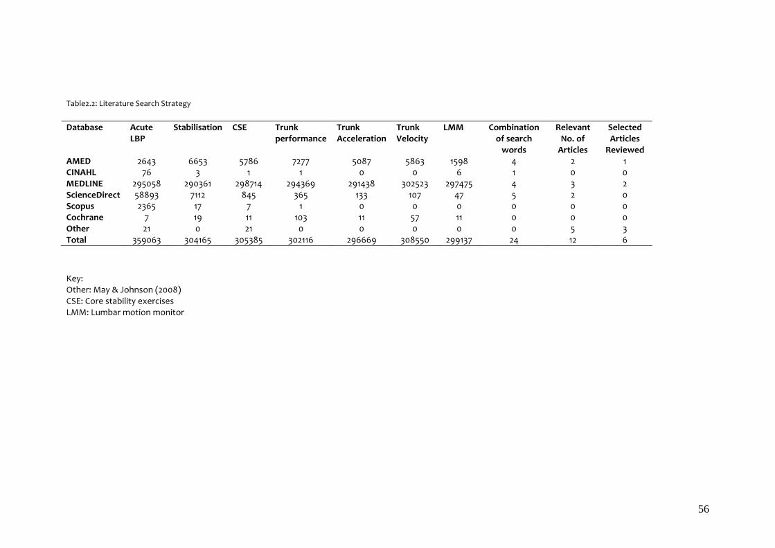
56
Table2.2: Literature Search Strategy Database Acute
LBP Stabilisation CSE Trunk
performance Trunk Acceleration
Trunk Velocity
LMM Combination of search
words
Relevant No. of
Articles
Selected Articles
Reviewed AMED 2643 6653 5786 7277 5087 5863 1598 4 2 1 CINAHL 76 3 1 1 0 0 6 1 0 0 MEDLINE 295058 290361 298714 294369 291438 302523 297475 4 3 2 ScienceDirect 58893 7112 845 365 133 107 47 5 2 0 Scopus 2365 17 7 1 0 0 0 0 0 0 Cochrane 7 19 11 103 11 57 11 0 0 0 Other 21 0 21 0 0 0 0 0 5 3 Total 359063 304165 305385 302116 296669 308550 299137 24 12 6
Key: Other: May & Johnson (2008) CSE: Core stability exercises LMM: Lumbar motion monitor

57
Table 2.3: Evaluation of methodological quality of the reviewed studies using PEDro rating Study 1 2 3 4 5 6 7 8 9 10 11 Score
Rasmussen-Barr et al. (2003) Y Y Y Y NS NS NS N N Y Y 5/10
Childs et al. (2004) Y Y Y Y NS Y Y N Y Y Y 8/10
Hicks et al. (2005) Y N N Y NS N NS Y N/A Y Y 5/10
Brennan et al. (2006) Y Y N Y N N Y N Y Y Y 6/10
Hebert et al. (2010) Y N N Y N N N Y N/A N Y 3/10
MacDonald et al. (2010) Y N N Y N N NS Y N/A Y Y 4/10
PEDro items: 1. Specification of eligibility criteria (Not included in the total score); 2. random allocation; 3. concealed allocation; 4. baseline comparability; 5. patient blinding; 6. therapist blinding; 7. assessor blinding; 8. at least 85% follow-up; 9. intention to treat analysis; 10. between group statistical comparisons; 11. point measures and measures of variability. Yes-Y; No-N; NS-Not stated; N/A-Not applicable

58
Table2.4: Literature review core stability exercises
Study Sample size and type Symptom Duration
Intervention and Outcome Measures
Follow up Period
Conclusions
Rasmussen-Barr et al. (2003) RCT 6/52 duration
47 participants Age range: 18-60 Randomisation by lot
>6 weeks +/- radicular pain
Stabilisation exercises (n=24) Manual treatment (stretches, traction, mobilisation, manipulation) (n=23) Pain (VAS) Disability (ODI and Disability Rating Index)
3 and 12 months Stabilisation exercise produced significantly better improvement in disability at 6/52 (50%), 3/12 (67%) and 12/12 (56%). Pain improved by 39% (6/52), 58% (3/12) and 61% (12/12). Participants in the manual treatment group had more episodes of reoccurrence long term.
Childs et al. (2004) RCT 4/52 duration
131 participants Age range: 18-60 ODI ≥ 30%
?<16 days. Only 46 (35%) had symptoms for this length of time. +/- radicular pain
Manipulation plus ROM exercise Low stress aerobic and lumbar spine strengthening Exercise Disability (Modified ODI) Pain (VAS)
1 and 4 weeks 6/12
Manipulation was more effective in reducing disability in patients who were positive on the clinical prediction rule compared to a similar patient receiving exercise
Hicks et al. (2005) Prospective cohort study 8/52 duration
54 participants Age range : > 18
Symptom duration -Not stated. Assumed to be acute? No radicular pain
‘standardised’ stabilisation exercises Disability (ODI)
Not stated A clinical prediction rule can be used to identify patients who will more likely respond to stabilisation exercises

59
Brennan et al. (2006) RCT 4/52 duration
123 participants (55% male, 45% female) Age range: 18-65 ODI ≥ 2.5%
<90 days +/- radicular pain
Manipulation Stabilisation exercises Specific exercises Disability (ODI)
4/52 1 year
Outcome of treatment of LBP can be improved by sub-grouping patients. ODI improved by 56%
Hebert et al. (2010) Cross sectional study Duration not stated
40 participants (30 prospective, 10 participants with ongoing Rx) Age range: 18-60 Positive prone instability test, aberrant movement, SLR >91 deg, lx hypermobility, self reported disability
Current episode of pain between T12 and the buttocks.
Nil Rehabilitative Ultrasound Imaging (RUSI)
Nil Decreased MF activation is more associated with predictive of factors to determine the success of stabilisation exercises than decreased TrA activation
MacDonald et al. (2010) Cross sectional study
13 participants with LBP and 14 healthy participants.
Unilateral LBP for 75% of the time. Can be of varying frequency but without a duration exceeding 3 months
Bilateral EMG recording of deep and superficial multifidus during both predictable and unpredictable trunk loading
Nil Both deep and superficial multifidus activity was subdued after remission of LBP compared to healthy participants. Superficial multifidus activity during predictable trunk loading was not affected.
Key: Randomised Controlled Trial (RCT) Visual Analogue Scale (VAS) Oswestry Disability Index (ODI) Range of movement (ROM) Electromyography (EMG) Multifidus (MF)

60
Rasmussen-Barr et al. (2003) compared the effect of stability exercises and
manual treatment as interventions for an episode of LBP for 6 weeks with follow up
periods of 3 and 12 months. The sample was made up of 47 participants aged
between 18 and 60 years who presented with an episode of LBP. Participants were
allocated to either a stability exercise or a manual treatment group. Randomisation
was achieved by allocating the first male and female participants to one of the
groups by lot and the others were then assigned separately but consistently to
either group.
The design of the study was very good as the authors used stratified
randomisation by sex as a method for group allocation of participants comparing
two treatments. Unfortunately this design does not provide unequivocal evidence
to be able to attribute with confidence, changes in outcome to the described
interventions. The absence of a control group further complicates the conclusion of
the study because it is not possible to take into account the natural resolution of
symptoms in the absence of intervention when there is no treatment. However, it is
possible that because the participants were randomised, natural recovery would be
equal between both groups. Furthermore the fact that the participants would have
different structural biomechanical responses associated with trunk strength,
flexibility and balance because of the different length of time of each episode
(Standaert et al., 2008) could have been a problem.
The variation in the number of manual treatments used in the study and the
lack of consistency in the choice of and length of manual treatment used may have
also diluted the true effect of the intervention. It is not possible, therefore, to
identify which of the interventions were of most benefit.

61
Because of the high rate of attrition, the authors concluded that the study
should be re-classified as a pilot study and suggested that the study should be
repeated with a bigger sample size. An alternative would have been to use an
intention to treat analysis which would have provided credible data analysis
(Altman, 1991). This problem was dealt with satisfactorily in another study
(Storheim et al., 2003).
Childs et al. (2004) compared specifically targeted lumbar strengthening
exercises with manipulation using a clinical prediction rule to identify patients most
likely to benefit from manipulation. 131 participants with an age range of 18-60 years
and who had an Oswestry Disability Index (ODI) score of a minimum of 30% with LBP
symptoms of 16 days or less were randomised into a manipulation group with range
of movement exercises and a group who performed targeted strengthening
exercises for the lumbar spine. Intervention was for a 4 week period with follow up
periods of 4 weeks and 6 months. Primary outcome measures were disability and
pain. They concluded that the decision process for the use of manipulation as a
form of intervention can be enhanced using the clinical prediction rule.
This study has its merits but only 46 (35%) of the participants had symptoms
for less than 16 days. This does suggest that the variation in the length of time
participants had been experiencing pain could result in recall bias because a
majority of the participants were being asked to reflect on their pain experience and
disability when they no longer were experiencing pain and/or disability (Chouinard
and Walter, 1995). This could account for the very wide confidence interval used to
suggest significance in the odds for successful outcome among patients who were
positive for the prediction rule and who were randomised into the manipulation

62
group [60.8 (95% CI 5.2-704.7)]. The data could have also been biased because some
participants who were randomised into the exercise group sought other forms of
treatment between the 4 week period (end of active intervention) and the 6 month
follow up. This was further compounded by the absence of a method to ensure
compliance of the exercise routine even though the authors suggest that the
strengthening exercises were isolated and targeted in accordance with previously
published literature.
Hicks et al. (2005) investigated the possibility of developing a clinical
prediction rule for successful use of stabilisation exercises for patients with LBP.
During an 8 week period 54 participants were recruited. The age of each participant
was greater than 18 years. They identified 4 main prognostic factors for a prediction
rule; a positive prone instability test, age less than 40, aberrant trunk movements
and straight leg raise greater than 91 degrees. However, there was the possibility
that the results from which the conclusions were derived could have been biased.
The absence of a control group does not allow a comparison of prognostic factors
and effects of intervention between the participants receiving treatment and
participants with no intervention and who’s LBP resolved naturally without
intervention. This would have provided substantiated proof of improvement due to
the intervention and added weight to the validity of the prognostic factors.
The exclusion criteria used during the recruitment process also did not
consider the mental state of the participants. Poor mental health and other
psychological factors such as depression can affect the outcome of intervention
(Standaert et al., 2008) and possibly motivation and compliance of the exercise
routine (Linton et al., 1996; Frank and DeSouza, 2001; Fritz and George, 2002;).
63
Although the authors used a Fear Avoidance Behaviour Questionnaire (FABQ) and a
Visual Analogue Scale (VAS) to measure fear of pain and subjective pain
respectively, it is not clear if any of the participants did not perform the prescribed
stability exercises for any reason because of their mental state and what was done
with these participants if they could be identified because this information was not
provided.
Hicks et al. (2005) used an inclinometer to quantify the available ROM in
participants for this study. Although the inclinometer may be reliable (Hicks et al.,
2005) there is no reference to the agreement of measure used to quantify its
reliability referred to by the authors. However the short confidence interval (0.86-
0.95) for which the reliability is described may negate this argument but the
suggestion that the straight leg raise test (SLR) and the sit-up test is reliable (k
range 0.48-0.77) is weak. The SLR itself was not clearly defined because the authors
did not accurately describe where the pain was elicited for it to be positive. Pain in
the back region from a SLR is not considered to be positive because it does not
invoke radicular pain but rather possible pain from secondary lumbar spine flexion
(Petty, 2006). It is entirely possible that restrictions at the end of the SLR may also
be caused by tight hamstring muscles. The relevance of the SLR was not adequately
provided within the paper. The ambiguity of the efficacy of the SLR as a prognostic
factor was equivocal by its relative poor reported sensitivity (0.28, C.I .13-.51).
The authors (Hicks et al., 2005) also suggest that only 18 (33.3%) of the 54
participants had success with the stabilisation exercises, by deduction a lot more
did not have a success. It is not clear if the number of reported episodes of LBP
could have influenced trunk behaviour of the participants through repeated

64
structural and functional adaptation. The authors did acknowledge this in the paper
but more discussion could have provided greater clarity.
Brennan et al. (2006) used a randomised controlled trial to investigate the
effect of intervention on sub-groups of non-specific LBP. 123 participants reporting
with an onset of non-specific LBP of not more than 90 days whose ages ranged
between 18-65 years and had a disability score of a minimum of 2.5% on the
Oswestry Disability Index (ODI) were randomised into three intervention groups-
manipulation, stabilisation exercises and specific exercises. The primary outcome
was changes in the ODI at 4 months and 1 year follow-up periods. The authors
concluded that non-specific LBP is in all probability a heterogeneous condition with
outcomes that are more successful if sub-grouping is used to guide treatment
decision-making. An instability test of central (posterior-anterior) PA translation of
vertebral segment was used to verify inter-segmental hypo-mobility as an inclusion
criterion for allocation to the manipulation group. This approach is problematic
because of poor agreement between assessors for the identification of a
comparable spinal level indicative of a problem (k=0.37) and the poor agreement for
the identification of a spinal level palpated (k=0.09) (Downey, Taylor et al. 2003).
This does mean that if, as reported outcomes rely on the successful sub-grouping of
patients there is a need to match reported symptoms to actual spinal segmental
levels of impairment and for this process to be consistent between assessors.
Indeed, a previous study has suggested that early baseline profiling for low back
pain can be useful in predicting long term pain and information gathered at 6 weeks
post onset can be useful in predicting both long term pain and disability (Wand et
al., 2009). Another issue is the description of what is ‘normal’ movement during the

65
stability test. The authors do not provide clarity. Without prior knowledge of the
spinal segment behaviour to AP pressure before the onset of the LBP it is not
possible to define ‘normal’ in context of the participants.
The interventions used by the Brennan et al’s. (2006) study also provided a
lack of specificity of poor discrimination between interventions. The quadruped
position was used as a starting position for all groups. Activity in this position would
have activated the tranversus abdominis and multifidus within both the stabilisation
and specific exercise groups. Without further clarity from the authors it is not
possible to identify the difference between the stabilisation and specific exercise
groups because a comparison against a benchmark was not possible. The natural
recovery of a control group would have provided this benchmark to which a
tangible meaning to the improvement reported within the study between the two
groups could be made.
Brennan et al. (2006) used the ODI alone as an outcome however, the initial
assessment included fear-avoidance beliefs and pain. Further valuable information
could have been provided if any relationship between the ODI scores and the fear
avoidance and pain scores could have been identified and compared throughout
the study. This could be useful because the effect of fear-avoidance on the
transition from acute to chronic LBP has been established (Fritz and George, 2002).
Hebert et al. (2010) investigated the relationship between prognostic factors
for clinical success of stabilisation exercises for the treatment of LBP and changes in
muscle activation in the multifidus and transversus abdominis muscles using
rehabilitative ultrasound imaging (RUSI). The prognostic factors have been
described earlier as a positive prone instability test, age less than 40, aberrant trunk

66
movements and straight leg raise greater than 91 degrees (Hicks et al., 2005). 40
participants were recruited from two similar studies, aged between 18 and 60 years
and had a current episode of LBP sufficient enough to precipitate a self report of
disability. The results demonstrated a relationship between the prognostic factors
and multifidus activation suggesting that stabilisation exercises increased muscle
activation. It was concluded that the activation of the multifidus muscle was
therefore more important than that of transversus abdominis for successful
management of LBP.
The results could have significant impact on the management of LBP but the
prognostic factors used in the study have not been validated (Hebert et al., 2010)
limiting the validity of the results. However, there are questions about the method
used to support the presence of each of those factors. Similar to Hicks et al, the
prone instability test like that used by Brennan et al may not be entirely reliable
unless there is prior agreement for the location of each level to be palpated.
Aberrant movement was described as any of the following; a sudden
acceleration/deceleration of movement during active flexion/extension or
movement outside the sagittal plane, the Gower movement (creeping up the thigh
during extension from a flexed position) or the reversal of the lumbo-pelvic rhythm
(bending of the knees on return to extension from the flexed position) and a painful
arc of trunk movement. Although reported to have a reasonable inter-rater
reliability (k=0.6), the tests are rather subjective and to try and demonstrate
objective quantifiable measurement could be problematic.
Segmental Posterior-Anterior movement as a mobility test has a poor inter-
rater reliability (k=0.3) but the authors consider this to be fair. This may be a slight

67
exaggeration. It is also not clear what the levels of agreement were for the RUSI
even though the authors report it to be good. A Bland-Altman plot would be very
useful to investigate if the reported correlation corresponds to good agreement
(Altman, 1991). The methodology used by Hebert et al. (2010) suggests that this
information is important to support the validity of the results.
One common limitation not identified by any other studies reviewed is the
effect of the age group on the generalisability of the results. Although all the
studies had a minimum age of 18, the upper limit of between 60 and 65 could have
been a problem. Although the age group most likely to present with an episode of
LBP in primary care is 18-55 years of age (Croft et al., 1998), changes in the structural
integrity of the spine will differ with advancing age (Gruber et al., 2007; Standaert,
et al., 2008). None of the authors interpreted their results in this context within
their work. Although older patients with chronic LBP may be reliably assessed by a
physical assessment even with the possible biomechanical and/or soft tissue
pathologies they may have (Weiner et al., 2006). It is not thought that younger
patients can be reliably assessed in the same way (Weiner et al., 2006).
The review suggests that changes in activity within Multifidus and
Transversus Abdominis muscles can be observed in acute non-specific low back
pain. Although it is suggested that core stability exercises (CSE) may reduce
disability caused by acute non-specific low back pain, the success CSE relies on
correctly identifying the patients that will respond to CSE very early on during an
onset of acute non-specific low back pain. This study was designed to reduce the
ambiguity that exists within the review. To do this the study will not only use strict
exclusion criteria but consider additional work to help substantiate any findings.

68
Little is known of the mechanism by which core stability exercises may work.
This study, will investigate the effect of a targeted trunk exercise routine using an
objective measure as an outcome. However, firstly the tool to be used (The lumbar
motion monitor) will be tested for its reliability by investigating the levels of
agreement of its measures. Secondly, basic trunk performance characteristics will
also be explored with the intention that the information will be used to interpret
the primary outcome data obtained by the study.
2.7 Trunk functional performance
There is a logical clinical reasoning process by which CSE are suggested to
work (Willardson et al., 2007) (Fig 2.1). The flow diagram demonstrates the
physiological effects of trunk neuromodulation and the role of stability within this
process. The mechanism by which CSE may influence the process is not entirely
clear but it is thought that CSE can restore a balance in fat infiltration within both
the Multifidus and Transversus Abdominis muscles and the balance between type I
and type II muscle fibre atrophy (Hebert et al., 2010). The maintenance of this
balance may be crucial to the efficiency of the multifidus and transversus abdominis
in establishing and maintaining trunk performance.
Fig. 2.1 Trunk core stability model (from Willardson 2007b)

69
Trunk functional performance can be used to quantify incapacity caused by
LBP (Marras and Wongsam, 1986; Marras et al., 1993; Marras and Mirka, 1993;
Marras et al., 2000) and different outcome measures have been used to describe
this trunk performance.
Cox et al. (2000) investigated the correlation between self reporting and
functional assessments of the trunk using spinoscopic assessments with the
Quebec Back Pain Disability Questionnaire (QBPDQ) and Visual Analogue Scale
(VAS). It was shown that trunk velocity and ROM are highly subjective and a better
indication of spinal dysfunction and a more robust measure independent of patient
influence is required (Cox et al., 2000). The study was a retrospective study over a 3
year period and involved 91 participants. Participants were referred by both
physicians and insurance companies for an assessment of work capacity, treatment
outcome following injury or surgery in the lumbar spine and residual function for a
disability claim. The selection criteria for this study was wide as the participants
only had to be able to walk and have an episode of non-specific LBP for a period of
10 weeks to be included. Outcome measures of range of movement (ROM),
velocity, range of lordosis (ROL) and estimate of inter-segmental movement (EISM)
were observed during flexion-extension and lateral bending movements with the
spine either loaded or unloaded. Loading was achieved by applying incremental
weights and it was up to participants to determine the level of maximum load. The
results suggest that the reliance upon subjective feedback to quantify levels of
disability is flawed because there is poor correlation between pain and trunk
performance.

70
Load has an effect on the interaction between simple and complex
coordination by encouraging concomitant muscular recruitment to cope with
increased levels of mechanical stress applied to the spine (Cox et al., 2000). This
process adds a further level of complexity to spinal coordination during functional
movements. Although a questionnaire may be more valuable in evaluating the
affective state caused by LBP it is not very useful for evaluating biomechanical
function. Velocity (rate of change in displacement) is strongly linked to subjective
self assessment (Cox et al., 2000) but it is not clear if the rate of change in velocity
or in this instance; acceleration during functional movement can be influenced by
subjective assessment of capability of performing that functional movement.
Repeated subtle adjustment movements may be required to complete a pre-
meditated task in a single plane with tolerated velocity. This may be an underlying
mechanism to provide stability. Patients with low back pain perform slower
movements than healthy patients (Marras et al., 1999). An inability to perform these
subtle adjustments may play a significant role in the predisposition to an onset of
acute LBP.
Trunk functional performance is a realistic indicator of recovery from LBP
(Marras and Wongsam, 1986; Marras et al., 1993; Marras and Mirka, 1993; Marras et
al., 2000; Marras et al., 1999). The natural course of recovery from LBP and the
quantification of recovery are dependent upon the outcome measure observed
(Ferguson et al., 2000). Kinematic functional performance measures are the most
sensitive to improvements during that process particularly during later stages of
recovery (Ferguson et al., 2000). The recurrence of LBP can be caused by a time lag
between resolution of reported symptoms and the attainment of full functional

71
performance resulting in errors in motor control (Panjabi, 2003). This mechanism
may be of significance as a potential cause of reoccurrence of LBP long after an
initial incident and this may underpin the effectiveness of CSE for subsets of
individuals with symptoms of LBP.
Ferguson et al. (2000) evaluated the functional performance of the trunk
reporting an onset of LBP. They used a sample of 32 participants and allocated them
to either an occupational or non-occupational group. The authors did not make it
clear as to how the participants were allocated and the occupational group had
more men than women (37.5%). This could have affected the functional testing
because gender may have influenced the results. The criterion for allocation was
solely if their injury was related to occupation or not. The authors justified this
method of allocation by suggesting that by controlling the diagnostic category
significant differences between the groups were avoided. Although the participants
were recruited within the first month of their symptoms being reported it was not
made clear how this was verified. The participants may have had symptoms long
before reporting to a physician. Anecdotal physiotherapy clinic evidence would
indicate this possibility. The effects of a time difference could affect the outcome
measures because the condition may have evolved over time and measures
observed during the assessment may therefore be different to what could be
observed earlier within the natural course of the condition (Panjabi, 2003).
To evaluate functional performance, Ferguson et al. (2000) used the Lumbar
Motion Monitor (LMM). Measurements were recorded during trunk flexion-
extension movements whilst the trunk was positioned in different degrees of
rotation to both the left and right (0, 15, 30 degrees). Varying degrees of rotation

72
provided graded levels of difficulty requiring different levels of motor coordination
(McGill et al., 2003) and changes in muscle recruitment patterns (Cholewicki et al.,
1997; Thomas et al., 1998; Granata and Marras, 2000; Granata et al., 2005). The
revised protocol for functional assessment using movement in the neutral position
within the sagittal plane without rotation only as described in a later study
(Ferguson and Marras 2004) is therefore more practical.
Ferguson et al. (2000) used a functional performance probability for the
participants in the study by normalising the data for age and gender and using an
existing model that distinguishes between asymptomatic and symptomatic patients
(Marras et al., 1999) to interpret the results. But the visual and oscillatory feedback
system used during the functional performance tests, like the Cox et al. (2000)
study may have introduced an element of subjectivity and bias because of the
reliance upon complete participant cooperation and willingness to perform the
tests to the best of their ability. In the absence of a clearer definition for the cause
of onset of the LBP within the participants other than whether it was work related
or not, the authors appear to suggest that all categorised injuries are similar in both
nature and influence on trunk movement characteristics. This cannot be strictly true
because changes in anatomical movement caused by biomechanical changes within
the structures may not be identical in either nature or scope. But what may be
possible is the ability of participants to influence their trunk movement. It has been
suggested that velocity and ROM are subjective (Cox et al., 2000). However the net
result of those biomechanical changes influence the severity, irritability and the
nature (SIN) of LBP playing a significant role in trunk function (Petty, 2006).

73
2.8 Trunk velocity
The ability for the trunk to change direction quickly may be important in the
prevention of structural trauma. It has been suggested that sudden or
unpredictable trunk loading is a common cause of LBP (MacDonald et al., 2010) and
following remission Multifidus muscle activity can remain subdued (MacDonald et
al., 2010). Activities such as rowing require a rapid change in trunk direction. It has
been demonstrated that the lower lumbar segments move into extension as quickly
as possible to avoid a position of maximum flexion during peak force production
during the stroke phase of rowing (Pollock et al., 2009). This mechanism is initiated
and executed by the trunk extensor muscles (Pollock et al., 2009). Velocity as an
outcome measure was observed in an investigation into muscle activation and trunk
kinematics during rowing by Pollock et al. (2009). Twelve participants performed a
standardised 2000m race simulation on an ergometer. Data was collected for 30
seconds during 250m splits. Participants were recruited irrespective of previous
injury status (Three had a previous trunk injury involving rib fractures, 3 had a
history of LBP). At the time of testing all participants reported to be healthy with no
injuries that impacted upon their training. However, the effect of a previous injury
may have left residual errors in motor control (Panjabi, 2003; Panjabi, 1994) and
possible changes in muscle recruitment patterns (Cholewicki et al., 1997; Granata
and Marras, 2000; Granata et al., 2005; Thomas et al., 2008). The use of a mix of
different training methods may also have had an influence on muscle coordination
(McGill et al., 2003). Different types of rowing requires different muscle recruitment
patterns and therefore different levels of muscle activity (Cholewicki et al., 1997;
Thomas et al., 1998; Granata and Marras, 2000; Granata et al., 2005). Pollock et al.

74
observed the angular velocity of the spine using a motion capture system. The
measures were calculated by differentiation and the data analysed using a custom
written software programme. Although the rowing action is performed in a sitting
position, the direction, range and end position of flexion closely mirror the flexed
position attained during flexion-extension movements in the sagittal plane when
standing.
In Chapter 1 (introduction) it was suggested that relative segmental mobility
of the lumbar spine increases sequentially from upper to lower sections of the
lumbar spine with the greatest amount of movement occurring at L4/5 (Kulig et al.,
2007). Segmental extension movement during the early stages of flexion (Pollock et
al., 2009) therefore occurs simultaneously as flexion occurs in the upper segments
of the spine. This mechanism influences structural deformation of the lumbar spine
and explains why during flexion, deformation of the lower intervertebral discs
occurs before that of the upper discs and during extension from the neutral
position deformation is mainly at L5/S1 (Kanayama et al., 1995).
Rowing is a sport that renders the trunk to movement similar to day to day
functional movement albeit with concentrated repetitions over a short period and
exposes the trunk to extreme loading. Understanding the mechanisms by which
injury is prevented during this sport can therefore provide an insight into the
mechanism by which the trunk copes with extreme loading during this activity.
During rowing the lower lumbar segments are moved into extension by extensor
muscle activity quickly to avoid a position of maximal flexion during peak force
production with most of the extension movement occurring between L3 and S1
(Pollock et al., 2009). However, the relative movement of the pelvis to the trunk is

75
also important in this process because peak pelvic angular velocity occurs at the
onset of co-activation (Pollock et al., 2009). The onset of co-activation is the period
between the onset of the first flexor muscle and the end of the last extensor muscle
burst (Pollock et al., 2009). This movement characteristic if replicated during trunk
flexion-extension movements may suggest a need to establish and maintain an
ability to initiate and maintain synergy between lumbar and pelvic structures in
response to changes in trunk position. The coordination of the extensors of the
spine and pelvis and peak extension angular velocity very early in the cycle before
peak loading after the ‘catch’ phase of the rowing stroke may be an effective
strategy to stabilise the spine (Pollock et al., 2009).
The relative position of the innominate bones in the sagittal plane influences
not only the size of the lumbar lordosis but also the cause of lumbar dysfunction
(Young et al., 2000). The lumbar lordosis will increase as the anterior tilt of the
pelvis is increased and vice versa (Young et al., 2000). The rowing position at the
beginning of the stroke positions the lumbar spine in minimal lordosis. The lordosis
increases as the trunk extends towards the point of peak force and maximum
velocity as described earlier. This relationship is observed in various postures of the
spine during sitting and standing (Knutson, 2002; Al-Eisa et al., 2006).
Pollock et al. (2009) like the previous studies of Cox et al. (2000) and
Ferguson et al. (2000) introduced a degree of subjectivity to the method by using a
self directed warm up and thus inconsistency within the method.
Data were collected every 250m of rowing for the duration of the study but
only the data from the first 250m was used for analysis. The authors argued that this
period represented steady state after the initial ‘‘push’’ and was likely to be before

76
the onset of significant fatigue. However, no evidence was provided to support this
assumption.
Low back pain produces less movement irrespective of the speed at which
the test is performed (McGregor and Hughes 2000). This therefore suggests that
during a routine objective assessment the actual range of movement measured and
the speed at which it is attained may not be indicative of impairment or suggestive
of underlying abnormal mechanisms (Cox et al., 2000). The speed of trunk flexion
does not influence the range attained in either symptomatic or asymptomatic
patients but the speed of extension is greater within asymptomatic participants
producing greater ranges of extension (McGregor and Hughes, 2000). The process
of trunk kinematic assessment is therefore best performed at the participants’ own
preferred speed (McGregor and Hughes, 2000).
The difference in ascent and descent velocities between asymptomatic and
symptomatic groups observed by McGregor and Hughes (2000) may have been as a
result of the different age range of the participants within the respective groups;
those with LBP were older. LBP resulted in less movement and speed of execution
of trunk flexion-extension and the consistency of measure of velocity favoured
measurements conducted at the preferred speed of the participant to perform the
task (McGregor and Hughes, 2000). However, there was no difference in this
consistency of measurement collected either at participants’ preferred or slowest
speed (McGregor and Hughes, 2000). The smaller measurements of the difference
in range of movement and mean velocity suggest that preferred speed is the most
reliable (McGregor, Hughes 2000).

77
The data sampling rate of 10Hz used by McGregor and Hughes is much
smaller than the sampling rate of between 40 and 120Hz used in most current
studies investigating trunk kinematics (Marras et al., 2000; Ferguson et al., 2000;
Granata and Marras, 2000; Giorcelli et al., 2001; Stodden et al., 2008). McGregor and
Hughes (2000) argued that this was adequate to assess simple planar movement
but did not provide evidence to support this claim. However, important information
could have been missed because the greater the sampling rate the more sensitive
the procedure. An ideal conversion of analogue signals to digital form can be
achieved if the sampling observes the Nyquist-Shannon theorem in which the
sampling rate should be greater than twice the maximum frequency of the signal
being sampled (Shannon, 1998). The authors also did not provide information on
how randomisation was achieved for the test protocol using variation of test
speeds or if there was a rest period between the tests.
An experimental crossover study (Giorcelli et al., 2001) investigated the
effects of wearing a belt on trunk kinematics. A comparison was made of lifting two
different sized boxes from floor level to a height of 79cms and 60 degrees to the
right of a neutral starting position. It was hypothesised that this was similar to
stacking shelves. The maximum sagittal flexion and maximum velocity during
flexion and extension movements of the trunk were reduced by wearing a belt
while lifting both boxes (Giorcelli et al., 2001). Significant reduction in right side
flexion and left rotation was only demonstrated when lifting the large box and
these parameters only become significant as the trunk begins to function with
increasing asymmetry caused by the increasing load (Giorcelli et al., 2001). It is
unclear if the same characteristics demonstrated in this study using asymptomatic
78
participants will be replicated with a symmetrical functional movement such as
flexion-extension. It is a possibility that similar responses produced by the belt may
be replicated by the Internal Oblique (IO) and external oblique (EO) abdominal
muscles of the trunk. In healthy individuals the loss of efficiency through muscle
recruitment patterns (McGill et al., 2003) and the affect of an increasing load (either
through repetition or actual weight) may cause uncontrolled increases in flexion,
rotation, lateral side flexion or various combinations of movement usually
associated with problem intervertebral discs (IVD) - with compromise to its
anatomical structure (Holm, 1996). The IO and EO by coactivation are actively
involved in maintaining trunk stability (Granata and Marras, 1993) but it is suggested
that the role of the IO and EO may not be significant in sagittal movements of the
spine (Granata and England, 2006) but it remains unclear if this is indeed true given
the possible functional similarities with a belt as previously described above. It is
unclear if CSE aimed at improving the integrity of the IO and EO could assist in the
restoration of the trunk kinematic characteristics to an acceptable level at which
they are required to function to ensure trunk stability.
The Transversus Abdominis and Multifidus muscles are often identified by
clinicians as the muscles that require improvement in function after an onset of LBP
that may cause early functional impairment (Hides et al., 2011). Furthermore it is
proposed that both the Transversus Abdominis and Multifidus work together to
provide trunk stability through its effects on intra-abdominal pressure and lumbar
segmental stiffness respectively (Hides et al., 2011).
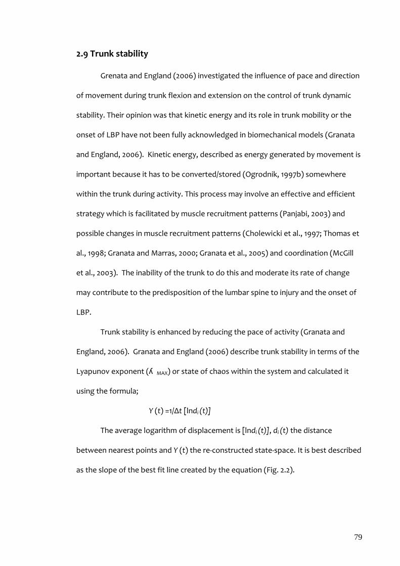
79
2.9 Trunk stability
Grenata and England (2006) investigated the influence of pace and direction
of movement during trunk flexion and extension on the control of trunk dynamic
stability. Their opinion was that kinetic energy and its role in trunk mobility or the
onset of LBP have not been fully acknowledged in biomechanical models (Granata
and England, 2006). Kinetic energy, described as energy generated by movement is
important because it has to be converted/stored (Ogrodnik, 1997b) somewhere
within the trunk during activity. This process may involve an effective and efficient
strategy which is facilitated by muscle recruitment patterns (Panjabi, 2003) and
possible changes in muscle recruitment patterns (Cholewicki et al., 1997; Thomas et
al., 1998; Granata and Marras, 2000; Granata et al., 2005) and coordination (McGill
et al., 2003). The inability of the trunk to do this and moderate its rate of change
may contribute to the predisposition of the lumbar spine to injury and the onset of
LBP.
Trunk stability is enhanced by reducing the pace of activity (Granata and
England, 2006). Granata and England (2006) describe trunk stability in terms of the
Lyapunov exponent (ʎ MAX) or state of chaos within the system and calculated it
using the formula;
Y (t) =1/Δt [lndi (t)]
The average logarithm of displacement is [lndi (t)], di (t) the distance
between nearest points and Y (t) the re-constructed state-space. It is best described
as the slope of the best fit line created by the equation (Fig. 2.2).

80
Fig. 2.2: The Lyapunov Exponent (from Granata and England 2006)
Flexion–extension movements of the trunk in both symptomatic and
asymptomatic individuals demonstrated that stability is greatest when performing
the movement asymmetrically (Fig. 2.3). This is shown by the Lyapunov exponent
(ʎ MAX), the greater the value the more unstable the movement.
Fig. 2.3: Effect of pace on the stability of the spine (from Granata and England 2006)
Fast asymmetrical flexion creates greater instability than performing a
symmetrical flexion slowly (Fig. 2.3). The differences in the stability demonstrated
were statistically significant between slow and fast movements (P< 0.001) in both
symmetric and asymmetric movement. This therefore suggests that neuromuscular
control of dynamic stability decreases significantly with increases in pace. The
increase in momentum requires an increase in neuromuscular activity to attenuate
kinematic instability as the trunk demonstrates changes in velocity and acceleration
(Marras and Mirka, 1993; Dolan and Adams, 1993; Granata and England, 2006).
Modulation of muscle forces during this process requires the recruitment of large
motor units with corresponding limitation in fine motor control during fast paced

81
movements (Granata and England, 2006). This may explain why seemingly
mundane, easy or common asymmetrical movements of the trunk can cause an
onset of LBP but the likelihood of onset is dependent upon the speed at which the
activity is executed. This may explain anecdotal evidence that suggests LBP is often
reported to occur whilst performing tasks that have been previously executed
successfully. If the muscle groups associated with ‘core stability’ are primarily
involved in fine motor control it is possible that restoration of this function will
enhance stability by altering asymmetrical movement characteristics.
The Lumbar spine accounts for approximately 70% of total trunk movement
during flexion-extension movements during lifting tasks in healthy individuals
(Granata and Marras, 2000) but the coordination of the important interdependent
relationship of the pelvis and lumbar spine (Young et al., 2000) during such tasks is
non-linear (Granata and Marras, 2000). The amount of lumbar spine involvement
will increase as the magnitude of the load increases (Granata and Marras, 2000).
Granata and Marras (2000) found that lumbar spine involvement is increased at
slower rates of performing a task such as lifting but is reduced at higher velocity
suggesting that there is a greater need for the pelvis to be involved to start the
extension component of the lifting task (Granata and Marras, 2000).
The lumbo-pelvic relationship described by Granata and Sandford (2000)
correlates with previous observations that lumbar disc distortion starts in the lower
lumbar region during extension (Kanayama et al., 1995). The lower segments take
more of the strain during the more vigorous part of the process- extension from
flexion. How this movement behaviour correlates with the extension observed in
the lower lumbar segments during the start of flexion (Pollock et al., 2009) remains

82
unclear but does explain why lower lumbar segments are more prone to injury than
the upper during lifting tasks whilst the upper segments are more involved when
the task involves either pushing or pulling (Plouvier et al., 2008) when shear forces
generated by the trunk oblique muscles (EO and IO) are primarily responsible
(Marras and Granata, 1995; Marras and Granata, 1997b).
2.10 Stability or robustness
Lumbar stability has no specific definition (Standaert et al., 2008) but
robustness has been used to describe the adaptability of the trunk to perturbation
(Reeves et al., 2007).
Trunk flexion requires summative lumbar segmental flexion (Kulig et al.,
2007). The performance of which is highly dependent upon its acceleration that
requires magnitude and direction (Ogrodnik, 1997c; Bloomfield, 2006). Because the
segment is restrained to a relatively fixed point by soft tissue such as ligaments, it is
presumed that movement occurs about a fixed point producing a pivot movement
(Fig. 2.4).
Fig. 2.4: Posterior view of a lateral pivot (F) and lateral view of a flexion pivot (E) of a spinal segment (from MacNab and McCulloch, 1990)
This movement behaviour of the spine is dependent upon perturbations
large enough to cause a change in position through a trajectory, which may not be
entirely unidirectional through range (Fig 2.5).
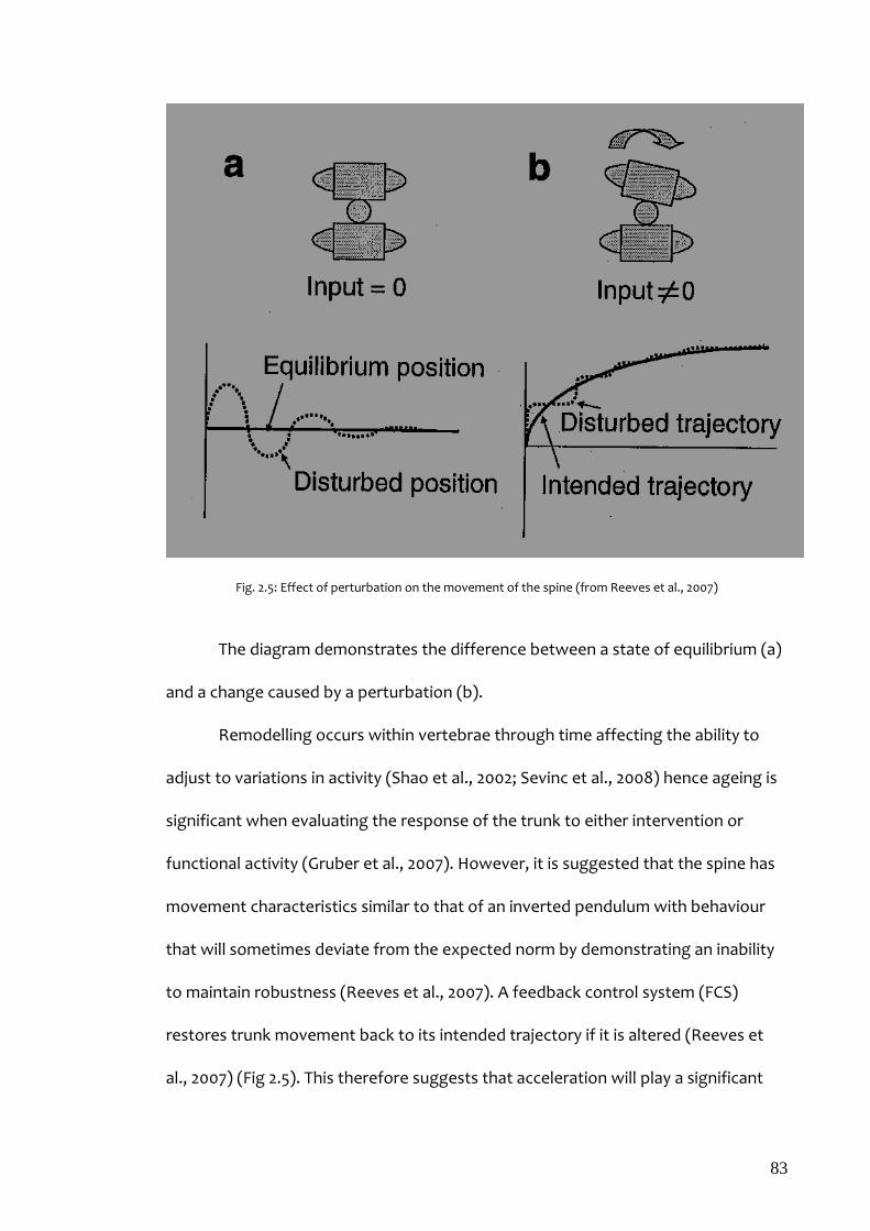
83
Fig. 2.5: Effect of perturbation on the movement of the spine (from Reeves et al., 2007)
The diagram demonstrates the difference between a state of equilibrium (a)
and a change caused by a perturbation (b).
Remodelling occurs within vertebrae through time affecting the ability to
adjust to variations in activity (Shao et al., 2002; Sevinc et al., 2008) hence ageing is
significant when evaluating the response of the trunk to either intervention or
functional activity (Gruber et al., 2007). However, it is suggested that the spine has
movement characteristics similar to that of an inverted pendulum with behaviour
that will sometimes deviate from the expected norm by demonstrating an inability
to maintain robustness (Reeves et al., 2007). A feedback control system (FCS)
restores trunk movement back to its intended trajectory if it is altered (Reeves et
al., 2007) (Fig 2.5). This therefore suggests that acceleration will play a significant

84
role in the ability of the spine to readjust itself and restore this intended sagittal
trajectory during flexion and extension. Acceleration into both lateral and rotational
directions will therefore also be of significance in the execution of the trajectory.
The feedback control system (Fig 2.6) suggests that the feedback from intrinsic
pathways (short range muscle stiffness and damping) is instantaneous while the
feedback from both the reflexive and voluntary pathways has a delay. However, all
components of the feedback control system can be used in part or in combination
to provide stability through force generation (Reeves et al., 2007).
Fig. 2.6: Feedback control system (from Reeves et al., 2007)
The mechanism by which the FCS provides tolerance to perturbation is
described as its robustness (Reeves et al., 2007) and it is this principle that this study
proposes to critique to offer an explanation for the mechanism by which CSE may
influence trunk performance in response to an onset of LBP. The absence/lack of

85
intervertebral stiffness provided by the passive sub-system (Panjabi, 1994) within
any direction of movement caused by perturbation implies that the counteracting
force is not proportional to the size of the displacement caused by the perturbation
suggesting a possible failure of the FCS (Reeves et al., 2007). The central nervous
system (CNS) responds to perturbation by increasing muscle activation (Reeves et
al., 2007) irrespective of the fact that it has been shown that LBP patients
demonstrate higher trunk muscle co-contraction than the healthy (Marras et al.,
2001; Lariviere et al., 2002; van Dieen et al., 2003). The difference between these
two groups has been shown to be of clinical significance (Reeves et al., 2007).
In principle, therefore, it has been proposed that there are two main
possibilities by which errors in neuromuscular control and loss of robustness can
cause LBP (Preus and Fung, 2005 cited in Reeves (1997));
1. Failure of the osteoligamentous structures of the spine creating
excessive segmental movement -beyond limits
2. Increased and sustained muscle contraction of muscle after a brief
period of instability
The second possibility could account for acute onset of LBP during functional
activity such as during trunk movements previously executed with success (Reeves
et al., 2007). However, this does not theorise why some episodes of acute LBP occur
well within the limits of trunk movement. Anecdotal evidence suggests that these
episodes are usually described as sharp twinges whilst performing a movement but
not always severe enough to cause an immediate cessation of function. There are
suspicions that changes in muscle recruitment in response to a perception of
instability may be the answer (Hodges et al., 2003; Moseley et al., 2004). It is
86
therefore proposed that robustness is directly related to trunk performance
requiring input from the FCS responding with a high degree of precision. The
resulting trunk movement behaviour in responding to perturbation has been
likened to an inverted pendulum (Stepan, 2009).
2.11 Inverted Pendulums
The human body is a multiple inverted pendulum of which the time delay
within the neural system required for control can be affected by age, physical and
mental state (Stepan, 2009). Three systems are recognised as providing control for
this complicated multiple inverted pendulum; the labyrinth (auditory system), eyes
(visual system) and mechanoreceptors (touch system) (Stepan, 2009). It is
suggested that the time delay affects the recruitment of the superficial component
of the multifidus and transversus abdominis (Stepan, 2009). Given the anatomy of
these muscles and the assumption that these muscle groups have a role in trunk
kinematics (Reeves et al., 2007), this delay could influence the ability of the trunk to
accelerate. This mechanism would suggest that the over all response cannot be
unduly influenced by subjectivity. In contrast once the trunk has accelerated, its
velocity in all probability becomes less independent. Although the deep component
of the multifidus creates stability by generating force without torque it is proposed
that the net effect of multifidus contraction is to maintain the relative position of
vertebrae (Reeves et al., 2007).
Two systems may describe the mechanism by which the trunk moves about
the pelvis. One system, a two-degree of freedom system best demonstrated by
balancing a stick on the hand, suggests that in order to maintain equilibrium a
control force (F) is required at the point of contact through a distance ‘χ’ (Fig.2.7).

87
The angle of displacement of the stick is denoted as ‘φ’ with ‘m’ the mass of the
stick and ‘ℓ’ its length (Stepan, 2009).
Fig. 2.7: Two-degree of freedom model for balance (from Stepan, 2009)
The second system is described as a one-degree of freedom model. This
model can best be demonstrated by trying to maintain balance whilst standing still.
Control is achieved through the ankle (Stepan, 2009) (Fig. 2.8). This model may
demonstrate a much clearer mechanism than the two-degree system because it
considers the influence of a control torque or moment (M) about the ankle (-M, M).
This system would probably fit more comfortably in describing movement of the
trunk about the sacrum in the absence of osteoligamentous connections.
Fig. 2.8: One-degree of freedom model for balance (from Stepan, 2009)
If either of the above systems are assumed to be true and the trunk moves
from a relatively fixed point, the sacrum (S), it will in all probability exhibit similar

88
characteristics of an inverted pendulum albeit with a degree of ‘chaos’ (Granata and
England, 2006) as illustrated in Fig. 2.9.
During flexion the trunk is displaced through an angle of θ in relation
to its starting point travelling over a displacement ‘A’ to ‘B’ represented by ds. Trunk
angular velocity (ω) is expressed as dθ/dt and its angular acceleration (α) (dω)/dt.
Fig. 2.9: Illustration of inverted pendulum movement of the Trunk
Another important characteristic of pendulum movement is simple harmonic
motion (SHM) (Ogrodnik, 1997c). SHM is rhythmic motion about a point, usually the
mid-point of the pendulum swing. It is proposed that this is the point during
functional movement when the trunk is in a state of ‘balance’ when the force
developed by muscle coordination equals the force developed by the perturbation.
Further work would be required to support this assumption but it is an interesting
possibility. It can be demonstrated in the lumbar spine during repetitive movements
whilst performing tasks; the trunk deviates and returns to a starting position (Fig.
2.9). If this holds true it would be possible to propose that from the waveform for
the movement;
Χt (angular displacement from midpoint) = A sin ωt (Fig. 2.10)
(t=time)

89
The maximum velocity of a pendulum is achieved at the midpoint of the
displacement when the amplitude of displacement is zero (Ogrodnik, 1997c).
According to Ogrodnik (1997c), the velocity (V) is therefore derived as;
V= dx(t)/dt………………………………………..(1)
Or
V= d(A sin ωt)/dt = Aω cos ωt…………………….(2)
(ω is the frequency and A is the amplitude of the sin wave) (Fig. 2.10) The acceleration (α) exhibited by such a movement can also be derived as;
α = dvx(t)/ dt……………..(3)
Or
α = d(Aωcos ωt) dt….………..…(4)
Or
α = -Aω2sin ωt…………………... (5)
Fig. 2.10: Simple harmonic motion wave (from Ogrodnik, 1997c)
This reinforces the importance of acceleration in initiating the change of
direction, a process less likely to be biased by subjectivity unlike velocity.
There is however, a need to translate angular movement into its linear
offshoot to appreciate how two fixed points on a vertebra may move in the
absence of the osteligamentous constraint from a point 1 to a point 2 (Fig. 2.11) or
from ‘A’ to ‘B’ (Fig 2.9).
The distance travelled by the point (ds) = rdθ……….. (6)

90
The vector velocity |v| = ds/dt………………………….. (7)
Or
|v|= Rdθ/dt……………………………………………… (8)
Or
|v| = r dθ/dt……………………………………………... (9)
Or
|v| = ωr…………………………………………………. (10)
Centripetal acceleration (ac) is required to attain a velocity once there is a
change in direction as a vertebra travels in a ‘circular’ path (Fig. 2.11).
Fig. 2.11: Velocity changes of vertebrae caused by angular movement (from Ogrodnik, 1997c)
If ds = rdθ…………………………………………………. (11) (Eq 6.)
ds/r = dv/|v|………………………………………. (12)
Or
dv = |v| ds/r………………………………………............... (13)
And
|ac| = |dv/dt|………………………………………………. (14)
|ac| = |v|/r ds/dt…………………………………………….. (15)
|ac| = |v|2/r……………………………………………….… (16)
Or
centripetal acceleration (ac) = ω2r……………………..… (17)

91
It is the average cumulative changes in acceleration that this method
quantifies and investigates (Fig. 2.11). Therefore force generated by movement is
proportional to ‘r’ and thus the greater the value of ‘r’ the greater the acceleration
and the force developed. This does suggest that the centripetal force is therefore
dependent upon the distance of the vertebra from the fixed point (sacrum)
suggesting that the closer the vertebra is to the fixed point (sacrum) the greater
the effect of cumulative centripetal force produced by trunk movement is
experienced. It is therefore proposed that this could be the mechanism by which
the L5/S1 segment has become specialised in terms of the relative size of L5 to other
vertebrae in the same region. A natural response to a role that is similar to a hinge,
upon which the trunk rotates.
2.12 Intra-abdominal pressure
Intra-abdominal pressure (IAP) is thought to play a significant role in the
stability of the spine (Cholewicki et al., 1999; Janda and Valenta, 2000). It may not
be possible to isolate this role from that of what is commonly termed as ‘core
stability’ muscle activity because of the interdependency of the muscle groups
concerned (Chapter One). A mathematical model has been devised to predict levels
of IAP and moment occurring at L4/5 movement segment during functional tasks
(Janda and Valenta, 2000).
IAP=IAP0 + 0.567735· M [Pa] (IAP0 - initial value of the intra-abdominal pressure during relaxed standing; M- moment produced at the L4/5 motion segment)
The generation of IAP is directly linked to anthropometric values, lumbar
lordosis and pelvic tilt during relaxed standing (Youdas et al., 2000) suggesting that
the ability to develop and maintain lumber/pelvic symmetry is an important way by
92
which excessive loading of the trunk can be dampened. However, IAP responds to
changes in posture and for instance it has been demonstrated that IAP is directly
related to respiratory activity (Cholewicki et al., 1999; Hodges and Gandevia, 2000).
2.13 Trunk acceleration
Higher order trunk movement characteristics are valid and reliable measures
to determine sincerity of effort (Marras et al., 2000). Velocity can be used to classify
trunk movement during controlled and uncontrolled sagittal flexion-extension,
uncontrolled rotation, uncontrolled side flexion, and repeatability of function
(Marras et al., 2000). Although it also influences the central set and acceleration
(Marras et al., 2000) it is only possible when there is a change in direction during the
task. This may also be true during a unidirectional task if a change in velocity is
required to respond to pain or discomfort at different points through the range of
activity.
Low Back Pain affects the characteristics of the central set or the
recruitment pattern of trunk muscles during movement (Marras et al., 2000). A
recruitment pattern of movement is developed and maintained by day to day
activity (Hseih et al., 1992) and is well developed for common tasks such as those
involving trunk flexion (Marras et al., 2000). The relationship between acceleration
and velocity during a movement can be demonstrated using a phase plane (Fig. 2.12)
using a set of Rho numbers to represent the distance in the phase plane space
between each point within it and the centroid of the phase plane.

93
The consistency of this plot with repeated cycles will therefore demonstrate
a sincerity of effort because the closer these phase planes are with repeated
measure the more the consistent the effort (Marras et al., 2000).
Physical factors may inhibit or change the characteristics of the central set
and produce inconsistent plots demonstrating changes in direction within the
tripartite coupling system (sagittal, coronal and axial planes) (Marras et al., 2000). It
is proposed that altered acceleration can be observed at those differing points
during the completion of a task such as flexion-extension. There is paucity in the
literature exploring this hypothesis and it is not clear which of the variables of
velocity or acceleration are the most sensitive to changes in direction but given that
acceleration is a factor of velocity it would appear that changes in acceleration
would be more descriptive of changes in both direction and dysfunction.
It is not clear if acceleration and velocity exhibit a similar behaviour during
trunk dynamic functional activity, especially when pain can influence trunk
characteristics. Anecdotal evidence does suggest that pain influences trunk mobility
and during an episode of LBP is reported to be at the beginning or end of changes in
trunk position. It has been reported that LBP reduces trunk acceleration (Marras
and Mirka, 1993). However, because of the direct relationship between acceleration
and velocity it is assumed that both measures will demonstrate a similar trend. It
Fig. 2.12: Phase plane of trunk movement (from Marras, 2000)

94
may be because of this assumption that there is a paucity of research into the
behaviour of trunk acceleration. But it is not clear if the pain reported at the
beginning or end of trunk movement is of the same magnitude experienced during
the actual movement within which the trunk is expected at some point to reach
peak levels of velocity. Hence it is proposed that if this pain is different both in
description and magnitude, acceleration or deceleration measures will have more
significance for outcome and expectation of intervention.
The subgroup of LBP patients most likely to respond to stabilisation
exercises are those with lumbar instability (Hicks et al., 2005) but the diagnosis of
instability remains controversial because its measurement and validity rely on static
observations (Teyhen et al., 2007). Using the same terminology to define
mechanical LBP is therefore a problem unless there is a valid means to observe such
instability during trunk movement. The pain experienced during flexion-extension
movements of the trunk is thought to be caused by a disruption to the passive
osteoligamentous system limiting movement (Teyhen et al., 2007). Loss of
neuromuscular control causes instability within the trunk causing the feeling of
‘’slipping’’ or ‘’catching’’ during movement (Panjabi, 1994). A flexion-relaxation
phenomenon, the period of electromyographic electrical silence of the lumbar
paraspinal muscles is evident at the end of range of trunk flexion (Teyhen et al.,
2007). This phenomenon is particularly poor in patients with LBP causing segmental
hypomobility with reduced linear displacement (Teyhen et al., 2007).
It is a theory that the ability of the trunk to harness efficient neuromuscular
control to produce trunk positional changes that will determine if the movement

95
will produce pain (Panjabi, 2003). Furthermore, it is proposed that this ability is
reliant on an ability to accelerate to and maintain an efficient velocity.
2.14 This thesis research questions
The main aim of this thesis was therefore to investigate the effect of core
stability exercises (CSEs) on the acceleration of the spine after an acute onset of
LBP.
The study was also designed to answer the following research questions;
Does the kinematics of the spine change with the onset of acute LBP?
Do CSEs reduce the levels of disability caused by LBP?
Do CSEs reduce pain caused by LBP?
Can CSEs be used effectively in primary care to prevent the increasing
prevalence of chronic LBP?
2.15 Conclusions
Higher order kinematics is a valid and reliable means of describing trunk
movement (Kroemer et al., 1990; Marras et al., 1990) but trunk velocity and range of
movement (ROM) can be subjective (Cox et al., 2000). Stability of the trunk during
movement is dependent upon the speed of execution and symmetry of the task
(McGregor and Hughes, 2000). However, tasks evaluating trunk performance are
best done when participants are evaluated performing tasks at their preferred
speed (McGregor and Hughes, 2000). Asymmetrical tasks require greater motor
control using large trunk muscles; a process that is accompanied by a corresponding
loss of fine motor control (Granata and England, 2006). The literature seems to
suggest that there is an assumption that both trunk acceleration and velocity will

96
demonstrate similar characteristics. However it is not clear if the ability to
accelerate mirrors the ability to maintain a desired velocity during displacement.
This mechanism is worthy of investigation. There is paucity in literature quantifying
the effects of core stability exercises using functional objective measures. This
study set out to evaluate changes in trunk performance as an effect of a course of
core stability exercises. It has been suggested that LBP does reduce trunk
acceleration but the behaviour of trunk acceleration in response to core stability
exercises as an intervention is not well understood. This is compounded by the fact
that previous studies purported to investigate the effects of CSE are not well
designed.

97
Chapter 3
Development of the method
3.1 Summary
This chapter describes the development of the rationale for the method
chosen to test the study hypothesis and answer the research questions described in
the previous chapter.
3.2 Operational definition of acute non-specific low back pain
Acute LBP is described as an episode of LBP that has lasted for up to 6 weeks
becoming chronic if it lasts longer (BackCare, 2007; Kinkade, 2007). It has also been
described as lasting for up to 3 months (Smith et al., 2002; Gullick, 2008). The ‘3
months’ definition was used in articles within a systematic review of core stability
exercises (May and Johnson, 2008) but the practicality of describing acute non-
specific low back pain as lasting for up to 3 months is most probably unacceptable
because of the secondary effects of LBP which are largely driven by a process of
central neuro-modulation (Jayson, 1996). The latest informed opinion is that an
acute episode of low back pain lasts up to 6 weeks (BackCare, 2007).
The National Institute for Health and Clinical Excellence (NICE) suggests that
non-specific LBP describes LBP which cannot be attributed to any specific cause and
also suggests that exercises and physical activity should be offered to patients with
non-specific LBP for a period of 12 weeks in the first instance (NICE, 2009).
98
3.3 Recruitment of participants
The study was approved by the School of Health Sciences & Social Care
Ethics Committee at Brunel University and Oxfordshire NREC ethics committee (Ref.
07/H0606/102) in September 2007.
3.3.1 Inclusion Criteria
Participants of either gender, male or female and aged between 18 and 55
were eligible for inclusion. This age group was chosen because they provide the
greatest number of consultations for LBP in primary care (Croft et al., 1998). Each
participant’s history of onset of non-specific LBP was to be no longer than 6 weeks’
duration, a period described as acute (BackCare, 2007).
3.3.2 Exclusion Criteria
Participants were excluded if they did not meet the inclusion criteria as
described above and demonstrated evidence through routine physiotherapy
assessment of any of the following;
Degenerative conditions affecting the spine- Loss of bone, joint and
intervertbral disc integrity influencing spinal mobility (Gruber et al., 2007).
Diabetes- Diabetes increases the risk of muscle strength impairment
(Bohannon, 2000) and affects the ability to perform the exercises required
by the protocol.
Pregnancy- Underlying mechanism for LBP during pregnancy is ambiguous
and may be multi-factorial (Kerr and Grahame, 2003) with approximately
50% of women reporting an incidence of LBP whilst pregnant (Kerr and
Grahame, 2003). Hormonal changes affect pelvic ligaments and cause an
increase in joint mobility (Kerr and Grahame, 2003).

99
Neurological involvement- Peripheral neurological damage will affect
muscle response to stimuli and exercise. A study on the ulnar nerve
conduction velocity of injured baseball pitchers suggested that although the
velocity appeared to be normal they demonstrated suboptimal performance
(Wei et al., 2005).
On-going spinal treatment- Other surgical and non-surgical treatment would
contaminate responses to intervention. Contamination of this type can
cause a cross-over effect (Altman, 1991).
On-going legal issues- subjective perception of pain and disability could be
affected by expected monetary gain from an injury (Standaert et al., 2008).
History of depression- Subjective perception of pain could be altered by
psychological factors (Standaert et al., 2008).
Histories of multiple recurrent episodes of low back pain- Biomechanical
properties of the structure of the spine are altered during repeat episodes
of LBP (Standaert et al., 2008).
Involvement in other research studies- Participation in multiple studies could
compromise safety. Attributable effects of intervention given in this study
will be ambiguous (Hicks, 1998).
English not being their first language- Misinterpretation of study procedure
and instruction would jeopardise results and the ability to provide informed
consent.
Participants were recruited from 5 locations within Hillingdon Primary Care Trust
(PCT). These locations included
1. Church Road Surgery, Cowley

100
2. The Warren Medical Centre, Uxbridge
3. Uxbridge Health Centre, Uxbridge
4. Eastcote Health Centre, Eastcote; Westmead Clinic, South Ruislip
5. Harefield Health Centre, Harefield
All the Centres operate with a ‘hub and spoke system’ with Laurel Lodge, the
hub, in Hillingdon coordinating physiotherapy provision across those Centres.
Referrals are generated from general practitioner (GP) surgeries within the area but
patients referred to a Centre may not necessarily be registered with a GP at the
Centre at which they will receive their physiotherapy treatment.
3.3.3 The sample size
The sample size presented within this work needed to be small enough to be
managed effectively within the constraints of true micro and macro environmental
pressures as described in the following section 3.3.4 but large enough to
demonstrate differences between the CSE and control groups (Section 4.3).
3.3.4 Challenges to recruitment
A number of challenges have affected the recruitment process. These include
the following;
The waiting list.
Fluctuations in the length of time patients were on the waiting list could
have been significant. This period may also differ according to each location from
which participants will be recruited. The waiting list was predicted to range
between 6 and 12 weeks depending upon the location. A significant number of
patients became ineligible for inclusion because they no longer met the definition of
acute non-specific LBP.

101
Local demands.
The demands of the Commissioners for healthcare delivery within primary
care resulted in changes in physiotherapy delivery within the primary care trust. As a
result physiotherapy staff were pressurised into implementing queue management
strategies to redress disparity in access to services and waiting times. These
strategies actively changed during the study to suit prevailing circumstances.
Staff turnover.
There was a continuous change in personnel within and between locations.
The structure of physiotherapy career pathways within the NHS precipitated this
tendency. This study protocol required physiotherapy staff to be actively involved
with the recruitment process. Non- clinical staff was also required to assist in
arranging convenient appointment times to ensure that participants’ visits for data
collection coincided with treatment times.
Personal Ethics.
Some physiotherapy practitioners faced personal dilemmas as they took the
view that patients who agreed to take part in the study were in effect ‘queue
jumping’. Practitioners who relied upon CSE as a primary form of intervention also
questioned the ethics and efficacy of withholding such intervention when they were
requested not to prescribe CSE because their patient was allocated to a control
group. Using alternative treatment methods, albeit for a short period of time (6
weeks) was not an entirely comfortable option for them.
Facilities.
All locations except Eastcote Health Centre operate within either a GP
surgery or a room within a Health Centre. These rooms did not offer enough space

102
within which to accommodate both the normal day to day function and additional
space required for data collection. Care was taken to ensure that visits coincided
with the least busy time of routine operational procedures.
Logistics
The study did not attract funding from either internal or external sources. All
the data for the study, was therefore collected by the researcher alone. This did
present difficulties involving travel to and between locations.
3.3.5 The randomisation process
Two hundred opaque envelopes with pieces of paper on which either
‘control’ or ‘experimental’ group were prepared by a colleague who was blinded to
the study. The colleague did not have contact with the participants or have any
other input to the study. The researcher did not take part in the process and had no
knowledge of the allocation of envelopes to groups. Once the envelopes were
sealed, they were shuffled and then had a sequence of numbers marked on the
exterior of the sealed envelope in a chronological order to denote the order in
which participants were recruited. Each sealed envelope was opened in the
sequence in which they were numbered by the participant as they are recruited.
This was done only after the participant had given their written consent to take part
in the study and after the participant was aware that it was not possible to change
from the group to which they would be potentially be allocated to. Neither the
researcher nor the participant was aware of the contents of the envelope until it
was opened by the participant. The participants were therefore not blinded to the
allocation.

103
3.4 Tools and outcome measures
3.4.1 Pain
There are a number of scales that are used to evaluate pain. These
include the Brief Pain Inventory (BPI) in either its short or long version (Daut and
Cleeland, 1982), Dallas Pain Questionnaire (DPQ) (Lawlis et al., 1989), Numeric Pain
Intensity Scale (NPIS) (McCaffery and Beebe 1993), Faces Pain Rating Scale (Bieri et
al., 1990), Wong-Baker Faces Pain Rating Scale (Wong and Whaley 1986), Aberdeen
Back Pain Scale (ABPS) (Ruta et al., 1994) and the Visual Analogue Scale (VAS)
(Huskisson, 1974). The VAS was used for this study because of its ease of use and
the ease to which the information was interpreted.
The Visual Analogue Scale (VAS), as a single item measure, is very sensitive to
changes in symptoms that can be rated (Paul-Dauphin et al., 1999). The VAS is
reliable and valid as an outcome measure (Crossley et al., 2004) and it is a good tool
for quantifying both pain and disability in conditions involving chronic
musculoskeletal pain but the measures for pain are more reliable than that for
disability (Boonstra et al., 2008). Because levels of disability correlate to levels of
pain it is realistic however, to assume that the reliability of measures will be similar
in the acute phase of a condition by relating consistently with levels of pain
(Boonstra et al., 2008). The VAS is also reliable when time variance is a factor (Badia
et al., 1999) especially with LBP (Olaogun et al., 2004).
The minimal clinically significant difference in the VAS pain score remains
unaffected by levels of pain (Kelly, 2001) which made the VAS a realistic choice for
this study to quantify pain during an acute onset of LBP. This is because pain is a
single variable/construct and there is the need to consider the complexity of the

104
recruitment process. ‘Pain on the day’ of testing was used to evaluate reported pain
because it minimised participants’ recall bias (Chouinard and Walter, 1995).
The VAS was denoted by a 100 mm line to indicate a range from a state of ‘no
pain’ (0 mm) to that of ‘very severe pain’ (100 mm). Increased scores indicated
more pain.
3.4.2 Disability
There is no preferred functional limitation outcome measure for low back
pain (The Chartered Society of Physiotherapy, 2004). However, a number of
outcome measures available include the Aberdeen Back Pain Scale (ABPS) (Ruta et
al., 1994), Quebec Back Pain Disability Scale (QBPDS) (Kopec et al., 1995), Oswestry
Disability Index (ODI) (Fairbank et al., 1980) and the Roland Morris Disability
Questionnaire (The Chartered Society of Physiotherapy, 2004). The Oswestry
Disability Index (ODI) and the Roland Morris Disability Questionnaire (RMDQ)
(Roland and Morris, 1983) are recommended for use by physiotherapists however
the setting and location should determine which one is more appropriate for the
circumstances (The Chartered Society of Physiotherapy, 2004). The RMDQ was used
in this study. The ODI was discounted because of its relative low internal
consistency (Cronbach’s а = 0.77) and because it has a mixture of both capacity and
performance based items (The Chartered Society of Physiotherapy, 2004). The
ABPS was not used because a relatively long time is required to complete it (The
Chartered Society of Physiotherapy, 2004) and the QBPDS was not used because
there is not enough evidence to support its use at present (The Chartered Society of
Physiotherapy, 2004).

105
The Roland-Morris Disability Questionnaire (RMDQ) is regarded as the best
scale to measure self reported disability (Deyo et al., 1998; Boonstra et al. 2008) and
it is both valid and reliable (Roland and Morris, 1983). This scale is recommended for
use in primary care studies (Roland and Morris, 1983; Kopec et al., 1995) and has a
high intra-class reliability of 0.8 (Kopec et al., 1995, Stratford et al., 1996; Dunn and
Croft, 2005). It also has a high degree of sensitivity (Hseih et al., 1992) and been
recommended for use without the need for further validation (Grotle et al., 2004;
Grotle et al., 2006).
The RMDQ as a tool can easily be used within a clinical environment. The
RMDQ consists of a 24-item back pain specific disability scale. The participants were
asked to answer ‘yes’ or ‘no’ to each question. The sum totals ‘yes’ score was
determined. The change in this score quantified the relative change in pain and
disability as a result of intervention.
The RMDQ results of this study will be interpreted with an understanding of
the minimal clinically important difference of the RMDQ for LBP. A rule for minimal
clinically important difference for RMDQ is shown below (Table 3.1) (Jordan et al.,
2006).
Table 3.1: Minimal clinically important difference rule for disability (from Jordan et al., 2006)
Definitely improved: Patients rating back pain as at least better at 6 months with a reduction > 30% on their RMDQ score.
Possibly improved: Patients with an RMDQ score >30% reduced at 6 months but have not rated their back pain as better.
Not improved: Patients with less than 30% reduction in RMDQ score at 6 months.
3.4.3 Trunk performance
Various methods of obtaining trunk kinematic evaluation have been
explored; video analysis (Wickstrom et al., 1996; Neumann et al., 2001; Chang et al.,
106
2003, Trott and Fisher, 2005; Wong and Lo, 2007), EMG activity (Capodaglio et al.,
1995; Oddsson et al., 1997; Chiou et al., 1999; Bonato et al., 2002; Pitcher et al., 2008)
and 3-D motion analysis (Nakajima et al., 2007; Pazos et al., 2007). The Lumbar
Motion Monitor (LMM) is however, potentially the most practical tool for use in a
clinical setting because it is highly portable and the time to set up the equipment is
much less labour intensive than the other aforementioned tools. The video analysis
and EMG evaluations may have allowed a greater freedom of movement in the
trunk than the LMM because of the harnesses required to secure the device to the
participant, however, these methods of analysis are labour intensive and require
high intra and inter-reliability measures rendering the procedures difficult to
transpose into the clinical environment.
The LMM provided objective measures of real time changes in trunk
kinematics to quantify LBP by measuring spinal kinematics (Marras and Wongsam
1986; Kroemer et al., 1990; Marras et al., 1990; Marras, 1996). Simultaneous
information on displacement, acceleration and velocity of the spine in three
dimensions during functional movement was obtained.
The LMM is an electrogoniometer consisting of a number of potentiometers
within an exoskeleton capable of detecting and measuring real time movement in 3-
dimensions (sagittal, frontal and coronal planes). The LMM was developed at Ohio
State University and it has become a useful tool in the quantification of triaxial
movement of the spine (Marras and Wongsam 1986).
3.4.3a Calibration of the Lumbar Motion Monitor
The LMM was originally demonstrated to be both valid and reliable with an
accuracy of measure of +/- 0.25% (Marras et al., 1990). The LMM has a sampling rate
107
of 60Hz and can measure between -35 to +65 degrees in the sagittal plane and
between -45 and +45 degrees in both the coronal and frontal planes (Lumbar
Motion Monitor, Industrial Analysis Desk Reference Chattanooga Group, Inc.).
Further calibration tests in a laboratory environment using a video motion analysis
demonstrated its accuracy to be within +/- 0.5 degrees (Parnianpour et al., 2001).
This was done by using a reference frame to +/- 3o degrees in the frontal and
coronal planes and to +/- 45 degrees in the sagittal plane (Parnianpour et al., 2001).
The manufacturers of the LMM calibrate the LMM before its inaugural use.
However, supplementary calibration tests within laboratory conditions for this
study demonstrated a 2% discrepancy between actual and recorded data (Appendix
2). This calibration test procedure was carried out by measuring the angle of
displacement of the LMM at different angles during both components of forward
bend and extension from flexion during a single flexion-extension movement
(Appendix 3: a-f). A comparison of the LMM programme output to actual LMM
plate angle was made. For this process, the subject was instructed to stand with the
feet at shoulder width apart and with the shoulders placed across the chest. A
single flexion-extension movement of the trunk was performed and still pictures
were taken to measure the angle of the LMM plates. These measurements were
taken in neutral and 6 other positions of flexion; 3 during forward bend movement
and 3 during backward extension movement of the trunk as it resumed the neutral
position.
The 2% difference between the measures suggests that the LMM produces
reliable data even when a thin layer of clothing exists between the LMM harnesses
and the skin.
108
3.5 The Lumbar Motion Monitor protocol
The original protocol devised by Marras et al. (1986) consisted of 5 tasks for
a single kinematic evaluation of trunk sagittal movement in 0 degrees or neutral, 15
and 30 degrees of rotation to the left and then right (Marras and Wongsam, 1986;
Marras, 1996; Gill and Callaghan, 1996). Movements in the sagittal plane with the
trunk in these degrees of rotation utilises different muscle recruitment patterns to
cope with the increase in the level of difficulty in performing the task (McGill et al.,
2003). The study protocol for this research did not consider difficulties performing
the task which may be complicated by underlying facet joint pathology (Boyling and
Jull 2004) but tested a revised version of the original protocol requiring only one
task to provide an evaluation of trunk sagittal movement in neutral (Ferguson and
Marras, 2004). This was done using a pilot study. The adjusted protocol resulting
from the pilot study was then used for this research project. For testing the
adjusted protocol participants stood with their feet shoulder width apart with the
arms loosely folded across the chest and performed flexion-extension movements
in neutral without rotation to either the right or left.
Although the LMM has been reported as reliable using the Intra-class
Correlation Coefficient (ICC) analysis of the data, levels of agreement between
measurements were not reported (Ferguson and Marras, 2004). For this study it
was therefore imperative that levels of agreement of the reliability for a single task
evaluation was demonstrated. This was important because all the data was
collected by one researcher who was not be blind to the study aims or objectives.

109
3.6 Reliability of the LMM using a single task method
3.6.1 Participants for the pilot study to evaluate its reliability
Approval for the pilot study was granted by the School of Health Sciences &
Social Care Ethics Committee at Brunel University and Oxfordshire NREC ethics
committee (Ref. 07/H0606/102).
Twenty participants were recruited by incidental sampling at Brunel
University between February and March 2008 and were classified as belonging to
one of two groups (Table 3.1). One group consisted of healthy participants, a mix of
both colleagues and students (male =5, female=5). This group had an average age of
38.3 years (SD 8.6) and did not have a recent history of LBP. No recent history was
defined as the 6 months prior to testing. This was judged to be sufficient enough to
avoid recall bias (Chouinard and Walter, 1995) and limit any possibility of any carry
over of previous treatment effects (Altman, 1991) and provide a wash-out period
where any effects of any previous intervention is reduced (Chapter 1 Peacock and
Peacock, 2011). The other group of participants were potential participants for the
main study (male = 4, female =6) and had an average age of 31.7 years (SD 7.5). All
participants in this group had been diagnosed with an acute onset of LBP within a 6
week period. A description of both groups of participants is provided below (Table
3.2).
Table 3.2: Pilot study group descriptive
Minimum Maximum Mean Std. Deviation
Healthy Participants (m=5, f=5)
Weight (Kgs) 54.5 99.0 71.2 14.4 Age (Yrs) 21 51 38.3 8.6
Height (cms) 154.0 183.0 171.9 11.2
LBP Participants (m=4, f=6)
Weight (Kgs) 45.0 113.0 74.5 21.3 Age (Yrs) 20 44 31.7 7.5
Height (cms) 157.0 191.0 171.3 12.0

110
3.6.2 Pilot study procedure
The Lumbar Motion Monitor (LMM) was used to collect data to explore the
intra-rater reliability of the equipment. The equipment consists of a light weight
exoskeleton within which are a set of potentiometers. The exoskeleton was
connected, via an umbilical lead to a laptop containing specialised software
(Lumbar ProSoft 2.0, NexGen Ergonomics, Canada) for data collection and analysis
(Fig. 3.1). The exoskeleton was positioned on the participant with a 2-piece harness,
one for the thorax and the other for the pelvis; both harnesses were secured in
place with Velcro strapping.
Before collecting data the anthropometric measurements (age, height and
weight) for the participants were entered into the computer software. The
potentiometers were then calibrated with the exoskeleton firmly in place within its
holding case. A zero- calibration check procedure using explicit instructions from
the manufacturers using the LMM software was performed before data was
collected from each participant and between each set of data.
A harness size (small, medium or large) which allowed for the exoskeleton to
be placed on the participant’s trunk without demonstrable movement whilst
standing erect in the neutral position was selected. The exoskeleton was then
attached and tightly secured in place onto the harness and tightly secured with the
locking mechanism provided.
For each set of data collected, each participant was asked to stand with their
feet shoulder width apart and their arms loosely folded across the chest. The
participant was instructed to perform trunk flexion-extension movements in a
sagittal plane for 8 seconds. The movement flexion-extension in the sagittal plane

111
without rotation to either side of midline were performed at the participants’
preferred speed (McGregor and Hughes, 2000; Al-Eisa et al., 2006) to ensure
natural trunk movement (Al-Eisa et al., 2006). No other instruction was given to the
participant. Previous studies that used the LMM (Marras and Wongsam, 1986;
Marras et al., 1990; Ferguson and Marras 2004) did not do this. No other
encouragement or stimulus was provided. Two sets of data (Test 1 and Test 2) were
collected with a 10 minute rest period in-between each measurement. This
prevented data contamination by cross over effects (Altman, 1991; Hicks, 1998).

112
Fig. 3.1: The Lumbar Motion Monitor exoskeleton

113
3.6.3 Results of the pilot study
The LBP group demonstrated slower acceleration compared to the healthy
participants within all kinematic variables during both tests 1 and 2 (Table 3.3) (Fig.
3.2). The LBP group were unable to increase trunk acceleration with repeated
measure (Fig. 3.2). There was no correlation between sex, height or weight with any
sagittal acceleration values.
Table 3.3: Pilot study group comparison (Acceleration)
Sagittal Acceleration
(Deg/s2)
Lateral Acceleration
(Deg/s2)
Rotation Acceleration
(Deg/s2)
Average (SD) Peak (SD) Average (SD) Peak (SD) Average (SD) Peak (SD)
Healthy T1 T2
215.9 (149.5) 253.9 (160.0)
627.1 (363.6) 674.5 (298.9)
42.6 (37.6) 33.6 (21.4)
175.5 (130.0) 130.4 (90.2)
20.8 (16.7) 28.6 (21.0)
88.9 (57.5) 106.6 (71.1)
LBP T1 T2
148.8 (103.1) 151.6 (114.3)
426.4 (177.3) 416.9 (208.3)
31.7 (17.9) 30.3 (22.3)
129.1 (60.6) 131.4 (79.3)
15.5 (7.3) 16.2 (11.5)
61.9 (24.3) 67.1 (34.9
The mean differences between measurements (T1 and T2) for all kinematic
variables were calculated and a two-way mixed ANOVA analysis (SPSS ver. 15 for
windows) was used to determine the intra-class correlation coefficient (ICC).
Fig. 3.2: Pilot study comparison of Sagittal Acceleration Test 1 Vs Test 2
Average sagittal acceleration demonstrated the highest ICC at 0.96 (C.I 0.90-
0.98) (Table 3.4). This suggests that average sagittal trunk acceleration is the most
0 50
100 150 200 250 300
Test 1 Ave
rage
Sag
itta
l Acc
eler
atio
n (d
eg/s
s)
Test 1 Test 2 Test 2 Healthy Group LBP Group
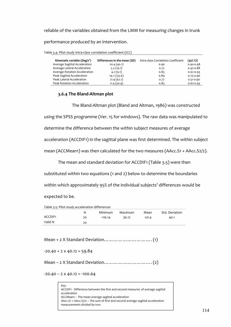
114
reliable of the variables obtained from the LMM for measuring changes in trunk
performance produced by an intervention.
Table 3.4: Pilot study Intra-class correlation coefficient (ICC)
Kinematic variable (Deg/s2) Differences in the mean (SD)
Intra-class Correlation Coefficient
(95% CI)
Average Sagittal Acceleration 20.4 (40.1) 0.96 0.90-0.98 Average Lateral Acceleration 5.2 (19.1) 0.72 0.42-0.88 Average Rotation Acceleration 4.2 (9.1) 0.83 0.62-0.93 Peak Sagittal Acceleration 19.1 (133.6) 0.89 0.75-0.96 Peak Lateral Acceleration 21.9 (62.1) 0.77 0.51-0.90 Peak Rotation Acceleration 11.4 (30.4) 0.83 0.62-0.93
3.6.4 The Bland-Altman plot
The Bland-Altman plot (Bland and Altman, 1986) was constructed
using the SPSS programme (Ver. 15 for windows). The raw data was manipulated to
determine the difference between the within subject measures of average
acceleration (ACCDIF1) in the sagittal plane was first determined. The within subject
mean (ACCMean1) was then calculated for the two measures (AAcc.S1 + AAcc.S2/2).
The mean and standard deviation for ACCDIF1 (Table 3.5) were then
substituted within two equations (1 and 2) below to determine the boundaries
within which approximately 95% of the individual subjects’ differences would be
expected to be.
Table 3.5: Pilot study acceleration differences
N Minimum Maximum Mean Std. Deviation ACCDIF1 20 -116.14 36.12 -20.4 40.1 Valid N 20
Mean + 2 X Standard Deviation…………………………. (1)
-20.40 + 2 x 40.12 = 59.84
Mean – 2 X Standard Deviation…………………………. (2)
-20.40 – 2 x 40.12 = -100.64
Key: ACCDIF1 - Difference between the first and second measures of average sagittal acceleration ACCMean1 – The mean average sagittal acceleration AAcc.S1 + AAcc.S2/2 – The sum of first and second average sagittal acceleration measurements divided by two

115
A plot of ACCDIF1 (y-axis) against the ACCMean1 (x-axis) was then drawn to
produce the Bland-Altman plot (Fig. 3.3)
Fig. 3.3: Bland-Altman plot showing limits of agreement of repeated measures for the pilot study
The plot (Fig. 3.3) suggests that the measures were not in very close
agreement. However, the high values for the ICC suggests that it remains
reasonable to use the LMM as a tool for measuring the primary outcome of trunk
acceleration.
3.6.5 Conclusions derived from the pilot study
It was reasonable to use the LMM as a tool for measuring the primary
outcome of sagittal trunk acceleration. The disparity in the level of agreement may
be because it is not at present possible to attach the LMM harness directly to
participants’ skin. To do so would be a potential health and safety hazard because
of possible cross infection between participants and/or possible skin irritation on
each participant. A small amount of unwanted movement may have therefore
occurred between the harness and the clothing worn by the participants. Another
reason for the disparity could be the fact that trunk performance will change with
100.00 200.00 300.00 400.00 500.00 600.00 ACCMean1 (Deg/s.s)
-100.00
-50.00
0.00
50.00
AC
CD
IF1
(Deg
/s.s
)
Mean difference = -20.40

116
repetition i.e a practice effect (Hicks, 1998) irrespective of whether the subjects
have LBP or not. Although the harness was applied over loose fitting clothes, this
did not directly interfere with lumbar spine movement; the 2% discrepancy
demonstrated earlier supports this assumption.
This pilot study has been published in full elsewhere (Aluko et al., 2011)
(Appendix 4).
3.7 Evaluation of trunk kinematics
No data is available which evaluates trunk performance within a sample
population. This information was useful for analysis of the results of this proposed
main study. A further pilot study was therefore required. Approval for this part of
the study was also granted by the School of Health Sciences & Social Care Ethics
Committee at Brunel University and Oxfordshire NREC ethics committee (Ref.
07/H0606/102) as an amendment.
3.7.1 Recruitment of participants
Participants were recruited from staff and students at Brunel University
between September 2009 and March 2010. A request for volunteers to take part in
the study was placed on the University intranet. Another verbal request was made
to the physiotherapy staff of the Musculoskeletal Physiotherapy Services, Hillingdon
Community Health based at Eastcote Health Centre. The combination of these
resulted in a total of 50 willing participants agreeing to take part in the study. Each
participant gave their written consent to take part.
A short questionnaire (Appendix 5) was used to gather a brief subjective
history for each participant to explore the history of their self reported LBP. This
questionnaire consisted of simple questions used during a standard subjective

117
history gathering process for all musculoskeletal conditions in a clinical setting and
therefore did not need to be piloted before use. The participants were classified as
belonging to either a healthy or LBP group according to that self reported history of
LBP. None of the participants reported LBP at the time of testing. A history of LBP
was described as an experience of an episode of acute non-specific LBP up to and
during the preceding 6 months prior to the date on which the participant was
measured as part of the evaluation of trunk kinematics study. 6 months was chosen
because it is long enough to prevent recall bias (Chouinard and Walter, 1995) and
limit cross over effects (Altman, 1991). On the date on which the participants were
tested none of the participants reported any disability in performing day to day
activities or underlying pathology that could have interfered with their trunk
performance. A description of the groups is shown in table 3.6.
Table 3.6: Database participant descriptive (Healthy participants, n=13; participants with LBP n=37) Presence of LBP N Mean Std. Deviation
LBP Age (Years) 37 33.2 12.3 Height (cms) 37 168.6 8.4 Weight (Kgs) 37 69.6 15.0
Last episode of LBP 37 3.1 2.2 Number of days LBP was
present (days) 36 27.0 86.7
No LBP Age (Years) 13 31.2 13.2
Height (cms) 13 167.7 10.5 Weight (Kgs) 13 66.3 16.9
Thirty seven of the 50 participants reported episodes of LBP in the preceding
6 months. These participants had a mean age of 33.2 (S.D 12.3) years, mean height
of 168.6 (S.D 8.4) cms and a mean weight of 69.6 (S.D 15.0) kgs. 13 of the fifty
participants reported no history of LBP and had a mean age of 31.2 (S.D 13.2) years,
mean height of 167.7 (S.D 10.5) cms and a mean weight of 66.3 (S.D 16.9) kgs. The
LBP group reported an average of 3.1 (S.D 2.2) weeks proceeding the date on which

118
they provided data as when they experienced an episode of acute non-specific LBP
and reported average duration of an episode to be 27 (S.D 86.7) days.
3.7.2 Procedure to evaluate trunk kinematics using the LMM
The LMM was applied as described in section 3.6.2 above. Data was
collected during one cycle of flexion-extension movement during a single 8 second
period (Section 3.6.2 above). Each participant was requested to perform the test
movement at their preferred speed. No other encouragement or stimulus was
provided to ensure as much natural trunk movement as possible (Al-Eisa et al.,
2006).
3.7.3 Statistical analysis
A measure of reliability was investigated using the two-way mixed model of
intra-class correlation coefficient to evaluate the proportion of the total variance
that was due to the variance between subjects alone.
A one way ANOVA analysis was used to investigate difference between and
within groups.
Pearson’s Correlation Coefficient (r) was used to investigate relationships
between factors.
All Analyses were conducted using SPSS (ver. 15 for Windows).
3.7.4 Results
74% of the participants reported a history of LBP. Data were collected during
a mean sagittal displacement of 52.7 (S.D 16.8) and 43.3 (S.D 15.0) degrees for the
participants with and without a history of LBP respectively. The distribution of data
was skewed as demonstrated by the asymmetrical distribution of the

119
measurements. This therefore required the data to be log-transformed before
analysis.
One way ANOVA (α=0.05) suggested that average sagittal acceleration was
not significantly different either between or within the groups (p= 0.4).
Participants who did not report a history of LBP demonstrated slower
performance (Table 3.7). This could possibly be because these participants had
better control of trunk movement during the measurement process compared to
the group who considered they were experiencing an episode of non-specific LBP.
Interestingly, none of the participants within this group were actively receiving
treatment for their back pain during the period of data collection. However it would
be expected that the reverse should hold true given the results demonstrated in
section 3.6.3 above.
Table 3.7: Group performance comparison (kinematic evaluation)
Presence of LBP N Mean Std.
Deviation LBP
Sagittal ROM (Max-Min PS) (Degs)
37 52.7 16.8
Average Sagittal Acceleration (Deg/S.S)
37 359.2 170.1
No LBP Sagittal ROM (Max-Min PS) (Degs) 13 43.3 15.0
Average Sagittal Acceleration (Deg/S.S) 13 309.7 150.4
Female participants demonstrated slower mean trunk performance than
males whether they reported a history of LBP or not but this was not significant
(Table 3.8).

120
Table 3.8: Mean sagittal acceleration (Deg/S2) by level of gender and the presence of low back pain Presence of LBP
( p=0.84) Yes No
Gen
der
( p=0
.27)
Male M= 440.4 S.D = 240.9
M= 364.8 S.D = 152.2
Female M= 336.8
S.D = 142.3 M = 293.1
S.D = 153.9
More females reported a history of LBP than males (M=8; F=29) and 41% of
this group of participants reported an episode within the last month prior to
testing.
Neither the length of time of the last reported episode prior to testing
(p=0.8) nor gender (p= 0.27) was significant in the mean sagittal acceleration
measures of participants.
Intra-Class Correlation Coefficient (ICC) analysis using a two-way mixed
method demonstrated the reliability of the single measure as 0.65 (95%CI 0.40-0.80)
and 0.58 (95%CI 0.70-0.85) for participants with and without a history of LBP
respectively.
Pearsons’ Correlation Analysis (r) suggests that there is a stronger
correlation between trunk displacement and the mean sagittal acceleration 0f 0.64
(p=0.01) and 0.57 (p=0.05) within participants with a history of LBP compared to the
healthy participants respectively. The displacement demonstrated by this group
was affected by the history of LBP.
A significant correlation between weight and height was demonstrated in
both the LBP group (r=o.49, p=0.01) and the group without LBP (r=0.87, p=0.01).
This suggests that the correlation for the participants who did not report a history
of LBP was stronger than in those reporting a history of LBP. A weight/age

121
correlation only existed in the participants with a history of LBP (r=0.34, p=0.05) but
was not statistically significant at p=0.05 in the other group (r=-0.29).
3.7.5 Conclusions derived from trunk kinematic evaluation
Within a sample of the population described here, the data suggests
that the prevalence of LBP can be demonstrated to be relatively high however a self
reported episode of LBP within this sample may not be reliable. From the sample
evaluated, more female participants reported an episode of LBP than males.
However, whether females reported a history of LBP or not within the sample they
consistently demonstrated slower average trunk performance. The reliability of the
LMM in both groups of participants without or with a history of LBP was similar but
the confidence interval within which the results are reported was smaller in the
group who did not report a history of LBP. The results demonstrated by the sample
in question may tentatively be applied in general, however, only a much bigger
study can provide substantive facts. This information is however used with this in
perspective to discuss trunk acceleration in relation to the findings of this research
study within chapter 6.
3.8 Development of the intervention
3.8.1 Core stability exercises
The ‘core’ is described as the lumbopelvic region (Willardson, 2007a;
Willardson, 2007b). Muscles that influence trunk performance are two groups
‘global’ (erector spinae, rectus abdominis) and ‘segmental’ or those that act across
spinal segments (multifidus and transversus abdominis) (Bergmark, 1989).
Stabilisation exercises improve the integrity of the segmental muscles (Willardson,
2007a; Willardson, 2007b). Exercise for the treatment and management of LBP is

122
common but the efficacy of exercise is doubtful (Linton et al., 1996). The
comparative effect of therapeutic exercise regimen with active exercises suggests
that there is no clinically significant difference (May and Johnson, 2008).
Stabilisation exercises may be useful for the management of chronic LBP
but the effects may not be more effective than other methods of management
involving activity (May and Johnson, 2008). The effect on acute LBP is not widely
reported. This may be because it is assumed that the effects of stabilisation
exercises in enhancing neuromuscular control and rectifying dysfunction
(Richardson et al., 1999; Norris, 2000; McGill, 2002) may not be required during an
acute phase of LBP.
There is no formal definition of core stability exercises or recommendation
for any specific grouping of CSE (Standaert et al., 2008). There is also no justification
for the choice, combination of, or the number of repetitions and frequency of the
chosen exercises (Standaert et al., 2008). However the clinical decision and
justification for using stability exercises to improve the integrity of the lumbar
multifidus has traditionally been underpinned by a set of belief systems (Table 3.9)
(MacDonald et al., 2006). Increases in cross sectional area of the multifidus can be
observed within 6 weeks following the introduction of CSE and this increase is
directly related to the frequency of the exercise (Sokunbi et al., 2008). Stabilising
exercises have been designed to encourage the activation of the deep fibres of the
multifidus through low loaded isometric activity with the spine in as much of a
natural position as possible (MacDonald et al., 2006; Standaert et al., 2008). The
exercise routine that was used in this study and derived from a software package

123
which accompanied work on core stability exercises (Norris, 2000) is attached
within the appendices (Appendices 4 and 5).
Table 3.9: Core stability exercises belief systems (MacDonald et al., 2006) Belief System Evidence
Deep multifidus rather than superficial fibres or the erector spinae responsible for spinal stability
Richardson & Jull, 1995; Richardson et al., 1999f
Deep multifidus has the most proportion of type 1 muscle fibres
Porterfield & DeRosa, 1991a,b; Richardson et al., 1999f
Isotonic activity is observed in the deep multifidus during trunk movements and gait
O’Sullivan et al., 1997; Richardson et al., 1999f; Taylor & Sullivan, 2000; Hides, 2004a
Deep multifidus and transversus abdominis co-contract during function
Richardson & Jull, 1995; O’Sullivan et al., 1997; Pool-Goudzwaard et al., 1998; Richardson et al., 1999b; Richardson et al., 2000; Taylor & Sullivan, 2000; Arokoski et al., 2001; Hides et al, 2004
Low back pain induces most changes within the deep multifidus
Norris , 1995a,b; Pool-Goudzwaard et al., 1998; Richardson et al.; 1999d; Arokoski et al., 2001; Hides 2004a,b
3.8.2 Exercise compliance
Compliance in Physiotherapy is important because the effect of intervention
is largely reliant upon it (Sluijs et al., 1993). Compliance plays a significant role in the
outcome of intervention (Linton et al., 1996). It does not significantly change the
perception of pain in LBP; although compliance with exercises can be improved
when pain intensity is negligible when compared to a control group (Linton et al.,
1996). The fear avoidance model of inactivity is the primary factor in restricting
levels of pain and thus by implication compliance (Linton et al., 1996; Frank and
DeSouza, 2001; Fritz and George, 2002). Exacerbation of pain may be linked to
attempts at becoming active (Linton et al., 1996).
The exercise routine that was used in this study was chosen because they
reflected the exercise routines which anecdotal evidence suggest are most
commonly used in clinical practice. They were also devised on the assumption that
compliance would be enhanced if participants understand the aetiology, onset of
124
their LBP and the behaviour of their pain. A good relationship with whoever is
providing the exercise instruction and the frequency and number of repetitions of
the exercise reflecting their lifestyle (allowing for omissions in the routine due to
unforeseen circumstances) will also improve exercise compliance (Linton et al.,
1996; Frank and DeSouza, 2001; Fritz and George, 2002).
Each participant that was randomised into the experimental group was
required to complete an exercise compliance sheet (Appendix 8). The participants
were to bring the sheets with them for each treatment session where the sheets
will be examined by the treating Physiotherapist. This served two purposes; to
suggest to the participant that their progress was being monitored closely and to
provide an incentive for compliance. Although each sheet represented the requisite
daily exercise compliment, participants were given only enough sheets for 3 weeks
at a time. Each participant in the experimental group was inclined to ask for more
sheets to complete the remaining 3 week period.
The participants in the experimental group were able to demonstrate the
frequency and the number of repetitions that were completed for each of the
exercises. The participants were told that if a set of the exercises were
inadvertently missed they were not to double up the number of repetitions for the
subsequent set of exercises. They were required to leave the corresponding section
on the sheet blank. This prevented anomalies in the data caused by the different
effects of either high or low repetition of exercises.
Other forms of compliance methods were not be used to avoid adding to the
participants’ perceived barrier to exercise by impinging on the available time that
they have to do the exercise routine (Sluijs et al., 1993).
125
3.9 The effects of missing data
Because of possible attrition and its possible effects on the results, an
‘intention to treat’ analysis (Altman, 1991) was used. As described in chapter 2, this
type of analysis allows direct comparison of the groups in response to the
intervention by maintaining the balance in respect to the subject characteristics
within each group (Chapter1 Peacock and Peacock, 2011). In order to achieve the
‘intention to treat’ analysis the last measurement obtained from a participant was
carried forward to replace any missing data. This is known as the Last Observation
Carried Forward (LOCF) (Howell, 1992). This procedure is often preferred (Peacock
and Peacock, 2011) however the trend of the data in this instance suggested that
this was a reasonable strategy.
3.10 Conclusions
This chapter has set out the method to answer the research questions
described in chapter 2. This chapter also defined and justified the use of the RMDQ,
VAS and the LMM to describe changes in pain and disability as a direct result of the
introduction of core stability exercises.
The following chapter describes the method derived from this chapter that
was used to answer the research questions and hypotheses posed in chapter 2.
126
Chapter 4
Method
4.1 Summary
This chapter describes the method used to collect the data for this study. It
provides an insight into the obstacles that the researcher encountered during the
study and the decisions made to ensure that the study remained valid. The biggest
threat to the success of the study was the frequent local changes to Physiotherapy
provision at the behest of the local Primary Care Trust. These changes resulted in a
number of amendments (4.6 below).
4.2 Introduction
It remains unclear if CSEs will make a significant difference in the treatment
of LBP or indeed if there is a defined subset of patients with non-specific LBP who
will respond more favourably than others (Brennan et al., 2006). This study
evaluates the effect CSEs have on an onset of acute non-specific LBP.
A significant problem with using CSE is the ambiguity of the underlying
mechanism that underpins the clinical decision making process that suggests its
use. This process can influence treatment outcome and any cost/benefit analysis
within both primary and secondary care. Better clarity can be achieved by providing
answers to the following research questions;
• Does an onset of acute LBP change trunk kinematics?
• Can CSEs reduce self reported disability caused by LBP?
• Do CSEs reduce LBP?

127
The study involved two groups of participants (Control and Experimental) who
were experiencing an acute episode of non-specific LBP and comparing the changes in
trunk average sagittal acceleration, reported pain and reported disability between
participants who had a CSE programme (experimental group) and those who did not
(control group).
4.3 The research design
The study was a randomised controlled trial and was approved by the School
of Health Sciences & Social Care Ethics Committee at Brunel University and
Oxfordshire NREC ethics committee (Ref. 07/H0606/102) in September 2007. The
study did not attract any internal or external funding and in view of the time
constraints it was decided that the study would proceed within the capabilities of
the researcher.
The objectives of this study were;
• To evaluate the effect of Core Stability Exercises (CSE) on trunk
performance during flexion-extension movements in the sagittal
plane. Performance was to be evaluated during a period of non-
specific acute LBP.
• To evaluate changes in disability caused by an acute onset of non-
specific LBP during a course of CSE.
• To evaluate the changes in pain during an acute episode of non-
specific LBP.
The hypotheses of the study tested were the following;
• CSE will improve average trunk acceleration when performing flexion-
extension movements in the sagittal plane.

128
• CSE will reduce pain during an episode of non-specific LBP.
• CSE will reduce self reported disability caused by an onset of acute
non-specific LBP
The study process is shown below (Fig 4.1).
Fig. 4.1: Study flow chart
4.4 The study sample size
A standard formula, N=2ks2/d2 (k=10.5 standard multiplier; s= standard
deviation and d= clinically significant difference) would have been the preferred
choice for calculating the sample size for this study in the first instance. The sample
size calculated would have provided a power of 90% with a significance of 5%.
However, the absence of a known clinically significant difference suggested that
this approach was not beneficial (Bland, 2009), since no research predating this

129
study provided a clinically significant difference for the primary outcome measure
for changes in mean sagittal acceleration in response to CSE. Although the trunk
produces an average sagittal acceleration of 470 deg/s2 (Marras et al., 1990) no data
existed to suggest a possible differential as a result of an intervention.
Because of the problems with recruitment (chapter 3), the alternative of an
estimation of the likely width of the confidence level to a power calculation was
used to interpret the findings (Bland, 2009). Clinical implementation of previous
research has been impeded by the inability of research to effectively demonstrate
its clinical importance; it is often lost in the emphasis of stating the power of the
research tests (Domholdt, 2005). This study had to be able to demonstrate its
meaningfulness to clinical practice by ensuring that the sample size presented in
this work was small enough to be managed effectively within the constraints of true
micro and macro environmental pressures and used confidence levels to report the
findings. This needed to be done without losing potentially important information
or giving misleading interpretation of results (Bland, 2009).
4.5 Study recruitment process
All patients referred between July 2008 and June 2010 for treatment of an
acute episode of non-specific LBP within Hillingdon Primary Care Trust in West
London were given the opportunity to take part in the study. The recruitment
process was continuous with participants randomised to groups as they were
recruited. Participants were approached during their first visit for an assessment
after being referred for physiotherapy treatment. Physiotherapy staff members in
the musculoskeletal outpatients departments of Hillingdon Community Health
providers who had agreed to be involved in the recruitment process made the initial
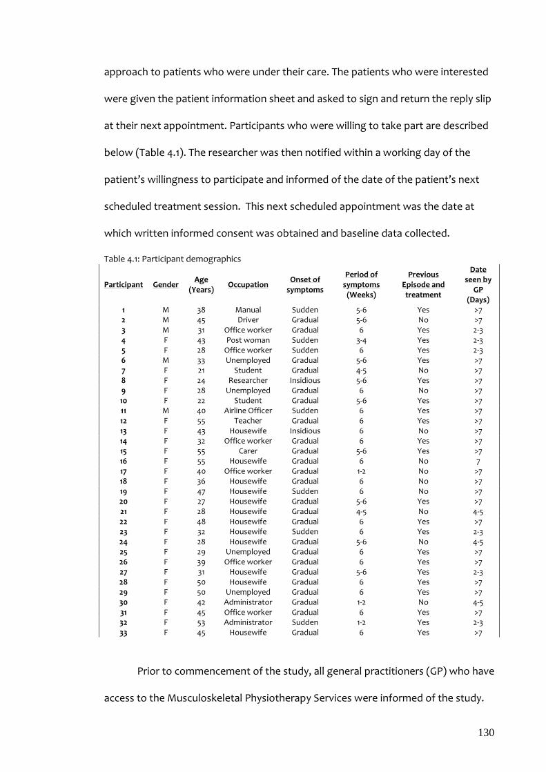
130
approach to patients who were under their care. The patients who were interested
were given the patient information sheet and asked to sign and return the reply slip
at their next appointment. Participants who were willing to take part are described
below (Table 4.1). The researcher was then notified within a working day of the
patient’s willingness to participate and informed of the date of the patient’s next
scheduled treatment session. This next scheduled appointment was the date at
which written informed consent was obtained and baseline data collected.
Table 4.1: Participant demographics
Participant Gender Age
(Years) Occupation
Onset of symptoms
Period of symptoms
(Weeks)
Previous Episode and treatment
Date seen by
GP (Days)
1 M 38 Manual Sudden 5-6 Yes >7 2 M 45 Driver Gradual 5-6 No >7 3 M 31 Office worker Gradual 6 Yes 2-3 4 F 43 Post woman Sudden 3-4 Yes 2-3 5 F 28 Office worker Sudden 6 Yes 2-3 6 M 33 Unemployed Gradual 5-6 Yes >7 7 F 21 Student Gradual 4-5 No >7 8 F 24 Researcher Insidious 5-6 Yes >7 9 F 28 Unemployed Gradual 6 No >7 10 F 22 Student Gradual 5-6 Yes >7 11 M 40 Airline Officer Sudden 6 Yes >7 12 F 55 Teacher Gradual 6 Yes >7 13 F 43 Housewife Insidious 6 No >7 14 F 32 Office worker Gradual 6 Yes >7 15 F 55 Carer Gradual 5-6 Yes >7 16 F 55 Housewife Gradual 6 No 7 17 F 40 Office worker Gradual 1-2 No >7 18 F 36 Housewife Gradual 6 No >7 19 F 47 Housewife Sudden 6 No >7 20 F 27 Housewife Gradual 5-6 Yes >7 21 F 28 Housewife Gradual 4-5 No 4-5 22 F 48 Housewife Gradual 6 Yes >7 23 F 32 Housewife Sudden 6 Yes 2-3 24 F 28 Housewife Gradual 5-6 No 4-5 25 F 29 Unemployed Gradual 6 Yes >7 26 F 39 Office worker Gradual 6 Yes >7 27 F 31 Housewife Gradual 5-6 Yes 2-3 28 F 50 Housewife Gradual 6 Yes >7 29 F 50 Unemployed Gradual 6 Yes >7 30 F 42 Administrator Gradual 1-2 No 4-5 31 F 45 Office worker Gradual 6 Yes >7 32 F 53 Administrator Sudden 1-2 Yes 2-3 33 F 45 Housewife Gradual 6 Yes >7
Prior to commencement of the study, all general practitioners (GP) who have
access to the Musculoskeletal Physiotherapy Services were informed of the study.

131
This was done with care so as not to change the referral behaviour. This was
important for the physiotherapy staff members who had volunteered to be actively
involved in the recruitment process because changes in referral patterns would
have had consequences on capacity, queue management and general treatment
outcomes.
All participants were referred from within primary care but there was a
variation between the times they had been experiencing symptoms before they
saw their GP. This variation ranged from between 2 and 7 days (Table 4.1). Although
this posed a threat to the recruitment process it was not possible to influence this
first stage of the care pathway.
An initial informal estimation of the waiting list suggested that referrals for
treatment of LBP constituted a significant majority of the referrals on the waiting
list for all locations included as a study site (Section 3.3.2). However, the length of
the waiting list suggested that a majority of those referrals would not meet the
inclusion criteria because they would have been waiting for more than 12 weeks.
The local physiotherapy staff members were co-opted into appraising all referrals as
they arrived at the locations and recruiting potential participants as quickly as
possible. Some physiotherapy staff had reservations about doing so because they
considered that those patients could be ‘jumping’ the queue. The Lead
Practitioner/Manager provided valuable support for the study by emphasising the
need for local research. This intervention was crucial in changing the view of those
staff members. However, a compromise was that a 3 month rather than a 1 year
follow up was more realistic. Six participants had been recruited before this
decision and had passed the time for the 3 month follow up. The data collected

132
from these participants at baseline, 3 weeks and 6 weeks were included in the
analysis of the results. Although they provided data at the 1 year follow up this data
was not included in any analysis within this study and the data that should have
been obtained at 3 months was considered to be missing data replaced with the last
observations carried forward (LOCF) (See section 3.9).
Changes in access policy and information management by the local Primary
Care Trust (PCT) presented a real threat 3 months into the study. A new IT system
(RIO) meant the physiotherapy staff no longer maintained control of their diaries
and all appointments were made from a central point. In an attempt to improve
access and drive down waiting times, the PCT decided that all patients with LBP
should have an initial one-to-one assessment and then be referred to ‘Core Stability
Classes’. The content of the classes included a mixture of exercises aimed at both
‘global’ muscle and ‘intersegmental’ muscle activity (Appendix 7 and 8). The
protocol for this group of participants is shown in fig 4.1. These classes were
provided on a weekly basis and each patient was offered a 5 week course of
treatment. Participant grouping for this study was therefore redefined. The control
group was redefined from the group that did not perform any exercises to that
which consisted of participants who attended the core stability classes only. The
experimental group constituted those participants who attended the core stability
classes but performed an additional exercise routine (Appendices 6 and 7). The
protocol for this group of participants is shown in fig 4.1. Care was taken to avoid
duplication of exercises and the use of specific words/phrases such as Transversus
Abdominis, Multifidus, core strengthening and stability were avoided during the
core stability classes. This was so that the chance of the physiotherapy staff

133
member introducing bias to the study was minimised and also to avoid potential
doubt of the impact of being allocated to a specific group within the minds of the
participants.
An advantage of these classes was that data were collected during those
class attendances however other contractual obligations of the researcher
prevented the researchers’ attendance at all the available classes and some
potential participants were missed to the recruitment process.
Local turnover of staff did not play a significant role in disrupting the
recruitment process. Staff co-opted to assist in the recruitment process was
experienced physiotherapists who had permanent non-rotational positions within
their respective departments. This meant that there was no need to re-visit the
various sites (Section 3.3.2) to maintain levels of interest within the departments to
remain actively involved in the recruitment process.
4.6 Randomisation and allocation process
Participants were allocated to groups using the method described in chapter
3. A colleague independent and blind to the study placed notes on which was
written either ‘experimental’ or ‘control’ group into opaque envelopes. These
envelopes were shuffled and then marked in numerical order.
4.7 Study procedure
Every participant received an initial assessment by a suitably qualified
member of staff at the nearest physiotherapy outpatient department to which they
were referred to by their GP. Once a participant was identified and agreed to take
part in the study, the physiotherapist completed a short history sheet (Appendix 11).

134
These details would have formed part of the standard musculoskeletal subjective
history procedure. This sheet provided basic details of the history of onset and
nature of the LBP. Each willing participant was then provided with a Participant
Information Sheet (Appendix 12). This took place after each participant was offered
a place in the core stability classes which was part of the routine care pathway for
an acute non-specific LBP patient. Each participant was asked to sign a return slip
confirming their willingness to participate in the study and requested to bring this
confirmation slip with them to their first core stability class where the researcher
met the participants for the first time. Eastcote Health Centre was the location for
data collection because it was easier for the participants to attend the same
location for measurement at which they were taking part in the CSE programme.
During the initial session, each participant was given the opportunity to ask
questions about the study and was given a further opportunity to withdraw.
Written informed consent was then obtained (Appendix 13). The participant was
then requested to open an envelope marked with the number which represented
the order in which they agreed to become a participant in the study. Neither the
participant nor the researcher was aware of the contents of the envelope prior to
this time. The researcher then re-affirmed the procedure for the study in relation to
the group to which the participant had been allocated. The participant was also
given a further opportunity to decline taking part in the study.
Baseline measurements of height, weight and age were collected and
recorded on the specialist software on a laptop to which the LMM was connected
via an umbilical cord through which two-way communication between the LMM
exoskeleton and the laptop occurred. Each participant was also required to

135
complete a RMDQ (Appendix 14) and VAS (Appendix 15). The RMDQ was not
required at 3 weeks (Fig 4.1) because it was not considered a long enough period to
identify any change in function. It was however, noted that it has been
demonstrated that cross sectional area of the multifidus can be increased within 6
weeks of CSE (Sokunbi et al., 2008).
The LMM had already been calibrated by the manufacturers before its initial
use (section 3.4.3). However, the ‘zero calibration’ procedure (section 3.4.3) was
performed before each measurement. A harness size (small, medium or large)
which allowed for the exoskeleton to be placed on the participant’s trunk without
demonstrable movement whilst standing erect in the neutral position was selected.
The exoskeleton was then attached and tightly secured in place onto the harness
and tightly secured with the locking mechanism provided.
The assessment of repeated measures demonstrated during reliability
testing of the equipment suggested that some movement can occur between the
LMM harness and the skin of the participant. It was, however, not possible to place
the LMM directly onto the participants’ skin because of health and safety
considerations. To reduce unwanted movement to a minimum all participants were
evaluated wearing a top made of as thin a material as possible and asked to wear a
similar garment for each evaluation.
Each participant was requested to perform as many sagittal trunk flexion-
extension movements as possible for 8 seconds. The movement was executed at
the participants’ preferred speed and within their preferred range of movement. No
encouragement verbal, non-verbal or otherwise was offered. No warm up exercise
was performed because the interest of the study lay within the natural muscle

136
recruitment process to effect functional movement without prior warning. All trunk
measurements were therefore also taken before each scheduled core stability class.
All participants completed the evaluation and none reported an exacerbation of
their symptoms.
Following the initial LMM evaluation, the participants allocated to the
experimental group were given their CSE instruction (Appendices 4 and 5) by the
researcher. The experimental group was required to perform 10 repetitions of 8 CSE
three times a day for 6 weeks. This was in anticipation that the participants would
be able to complete the routine at least twice a day given their individual personal
circumstances involving work commitments and family life. The participants in this
group were encouraged to keep an account using a diary (Appendix 8) to
demonstrate their compliance with the task. This diary was inspected by the
treating physiotherapist at each visit either for treatment or attendance at the core
stability classes. The participants in the control group were asked to continue with
the classes alone. Participants in the control group received the same face-to-face
contact time as those in the experimental group.
Trunk performance of all 33 participants using the LMM procedure was
evaluated at 0, 3 and 6 weeks. A further evaluation was made at 3 months for all
participants excluding the first 6 participants, three of whom provided an
evaluation at 1 year follow up after cessation of the intervention (Fig 4.1). All but the
6 week and 3 month follow-up evaluations coincided with regular attendance at the
core stability classes. However, all participants were asked to continue their routine
as advised by their treating physiotherapist until their follow up at three months.

137
4.8 Ethical considerations
The original protocol submitted for ethical approval was changed on three
occasions to meet volatile circumstances encountered during the study. These
circumstances were out of the control of the researcher. All amendments were
substantial and therefore required resubmission to the Oxfordshire NREC ethics
committee (Appendix 16)
Procedures were put in place to allow for ethical considerations during the
study (Table 4.2).
Table 4.2: Study ethical considerations Ethical Consideration Consequences Procedures
1. Participants changed their mind about taking part in the study
High rates of attrition Participants were advised at every step that they could withdraw without prejudice at any point in the study.
2. Bias Participants in the control group could have felt ‘disadvantaged’ in not having an extra exercise routine
Each participant in the control group was told that at the end of the study they would be offered the same exercise routine.
3. Adverse response to exercises Increase in subjective pain and discomfort
All participants were instructed to stop any of the exercises at any time they considered their symptoms to have increased.
4. LMM assessments Increase in pain with measure of outcome
Participants were instructed to perform all test movements at their own preferred speed and range
4.9 Potential sources of error
Potential sources of error during the study and strategies adopted to
minimise potential influence on the outcome of the study included the following;

138
• The success of the study was dependent upon the researcher. To
minimise bias the researcher avoided any involvement in the
recruitment process, the randomisation of the sample or allocation of
participants into groups.
• Although all raw data for each of the outcome measures using the
VAS, RMDQ and LMM were collected by the researcher, the VAS
score measurement and the evaluation of the RMDQ were carried out
by a colleague independent of the study. This was to ensure that any
indirect influences that could have been introduced by the researcher
were addressed.
• Although the experimental group received exercise instruction from
the researcher care was taken to give equal face-to-face time to the
control group participants.
• The researcher avoided any cues/prompting during any of the
outcome measurement procedures. This was particularly important
during the LMM evaluation.
• Similar contact was made with all the participants throughout the
study irrespective of the group into which they were allocated.
• Evaluating the intra-rater reliability of the LMM demonstrated a
discrepancy between repeated measures (section 3.6.3). Participants
were evaluated wearing a similar thin layer of clothing for each
evaluation.
• LMM evaluation was not preceded by a ‘practice’ run to minimise
exaggeration of trunk performance resulting from ‘practice effect’.

139
• Participants in the exercise group may not have been entirely
accurate with the exercise diary. An assumption was made that the
exercise routine would be completed at least twice a day.
4.10 Data preparation
The following procedures were used to prepare the data before analysis;
The VAS score was measured in centimetres between a minimum of 0
(no pain) to a maximum of 10 (Worst pain). The score was directly
proportional to the amount of pain reported. This figure was
imported directly and without manipulation for statistical analysis.
The RMDQ was scored out of a maximum of 24 points. The level of
self reported disability due to LBP was directly proportional to the
score out of 24. This figure was imported directly and without
manipulation for statistical analysis.
Trunk displacement was defined as the difference between the
maximum and minimum trunk position for each evaluation. This
figure was imported directly for statistical analysis.
Average trunk acceleration was provided as an absolute number by
the LMM software and did not require manipulation. This figure was
imported directly for statistical analysis.
The CSE routine was recorded by the degree of compliance. The
exercise routine was considered to be done if the routine was
completed a minimum of twice a day. The total number of days for
which the exercise was completed was then directly imported to the

140
statistical software package without further transformation and used
for analysis.
Where data were log-transformed to give a normal distribution, the
results of comparisons between transformed means were back-
transformed to give the ratio of geometric means.
4.11 Procedure for testing the hypotheses
All analyses to test the study hypotheses were conducted using SPSS
(ver. 15 for windows) (Table 4.3) using an intention to treat analysis.
Table 4.3: Statistical tools to test the study hypotheses Study Null Hypothesis
Outcome Measure used Statistical Analysis
1. Pain experienced during an acute onset of LBP will not be reduced with CSE
Pain VAS score
Two way mixed ANOVA analysis
(α=0.05) of differences between and within both the
control and experimental groups
2. Self reported disability during an acute episode of LBP will not be reduced by CSE
Disability RMDQ
3. Sagittal trunk performance during an acute episode of LBP will not be improved by CSE
Trunk performance Average sagittal trunk performance
A regression analysis was used to investigate the causal relationship
between CSE and outcome measures
Pearson’s correlation co-efficient analysis (r) was used to identify
relationships between the 3 outcome variables.
Intra-Class Correlation Coefficient (ICC) analysis using a two-way
mixed method was used to evaluate the reliability of the measure.

141
4.12 Conclusions
The method described in this chapter was designed to answer the research
questions and test the generated hypotheses posed in chapter 2. The outcome
measures used provided data to adequately achieve this goal. Reasonable measures
were taken to reduce sources of error that could affect the results of the study but
total eradication of error could not be entirely guaranteed.
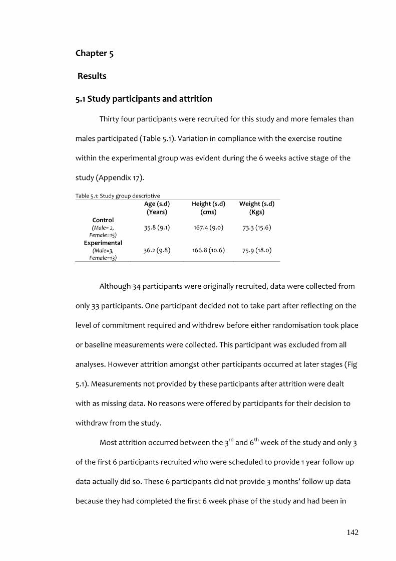
142
Chapter 5 Results 5.1 Study participants and attrition
Thirty four participants were recruited for this study and more females than
males participated (Table 5.1). Variation in compliance with the exercise routine
within the experimental group was evident during the 6 weeks active stage of the
study (Appendix 17).
Table 5.1: Study group descriptive
Age (s.d) (Years)
Height (s.d) (cms)
Weight (s.d) (Kgs)
Control (Male= 2,
Female=15) 35.8 (9.1) 167.4 (9.0) 73.3 (15.6)
Experimental (Male=3,
Female=13) 36.2 (9.8) 166.8 (10.6) 75.9 (18.0)
Although 34 participants were originally recruited, data were collected from
only 33 participants. One participant decided not to take part after reflecting on the
level of commitment required and withdrew before either randomisation took place
or baseline measurements were collected. This participant was excluded from all
analyses. However attrition amongst other participants occurred at later stages (Fig
5.1). Measurements not provided by these participants after attrition were dealt
with as missing data. No reasons were offered by participants for their decision to
withdraw from the study.
Most attrition occurred between the 3rd and 6th week of the study and only 3
of the first 6 participants recruited who were scheduled to provide 1 year follow up
data actually did so. These 6 participants did not provide 3 months’ follow up data
because they had completed the first 6 week phase of the study and had been in

143
abeyance beyond the 3 months’ follow up period before the decision was made to
reduce the follow up from 1 year to 3 months. The absent data was therefore
treated within the missing data protocol described in Chapter 3. The one year follow
up data was not used for any data analysis. Of the remaining 25 participants only 11
completed the whole study; 5 and 6 participants in the experimental and control
groups respectively.
Fig.5.1: Description of sample and attrition
Three participants, following a discussion with their treating
physiotherapists decided that they may be disadvantaged by taking part in the
study. One participant developed an unrelated orthopaedic problem in the lower
limb and decided to end involvement following a tertiary referral to a consultant.
The rest of the attrition is unaccounted for because the participants did not provide

144
a reason and in keeping with the research protocol consent form were not pressed
to provide any further information.
5.2 Missing data analysis
The missing data (section 3.9) was replaced with the Last Observation
Carried Forward (LOCF) (Howell, 1992). This decision was based upon the fact that it
allowed an analysis over the whole time frame of the study and assumed that the
data carried forward followed the trend of the data preceding it. The reason for this
approach was because the trend of the raw data demonstrated either a sequential
improvement or status quo in the outcome measures within both groups of
participants. It was therefore assumed that the missing data could be replaced with
the data that proceeded without the fear that the results would therefore be over
optimistic. It was therefore assumed that bias would be kept to a minimum as the
artificial inflation of the effect of intervention was avoided.
5.3 Description of the data The trend of the data collected is demonstrated in figs 5.2-5.7 below and a
comparison of the data between the data including and excluding missing data is
also demonstrated in figs 5.9-5.13).The number at the top of each graph denotes the
number of each participant during the allocation to group process.

145
Fig. 5.2: Mean trunk sagittal acceleration (Control group)

146
ig. 5.3: Mean trunk sagittal acceleration (experimental group)

147
Fig. 5.4: Mean pain scores (control group)

148
Fig. 5.5: Mean pain score (experimental group)
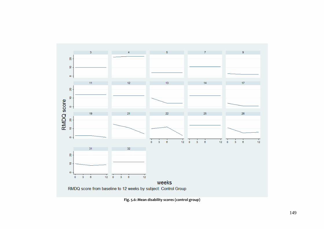
149
Fig. 5.6: Mean disability scores (control group)

150
Fig. 5.7: Mean disability scores (experimental group)

151
Fig. 5.8: Mean trunk sagittal acceleration (control group) without missing data

152
Fig. 5.9: Mean sagittal trunk acceleration (experimental group) without missing data

153
Fig. 5.10: Mean pain scores (control group) without missing data

154
Fig. 5.11: Mean pain scores (experimental group) without missing data

155
Fig. 5.12: Mean disability scores (control group) without missing data

156
Fig. 5.13: Mean disability scores (experimental group) without missing data

157
5.4 Data transformation
The data for the outcome measures of mean sagittal acceleration, pain and
disability did not demonstrate a normal distribution (Figs 5.2-5.4) and were
transformed using the logarithm for analysis. The data were back transformed to
provide meaningful results on the original scale. For a comparison of means this
gives the ratio of geometric means rather than the difference of arithmetic means.
5.5 Range of movement
The experimental group (m=3, f=13) with a mean age of 36.2 (9.8) years,
mean height of 166.8 (10.6) cms and mean weight of 75.9 (18.0) kgs demonstrated
greater range of movement during testing at every stage of evaluation including
baseline (Table 5.2) when compared to the control group (m=2,f=15) with a mean
age of 35.8 (9.1) years, mean height 167.4 (9.0) cms and mean weight of 73.3 (15.6)
kgs. The mean difference between the groups was greatest at 3 months even when
the difference between the groups narrowed halfway through the study. This was
demonstrated even when the mean range remained relatively constant within the
experimental group.
Table 5.2: Between group differences in range of movement (ROM)
Control Experimental % Difference
between groups Mean ROM at 0 weeks [(degs (SD)]
37.6 (11.2) [n=17] 41.3 (14.0) [n=16] 9.8
Mean ROM at 3 weeks [(degs (SD)]
34.7 (8.7) [n=12] 42.1 (18.3) [n= 13] 21.3
Mean ROM at 6 weeks [(degs (SD)]
38.3 (13.4) [n=9] 41.1 (16.8) [n=8] 7.3
Mean ROM at 3 months [(degs (SD)] 35.9 (10.2) [n=6] 41.9 (15.7) [n=5] 16.7
5.6 Trunk sagittal acceleration
The experimental group consistently demonstrated greater mean sagittal
acceleration than the control group throughout the study (Table 5.3). The

158
differences, however were not statistically significant at the start of the study (two-
sample t-test, t= -0.2, 31 d.f, p=0.83) 3 weeks (two-sample t-test-1.1, 31 d.f., p=0.28), 6
weeks (two-sample t-test, t= -0.5, 31 d.f., p=0.64) or at 3 months (two-sample t-test,
t=-0.9, 31d.f., p=0.41).
The control group had better trunk performance at the 3 week stage. At the
end of the 6 week active intervention period the control and experimental groups
demonstrated overall increases of 15.1 and 29.4% respectively which increased at the
3 month follow up evaluation to 13.0 and 37.0% respectively. A comparison of
sagittal acceleration at 3 months to week 6 shows that the control group
demonstrated a reduction in performance.
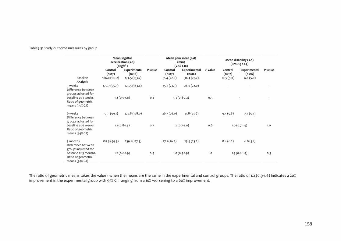
158
Table5.3: Study outcome measures by group
Mean sagittal
acceleration (s.d) (deg/s2 )
Mean pain score (s.d)
(mm) (VAS 1-10)
Mean disability (s.d)
(RMDQ 0-24)
Control Experimental P value Control Experimental P value Control Experimental P value (n=17) (n=16) (n=17) (n=16) (n=17) (n=16)
Baseline 166.0 (110.2) 174.5 (133.7) 31.4 (22.0) 36.4 (23.2) 10.5 (5.0) 8.6 (5.0) Analysis
3 weeks 170.7 (95.5) 225.5 (163.4) 25.3 (23.5) 26.0 (22.0) - - - Difference between groups adjusted for baseline at 3 weeks. Ratio of geometric means (95% C.I)
1.2 (0.9-1.6) 0.2 1.3 (0.8-2.2) 0.3 - -
6 weeks 191.1 (99.1) 225.8 (178.0) 26.7 (26.0) 31.8 (23.6) 9.4 (5.8) 7.4 (5.4) Difference between groups adjusted for baseline at 6 weeks. Ratio of geometric means (95% C.I)
1.1 (0.8-1.5) 0.7 1.2 (0.7-2.0) 0.6 1.0 (0.7-1.5) 1.0
3 months 187.5 (99.5) 239.1 (177.5) 27.1 (26.7) 25.9 (23.2) 8.4 (6.2) 6.8 (5.1) Difference between groups adjusted for baseline at 3 months. Ratio of geometric means (95% C.I)
1.2 (0.8-1.9) 0.9 1.0 (0.5-1.9) 1.0 1.3 (0.8-1.9) 0.3
The ratio of geometric means takes the value 1 when the means are the same in the experimental and control groups. The ratio of 1.2 (0.9-1.6) indicates a 20% improvement in the experimental group with 95% C.I ranging from a 10% worsening to a 60% improvement.

159
At 3 weeks the improvement in trunk sagittal acceleration in the
experimental group was 20% greater than in the control group but this was not
statistically significant (95% CI 0.9-1.6, p=0.2). Similar improvements of 10% and 20%
respectively were observed at 6 weeks (95% CI 0.8-1.5, P=0.7) and 3 months (95%CI
0.8-1.9, P= 0.9) but again these were not statistically significant (Table 5.3).
5.7 Pain
Mean pain scores were similar in both groups at each stage of the
study; the differences in mean pain scores between the groups adjusted for
baseline were not statistically significant at 3 weeks (30%) (95%CI 0.8-2.2, p= 0.3), 6
weeks (20%) (95%CI0.7-2.0, p=0.6) or 3 months (0%) (95%CI 0.5-1.9, p=1.0) (Table 5.3).
Analysis of VAS scores between 3 months and 6 weeks was not possible
because of the missing data and the small number of the sample.
5.8 Disability
The data suggests that the differences in disability scores between the
groups adjusted for baseline were also statistically insignificant at 6 weeks (0%)
(95%CI 0.7-1.5, p= 1.0) and 3 months (30%) (95%CI 0.8-1.9, p= 0.3) (Table 5.3).
5.9 Between variable relationships
Anecdotal clinical evidence suggests that there would be logical
relationships between the variables measured. However, the results do not
demonstrate this at any of the time points of the study (Table 5.4).
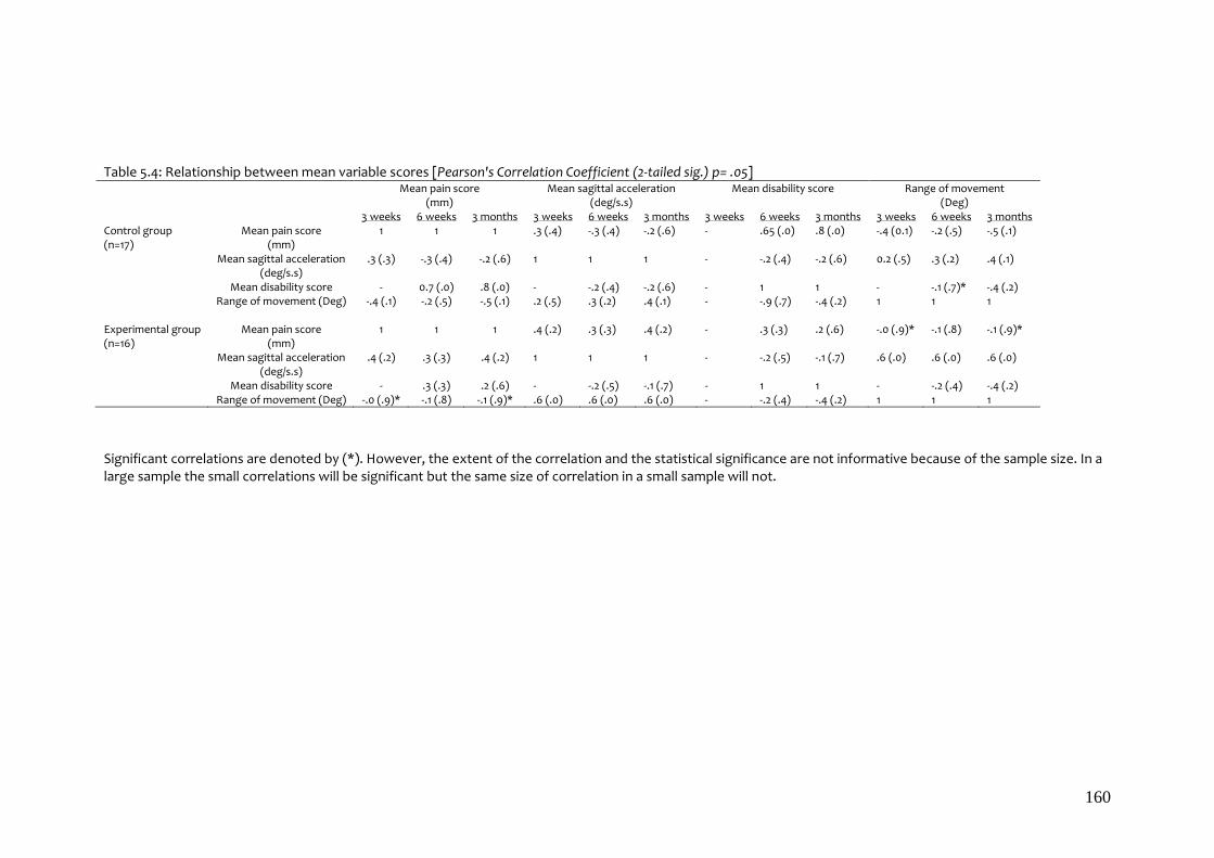
160
Table 5.4: Relationship between mean variable scores [Pearson's Correlation Coefficient (2-tailed sig.) p= .05] Mean pain score
(mm) Mean sagittal acceleration
(deg/s.s) Mean disability score Range of movement
(Deg) 3 weeks 6 weeks 3 months 3 weeks 6 weeks 3 months 3 weeks 6 weeks 3 months 3 weeks 6 weeks 3 months Control group (n=17)
Mean pain score (mm)
1 1 1 .3 (.4) -.3 (.4) -.2 (.6) - .65 (.0) .8 (.0) -.4 (0.1) -.2 (.5) -.5 (.1)
Mean sagittal acceleration (deg/s.s)
.3 (.3) -.3 (.4) -.2 (.6) 1 1 1 - -.2 (.4) -.2 (.6) 0.2 (.5) .3 (.2) .4 (.1)
Mean disability score - 0.7 (.0) .8 (.0) - -.2 (.4) -.2 (.6) - 1 1 - -.1 (.7)* -.4 (.2) Range of movement (Deg) -.4 (.1) -.2 (.5) -.5 (.1) .2 (.5) .3 (.2) .4 (.1) - -.9 (.7) -.4 (.2) 1 1 1 Experimental group (n=16)
Mean pain score (mm)
1 1 1 .4 (.2) .3 (.3) .4 (.2) - .3 (.3) .2 (.6) -.0 (.9)* -.1 (.8) -.1 (.9)*
Mean sagittal acceleration (deg/s.s)
.4 (.2) .3 (.3) .4 (.2) 1 1 1 - -.2 (.5) -.1 (.7) .6 (.0) .6 (.0) .6 (.0)
Mean disability score - .3 (.3) .2 (.6) - -.2 (.5) -.1 (.7) - 1 1 - -.2 (.4) -.4 (.2) Range of movement (Deg) -.0 (.9)* -.1 (.8) -.1 (.9)* .6 (.0) .6 (.0) .6 (.0) - -.2 (.4) -.4 (.2) 1 1 1
Significant correlations are denoted by (*). However, the extent of the correlation and the statistical significance are not informative because of the sample size. In a large sample the small correlations will be significant but the same size of correlation in a small sample will not.

161
5.10 Conclusions
The results do not decisively demonstrate that CSEs do improve trunk
performance, reduce disability and pain.
A comparison of the experimental and control groups showed that the mean
sagittal acceleration of the trunk was 20% greater at 3 weeks, 10% greater at 6 weeks
and 20% greater at 3 months but none of the differences were statistically
significant.
A comparison of the mean pain scores between the groups was
demonstrated to be 30% at 3 weeks, 20% at 6 weeks and 0% at 3 months but none of
the differences were statistically significant.
A comparison of the mean disability scores between the groups was
demonstrated to be 0% at 6 weeks and 30% at 3 months but none of the differences
were statistically significant.
Although the results do not provide exhaustive evidence of the effect of CSE
there does appear to be improvement which may become more significant with a
much larger study because the larger the sample size the greater the power of the
study and it will be more probable that significant differences between groups
could be demonstrated.

162
Chapter 6
Discussion
6.1 Introduction
This study set out to test the hypotheses that pain, disability and trunk
performance impaired by an onset of non-specific Low Back Pain (LBP) can be
improved by Core Stability Exercises (CSE). The hypotheses was tested using a
randomised controlled trial conducted on a sample of patients referred for
physiotherapy treatment at Musculoskeletal Physiotherapy Services, Hillingdon
Community Health following an acute onset of non-specific LBP.
This chapter affirms the originality of this study and contribution to the
existing knowledge. It discusses the results generated by the study and examines
the meaning of these results in the context of existing knowledge of trunk
behaviour as influenced by an onset of acute non-specific LBP. Furthermore, it
examines the implication of the results on possible management of acute non-
specific LBP in future clinical practice.
There were some important limitations to the design and conduct of the
study that meant that the researcher could not be blinded and it was not possible to
recruit a very large sample. The chapter will explore the implications of this design
in view of these constraints and discuss the consequences of the design on the
results.
Finally the chapter will discuss the limitations within the study and highlight
the direction in which supplementary work can be done to augment the conclusions
of this study.

163
6.2 Originality of the study
Although the effects (Wong and Deyo, 2001; Pahl et al., 2006) and costs
(BackCare, 2007; Dagenais et al., 2008) of LBP are well documented effective
control of LBP remains elusive. One of the causes of this is the inability to quantify
real time changes in trunk performance and relate those changes to subjective
measures of the effects of an onset of LBP.
Physiotherapy practitioners like osteopathy and chiropractic practitioners
have relied upon manipulation/mobilisation to rectify aberrant lumbar segment
movement patterns suggestive as being the cause of an onset of LBP. The
effectiveness of these techniques has been compared to other non-manual
interventions and found to be effective only for specific stages of LBP (Assendelft
et al., 2003; Haas et al., 2004; Ernst, 2007). The effectiveness of non-specific
exercises has also been compared to specific exercises and it is suggested that
there is no difference (May and Johnson, 2008). All the studies have relied upon
subjective measures as either a primary or significant outcome upon which the
results have been interpreted. This study used a Lumbar Motion Monitor (LMM) to
demonstrate objective real time measures of trunk performance.
The validity and reliability of the LMM, however, had only been established
using a method that was impractical for clinical use and although a revised
streamlined method had been shown to be reliable, the agreement for the
measurement used to demonstrate its reliability had never been demonstrated. The
level of agreement was explored within this study and a subsequent paper was
published (Aluko et al., 2011) (Appendix 4). This was important because it provided

164
evidence of reliability allowing this measure to be used more confidently for this
study and by future researchers and clinicians.
No previous evidence was available to interpret trunk behaviour within a
sample population. Within this study trunk behaviour of 50 volunteers was
quantified and evaluated. A paper demonstrating these findings has also been
submitted for publication (Appendix 18). The findings of this study (Appendix 18)
suggest that a self reported episode of LBP may not be a reliable indicator of
changes in real time measures of trunk performance. Changes in trunk performance
due to LBP may also not be cumulative. Furthermore females within the sample
demonstrated slower trunk performance. These findings are used to discuss the
results of this study.
Although CSEs have become a popular intervention for the treatment of LBP
(Willardson, 2007a; Willardson, 2007b) no previous research has explored the
mechanism by which they may work. This study was designed with the purpose of
doing just that by comparing the changes in subjective measures with an objective
measure and investigating if a correlation exists. No existing published research has
as yet explored a relationship between self reported measures of either pain or
disability with measures of trunk kinematics. This work has attempted to do this by
using the Lumbar Motion Monitor.
In order to ensure the effectiveness of the CSE all the participants in the
experimental group completed a diary to demonstrate their compliance (Appendix
17). The data suggested that compliance was highest in the early stages of the
study. This could account for the observed rapid mean changes within this group.
Furthermore it demonstrated that compliance is essential for the true benefits of

165
CSE to be seen. This observation may be a key element for why previous
comparative intervention studies for the management of LBP have not been able to
demonstrate significant differences.
6.3 Contribution to existing knowledge
This study demonstrated a 20% difference in mean trunk sagittal acceleration
at 3 weeks, 10% at 6 weeks and 20% at 3 months between the experimental and the
control groups which was not statistically significant. This study therefore did not
conclusively suggest that CSE can improve trunk performance. Similarly the
difference in the reduction in pain between the two groups was a statistically
insignificant 30% at 3 weeks, 20% at 6 weeks and 0% at 3 months. The difference in
the mean disability scores between the 2 groups of 0% at 6 weeks and 30% at 3
months was equally statistically insignificant. The lack of statistical significance may
in part be due to the small sample size and is suggestive of the need to conduct this
study on a much bigger sample. However, the improvement demonstrated within
these outcomes suggests an improved clinical response to an onset of acute non-
specific LBP may be achieved if CSE are commenced quickly within an episode and
may result in a reduction in the likelihood of an acute onset becoming chronic. It is
proposed that a direct impact of such improvement to patients may be increased
empowerment, quicker resolution of symptoms and improvement in well being.
The advantage to the economy at large may therefore be a reduction in the cost of
treatment and therefore a reduction in the burden of LBP on healthcare delivery.
This study provides evidence to suggest a new method to quantify the
effects of an acute onset of non-specific LBP using the Lumbar Motion Monitor to
evaluate trunk sagittal acceleration. A mechanism by which CSEs work may be by

166
improving trunk sagittal acceleration. The results of this study therefore suggest
that the NICE guideline of encouraging activity and exercise as soon as possible
(NICE, 2009) have credence and adds to the evidence-base of practice.
6.4 Results
Trunk displacement demonstrated by participants during data collection
improved within both groups of participants. However, the amount of increase was
more evident in participants in the experimental group. Although the range of trunk
movement during an episode of LBP can be subjective (Cox et al., 2000) the results
lend some support to the hypothesis that LBP produces less movement (McGregor
and Hughes, 2000). However, the increases were not statistically significant and
could be attributed to chance alone. The experimental group may have
demonstrated greater increases because of specificity of the intervention compared
to the control group.
Although trunk displacement during an episode of LBP can be subjective, the
results of this study do suggest that this may only be applicable where a specific
range is anticipated for use during trunk kinematic evaluation and therefore
becomes an emphasis on the sincerity of effort (Ferguson et al., 2000). This study
did not have specific start or end points for trunk movement during the LMM
evaluation, participants were therefore not able to regulate movement in
anticipation of these points and thus influence their effort to complete the task.
Clinical practice involves emphasis upon the range of movement attainable within
the patient’s tolerance during an assessment. The method used in this study
attempted to replicate this. The participants were encouraged to focus on the
number of repetitions that could be performed during the data collecting period of

167
8 seconds without a fixed starting or end point. This approach gave each participant
control and ownership of the test, consequently, the range of movement
demonstrated was less likely to be manipulated. They were also informed that the
study involved an assessment of the quality of movement rather than range. The
range was therefore more likely to represent the capability of the participants at
the time of testing.
6.4.1 Trunk sagittal acceleration
There is a paucity of literature investigating trunk acceleration. There are
however studies that evaluate trunk velocity (Marras et al., 2000; Cox et al., 2000;
McGregor and Hughes, 2000) only one study specifically suggests that acceleration
may be sensitive to sincere conditions (trunk movement profiles reflecting its
musculoskeletal status) at the point where the participant changes direction
(Marras et al., 2000). The work of Marras et al has over the years investigated the
quantification of 3-dimensional trunk movement. The work (Marras et al., 2000)
demonstrated that an acceleration profile may be highly repeatable for sincere
conditions. It has also been demonstrated that kinematic functional performance
measures are sensitive to improvements during recovery from LBP (Ferguson et al.,
2000). The results of this study demonstrate improvement in trunk performance
defined as trunk sagittal acceleration, in participants in both the control and
experimental groups. Although it is possible that some of this improvement could
be due to the natural course of recovery the experimental group did show greater
improvement in trunk performance at the end of the active 6 week period of the
study. The improvement continued at the 12 week follow up within this group. The
improvement at 6 weeks in the control group however was not maintained as this

168
group demonstrated a reduction at the 12 week follow up. These findings are
consistent with the findings of those previous studies (Marras et al., 2000; Ferguson
et al., 2000). It may therefore be possible to suggest that CSE can improve and
maintain trunk performance more efficiently than the natural course of recovery
following an onset of acute LBP. This however, has to be put in context of the
statistically insignificant difference between the groups.
The pilot study suggested that acute non-specific LBP may reduce trunk
performance; which cannot be improved by repetition (Aluko et al., 2011). This
supports a previous finding (Marras et al., 2000) however, this may only be
reproduced in a clinical environment if the participant actually has an episode of
LBP. A subjective opinion for the presence or absence of LBP may not be strictly
reliable (Appendix 18).
The clinical assessment of trunk movement is not functional but rather a
global unidirectional anatomical movement (Petty, 2006). Most episodes of LBP
occur whilst performing movements involving complex tasks requiring a wide array
of muscle recruitment patterns (McGill et al., 2003; McGreary et al., 2003) however,
the data for this study was obtained using a global unidirectional anatomical
movement which was adapted to increase the level of difficulty by using repetitions
to create instability within the movement segments of the lumbar spine (Reeves et
al., 2007).
The results of the study do support the suggestion that improved trunk
muscle activity and co-contraction increases trunk acceleration (Granata and
England, 2006). The mean sagittal acceleration in the experimental group was
consistently greater than in the control group. The difference between the groups
169
at key milestones (3, 6 and 12 weeks) was also not statistically significant. However,
the insignificance could be attributed to the sample size. The sample size used in
this study was similar to that used in a previous study that used a sample size of 33
to investigate the effect of stabilisation instruction on lumbar acceleration using a
uniaxial accelerometer (Webber and Kriellaars, 2004). They were able to
demonstrate statistically significant reduction in lumbar acceleration induced by
limb movement (Webber and Kriellaars, 2004). It is possible that the difference in
results was because the Webber and Kriellaars’ study was observing the effect of
limb movement rather than adjacent lumbar musculature on trunk acceleration.
The reasons for the insignificant differences in the pain and disability scores
demonstrated in this study may also be because an assumption was made that all
reported scoring was sincere. All participants, however, had been assessed and
considered to have a true episode of LBP prior to agreement to take part in the
study. If there were any doubt about the sincerity of the episode, it would be
expected that this would be reflected in the rate of attrition. Insincerity of the
presence of LBP would be expected to be proportional to the willingness to
participate in the study. Attrition, however, was similar within both groups (chapter
5) suggesting that the likelihood of this having affected the results was very low. It
is, however possible that the initial referral for treatment by the general practitioner
was based more on subjective assessment of the severity of disability and/or pain.
This assumption is based upon previous suggestions that disability is reliant upon
the level of distress (Proctor et al. (2000) cited in Johnstone et al. (2004)). This may
therefore have had an influence on the urgency and timing of their referral from

170
their GP and ensuing prevalent belief systems (Coudeyre et al., 2006; Cayea et al.,
2006; Fullen et al., 2008).
Both groups demonstrated increases in mean acceleration at the end of the
study. This is in agreement with previous findings (Marras et al., 2000; Aluko et al.,
2011) and agrees with the suggestion that LBP causes segmental hypomobility
(Teyhen et al., 2007). However, the increase in acceleration was much greater
within the experimental group at 12 weeks. This does suggest that CSE may not only
improve trunk acceleration but may also help in maintaining any increase. The
reduction in mean acceleration within the control group between 6 and 12 weeks
following the end of the active period of the study further strengthens this
argument.
Although there is very little evidence to suggest that LBP affects a particular
gender more than the other (Walsh et al., 1992; Fritz and George, 2002), this
hypothesis could not be tested because of the small number of male participants
recruited. However, previous results have already shown that females may
consistently demonstrate slower trunk performance (Appendix 18). The reason for
the small number of male participants is not clear. Significance in weight and height
was not demonstrated in this study. The possibility exists that pelvic pathologies
(Frank and DeSouza, 2001) may have a significant affect on general trunk
performance within females but it has not yet been demonstrated how these
pathologies impact on trunk movement.
An increase in displacement has been reported to be accompanied by an
increase in velocity (Cox et al., 2000) and thus by default an increase in acceleration.
This trend is not demonstrated by the results of this study. Although both groups

171
demonstrate an increase in mean acceleration over time, mean displacement does
not follow the same trend. There was a negative correlation within the control
group between the start of the study and 12 weeks. An exception was between 6
and 12 weeks when the mean acceleration actually decreased within the control
group. A possible explanation for this negative correlation could be due to the fact
that the previous study (Cox et al., 2000) was retrospective and relied upon patients
referred from insurance companies and who may have had other reasons for being
motivated to demonstrate good functional movement during the assessment
irrespective of the discomfort they were experiencing. Participants in this study
were not under the same pressure, fear avoidance (Fritz and George, 2002)
however may have inadvertently been reinforced by the encouragement
participants were given to concentrate on the number of repetitions rather than the
range. This may have given the impression that the range of movement required to
perform the test would not aggravate their symptoms. This fact was repeatedly
given in the patient information sheet (Appendix 12) and at each point of contact
during which data was obtained. The experimental group participants may not have
responded this way because they reported a lower mean disability score at the start
of the study.
Mean trunk acceleration changed at different stages of the study. The
control and experimental groups demonstrated improvements of 2.8% and 29.2%
respectively at 3 weeks, 15.1% and 29.4% respectively at 6 weeks and 13% and 37%
respectively at 12 weeks. This does suggest that the affects of CSE on sagittal trunk
acceleration may be rapid. Previous studies have suggested that specific exercises
for the treatment of acute LBP are not significantly better than any other

172
intervention (May and Johnson, 2008). The results of this study do not change this
view.
The results of this study suggest that the significance of the effects of CSE in
the early stages following an onset of acute LBP may be underestimated because
trunk muscle activity and co-contraction has been shown to increase with trunk
velocity and acceleration (Granata and England, 2006). The Multifidus (MF) and
Transversus Abdominis (TrA) are accredited with the facilitation of this mechanism
(Bergmark, 1989). The rapid response demonstrated within the experimental group
suggests that there is a rapid increased ability for the trunk to respond to
perturbation by increasing trunk sensitivity to positional change (Reeves et al.,
2007). Trunk stiffness reported to be a result of LBP (Owens et al., 2007) is more of
a structural response to pain and therefore supports the supposition that LBP
produces slower trunk movements (Marras et al., 1999). The ability to accelerate
quickly to change in trunk sagittal movement is demonstrated by the results of the
pilot study that preceded this study (Aluko et al., 2011). This finding fits well with
earlier findings that suggest that the MF and TrA are affected very quickly following
an onset of LBP (Hodges and Richardson, 1996; Hodges and Richardson, 1997) and
improvement in cross sectional area in the MF can be observed within 6 weeks
during of a course of CSE (Sokunbi et al., 2008). The strength of a healthy muscle is
directly proportional to its cross sectional area (Jones and Round, 1990) improving
the cross sectional area of the MF and TrA through targeted exercise may therefore
be beneficial.
Improvement in muscle recruitment and coordination which accompanies
increases in trunk acceleration is supported by the findings of Pollock et al. (2009)
173
that suggest that to avoid excessive loading of the lower lumbar segments, these
segments rapidly move into extension during the stroke phase of rowing. The
demonstrated improvement in trunk sagittal acceleration suggests that this
mechanism is critical for normal trunk function when the lower lumbar segments
are incrementally loaded during flexion.
The increase in trunk sagittal acceleration may also increase the range of
lordosis (ROL) because the range of lordosis should be directly proportional to the
changes in acceleration (Cox et al., 2000). It has been shown that lumbar lordosis
alters according to posture (Knutson, 2002; Al-Eisa et al., 2006). This would not be
true if there was structural trunk stiffness caused by underlying natural pathology
such as natural degenerative change (Gruber et al., 2007; Standaert et al., 2008). An
increase in ROL during loading facilitates trunk efficiency during any task involving
lifting, carrying or pushing/pulling. This is pertinent because pushing/pulling
activities are a main cause of LBP associated with lumbar disc involvement (Plouvier
et al., 2008; Marras et al., 2009). Anterior-posterior (AP) shear force across the
lumbar spine is increased by an increase in speed of activity and during pushing
activities the AP shear force is in the opposite direction to the push and occurs
mainly at L5 (Marras et al., 2009).
The inability for the control group to demonstrate a similar trend may be
because the muscle recruitment and coordination mechanism where not engaged
by core stability exercise training. The net result of this non-engagement may be the
reason why any gains achieved within the first 3 weeks of the study is lost at the 6
week stage. The net increase in trunk sagittal acceleration demonstrated at the 3
month stage may be attributable to the participant returning to normal daily

174
activity. A follow up period of 18-24 months may provide significant evidence of any
correlation between any re-occurrence of LBP and the continued absence of this
muscle recruitment and coordination mechanism. It has been suggested that
reoccurrence is linked to stress, anxiety, mood, cognitive function and pain
behaviour (Johnstone et al., 2004) however, it is highly suggestive that a
precipitating factor for the onset of these risk factors is the underlying inability for
the trunk to adequately compensate for the effects of axial loading on the trunk
with consequent frequent and/or periodic episodes of LBP.
The use of a belt has been shown to reduce trunk velocity (Giorcelli et al.,
2001) and because acceleration is a factor of velocity it is possible to assume that it
will also be reduced. The action of a belt is similar to the action of the global trunk
muscles. It is not clear how such a support will influence trunk acceleration in the
long term. An increase in the perception of support may increase trunk robustness
rather than stiffness in the immediate term but the cumulative effect must become
evident when the belt is removed. The use of an artificial support may interfere with
the natural muscle coordination required to reduce the natural perception of
instability (Hodges et al., 2003; Moseley et al., 2004) or indeed the suggested chaos
within the trunk during movement (Granata and England, 2006).
6.4.2 Pain
Although there was no demonstrable statistical difference in pain
score between the groups, within the first 3 weeks of the study the experimental
group demonstrated a larger mean reduction in reported pain at 3 and 12 weeks.
The difference in pain however can be considered to be clinically significant (Kelly,
2001). The experimental group achieved the greatest reduction within the first 3

175
weeks compared to the control group that demonstrated a slight increase in pain
between the end of the study and the follow up at 12 weeks. This result parallels the
behaviour of trunk acceleration suggesting that most of the change occurred within
the first 3 weeks. However, the study has not demonstrated that there is a causal
relationship between the changes in trunk acceleration and pain although a
negative correlation between pain and trunk performance has been previously
reported (Cox et al., 2000).
It has been suggested that the pain adaptation model reduces trunk velocity
(van Tulder et al., 2000). The findings of this study therefore suggest that non-
specific LBP may induce the pain-spasm-pain model (van Dieen et al., 2003) rather
than the pain adaptation model because the results of this study demonstrate an
increase in acceleration accompanied by a reduction in pain (van Dieen et al., 2003).
Clinically this could be very important because treatment/intervention for
the pain experienced by an onset of non-specific LBP could be more effective if it is
aimed at reducing the excitability of the α-neurons or reducing muscle spindle
activity (Johansson and Sojka, 1991). It is possible that CSE act favourably in
enhancing both of these activities of α-neurons or reducing muscle spindle activity.
If the TrA and MF are actively involved in stability and respond to trunk
perturbation, they will need a high proportion of both fast and slow twitch motor
units. They will also require low activation thresholds to facilitate an ease of
response to pre-empt trunk perturbation (Barr et al., 2005) and be able to produce
prolonged contractions with a relatively low force with low fatigue resistance
(Watkins, 1999). The results of this study do suggest that the behaviour of the MF in
particular in response to CSE may fit this assumption. The findings of the pilot study

176
(Chapter 3) demonstrate the possibility that trunk acceleration will increase as
recovery develops (Aluko et al., 2011) and these muscles regain normal function.
Previous evidence suggests that the deep MF have a high proportion of type 1 fibres
compared to the superficial MF (Richardson et al., 1999; MacDonald et al., 2006). It
could therefore be speculated that the superficial and deep fibres of the MF have,
apart from their similar functions, quite specific functions pertaining to the load
placed upon them by trunk displacement. This speculation may be supported by
previous work that does suggest that although both the deep MF and the
superficial MF produces trunk torque, the deep MF may be primarily involved in
generating compressive forces with minimal associated torque (MacDonald et al.,
2006). It may be that the CSE routine facilitates this ability to generate the torque
efficiently without compromising the structure of the spine.
The results do not demonstrate differences within gender, age, weight or
height or correlate any changes with subjective pain. But the difference in
structural build between the genders may play a significant role in trunk sagittal
acceleration in the fact that females may be more likely to report an episode of
acute non-specific LBP (Aluko et al. in preparation) (Appendix 18) than males and
because males are more likely to have more muscle bulk because of the higher
levels of testosterone (Jones and Round, 1990). The larger muscle size may suggest
that males may also have higher thresholds at which the α-neurons are excited or
have a greater ability to reduce muscle spindle activity. This assumption does
require further enquiry. Exercises designed to increase muscle bulk may not be
effective because previous work has suggested that the increase in strength during
muscle training is disproportionate to the increase in its cross-sectional area (CSA)

177
(Jones and Round, 1990; Hansen et al. 2006). Exercises used in physiotherapy are
therapeutic and may include an aim of restoring muscle coordination. The TrA and
MF, may be affected by an increase in fatty tissue content and/or reduction in trunk
intra-abdominal pressure (IAP). The efficiency of the TrA and MF may be reduced in
either circumstance; furthermore anecdotal evidence suggests that low back pain is
often associated with obesity or Body Mass Index (BMI) and it is often referred to
by general practitioners in referrals of patients to physiotherapy reporting with an
acute episode of low back pain and obesity is recognised to have a negative effect
in reducing LBP induced disability (Yildirim et al., 2007). This may change in the near
future because it is now suggested that a better barometer for obesity could be
trunk circumference rather than the Body Mass Index (Lean et al., 1995).
The lack of statistical power for the study precipitated the absence of a
demonstrable relationship between weight and pain. However, it could be
suggested that there ought to be a direct correlation between anthropometric
measures and trunk performance because there is an established correlation
between poor IAP and moment arm of the trunk during movement (Janda and
Valenta, 2000). It is believed that IAP is proportional to anthropometric measures
(Youdas et al., 2000). An increase in waist circumference is expected to decrease
lumbar lordosis and therefore influence adaptive lengthening of the lumbar MF and
its subsequent loss of efficiency. Weak MF may reduce the ability to maintain a
natural lumbar lordosis when the spine experiences axial loading (chapter 1). The
importance of this mechanism may be linked to the early changes observed within
the MF during an onset of LBP (Hodges and Richardson, 1996; Hodges and
Richardson, 1997). The TrA by virtue of its anatomical location may remain relatively
178
unaffected unless there is an increase in waist circumference. This is probably why
CSE is thought to be effective because it aims at restoring ‘normal’ activation of the
MF (Hodges and Richardson, 1996; Hodges and Richardson 1997). All the core
stability exercises used in this study included activity designed to increase and
maintain tension within the MF during the exercise routine by instructing the
participant to ‘draw the navel towards the spine and hold’ during each activity
(Appendices 4 and 5).
Bed rest not exceeding 4 days has been a recommendation as a first
treatment intervention for an acute onset of non-specific LBP (Rozenberg et al.,
2003). The reduction in pain scores demonstrated during the study suggests that
CSE may be a more effective alternative. Unlike bed rest, CSE may help maintain a
normal level of activity as much as possible and therefore reduce days of sick leave
(Smith et al., 2002; Rozenberg et al., 2003; Kinkade, 2007). However, requesting a
patient who presents with an acute onset of non-specific LBP to commence a
regime of CSE may not be appreciated because even the request to resume normal
activity reduces patient satisfaction (Atlas and Deyo, 2001). The use of CSE,
however, may be very useful to prevent catastrophisation of the condition and
possible associated bouts of depression (Johnstone et al., 2004), facilitate fear
avoidance techniques (Fritz and George, 2002) and ferment a positive approach to
treatment (Frank and DeSouza, 2001). Unnecessary tertiary referrals could be
minimised (Lauchlan, 2005) and it could be speculated that in doing so it is possible
to reduce the cost of healthcare delivery.
Within chapter 3, a sample of the participants were asked if they had pain at
the time of testing, episodes of LBP and when the last episode was experienced
179
(Appendix 5). The results suggest that any correlation between self reported pain
and the participant responses may not be reliable (Appendix 18). The pain reported
by the high proportion of females in that part of this study may be associated with
pelvic pathology that is difficult to exclude by history alone (Frank and DeSouza,
2001). It may therefore be difficult for example to differentiate between LBP
associated with the female pelvic pathology and pain associated with real time
structural/mechanical dysfunction. Recall bias (Chouinard and Walter, 1995) may
also be problematic because invariably most sufferers of LBP may have difficulty
describing pain that occurred in the past in the context of an active subjective
history interview. Asking patients to describe their pain on the day does not
adequately describe the fluctuation in intensity that may occur between
assessments. These fluctuations may have had an impact on the exercise
compliance demonstrated by the reduction during the second half of the study
(Appendices 15).
6.4.3 Disability
The results suggest that the trigger for the pain-spasm-pain model to be
activated may be a structural change capable of reducing trunk sagittal acceleration
with reduced TrA and MF efficiency. However, it is recognised that the sensitivity of
the RMDQ in quantifying disability may be not be as high because the participants
within this study may not be ‘back pain disabled’ because they did not have the
pain long enough to decide that certain movements caused pain unlike individuals
with chronic LBP (Beurskens et al., 1996).
The difference in disability between the groups was not only statistically
insignificant but also clinically insignificant because the difference was less than 30%

180
(Jordan et al., 2006). However, the participants within the control group appeared
to maintain a higher mean disability scores than the participants in the experimental
group throughout the study. This does suggest that CSE may facilitate a reduction in
disability however the depth of the facilitation is not effectively demonstrated.
The result in isolation does suggest that CSE may improve disability,
however, a possible reason for the insignificant result may be that although all
participants were asked to answer the RMDQ on the basis of their pain on the day,
participants were keen to express their opinion of the pain for the duration
between testing believing that their representation of disability on the day did not
reflect what they perceived to be their true experience. Recall bias (Chouinard and
Walter, 1995) would therefore become an issue as they may not have been able to
accurately quantify their experience. This possibility is enhanced by the fact that all
participants enquired at every visit and on more than one occasion during each
testing period if they were supposed to report how their pain was in real time or
how it had been. They sometimes emphasised the point by trying to suggest that
their disability had been much worse than at the time of measurement.
The results do support previous findings that stabilisation exercises can be
used effectively to effect changes in disability; the magnitude of which has been
used to develop a clinical prediction rule to determine which patients are most likely
to respond following an onset of LBP (Hicks et al., 2005).
The trend of the result is not in keeping with two previous studies that
demonstrated significant improvement in disability after intervention at 6 weeks
(50%) and at 3 (67%) and 12 month (56%) follow up periods (Rasmussen-Barr et al.,
2003b). The other study demonstrated a 43.2% improvement after 4 weeks

181
(Brennan et al., 2006). It may be that this study was more rigorous than the
aforementioned studies but this has to be put in context of the smaller sample size.
However, both studies (Rasmussen-Barr et al., 2003b; Brennan et al., 2006) used the
Oswestry disability Index (ODI), the results of which may have been skewed
because of its low internal consistency (The Chartered Society of Physiotherapy,
2004). The significance of the results may therefore be lower than that reported.
The results also differ from the results of Childs et al. (2004) which suggests
that like for like on the clinical prediction rule for the treatment of LBP comparing
manipulation and lumbar strengthening exercises, manipulation is better than
exercise (Childs et al., 2004). Their observation was made using a modified ODI. The
discrepancy may be because the exercises used in the study were not specific
enough to engage either the TrA or MF either in isolation or together, to effect a
change in trunk kinematics. There is, however, a view that suggests that it should
not matter because any spinal exercise is a stability exercise (McGill et al., 2003) but
the effect of LBP on trunk sagittal acceleration may suggest otherwise (Marras et
al., 2000; Aluko et al., 2011).
The missing data had an effect on any within and between group analyses by
gender, weight and height. It was therefore not possible to draw any inference
from the results. However, it would be expected that there should be a trend given
the difference in body shape and muscle size with respect to gender and age (Jones
and Round, 1990). It has already been suggested that obesity has a negative effect
reducing LBP induced disability (Yildirim et al., 2007).
There were an equal number of dropouts excluding the pre-randomised drop
out (control 11, experimental 11) by the end of the study (Fig. 5.1). However, one

182
participant dropped out because another unrelated problem required orthopaedic
intervention and the view was taken that further participation might aggravate the
problem requiring surgery. Two other participants could not commit to the time to
be continually involved in the study as their personal circumstances changed and
the remainder did not offer any explanation other than that a decision had been
made to terminate their involvement in the study. Further qualitative analyses may
provide more insight into the underlying reasons for the attrition which may be
valuable for clinical purposes in understanding compliance with acute LBP home
exercise programmes or attendance for treatment. However, a future study may
benefit from recognising the complex lifestyles participants may have and the need
to factor these complexities into the development of the method stage of the
study. Funding may also allow better flexibility for data collection that could suit the
participants better.
6.4.4 Variable relationships
6.4.4.1 Start of the study to 3 weeks
The increase in range of movement demonstrated was to be expected
however, the sincerity of effort can be in doubt. Albeit, the results do suggest that
during the first 3 weeks of the study all activity did produce an increase in
discomfort within both groups. This was not unexpected because the participants’
behaviour was changed from that of fear-avoidance when patients avoid activity
because of the fear of exacerbating pain, to one of activity. However, the study may
suggest that the CSE was less aggressive because less pain was provoked. The
experimental group were able to demonstrate the most improvement in mean pain
scores within this short period of time. It is plausible that the CSE were specific
183
enough to isolate both movement at lumbar segment level and restrict its affect to
the immediate structures i.e the MF, that supports those segments being mobilised.
The finding that as movement increased there was a demonstrable increase in trunk
sagittal acceleration when the activity reduced spasm within the MF to facilitate
trunk flexion may support this view.
During the first 3 weeks of the study it was not possible from the results to
suggest that there was a difference between genders because of the small number
of male participants. However, it has already been proposed that women of
different age and racial groups demonstrate different amounts of trunk movement
(Trudelle-Jackson et al., 2010). The results do not suggest an inference about male
participants but there may be a hint of a trend that within both groups male
participant trunk range of movement was less influenced by CSE. This was equally
true for the older and the heavier male participants within both groups. This would
therefore suggest that in the early stages of CSE female patients may experience a
far more noticeable improvement in trunk flexibility than a similar male cohort.
Conversely, heavier females may have a slower response to CSE than their slimmer
counterparts. However, the effect of hormones on range of movement within the
female participants is not known (Kerr and Grahame, 2003) and variations in the
menstrual cycle within these participants may be expected. An earlier study in the
United States of America has found an increase in headaches with the onset of a
menstrual cycle in women (Johannes et al., 1995) and a similar study investigating
the effects of oestrogen levels on temporomandibular pain also found that this type
of pain in women increased when there were low levels of oestrogen (LeResche et
al., 2003). This may indicate that the effect of hormonal changes on reported pain

184
and disability resulting from an acute onset of non-specific LBP remains unclear and
is worthy of further investigation. Female participants in this study were not asked
about the timing of their menstrual cycle or the stage at which they felt they were
at within it.
There was no correlation between changes in trunk sagittal acceleration and
any of the other variables at this stage of the study. However, this does suggest
that weight loss may not influence the effect of CSE. The implication of which is that
asking a patient to lose weight as a pre-requisite for treatment of an onset of LBP
using CSE may not be valid or even possible.
6.4.4.2 3 weeks to 6 weeks
During this period, the increase in range of movement demonstrated was
associated with an increase in both pain and trunk sagittal acceleration. During this
phase of the study improvements in disability are expected (Sokunbi et al., 2008).
The reduction in disability demonstrated by the end of the 6 week period (Table
5.10) appears to support this suggestion. It is possible that the reduction in disability
may be a direct consequence of an increase in both the range of movement and
trunk acceleration.
This mechanism is facilitated by improvement in trunk robustness (Reeves et
al., 2007) as the ability of the trunk to change direction in response to perturbation
improves. A CSE programme may facilitate this mechanism by improving fine motor
control to enhance trunk functional stability by reducing its state of chaos (Marras
and Mirka 1993; Dolan and Adams, 1993; Granata and England, 2006) as can be
demonstrated by a reduction in the trunk Lyapunov exponent (ʎ MAX) (Granata and
England, 2006). This is all the more important because the study involved a

185
symmetrical movement of flexion-extension, a direction within which trunk
instability is greater than if it was performed with a degree of rotation (Granata and
England, 2006). The possible improvement in stability demonstrated by this study is
therefore of high clinical importance.
The increase in pain demonstrated during this period may be as a result of an
exacerbation of the ‘trigger’ of the pain-spasm-pain model (van Dieen et al., 2003)
induced by the increase in range of movement and the ensuing increased inter-
segmental movement. Although movement and sagittal acceleration improved
there is no evidence to suggest that structural dysfunction had been reversed at
this stage. This finding is in keeping with previous findings to suggest that
performance and pain have a negative correlation (Cox et al., 2000). The structural
dysfunction may include facet joints, Intervertebral discs (IVD) or ligament damage
caused by abnormal force, strain and stress applied to the structures (Atlas and
Deyo, 2001). It is not clear what the effect of the timescale for the repair of these
structures has on trunk performance and it remains a possible direction for future
research.
6.4.4.3 6 weeks to 12 weeks
This period is a relatively inactive period of the study. All intervention had
ceased during this phase and it was assumed that all the participants continued with
their normal active routine similar to what it was prior to the onset of their episode
of pain. All participants had been discharged by their treating physiotherapist.
However, the rate of attrition during this period made it impossible to draw
inferences from the data. However, it could be that the rate of attrition was directly
related to the participants’ wellbeing. The premise for this assumption was that
186
none of the participants (to the researcher’s knowledge) reported any reoccurrence
or returned to their treating physiotherapist requesting further advice or treatment.
However, it could be that the timescale of 3 months was too narrow to assess
medium term changes following the cessation of an intervention. Previous studies
had a range of follow up periods of 6 months (Childs et al., 2004) to a year
(Rasmussen-Barr et al., 2003b; Brennan et al., 2006). The original protocol for this
study did have a one year follow up period but enforced changes meant that a 3
month follow up was more realistic given the constraints of the study. Although it
has been demonstrated that the majority of the change had occurred within the
first 3 weeks of the study and the difference between participant groups
maintained at 6 weeks, the long term effects of the intervention may be best
judged on trunk performance after a year. However, a control over attrition would
need to be improved to ensure that the sample remains truly representative of the
participants of the study by maintaining strict inclusion criteria.
6.5 Key findings
From the above discussion the following key findings of this study were
deduced;
Trunk sagittal acceleration may be sensitive to an onset of acute non-
specific LBP.
The effects of CSE on the observed changes within 3 weeks of
intervention are not conclusive. However, the possible clinical
importance of CSE cannot be ignored and requires further
investigation.

187
The increase in trunk sagittal acceleration and thus its performance
was not demonstrated conclusively shown to be as a result of CSE.
The reduction in disability caused by acute non-specific LBP observed
was not conclusively shown to be as a result of CSE.
The reduction in pain induced by trunk dysfunction observed was not
conclusively shown to be as a result of CSE.
There may be a positive correlation between increased trunk sagittal
acceleration and the range within which the trunk is displaced.
Within the process of restoration of trunk function following an onset
of non-specific LBP, there is a phase within which there may be a
negative correlation between the improvement in trunk range of
movement and sagittal acceleration and subjective reported pain.
These findings do not in general agree with the suggestion that CSE may not
be of benefit to acute LBP patients (Atlas and Deyo, 2001). They do however
suggest that they may encourage normal activity as quickly as possible (Smith et al.,
2002; Rozenberg et al., 2003; Kinkade, 2007). The study is inconclusive in suggesting
that CSE affects trunk performance and thus it remains to be proved if they are
effective in preventing acute non-specific LBP from becoming chronic thereby
reducing tertiary referrals that may be either unnecessary or expensive (Lauchlan,
2005).
6.6 Mechanism by which CSE may work
Core Stability Exercise may be beneficial for people seeking physiotherapy
for an episode of acute non-specific low back pain. The literature review and
ensuing discussion of the research results provides an understanding of a possible

188
mechanism by which acute non-specific LBP is commenced, the effect it has on
trunk performance and trunk response to early intervention using CSE.
Before the study commenced there was a difficult debate between the
researcher, peers and colleagues independent of the study as to the type of
movement that occurs within a lumbar segment. The debate was complicated by
the fact that the movement in question involved relatively small displacements
based on whether the movement is linear or angular in nature. It was finally
concluded that the movement has angular characteristics because the movement
was more about deformation of the intevertebral discs, gliding/sliding of the facet
joints and the tilting of the vertebral body about an axis (Fig 2.4).
Non-specific acute non-specific low back pain as a symptom is elicited by a
structural dysfunction (Atlas and Deyo, 2001). The stress and strain that precipitates
dysfunction may or may not be influenced by underlying pathology (Gruber et al.,
2007) or hormonal changes (Kerr and Grahame, 2003). However, it is assumed that
underlying pathology will reduce structural tolerance to forces applied across the
structure and therefore could make it more susceptible to damage.
Newton’s Laws of Motion suggest that the force and the acceleration
required to produce it are directly proportional (Ogrodnik, 1997c) and it has been
shown anecdotally and demonstrated that trunk acceleration is reduced by an
onset of acute LBP (Marras et al., 2000; Aluko et al. 2011). But what may be now
possible is to suggest that trunk performance is not likely to improve with repeated
measure without intervention (Aluko et al., 2011). At the point of onset of an
episode of non-specific LBP, local spasm within the MF muscle caused within the
189
pain-spasm-pain model (van Dieen et al., 2003) may oppose trunk flexion through
stiffness and in the process reduce trunk acceleration.
The results of this study suggest that early intervention using CSE may
improve trunk performance by increasing its acceleration (at least within the
sagittal plane). It is suggested that it may achieve this by facilitating trunk
robustness (Reeves et al., 2007) by improving trunk muscle recruitment and
coordination of activity (McGill et al., 2003). The significance of the stability exercise
programme as an intervention for an episode of acute low back pain is however
dependent upon the muscle group the CSE are intended to target. The CSE used in
this study isolated both the TrA and MF in accordance to current suggestions that
these are the primary muscles activated first to provide trunk stability (Barr et al.,
2005) and are most affected by an onset of LBP (Hodges and Richardson, 1996;
Hodges and Richardson, 1997). However it has been suggested that the MF muscle
is more important that the TrA because greater structural change can be observed
within this muscle group within a relatively short time (Sokunbi et al., 2008)
following an onset of acute non-specific low back pain.
The anatomy of the MF (Figs. 1.4 and 1.5) suggests that the trunk may be
controlled like an inverted pendulum using a mechanism similar to a two-degree of
freedom of balance (Fig. 2.7) (Stepan, 2009). The movement of this inverted
pendulum requires constant regulation to initiate and maintain movement within a
single plane. The evidence may suggest that this mechanism may be provided by
the torque produced by the multifidus (MacDonald et al., 2006). Gross pendulum
movement is observed in a single plane however, this movement may require
constant subtle adjustment in trunk kinematics in 3-dimensions (Marras and

190
Wongsam 1986) during the whole gross trunk displacement. The importance of a 3-
dimensional movement assessment during a functional evaluation of an episode of
acute non-specific low back pain therefore becomes apparent. 3-dimensional
evaluation of trunk behaviour can therefore be a more useful method of quantifying
changes trunk behaviour caused by an episode of acute non-specific low back pain
(Marras and Wongsam, 1986; Kroemer et al., 1990; Marras et al., 1990).
Core stability exercises may also facilitate the stability mechanism of the
trunk during functional activity by facilitating the restoration of the feedback
control system (Reeves et al., 2007) within the two-degree of freedom model for
balance. Improvement in the mechanism of this model therefore reduces trunk
chaos (Granata and England, 2006).
Good trunk stability may also require good relative movement of the sacrum
(and the pelvis) upon which the inverted pendulum system sits and therefore
requires good lumbar-pelvic coordination (LPC) during trunk movement (Granata
and Marras, 2000). The LPC has been demonstrated to be repeatable and
consistent in the absence of LBP (Granata and Marras, 2000). This consistency may
enable the multifidus to work at a fairly regular length consistent with Hookean law
(Chapter 2) with the elastic limit never exceeded. The core stability exercises may
have improved the integrity of the TrA and synonymously restored/maintained the
multifidus length.
6.7 Study limitations
The absence of external funding influenced the length of the study and
meant that there was a degree of risk of bias within the study. The Researcher not
only collected all the data and analysed the data a month after the 3 month follow

191
up session, but also administered the first CSE instruction for those in the
experimental group. A colleague independent to this study however collated the
raw VAS and RMDQ scores.
The specialist equipment used to measure the primary outcome would have
required both training for participating physiotherapists and additional costs for the
acquisition of additional LMM units. However, if there had been multiple data
collection points inter-rater reliability could have posed difficulties because
individuality amongst the physiotherapists within the process could have
threatened consistency.
The study results refer to only one plane of movement however functional
movement is three dimensional (Marras and Wongsam, 1986). The test movement
used in this study was within the sagittal plane only. Data representing sagittal
movement characteristics may therefore be too restrictive to interpret the true
natural trunk behaviour during functional movement. It is within this context that
the acceleration profile may be highly repeatable for sincere conditions (Ferguson
et al., 2000), but only at the point where there is a change in direction (Ogrodnik,
1997c). It is therefore assumed that minor adjustments within the frontal and
rotational planes provide the constant change in direction to maintain consistent
global movement within a desired direction. This is consistent with the idea that this
occurs when a recruitment pattern of muscle fibres (central set) for movement is
established (Marras et al., 2000). The relationship between movement
characteristics of side flexion and rotation with sagittal movement therefore does
need to be better understood.
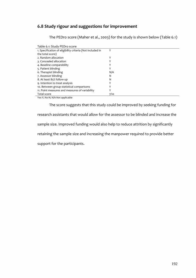
192
6.8 Study rigour and suggestions for improvement
The PEDro score (Maher et al., 2003) for the study is shown below (Table 6.1)
Table 6.1: Study PEDro score 1. Specification of eligibility criteria (Not included in the total score)
Y
2. Random allocation Y 3. Concealed allocation Y 4. Baseline comparability Y 5. Patient blinding Y 6. Therapist blinding N/A 7. Assessor blinding N 8. At least 85% follow-up N 9. Intention to treat analysis Y 10. Between group statistical comparisons Y 11. Point measures and measures of variability Y Total score 7/10 Yes-Y; No-N; N/A-Not applicable
The score suggests that this study could be improved by seeking funding for
research assistants that would allow for the assessor to be blinded and increase the
sample size. Improved funding would also help to reduce attrition by significantly
retaining the sample size and increasing the manpower required to provide better
support for the participants.
193
Chapter 7
Study conclusions and recommendations for future research
This study set out to investigate the effect of CSEs on trunk sagittal
acceleration following an onset of acute non-specific low back pain. The findings
suggest that there may be a positive effect of increasing trunk sagittal acceleration
however the changes were not statistically significant. Increasing the sample size
may improve the level of significance demonstrated in this study.
It is also suggested that it is possible to use the lumbar motion monitor
efficiently and effectively to quantify changes in trunk sagittal acceleration during
the natural course of recovery following an acute onset of low back pain within a
clinical environment.
The reported early changes in trunk sagittal acceleration may suggest that it
is possible to detect a true onset of LBP. This may be achieved by quantifying the
change in trunk acceleration through repeated functional testing within a short
time span. This process will need baseline measurements for direct comparison
from an individual. But an accurate demonstration of an onset of non-specific LBP
will also require a statistically significant difference in trunk sagittal acceleration
below which a change can be considered to be negligible. Further work is required
to identify such a threshold and the specificity and sensitivity of such a method will
need further investigation. However, the benefits of such advancement in
knowledge will be a positive step in reducing the real costs of an onset of non-
specific LBP and the detail could be commercially sensitive.

194
This study has demonstrated the importance of the MF in trunk stability,
however, further research is required to unlock the impasse and further explore the
difference in function between the deep and superficial fibres. This is because the
deductions arrived at from this study do not sit comfortably with the evidence that
suggests that the deep MF has a high proportion of type 1 fibres compared to the
superficial MF. The only conceivable explanation is that the superficial and deep
fibres of the multifidus may have different functions. A recent study does suggest
that the deep and superficial fibres of the MF do have different functions and differ
between asymptomatic and symptomatic participants.
Further clarification of the role of the TrA within the stability mechanism is
also required. Although the TrA may work in synchrony with the MF, the anatomical
location and function of the TrA may suggest that it is more directly involved with
the regulation of IAP more so than the regulation of either actual involuntary or
voluntary inter-segmental movement.
Further research is also required to investigate the effects of pelvic
pathology on the onset of non-specific LBP experienced by females. True
mechanical LBP can be easily confused with the low back pain associated with
pelvic pathology by experienced and less experienced clinicians alike. It is not
known if there is a change in trunk kinematics during bouts of LBP that are
associated with internal viscera and if those changes are permanent and
cumulative. It is suggested that such pain will not respond to CSE.
It is also not clear if the timescale required for the repair of any structural
deficit has an effect on trunk kinematics. Non-specific LBP (excluding idiopathic
LBP) is usually initiated by structural dysfunction. It is not clear if the improvement

195
in outcome measured in this study correlates with the repair of the structures
involved during previous or repeated episodes of low back pain.
196
References
ABBOTT, J.H., 2008. Spinal manipulative therapy for acute low back pain. Journal of Manual & Manipulative Therapy, 16(4), pp. 204-207.
ABENHAIM, L., ROSSIGNOL, M., VALAT, J.P., NORDIN, M., AVOUAC, B. and BLOTMAN, F., 2000. The role of activity in the therapeutic managment of back pain. Report of the International Paris Task Force on Back Pain. Spine, 25, pp. 1.
ABUMI, K., PANJABI, M.M., KRAMER, K.M., DURANCEAU, J., OXFORD, T.R. and CRISCO, J.J., 1990. Biomechanical evaluation of lumbar stability after guided facectomies. Spine, 15(11), pp. 1142.
ALAM, A., 2002. Radiological evaluation of lumbar intervertebral instability. Ind. J Aerospace Med, 46(2), pp. 48.
AL-EISA, E., EGAN, D., DELUZIO, K. and WASSERSUG, R., 2006. Effects of pelvic asymmetry and low back pain on trunk kinematics during sitting: A comparison with standing. Spine, 31(5), pp. 135.
ALTMAN, D.G., 1991. Practical statistics for medical research. 1st edn. London: Chapman & Hall.
ALUKO, A.A., DESOUZA, L.H. and PEACOCK, J., 2011. Evaluation of trunk acceleration in healthy individuals with Low Back Pain. International Journal of Therapy and Rehabilitation, 18(1), pp. 18.
ANAES., 2000. Diagnostic and therapeutic management of common lumbago and sciatica of less than 3 months duration. Recommendations of the ANAES. Agence Nationale d'Accreditation et d'Evaluation en Sante. J. Radiol, 81, pp. 1665.
ASSENDELFT, W., MORTON, S.C., YU, E.I., SUTTORP, M.J. and SHEKELLE, P.G., 2003. Spinal manipulative therapy for low back pain: A meta-analysis of effectiveness relative to other therapies. Annals of Internal Medicine, 138(11), pp. 871.
ATLAS, J.S. and DEYO, R.A., 2001. Evaluating and managing acute low back pain in the primary care setting. Journal of General Internal Medicine, 16, pp. 120.
ATLAS, J.S., DEYO, R.A., PATRICK, D.L., CONVERY, K., KELLER, R.B. and SINGER, D.E., 1996. The Quebec Task Force classification for spinal disorders and the severity, treatment and outcomes of sciatica and lumbar spinal stenosis. Spine, 21, pp. 2885.
AULTMAN, C.D., DRAKE, J.D.M., CALLAGHAN, J.P. and MCGILL, S.M., 2004. The effect of static torsion on the compressiive strength of the spine. Spine, 29(15), pp. E304.
197
BACKCARE, 2007-last update, Back Facts. Available: http://www.backpain.org/b_pages/backfacts-2007.php [03/21, 2007].
BADIA, X., MONSERRAT, S., ROSET, M. and HERDMAN, M., 1999. Feasibility, validity and test-retest reliability of scaling methods for health states: The visual analogue scale and the time trade-off. Quality of Life Research, 8, pp. 303.
BARR, K.P., GRIGGS, M. and CADBY, T., 2007. Lumbar Stabilization. A review of Core Concepts and Current Literature. American Journal of Physical Medicine & Rehabilitation, 86(1), pp. 72.
BARR, K.P., GRIGGS, M. and CADBY, T., 2005. Lumbar Stabilization: Core concepts and current literature, Part 1. American Journal of Physical Medicine & Rehabilitation, (June), pp. 473.
BASMAJIAN, J.V. 1976. Primary anatomy. 7th Edition, Williams & Wilkins USA.
BERGMARK, A., 1989. Stability of the lumbar spine. A study in mechanical engineering. Acta Orthop Scand Suppl., 230, pp. 1.
BEURSKENS, A.J.H.M, de Vet, H.C.W. and KÖK, A.J.A., 1996. Responsiveness of functional status in low back pain: a comparison of different instruments. Pain, 65, pp. 71-76.
BIERI, D., REEVE, R., CHAMPION, G., ADDICOAT, L., ZIEGLER, J., 1990. The faces pain scale for the assessment of the severity of pain experienced by children: development, initial validation and preliminary investigation for the ratio scale properties. Pain. 41, pp. 139-150.
BLAND, J.M., 2009. The tyranny of power: is there a better way to calculate sample size? BMJ, 339, pp. 1133.
BLAND, J.M., ALTMAN, D.G., 1986. Statistical methods for assessing agreement between two methods of clinical measurement. The Lancet, 327(8476), pp. 307-310.
BLOOMFIELD, L., 2006. How things work. The physics of everyday life. 3rd edition, John Wiley & Sons, USA.
BOHANNON, R.W., 2000. Muscle strength impairments in Diabetes Mellitus: a brief review. Clinical Exercise Physiology, 2(4), pp. 185-188.
BONATO, P., BOISSY, P., CROCE, U.D. and ROY, S.H., 2002. Changes in the surface EMG signal and the biomechanics of motion during a repetitive lifting task. IEEE Trans Neural Syst Rehabil Eng, 10(1), pp. 38-47.
BOONSTRA, A.M., SCHIPHORST PREUPER, H.R., RENEMAN, M.F., POSTHUMUS, J.B. and STEWART, R.E., 2008. Reliability and validity of the visual analogue scale for

198
disability in patients with chronic musculoskeletal pain. International Journal of Rehabilitation Research, 31, pp. 165.
BORKAN, J.M., KOES, B.W., REIS, S. and CHERKIN, D.C., 1998. An agenda for primary care research on low back pain. Spine, 23(18), pp. 1992.
BOYLING, D.B. and JULL, G.A., eds, 2004. Grieve's Modern Manual Therapy. The vertebral Column. 3rd edn. London: Elsevier Ltd.
BRENNAN, G., P., FRITZ, J.M., HUNTER, S.J., THACKERAY, A. and ERHARD, R.E., 2006. Identifying subgroups of patients with acute/subacute "non-specific" low back pain. Results of a randomised clinical trial. Spine, 31(6), pp. 623.
CALLAGHAN, J.P., GUNNING, J.L. and MCGILL, S.M., 1998. The relationship between lumbar spine load and muscle activity during extensor exercises. Physical Therapy, 78(1), pp. 8.
CAMPBELL, C. and MUNCER, S.J., 2005. The causes of low back pain: a network analysis. Social Science & Medicine, 60(2), pp. 409-419.
CAPODAGLIO, P., NILSSON, J. and JURISIC, D.H., 1995. Changes in paravertebral EMG spectrum parallel to strength increases after rehabilitation in chronic low back pain patients. Clinical rehabilitation, 9(4), pp. 354-362.
CARRAGEE, E., ALAMIN, T., CHENG, I., FRANKLIN, T., VAN DEN HAAK, E. and HURWITZ, E., 2006. Are first-time episodes of serious LBP associated with new MRI findings? The Spine Journal, 6(6), pp. 624-635.
CARREON, L.Y., DJURASOVIC, M., GLASSMAN, S.D. and SAILER, P., 2007. Diagnostic accuracy and reliability of fine-cut CT scans with reconstructions to determine the status of instrumented posterolateral fusion with surgical exploration as reference standard. Spine, 32(8), pp. 892.
CAYEA, D., PERERA, S. and WEINER, D.K., 2006. Chronic low back pain in older adults: What physicians know, what they think they know, and what they should be taught. Journal of the American Geriatrics Society, 54(11), pp. 1772-1777.
CHANG, C.C., HSIANG, S., DEMPSEY, P.G. and MCGORRY, R.W., 2003. A computerized video coding system for biomechanical analysis of lifting tasks. International Journal of Industrial Ergonomics, 32(4), pp. 239-250.
CHILDS, J.D., FRITZ, J.M., FLYNN, T.W., IRRGANG, J.J., JOHNSON, K.K., MAJKOWSKI, G.R. and DELLITO, A., 2004. A clinical prediction rule to identify patients with low back pain most likely to benefit from spinal manipulation. A validation study. Annals of Internal Medicine, 141(12), pp. 921.

199
CHIOU, W.K., LEE, Y.H. and CHEN, W.J., 1999. Use of the surface EMG coactivational pattern for functional evaluation of trunk muscles in subjects with and without low back pain. International Journal of Industrial Ergonomics, 23(1-2), pp. 51-60.
CHOLEWICKI, J., JULURU, K. and MCGILL, S.M., 1999. Intra-abdominal pressure mechanism for stabilizing the lumbar spine. Journal of Biomechanics, 32(1), pp. 13-17.
CHOLEWICKI, J., PANJABI, M.M. and KAHACHALRYAN, A., 1997. Stabilising function of trunk flexor-extensor muscles around a neutral zone posture. Spine, 22, pp. 2207.
CHOLEWICKI, J., POLZHOFER, G., K. and RADEBOLD, A., 2000. Postural control of trunk during unstable sitting. Journal of Biomechanics, 33, 1733.
CHOUINARD, E. and WALTER, S., 1995. Recall bias in case-control studies: An empirical analysis and theoretical framework. Journal of clinical epidemiology, 48(2), pp. 245-254.
CLAUDE, L.N., SOLOMONOW, M., ZHOU, B.H., BARATTA, R.V. and ZHU, M.P., 2003. Neuromuscular dysfunction elicited by cyclic lumbar flexion. Muscle Nerve, 27, pp. 348.
COLACHIS, S.C. and STROHM, B.R., 1969. Effects of intermittent traction on separation of lumbar vertebrae. Archives of Physical Medicine and Rehabilitation, 50, pp. 251.
COLLINS, J. A. and FAUSER, B. C. J. M., 2005. Balancing the strengths of systematic and narrative reviews. Human Reproduction Update. 11(2). pp. 103.
COUDEYRE, E., RANNOU, F., TUBACH, F., BARON, G., CORIAT, F., BRIN, S., REVEL, M. and POIRAUDEAU, S., 2006. General Practitioners' fear-avoidance beliefs influence thier management of patients with low back pain. Pain, 124, pp. 330.
COX, M.E., ASSELIN, S., GRACOVETSKY, S.A., RICHARDS, M.P., NEWMAN, N.M., KARAKUSEVIC, V., ZHONG, L. and FOGEL, J.N., 2000. Relationship between functional evaluation measures and self-assessment in nonacute low back pain. Spine, 25(14), pp. 1817.
CRIPTON, P.A., JAIN, G.M., WITTENBERG, R.H. and NOLTE, L., 2000. Load sharing characteristics of stabilised lumbar spine segments. Spine, 25(2), pp. 170.
CRISCO, J.J., 1989. The biomechanical stability of the human lumbar spine: Experimental and theoretical investigations [Doctoral dissertation], Yale University.
CROFT, P.R., MACFARLANE, G.J., PAPAGEORGIOU, A.C., THOMAS, E. and SILMAN, A.J., 1998. Outcome of Low Back Pain in General Practice. British Medical Journal, 316, pp. 1356.

200
CROSSLEY, K.M., BENNELL, K.L., COWAN, S.M. and GREEN, S., 2004. Analysis of outcome measures for persons with patellofemoral pain: which are reliable and valid? Arch Phys Med Rehabil, 85, pp. 815.
DAGENAIS, S., CARO, J. and HALDERMAN, S., 2008. A systematic review of low back pain cost of illness studies in the United States and internationally. The Spine Journal, , pp. 8.
DAUT, R.L., CLEELAND, C.S., 1982. The prevalence and severity of pain in cancer. Cancer. 50, pp. 1913-1918.
DEYO, R.A., BATTIE, M., BEURSKENS, A. J. H. M., BOMBARDIER, C., CROFT, P., KOES, B., MALMIVAARA, A., ROLAND, M., VON KORFF, M. and WADDELL, G., 1998. Outcome Measures for Low Back Pain Research: A Proposal for Standardized Use. Spine, 23(18), pp. 2003-2013.
DOLAN, P. and ADAMS, M.A., 1993. The relation between EMG activity and extensor moment generation in the erector spinae muscles during bending and lifting activities. Journal of Biomechanics, 26, pp. 513.
DOMHOLDT, E., 2005. Rehabilitation Research: Principles and applications. Elsevier Saunders.
DOODY, C. and MCATEER, M., 2002. Clinical reasoning of expert and novice Physiotherapists in an outpatient orthopaedic setting. Physiotherapy, 88(5), pp. 258.
DOWNEY, B., TAYLOR, N. and NIERE, K., 2003. Can Physiotherapists agree on which lumbar level to treat based on palpation? Physiotherapy, 89(2), pp. 74.
DUNN, K.M. and CROFT, P.R., 2005. Classification of low back pain in primary care; using "bothersomeness" to identify the most severe patients. Spine, 30, pp. 1887.
ERNST, E., 2007. Adverse effects of spinal manipulation: a systematic review. Journal of the Royal Society of Medicine, 100, pp. 330.
FAIRBANK, J.C.T., COUPER, J., DAVIES, J.B., O'BRIEN, J.P., 1980. The Oswestry low back pain disability questionnaire. Physiotherapy. 66, pp. 271-273.
FERGUSON, S.A., GALLAGHER, S. and MARRAS, W.S., 2003. Validity and reliability of sincerity test for dynamic trunk motions. Disability and Rehabilitation, 25(4/5), 236.
FERGUSON, S.A. and MARRAS, W.S., 2004. Revised protocol for the kinetic assessment of impairement. The Spine Journal, 4, pp. 163.
FERGUSON, S.A., MARRAS, W.S. and GUPTA, P., 2000. Longitudinal Quantitative Measures of the Natural Course of Low Back Pain Recovery. Spine, 25(15), pp. 1950-1956.

201
FERRIERA, P.H., FERRIERA, M.L., MAHER, C.G., HERBERT, R.D. and REFSHAUGE, K., 2006. Specific stabilisation exercise for spinal and pelvic pain: A systematic review. Australian Journal of Physiotherapy, 52, pp. 79.
FRANK, A.O. and DESOUZA, L.H., 2001. Conservative Management of Low back Pain. International Journal of Clinical Practice, 55(1), pp. 21.
FRITZ, J.M., ERHARD, R.E. and HAGEN, B.F., 1998. Segmental instability of the lumbar spine. Physical Therapy, 78(8), pp. 889.
FRITZ, J.M. and GEORGE, S.Z., 2002. Identifying psychosocial variables in patients with acute work-related low back pain: the importance of fear-avoidance beliefs. Physical Therapy, 82(10), pp. 973-983.
FULLEN, B.M., BAXTER, G.D., O'DONOVAN, B.G.G., DOODY, C., DALY, L. and HURLEY, D.A., 2008. Doctors' attitudes and beliefs regarding acute low back pain management: A systematic review. Pain, 136, pp. 388.
GARDNER-MORSE, M.G. and STOKES, I.A.F., 2004. Structural behavior of human lumbar spinal motion segments. Journal of Biomechanics, 37(2), pp. 205-212.
GEDALIA, U., SOLOMONOW, M., ZHOU, B.H., BARATTA, R.V., LU, Y. and HARRIS, M., 1999. Biomechanics of increased exposure to lumbar injury caused by cyclic loading. Part 2. Recovery of reflexive muscular stability with rest. Spine, 24, pp. 2461.
GEORGE, S.Z. and DELITTO, A., 2005. Clinical examination variables discriminate among treatment-based classification groups: a study of construct validity in patients with acute low back pain. Physical Therapy, 85(4), pp. 306-314.
GILES, L.G. and SINGER, K.P., eds, 1997. Clinical anatomy and management of low back pain. 1 edn. Oxford: Reed Educational and Professional Publishing Ltd.
GILL, K.P. and CALLAGHAN, M.J., 1996. Intratester and intertester reproducibility of the lumbar motion monitor as a measure of range, velocity and acceleration of the thoracolumbar spine. Clinical Biomechanics, 11(7), pp. 418.
GIORCELLI, R.J., HUGHES, R.E., WASSELL, J.T. and HSIAO, H., 2001. The effect of wearing a back belt in spine kinematics during asymmetric lfting of large and small boxes. Spine, 26(16), pp. 1794.
GRANATA, K.P. and ENGLAND, S.A., 2006. Stability of dynamic trunk movement. Spine, 31(10), pp. 271.
GRANATA, K.P., LEE, P.E. and FRANKLIN, T.C., 2005. Co-contraction recruitment and spinal load during isometric trunk flexion and extension. Clinical Biomechanics, 20, pp. 1029.

202
GRANATA, K.P. and MARRAS, W.S., 2000. Cost benefit of muscle co-contraction in protecting against spinal instability. Spine, 25, pp. 1398.
GRANATA, K.P. and MARRAS, W.S., 1993. An EMG-assisted model of loads on the lumbar spine during asymmetric trunk extensions. Journal of Biomechanics, 26, pp. 1429.
GRAVEN-NIELSON, T., SVENSSON, P. and ARENDT-NIELSON, L., 1997. Effects of experimental muscle pain on muscle activity and co-ordination during static and dynamic motor function. Electroencephalography and Clinical Neurophysiology, 105, pp. 156.
GRIEVE, G., P., 1984. Mobilisation of the Spine. 4th edn. London: Churchill Livingstone.
GROTLE, M., BROX, J.I. and VØLLESTAD, N.K., 2006. Reliability, validity and responsiveness of the fear-avoidance beliefs questionnaire: methodological aspects of the Norwegian version. Journal of Rehabilitation Medicine, 38(6), pp. 346-353.
GROTLE, M., BROX, J.I. and VØLLESTADD, N.K., 2004. Functional status and disability questionnaires: What do they assess? A systematic review of back specific outcome questionnaires. Spine, 30, pp. 130.
GRUBER, H.E., NORTON, H.J., SUN, Y. and HANLEY JR, E.N., 2007. Crystal deposits in the human intervertebral disc: implications for disc degeneration. The Spine Journal, 7(4), pp. 444-450.
GUILLEMIN, F., 2005. Incidence and prevalence: two demanding indicators. Joint Bone Spine, 72, pp. 7.
GULLICK, D.W., 2008. Acute non-specific back pain management in the emergency setting: A review of the literature. Australasian Emergency Nursing Journal, 11, pp. 13.
HAAS, M., GOLDBERG, B., AICKIN, M., GANGER, B. and ATTWOOD, M., 2004. A practice-based study of patients with acute and chronic low back pain attending primary care and chiropractic physicians: Two-week to 48-month follow-up. Journal of Manipulative and Physiological Therapeutics, 27(3), pp. 160.
HAGEN, K.B., HILDE, G., JAMVEDT, G. and WINNEM, M.F., 2005. Bed rest for acute low back pain and sciatica. The Cochrane database of systematic reviews, (2),.
HAGEN, K.B., HILDE, G., JAMVEDT, G. and WINNEM, M.F., 2002. The Cochrane review of advice to stay active as a single treatment for low back pain and sciatica. Spine, 27, pp. 1736.
HANSEN, L., DE ZEE, M., RASMUSSEN, J., ANDERSEN, T.B., WONG, C. and SIMONSEN, E.B., 2006. Anatomy and biomechanics of the back muscles in the lumbar spine with reference to biomechanical modelling. Spine, 31(17), pp. 1888.

203
HEBERT, J.J., KOPPENHAVER, S.L., MAGEL, J.S. and FRITZ, J.M., 2010. The relationship of Transversus Abdominis and lumbar Multifidus activation and prognostic factors for clinical success with a stabilization exercise program: A cross-sectional study. Archives of Physical Medicine and Rehabilitation, 91(1), pp. 78-85.
HEUER, F., SCHMITT, H., SCHMIDT, H., CLAES, L. and WILKE, H., 2007. Creep associated changes in intervertebral disc bulging obtained with a laser scanning device. Clinical Biomechanics, 22(7), pp. 737-744.
HICKS, C.M., 1998. Practical Research Methods for Physiotherapists. 1 edn. London: Churchill Livingstone.
HICKS, G.E., FRITZ, J.M., DELITTO, A. and MCGILL, S.M., 2005. Preliminary development of a clinical prediction rule for determining which patients with low back pain will respond to a stabilization exercise program. Archives of Physical Medicine and Rehabilitation, 86(9), pp. 1753-1762.
HIDES, J., STANTON, W., MENDIS, M.D., and SEXTON, M., 2011. The relationship of transversus abdominis and lumbar multifidus clinical muscle tests in patients with chronic low back pain. Manual Therapy, 16, pp.573-577.
HODGES, P.W. and GANDEVIA, S.C., 2000. Changes in intra-abdominal pressure during postural and respiratory activation of the human diaphragm. J Appl. Physiol., 89, pp. 967.
HODGES, P., MOSELEY, G.L., GABRIELSSON, A. and GANDEVIA, S.C., 2003. Experimental muscle pain changes feed forward postural responses of the trunk muslces. Exp. Brain Res., 151, pp. 262.
HODGES, P. and RICHARDSON, C., 1997. Contraction of the abdominal muscles associated with movement of the lower limb. Physical Therapy, 77, pp. 132.
HODGES, P. and RICHARDSON, C., 1996. Inefficient muscular stabilisation of the lumbar spine associated with low back pain: A motor control evaluation of transversus abdominis. Spine, 21, pp. 2640.
HOLDSWORTH, L.K., WEBSTER, V.S. and MCFADYEN, A.K., 2006. Are patients who refer themselves to physiotherapy different from those referred by GP's? Results of a national trial. Physiotherapy, 92, pp. 26.
HOLM, S., 1996. Nutrition and pathophysiological aspects of the lumbar intervetebral disc in the lumbar spine. 2nd edn. Philadelphia: WB Saunders Company.
HOWELL, D., 1992. Statistical Methods tor Psychology. 3rd edn.. PWS Kent. Boston. MA.

204
HSEIH, C.Y., PHILLIPS, R.B., ADAMS, A.H. and POPE, M.H., 1992. Functional outcomes of low back pain: Comparison of four treatment groups in a randomised controlled trial. Journal of Manipulative & Physiological Therapies, 15(1), pp. 4.
HUSKISSON, C.E., 1974. The measurement of pain. The Lancet. 9(2), pp. 1127-1131.
IANUZZI, A., LITTLE, J.S., CHIU, J.B., BAITNER, A., KAWCHUK, G. and KHALSA, P.S., 2004. Human lumbar facet joint capsule strains: I. During physiological motions. The Spine Journal, 4(2), pp. 141-152.
JACKSON, D.A., 2001. How is low back pain managed? Physiotherapy, 87(11), pp. 573.
JANDA, S. and VALENTA, J., 2000. Mathematical model of the lumbar spine loads. Strojnicky Casopis, 51(2).
JAYSON, M.I.V., 1996. Back Pain. British Medical Journal, 313, pp. 355.
JOHANNES, C.B., LINET, M.S., STEWART, W.F., CELENTANO, D.D., LIPTON, R.B., and SZKLO, M., 1995. Relationship of headache to phase of the menstrual cycle among young women: a daily diary study. Neurology. 45(6), pp. 1076-82.
JOHANSSON, H. and SOJKA, P., 1991. Pathophysiological mechanisms involved in genesis and spread of muscular tension in occupational muscle pain and in chronic musculoskeletal pain syndromes: A hypothesis. Medical Hypotheses, 35(3), pp. 196.
JOHNSTONE, R., DONAGHY, M. and MARTIN, D., 2004. A pilot study of a cognitive-behavioural therapy approach to physiotherapy, for acute low back pain patients, who show signs of developing chronic pain. Advances in Physiotherapy, 4(4), pp. 182-188.
JONES, D.A. and ROUND, J.M., 1990. Histochemistry, contractile properties and motor control. Skeletal muscle in health and disease. 1st edn. Manchester: Manchester University Press, pp. 55.
JORDAN, K., DUNN, K.M., LEWIS, M. and CROFT, P., 2006. A minimal clinically important difference was derived for the Roland-Morris Disability Questionnaire for low back pain. Journal of Clinical Epidemiology, 59(1), pp. 45-52.
KANAYAMA, M., TADANO, S., KANEDA, K., UKAI, T., ABUMI, K. and ITO, M., 1995/6. A cineradiographic study on the lumbar disc deformation during flexion and extension of the trunk. Clinical Biomechanics, 10(4), pp. 193-199.
KELLY, A., 2001. The minimum clinically significant difference in visual analogue scale pain scores does not differ with severity of pain. Emerg Med Journal, 18, pp. 205.
KENT, P. and KEATING, J., 2004. Do Primary care clinicians think that non-specific low back pain is one condition? Spine, 29, pp. 1022.

205
KERR, R. and GRAHAME, R., eds, 2003. Hypermobility Syndrome: Recognition and management for physiotherapists. 1 edn. London: Butterworth Heinemann.
KINKADE, S., 2007. Evaluation and treatment of acute low back pain. American Family Physician, 75, pp. 1181.
KNUTSON, G.A., 2002. Incidence of foot rotation, pelvic crest unlevelling, and supine leg length alignment asymmetry and their relationship to self-reported back pain. J Manipulative Physiol Ther, 25, pp. 110.
KOES, B.W., VAN TULDER, M.W. and THOMAS, S., 2006. Diagnosis and treatment of low back pain. BMJ, 332(7555), 1430-1434.
KOPEC, J.A., ESDAILE, J.M., ABRAHAMOWICZ, M., ABENHAIM, L., WOOD-DAUPHINEE, S. and LAMPING, D.L., 1995. The Quebec Back Pain Disability Scale. measurement properties. Spine, 20, pp. 341.
KOUMANTAKIS, G.A., WATSON, P.J. and OLDHAM, J.A., 2005. Trunk muscle stabilisation training plus general exercise versus general exercise only: Randomised controlled trial of patients with recurrent low back pain. Physical Therapy, 85(3), pp. 209.
KRISMER, M. and VAN TULDER, M., 2007. Low back pain (non-specific). Best Practice & Research Clinical Rheumatology, 21(1), pp. 77-91.
KROEMER, K.H.E., MARRAS, W.S., MCGLOTHLIN, D.R. and NORDIN, M., 1990. Towards understanding human dynamic motor performance. Industrial Ergonomics Journal, 6, pp. 199.
KULIG, K., POWERS, C.M., LANDEL, R.F., CHEN, H., FREDERICSON, M., GUILLET, M. and BUTTS, K., 2007. Segmental lumbar mobility in individuals with low back pain: in vivo assessment during manual and self-imposed motion using dynamic MRI. BMC Musculoskeletal Disorders, 8(8), pp. 1471.
LANGRANA, N.A., KALE, S.P., EDWARDS, W.T., LEE, C.K. and KOPACZ, K.J., 2006. Measurement and analyses of the effects of adjacent end plate curvature on vertebral stresses. The Spine Journal, 6, pp. 267.
LARIVIERE, C., GAGNON, D. and LOISEL, P., 2002. A biomechanical comparison of lifting techniques between subjects with and without chronic low back pain during free style lifting and lowering tasks. Clinical Biomechanics, 17, pp. 89.
LASLETT, M., MCDONALD, B., APRILL, C.N., TROPP, H. and OBERG, B., 2006. Clinical predictors of screening lumbar zygapophyseal joint blocks: development of clinical prediction rules. The Spine Journal, 6, pp. 370.
LAU, P.M., CHOW, D.H. and POPE, M.H., 2008. Early physiotherapy intervention in an Accident and Emergency department reduces pain and improves satisfaction for

206
patients with acute low back pain: a randomised trial. Australian Journal of Physiotherapy, 54(4), pp. 243-249.
LAUCHLAN, D., 2005. Acute low back pain in adolescence: a review of the recent literature with regard to clinical guidelines. Clinician in Management, 13(3), pp. 165-169.
LAWLIS, G.F., CUENCAS, R., SELBY, D., and MCCOY, C.E.,1989. The development of the Dallas Pain Questionnaire: an assessment of the impact of spinal pain on behaviour. Spine. 14(5), pp. 511-516.
LEAN, M.E.J., HAN, T.S. and MORRISON, C.E., 1995. Waist circumference as a measure for indicating need for weight management. British Medical Journal, 311, pp. 158.
LEE, R.Y.W. and EVANS, J.H., 2001. Loads in the lumbar spine during traction therapy. Australian Journal of Physiotherapy, 47, pp. 102.
LeRESCHE, L., MANCI, L., SHERMAN, J.J., GANDARA, B., and DWORKIN, S. F., 2003. Changes in temporomandibular pain and other symptoms across the menstrual cycle. Pain. 106(3), pp. 253-261.
LIEBSCHNER, M.A.K., KOPPERDAHL, D.L., ROSENBERG, W.S. and KEAVENY, T.M., 2003. Finite element modeling of the human thoracolumbar spine. Spine, 28(6), pp. 559-565.
LINTON, S.J., BOERSMA, K., JANSSON, M., SVARD, L. and BOTVALDE, M., 2005. The effects of cognitive behavioral and physical therapy preventive interventions on pain related sick leave: A randomised controlled trial. Clinical Journal of Pain, 21(2), pp. 109.
LINTON, S.J., HELLSING, A. and BERGSTROM, G., 1996. Exercise for workers with musculoskeletal pain: Does enhancing compliance decrease pain? Journal of Occupational Rehabilitation, 6(3), pp. 177.
LITTLE, J.S., IANUZZI, A., CHIU, J.B., BAITNER, A. and KHALSA, P.S., 2004. Human lumbar facet joint capsule strains: II. Alteration of strains subsequent to anterior interbody fixation. The Spine Journal, 4(2), pp. 153-162.
LU, D., SOLOMONOW, M., ZHOU, B.H., BARATTA, R.V. and LI, L., 2004. Frequency-dependent changes in neuromuscular responses to cyclic lumbar flexion. Journal of Biomechanics, 37, pp. 845.
LURIE, J.D., 2005. What diagnostic tests are useful for low back pain? Best Practice & Research Clinical Rheumatology, 19(4), pp. 557.
MACDONALD, D.A., MOSELEY, G.L. and HODGES, P.W., 2006. The lumbar multifidus: Does the evidence support clinical beliefs. Manual therapy, 11, pp. 254.

207
MACDONALD, D., MOSELEY, G.L. and HODGES, P.W., 2010. People with recurrent low back pain respond differently to trunk loading despite remission from symptoms. Spine, 35(7), pp. 818-819.
MACFARLANE, G.J., JONES, G.T. and HANNAFORD, P.C., 2006. Managing low back pain presenting to primary care: Where do we go from here? Pain, 122, pp. 219.
MACNAB, I. and MCCULLOCH, J., 1990. In: I. MACNAB and J. MCCULLOCH, eds, Backache. 2 edn. USA: .
MAHER, C.G., SHERRINGTON, C., HERBERT, R. D., MOSELEY, A. M., and ELKINS, M., 2003. Reliability of the PEDro scale for rating quality of randomised controlled trials. Physical Therapy, 83(8), pp. 713-721.
MARRAS, W.S., 1996. Quantification of motion characteristics in low back disorders. Journal of Rehabilitation Research and Development, 33, pp. 186.
MARRAS, W.S., DAVIS, K.G. and MARONITIS, A.B., 2001. A non-MVC EMG normalisation technique for the trunk musculature: Part 2. Validation and use to predict spinal loads. J. Electromyog. Kinesiol., 11, pp. 11.
MARRAS, W.S., FATHALLAH, F.A., MILLER, R.J., DAVIS, S.W, and MIRKA, G.A., 1992. Accuracy of a three-dimensional lumbar motion monitor for recording dynamic trunk motion characteristics. International Journal of Industrial Ergonomics, 9, pp 75.
MARRAS, W.S., FERGUSON, S.A., GUPTA, P., BOSE, S., PARNIANPOUR, M., KIM, J. and CROWELL, R.R., 1999. The Quantification of low back disorder using motion measures: Methodology and validation. Spine, 24(20), pp. 2091.
MARRAS, W.S., FERGUSON, S.A. and SIMON, S.R., 1990. Three dimensional dynamic motor performance of the normal trunk. International Journal of Industrial Ergonomics, 6, pp. 211.
MARRAS, W.S. and GRANATA, K.P., 1997b. Spine loading during trunk lateral bending motions. J. Biomech, 30, pp. 697.
MARRAS, W.S. and GRANATA, K.P., 1995. A biomechanical assessment and model of axial twisting in the thoracolumbar spine. Spine, 20, pp. 1440.
MARRAS, W.S., KNAPIK, G.G. and FERGUSON, S., 2009. Loading along the lumbar spine as influenced by speed, control, load magnitude and handle height during pushing. Clinical Biomechanics, 24, pp. 155.
MARRAS, W.S., LEWIS, E.K., FERGUSON, S.A. and PARNIANPOUR, M., 2000. Impairment magnification during dynamic trunk motions. Spine, 25(5), pp. 587-595.

208
MARRAS, W.S. and MIRKA, G.A., 1993. Electromyographic studies of the lumbar trunk musculature during the generation of low level trunk acceleration. Journal of Orthopaedic Research, 11, pp. 811.
MARRAS, W.S., PARNIANPOUR, M., FERGUSON, S., KIM, J., CROWELL, R. and SIMON, S., 1993. Vienna Physical Medicine Award 1993. Quantification and classification of low back disorders based on trunk motion. European Journal Physical Medicine, 3, pp. 218.
MARRAS, W.S. and WONGSAM, P.E., 1986. Flexibility and velocity of the normal and impaired lumbar spine. Archives of Physical Medicine and Rehabilitation, 67, pp. 213.
MARTINI, F.H., OBER, W.C., GARRISON, C.W., WELCH, K. and HUTCHINGS, R.T., 1995. The axial skeleton in fundamentals of anatomy and physiology. 3rd edn. New Jersey: Prentice Hall.
MAY, S. and JOHNSON, R., 2008. Stabilisation exercises for low back pain: A systematic review. Physiotherapy, 94, pp. 179.
MCCAFFERY, M. and BEEBE, A., 1993. Pain: clinical manual for nursing practice. Baltimore: V.V. Mosby Company
MCGEARY, D.D., MAYER, T.G., GATCHEL, R.J., ANAGNOSTIS, C. and PROCTOR, T.J., 2003. Gender-related differences in treatment outcomes for patients with musculoskeletal disorders. The Spine Journal, 3, pp. 197.
MCGILL, S., 2002. Low back disorders. Evidence-based prevention and rehabilitation. Champaign: Human Kinetics.
MCGILL, S.M., GRENIER, S., KAVCIC, N. and CHOLEWICKI, J., 2003. Coordination of muscle activity to assure stability of the lumbar spine. Journal of Electromyography and Kinesiology, 13, pp. 353.
MCGREGOR, A.H. and HUGHES, S.F., 2000. The effect of test speed on the motion characteristics of the lumbar spine during an A-P flexion-extension test. Journal of Back and Musculoskeletal Rehabilitation, 14, pp. 99.
MCKENZIE, R.A., 1981. The lumbar spine: Mechanical diagnosis and therapy. 1st edn. New Zealand: Spinal Publications.
MOFFETT, J. and MCLEAN, S., 2006. The role of physiotherapy in the management of non-specific back and neck pain. Rheumatology, 45, pp. 371.
MORGAN, F.P. and KING, T., 1957. Primary instability of lumbar vertebrae as common cause of low back pain. J. Bone Joint Surg. Br., 39, pp. 6.
MOSELEY, G.L., NICHOLAS, M.K. and HODGES, P.W., 2004. Does anticipation of back pain predispose to back trouble. Brain, 127, pp. 2339.

209
NACHEMSON, A., 1981. Disc pressure measurements. Spine, 6, pp. 93.
NACHEMSON, A. and MORRIS, J.M., 1964. In Vivo measurements of intradiscal pressure in the lower lumbar discs. Journal Bone Joint Surgery, 46A(5), pp. 1077.
NAJM, W.I., SEFFINGER, M.A., MISHRA, S.I., DICKERSON, V.M., ADAMS, A., REINSCH, S., MURPHY, L.S. and GOODMAN, A.F., 2003. Content validity of manual spinal palpatory exams-A systemic review. BMC Complimentary and Alternative Medicine, .
NAKAJIMA, A., KAWAKAMI, N., IMAGAMA, S., TSUJI, T., GOTO, M. and OHARA, T., 2007. Three -dimensional analysis of formation failure in congenital scoliosis. Spine, 32(5), pp. 562.
NEUMANN, W.P., WELLS, R.P., NORMAN, R.W., KERR, M.S., FRANK, J. and SHANNON, H.S., 2001. Trunk posture: reliability, accuracy, and risk estimates for low back pain from a video based assessment method. International Journal of Industrial Ergonomics, 28(6), pp. 355-366.
NHS, October, 2005, 2005-last update, Back pain-Lower, Prodigy Knowledge Guidance [Homepage of National Library for Health], [Online]. Available: www.prodigy.nhs.uk/ProdgyKnowledge [02/09, 2007].
NICE, May, 2009, Low back pain, Early management of persistentent non-specific low back pain [Home page National Institute for Health and Clinical Excellence ], [Online]. Available: www.guidance.nice.org.uk [accessed 02/12/11]
NORRIS, C.M., 2000. Back stability. Champaign: Human Kinetics.
NORRIS, C.M., 1995/2. Spinal stabilisation: 2. Limiting factors to end-range motion in the lumbar spine. Physiotherapy, 81(2), pp. 64-72.
ODDSSON, L.I., GIPHART, J.E., BUIJS, R.J., ROY, S.H., TAYLOR, H.P. and LUCA, C.J., 1997. Development of new protocols and analysis procedures for the assessment of LBP by surface EMG techniques. Journal of Rehabilitation Research and Development, 34(4), pp. 415-426.
OGRODNIK, P.J., 1997a. Behaviour of materials. Fundamental Engineeering Mechanics. 1 edn. Malaysia: Addison Wesley Longman Ltd, pp. 179.
OGRODNIK, P.J., 1997b. Conservation of energy. In: P.J. OGRODNIK, ed, Fundamental Engineering Mechanics. 1 edn. Malaysia: Addison Wesley Longman Ltd, pp. 84.
OGRODNIK, P.J., 1997c. Velocity and acceleration. In: P.J. OGRODNIK, ed, Fundamental Engineering Mechanics. 1 edn. Malaysia: Addison Wesley Longman Ltd, pp. 21.
210
OLAOGUN, M.O.B., ADEDOYIN, R.A., IKEM, I.C. and ANIFALOBA, O.R., 2004. Reliability of rating low back pain with visual analogue scale and a sematic differential scale. Physiotherapy Theory and Practice, 20, pp. 135.
OSTELO, R. W. J. G., VAN TULDER, M.W., VLAEYEN, J.W.S., LINTON, S.J., MORLEY, S.J. and ASSENDELFT, W., 2005. Behavourial treatment for chronic low back pain. The Cochrane Database of Systematic Reviews, (2),.
O'SULLIVAN, P.B., 2000. Lumbar segment 'instability': clinical presentation and specific stabilizing exercise management. Manual Therapy, 5(1), pp. 2.
OWENS, E.F., DEVOCHT, J., WILDER, D.G., GUDAVALLI, M.R. and MEEKER, W.C., 2007. The reliability of a posterior-to-anterior spinal stiffness measuring system in a population of patients with low back pain. Journal Manipulative Physiol. Ther., 30, pp. 116.
PAHL, M., BRISLIN, B., BODEN, S., HILIBRAND, A.S., VACCARO, A., HANSCOM, B. and ALBERT, T.J., 2006. The impact of four common lumbar spine diagnoses upon overall health status. The Spine Journal, 6, pp. 125.
PANJABI, M.M., 2003. Clinical spinal instability and low back pain. J. Electromyog. Kinesiol., 13, pp. 371.
PANJABI, M.M., 1994. Lumbar Spine Instability: A biochemical challenge. Current Orthopaedics, 8, pp. 100.
PAPAGEORGIOU, A.C., CROFT, P.R., THOMAS, E., FERRY, S., JAYSON, M.I.V. and SILMAN, A.J., 1996. Influence of previous pain experience on the episode incidence of low back pain: results from the South Manchester Back Pain Study. Pain, 66, pp. 181.
PARNIANPOUR, M., GOSH, K., BARIN, K. and MARRAS, W.S., 2001. Quantification of trunk motion in response to complex platform perturbations while holding weights in an upright posture. Biomed. Eng Appl Basis Comm, 13, pp 33.
PATEL, V., 2004. Diagnostic modalities for low back pain. Seminars in Pain Medicine, 2(3), pp. 145-153.
PAUL-DAUPHIN, A., GUILLEMIN, F., VIRION, J. and BRIANCON, S., 1999. Bias and precision in visual analogue scales: A randomised controlled trial. American journal of Epidemiology, 150(10), pp. 1117.
PAZOS, V., CHERIET, F., DANSERAU, J., RONSKY, J., ZERNICKE, R.F. and LABELLE, H., 2007. Reliability of trunk shape measurements based on 3-D surface reconstructions. European Spine Journal, No;16(11), pp. 91.
PEACOCK, J.L., PEACOCK, P.J., 2011. Oxford handbook of medical statistics. 1st edn. Oxford: Oxford University Press.
211
PETTY, N.J., 2006. Neuromusculoskeletal Examination and Assessment: A handbook for Therapists. 3rd edn. London: Churchill Livingstone.
PITCHER, M.J., BEHM, D.G. and MACKINNON, S.N., 2008. Reliability of electromyographic and force measures during prone isometric back extension in subjects with and without low back pain. Applie Physiology Nutrition and Metabolism, 33(1), pp. 52-60.
PLOUVIER, S., RENAHY, E., CHASTANG, J.F., BONENFANT, S. and LECLERC, A., 2008. Biomechanical strains and low back disorders: quantfying the effects of the number of years of exposure on various types of pain. Occupational and Environmental Medicine, 65, pp. 268.
POLLOCK, C.L., JENKYN, T.R., JONES, I.C., IVANOVA, T.D. and GARLAND, S.J., 2009. Electromyography and kinematics of the trunk during rowing in elite female rowers. Medicine and science in sports and exercise, 41(3), pp. 628-636.
POPE, M.H. and PANJABI, M.M., 1985. Biomechanical definitions of spinal instability. Spine, 10, pp. 255.
RASMUSSEN-BARR, E., NILSSON-WIKMAR, L. and ARVIDSSON, I., 2003. Stabilizing training compared with manual treatment in sub-acute and chronic low-back pain. Manual therapy, 8(4), pp. 233-241.
REEVES, N.P., NARENDRA, K.S. and CHOLEWICKI, J., 2007. Spine stability: The six blind men and the elephant. Clinical Biomechanics, 22, pp. 266.
RICHARDSON, C., JULL, G., HODGES, P. and HIDES, J., 1999. Therapeutic exercises for spinal segmental stabilisation in low back pain. Edinburgh: Churchill Livinstone.
ROLAND, M. and MORRIS, R., 1983. A study of the natural history of back pain. Part 1: Development of a reliable and sensitive measure of disability in low back pain. Spine, 8(2), pp. 141.
ROZENBERG, S., DELVAL, C., REZVANI, Y. and ET AL., 2003. Bed rest and normal daily activity were equivalent for acute low back pain. Journal of Bone & Joint Surgery, 85-A(5), pp. 975.
RUTA, D.A., GARRATT, A.M, WARDLAW, D. and RUSSELL, I.T, 1994. Developing a valid and reliable measure of health outcome for patients with low back pain. Spine, 19. pp. 1887-1896.
SAMANTA, J., KENDALL, J. and SAMANTA, A., 2003. 10 minute consultation: Chronic low back pain. British Medical Journal, 326, pp. 535.
SCHMIDT, H., HEUER, F., CLAES, L. and WILKE, H., 2008. The relation between the instantaneous center of rotation and facet joint forces – A finite element analysis. Clinical Biomechanics, 23(3), pp. 270-278.
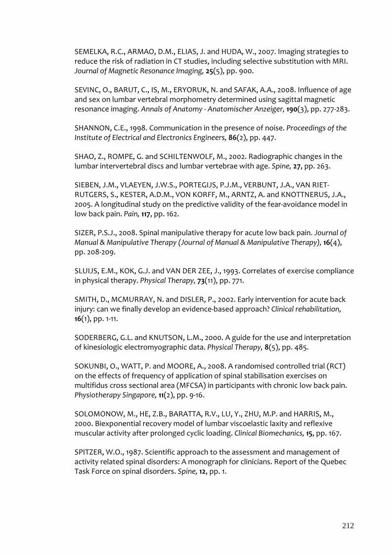
212
SEMELKA, R.C., ARMAO, D.M., ELIAS, J. and HUDA, W., 2007. Imaging strategies to reduce the risk of radiation in CT studies, including selective substitution with MRI. Journal of Magnetic Resonance Imaging, 25(5), pp. 900.
SEVINC, O., BARUT, C., IS, M., ERYORUK, N. and SAFAK, A.A., 2008. Influence of age and sex on lumbar vertebral morphometry determined using sagittal magnetic resonance imaging. Annals of Anatomy - Anatomischer Anzeiger, 190(3), pp. 277-283.
SHANNON, C.E., 1998. Communication in the presence of noise. Proceedings of the Institute of Electrical and Electronics Engineers, 86(2), pp. 447.
SHAO, Z., ROMPE, G. and SCHILTENWOLF, M., 2002. Radiographic changes in the lumbar intervertebral discs and lumbar vertebrae with age. Spine, 27, pp. 263.
SIEBEN, J.M., VLAEYEN, J.W.S., PORTEGIJS, P.J.M., VERBUNT, J.A., VAN RIET-RUTGERS, S., KESTER, A.D.M., VON KORFF, M., ARNTZ, A. and KNOTTNERUS, J.A., 2005. A longitudinal study on the predictive validity of the fear-avoidance model in low back pain. Pain, 117, pp. 162.
SIZER, P.S.J., 2008. Spinal manipulative therapy for acute low back pain. Journal of Manual & Manipulative Therapy (Journal of Manual & Manipulative Therapy), 16(4), pp. 208-209.
SLUIJS, E.M., KOK, G.J. and VAN DER ZEE, J., 1993. Correlates of exercise compliance in physical therapy. Physical Therapy, 73(11), pp. 771.
SMITH, D., MCMURRAY, N. and DISLER, P., 2002. Early intervention for acute back injury: can we finally develop an evidence-based approach? Clinical rehabilitation, 16(1), pp. 1-11.
SODERBERG, G.L. and KNUTSON, L.M., 2000. A guide for the use and interpretation of kinesiologic electromyographic data. Physical Therapy, 8(5), pp. 485.
SOKUNBI, O., WATT, P. and MOORE, A., 2008. A randomised controlled trial (RCT) on the effects of frequency of application of spinal stabilisation exercises on multifidus cross sectional area (MFCSA) in participants with chronic low back pain. Physiotherapy Singapore, 11(2), pp. 9-16.
SOLOMONOW, M., HE, Z.B., BARATTA, R.V., LU, Y., ZHU, M.P. and HARRIS, M., 2000. Biexponential recovery model of lumbar viscoelastic laxity and reflexive muscular activity after prolonged cyclic loading. Clinical Biomechanics, 15, pp. 167.
SPITZER, W.O., 1987. Scientific approach to the assessment and management of activity related spinal disorders: A monograph for clinicians. Report of the Quebec Task Force on spinal disorders. Spine, 12, pp. 1.

213
STANDAERT, C.J. and HERRING, S.A., 2007. Expert opinion and controversies in musculoskeletal and sports medicine: Core stabilization as a treatment for low back pain. Archives of Physical Medicine and Rehabilitation, 88, pp. 1734.
STANDAERT, C.J., WEINSTEIN, S.M. and RUMPELTES, J., 2008. Evidence-informed management of chronic low back pain with lumbar stabilisation exercises. The Spine Journal, 8, pp. 114.
STANLEY, I., MILLER, J., PINNINGTON, M.A., ROSE, G. and ROSE, M., 2000. Uptake of prompt access physiotherapy for new episodes of back pain presenting in primary care. Physiotherapy, 87(2), pp. 60.
STEPAN, G., 2009. Delay effects in the human sensory system during balancing. Philosophical Transactions of The Royal Society A, 367, pp. 1195.
STODDEN, D.F., CAMPBELL, B.M. and MOYER, T.M., 2008. Comparison of trunk kinematics in trunk training exercises and throwing. The Journal of Strength and Conditioning Research, 22(1), pp. 112.
STRATFORD, P.W., FINCH, E., SOLOMON, P., BINKLEY, J., GILL, C. and MORELAND, J., 1996. Using the Roland-Morris Questionnaire to make decisions about individual patients. Physiotherapy Canada, 48, pp. 107.
SUZUKI, H., CONWIT, R.A., STASHUK, D., SANTARSIERO, L. and METTER, E.J., 2002. Relationship between surface detected EMG signals and motor unit activation. Medicine and Science in Sports and Exercise, 1(9), pp. 1509.
TABOR, G., COLLINS, T., RICHTEROVA, J., DEJUNIAT, E. and BERESFORD-WEST, T., 2005-last update, Automating the generation of 3D finite element models based on medical imaging data. Available: www.simpleware.com/resources/files/DHM06DHM-51.pdf [September/1st, 2008].
TAYLOR, N., GOLDIE, P. and EVANS, O., 2004. Movements of the pelvis and lumbar spine during walking in people with acute low back pain. Physiotherapy Research International, 9(2), pp. 74-84.
TEYHEN, D.S., FLYNN, T.W., CHILDS, J.D. and ABRAHAM, L.D., 2007. Arthrokinetics in a subgroup of patients likely to benefit from a lumbar stabilisation exercise program. Physical Therapy, 87(3), pp. 313.
THE CHARTERED SOCIETY OF PHYSIOTHERAPY, 2004. PPA recommendations for low back pain related functional limitation outcome measures. The Chartered Society of Physiotherapy.
THOMAS, J.S., FRANCE, C.R., LAVENDER, S.A. and JOHNSON, M.H., 2008. Effects of fear of movement on spine velocity and acceleration after recovery from low back pain. Spine, 33(5), pp. 564-570.

214
THOMAS, J.S., LAVENDER, S.A., CORCOS, D.M. and ANDERSSON, G.B., 1998. Trunk kinematics and trunk muscle activity during a rapidly applied load. Journal of Electromyography and Kinesiology, 8, pp. 215.
TROTT, M. and FISHER, K., 2005. Using Video Assessment for Clients with Chronic Low Back Pain: is it Reliable? British Journal of Occupational Therapy, 68(12), pp. 538-544.
TRUDELLE-JACKSON, E., FLEISHER, L.A., BORMAN, N., MORROW, J.R. and FRIERSON, G., 2010. Lumbar spine flexion and extension extremes of motion in women of different age and racial groups. The WIN study. Spine, 35(16), pp. 1539.
TWOMEY, L.T., 1985. Sustained lumbar traction: an experimental study of long spine segments. Spine, 10, pp. 146.
URBAN, J.P.G. and ROBERTS, S., 2003. Degeneration of the intervertebral disc. Arthritis Research & Therapy, 5(3), pp. 120.
VAN DEN BOSCH, M. A. A. J., HOLLINGWORTH, W., KINMONTH, A.L. and DIXON, A.K., 2004. Evidence against the use of lumbar spine radiography for low back pain. Clinical Radiology, 59(1), pp. 69-76.
VAN DEN HOOGEN, H. M., KOES, B.W., VAN EIJK, K.T. and BOUTER, L.M., 1995. On the accuracy of history, physical examination, and erythrocyte sedimentation rate in diagnosing low back pain in general practice. A criteria based review of the literature. Spine, 20(3), pp. 318.
VAN DIEEN, J.H., CHOLEWICKI, J. and RADEBOLD, A., 2003. Trunk muscle recruitment patterns in patients with low back pain enhance the stability of the lumbar spine. Spine, 28, pp. 834.
VAN DIEEN, J.H., SELEN, L.P.J. and CHOLEWICKI, J., 2003. Trunk muscle activation in low back pain patients, an analysis of the literature. Journal of Electromyography and Kinesiology, 13, pp. 333.
VAN TULDER, M.W., BECKER, A., BEKKERING, T., BREEN, A.C., GIL DELREAL, M.T., HUTCHINSON, A., KOES, B., LAERUM, E., MAELMIVAARA, A., CARTER, T., KRYGER-BAGGESEN, P., NACHEMSON, A., NIEHUS, W., ROUX, E. and ROZENBERG, S., , European guidelines for the management of acute non-specific low back pain in primary care. Available: http://www.backpaineurope.org/web/html/wg1_results.html [October 14th, 2008].
VAN TULDER, M.W., MALMIVAARA, A.V., ESMAIL, R. and KOES, B.W., 2000. Exercise Therapy for low back pain: a systematic review within the framework of the cochrane collaboration back pain review group. Spine, 25, pp. 2784.
WADDELL, G., 1987. 1987 Volvo award in clinical sciences. A new clinical model for the treatment of low back pain. Spine, 12, pp. 632.
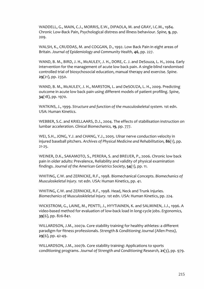
215
WADDELL, G., MAIN, C.J., MORRIS, E.W., DIPAOLA, M. and GRAY, I.C.M., 1984. Chronic Low-Back Pain, Psychological distress and illness behaviour. Spine, 9, pp. 209.
WALSH, K., CRUDDAS, M. and COGGAN, D., 1992. Low Back Pain in eight areas of Britain. Journal of Epidemiology and Community Health, 46, pp. 227.
WAND, B. M., BIRD, J. H., McAULEY, J. H., DORE, C. J. and DeSouza, L. H., 2004. Early intervention for the management of acute low back pain. A single-blind randomised controlled trial of biosychosocial education, manual therapy and exercise. Spine. 29(21), pp. 2350.
WAND, B. M., McAULEY, J. H., MARSTON, L. and DeSOUZA, L. H., 2009. Predicting outcome in acute low back pain using different models of patient profiling. Spine, 34(18), pp. 1970.
WATKINS, J., 1999. Structure and function of the musculoskeletal system. 1st edn. USA: Human Kinetics.
WEBBER, S.C. and KRIELLAARS, D.J., 2004. The effects of stabilisation instruction on lumbar acceleration. Clinical Biomechanics, 19, pp. 777.
WEI, S.H., JONG, Y.J. and CHANG, Y.J., 2005. Ulnar nerve conduction velocity in injured baseball pitchers. Archives of Physical Medicine and Rehabilitation, 86(1), pp. 21-25.
WEINER, D.K., SAKAMOTO, S., PERERA, S. and BREUER, P., 2006. Chronic low back pain in older adults: Prevalence, Reliability and validity of physical examination findings. Journal of the American Geriatrics Society, 54(1), pp. 11.
WHITING, C.W. and ZERNICKE, R.F., 1998. Biomechanical Concepts. Biomechanics of Musculoskeletal Injury. 1st edn. USA: Human Kinetics, pp. 41.
WHITING, C.W. and ZERNICKE, R.F., 1998. Head, Neck and Trunk Injuries. Biomechanics of Musculoskleletal Injury. 1st edn. USA: Human Kinetics, pp. 224.
WICKSTROM, G., LAINE, M., PENTTI, J., HYYTIAINEN, K. and SALMINEN, J.J., 1996. A video-based method for evaluation of low-back load in long-cycle jobs. Ergonomics, 39(6), pp. 826-841.
WILLARDSON, J.M., 2007a. Core stability training for healthy athletes: a different paradigm for fitness professionals. Strength & Conditioning Journal (Allen Press), 29(6), pp. 42-49.
WILLARDSON, J.M., 2007b. Core stability training: Applications to sports conditioning programs. Journal of Strength and Conditioning Research, 21(3), pp. 979.

216
WILLIAMS, M., SOLOMONOW, M., ZHOU, B.H., BARATTA, R.V. and HARRIS, M., 2000. Multifidus spasms elicited by prolonged lumbar flexion. Spine, 25, pp. 2916.
WISLEDER, D., SMITH, M.B., MOSHER, T.J. and ZATSIORSKY, V., 2001. Lumbar spine mechanical response to axial compression load In vivo. Spine, 26(18), pp. E403.
WONG, D., WHALEY, L., 1986. Clinical handbook of paediatric nursing. St Louis: C. V. Mosby Company.
WONG, E.Y. and DEYO, R.A., 2001. Acute low back pain. Prim Care Update Ob/Gyns, 8, pp. 171.
WONG, G., GREENHALGH, T. and PAWSON, R., 2010. Internet-based medical education: a realist review of what works, for whom and in which circumstances. BMC Medical Education, 10:12.
WONG, W.Y. and LO, K.H., 2007. Clinical applications of sensors for human posture and movement analysis: A review. Prosthetics and orthotics international, 31(1), pp. 62-75.
YILDIRIM, Y., KARA, B., GENC, A., BASKURT, Z., BASKURT, F. and ERBAYRAKTAR, S., 2007. Evaluation of functional results according to body mass index in patients with acute non-specific low back pain. Journal of Back & Musculoskeletal Rehabilitation, 20(2-3), pp. 49-53.
YOUDAS, J.W., GARRETT, T.R., EGAN, K.S. and THERNEAU, T.M., 2000. Lumbar Lordosis and pelvic inclination in adults with chronic low back pain. Physical Therapy, 80(3), pp. 261.
YOUNG, R.S., ANDREW, P.D. and CUMMINGS, G.S., 2000. Effect of simulating leg length inequality on pelvic torsion and trunk mobility. Gait Posture, 11, pp. 217.
YU, J., 2001. Case studies. In: D.M. YOUSEM, ed, Case Review: Musculoskeletal Imaging. 1st edn. Missouri, USA: Mosby, Inc., pp. 6-13.

217
Appendix 1: Extraction from the Department of Works and Pensions musculoskeletal injury data 2006
Musculoskeletal and Connective Tissue
Long term WDL 7,313 7,118 19,154 5,446 15,464 19,659 10,323 6,191 7,409 9,442 13,608 121,207
Occurrences 120 123 226 96 230 304 164 107 126 133 214 1,843
Short term WDL 2,936 3,483 5,855 2,777 5,534 7,113 3,702 3,691 2,690 2,844 4,293 44,916
Occurrences 884 1,043 808 693 1,468 1,843 1,072 1,071 853 887 1,289 12,911
Sub-total WDL 10,249 10,601 25,009 8,223 20,997 26,771 14,024 9,882 10,099 12,286 17,981 166,122
Sub-total Occurrences 1,004 1,166 2,034 789 1,698 2,147 1,236 1,178 979 1,020 1,503 14,754
Notes: 1. The “short term “category represents all those spells lasting 28 calendar days or less (full time equivalency), and “long term” all those over 28 days. 2. The period covered by the data is from 1 July 2005 and June 2006. 3. WDL= Working Days Lost
DWP Overall Region/Nation Illness Category Duration East
Midlands East Of England
London North East
North West
Scotland South East
South West
Wales West Midlands
Yorks and Hum
Grand Total

218
Appendix 2: Laboratory calibration of the LMM

219
Appendix 3
a
b

220
c
d
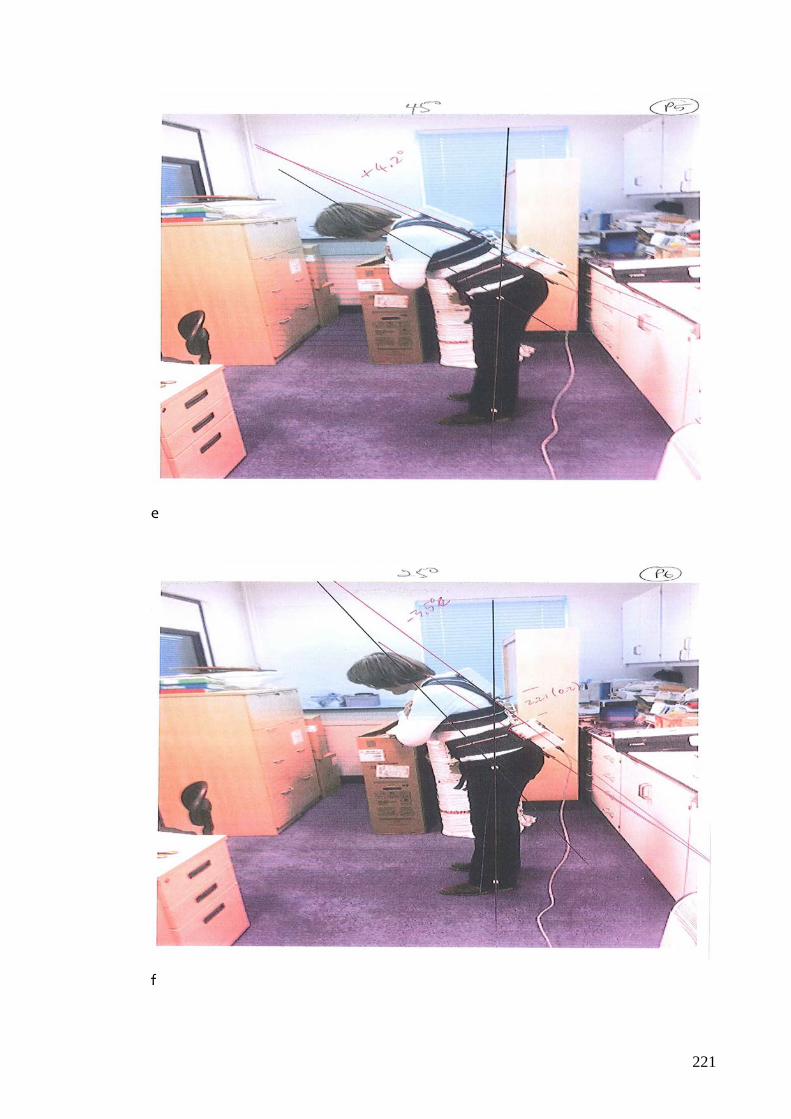
221
e
f

222
g

Appendix 4: Published pilot study: Evaluation of trunk acceleration in healthy Individuals and those with low back pain
Appendix 4 has been removed from this thesis due to the publisher’s copyright restrictions. As a result pages 223 – 230 of the thesis are not presented here.
The contents that has been removed is an article with the following citation:
(2011) Aluko, A., DeSouza, L. and Peacock, J., Evaluation of Trunk acceleration in healthy individuals and those with low back pain. International Journal of Therapy and Rehabilitation, 18(1): 18-25.
To obtain a copy of the article please go to http://www.ijtr.co.uk/cgi-bin/go.pl/library/article.cgi?uid=80927;article=IJTR_18_1_18_25 where the article is available for subscribers or available to purchase.

231
Appendix 5: Short non-standardised questionnaire

232
Appendix 6: Study stability exercises 1

233
Appendix 7: Study stability exercises 2

234
Appendix 8: Exercise compliance sheet

235
Appendix 9: Core stability exercise class exercise sheets used by Hillingdon
Community Health sheet level 1

236

237

238

239
Appendix 10: Core stability exercise class exercise sheets used by Hillingdon
Community Health sheet level 2

240
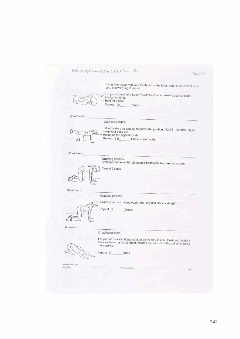
241

242

243
Appendix 11: Short history sheet used by recruiting Physiotherapists

244
Appendix 12: Patient information sheet

245

246

247

248
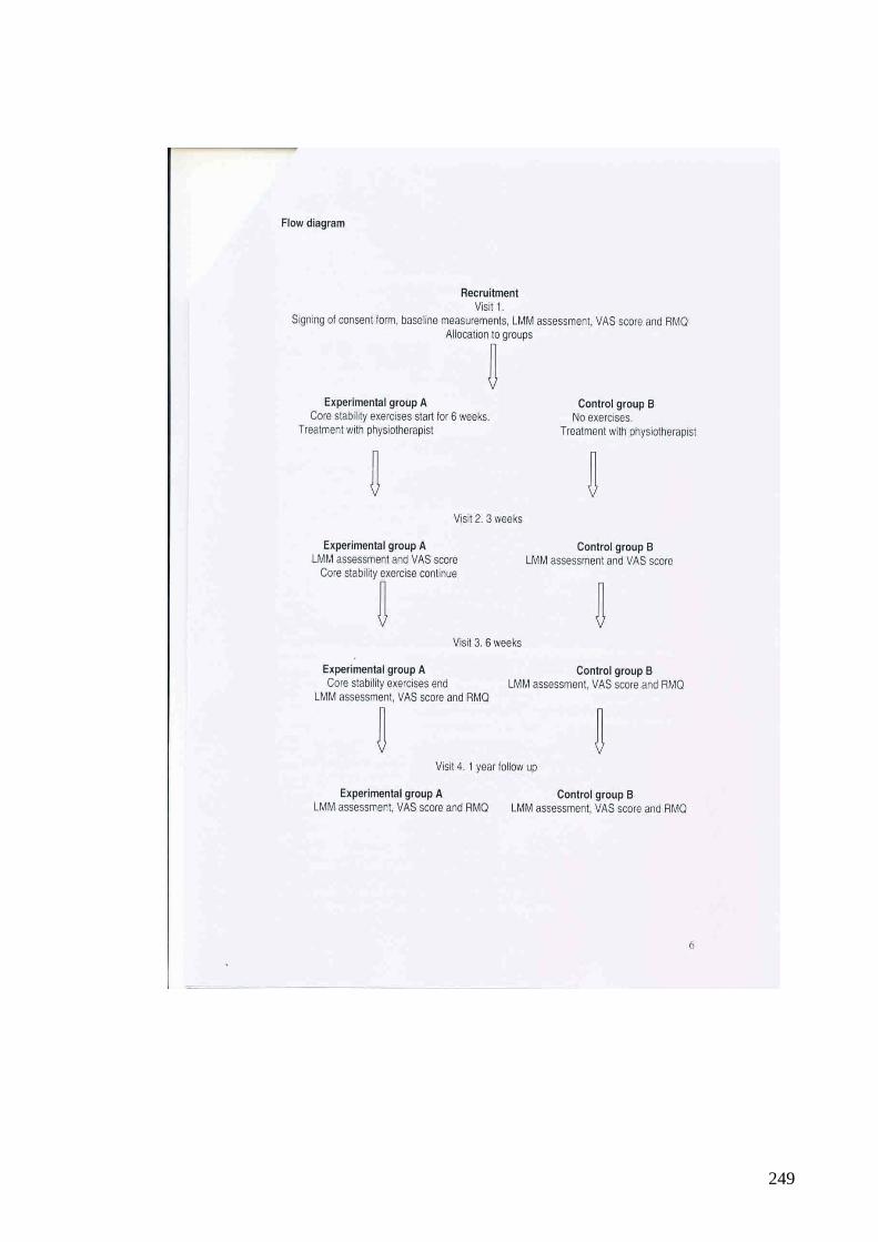
249

250

251
Appendix 13: Consent form

252

253
Appendix 14: Study Roland Morris Disability Questionnaire

254

255
Appendix 15: Study Visual Analogue Scale

256
Appendix 16: Approved amendments to study

257

258

259

260

261

262

263

264
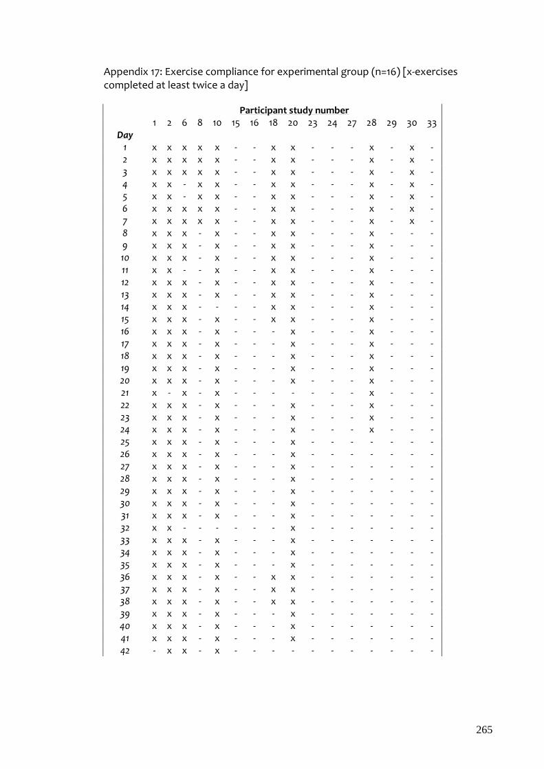
265
Appendix 17: Exercise compliance for experimental group (n=16) [x-exercises completed at least twice a day]
Participant study number 1 2 6 8 10 15 16 18 20 23 24 27 28 29 30 33
Day 1 x x x x x - - x x - - - x - x - 2 x x x x x - - x x - - - x - x - 3 x x x x x - - x x - - - x - x - 4 x x - x x - - x x - - - x - x - 5 x x - x x - - x x - - - x - x - 6 x x x x x - - x x - - - x - x - 7 x x x x x - - x x - - - x - x - 8 x x x - x - - x x - - - x - - - 9 x x x - x - - x x - - - x - - - 10 x x x - x - - x x - - - x - - - 11 x x - - x - - x x - - - x - - - 12 x x x - x - - x x - - - x - - - 13 x x x - x - - x x - - - x - - - 14 x x x - - - - x x - - - x - - - 15 x x x - x - - x x - - - x - - - 16 x x x - x - - - x - - - x - - - 17 x x x - x - - - x - - - x - - - 18 x x x - x - - - x - - - x - - - 19 x x x - x - - - x - - - x - - - 20 x x x - x - - - x - - - x - - - 21 x - x - x - - - - - - - x - - - 22 x x x - x - - - x - - - x - - - 23 x x x - x - - - x - - - x - - - 24 x x x - x - - - x - - - x - - - 25 x x x - x - - - x - - - - - - - 26 x x x - x - - - x - - - - - - - 27 x x x - x - - - x - - - - - - - 28 x x x - x - - - x - - - - - - - 29 x x x - x - - - x - - - - - - - 30 x x x - x - - - x - - - - - - - 31 x x x - x - - - x - - - - - - - 32 x x - - - - - - x - - - - - - - 33 x x x - x - - - x - - - - - - - 34 x x x - x - - - x - - - - - - - 35 x x x - x - - - x - - - - - - - 36 x x x - x - - x x - - - - - - - 37 x x x - x - - x x - - - - - - - 38 x x x - x - - x x - - - - - - - 39 x x x - x - - - x - - - - - - - 40 x x x - x - - - x - - - - - - - 41 x x x - x - - - x - - - - - - - 42 - x x - x - - - - - - - - - - -

Appendix 18: Submitted paper under review- Quantification of trunk performance in a sample population
Appendix 18 has been removed from this thesis due to the fact that the paper is currently under review and neither the publisher nor the copyright restrictions are known. As a result pages 266 – 278 of the thesis are not presented here.
The contents that has been removed is an article with the following citation:
(n.d.) Aluko, A., DeSouza, L. and Peacock, J., Quantification of trunk performance in a sample population. Submitted.