the effect of androgens and estrogens on human sebaceous … · 2017-01-30 · effect of androgens...
TRANSCRIPT

THE EFFECT OF ANDROGENS AND ESTROGENS ON HUMANSEBACEOUS GLANDS*
JOHN S. STRAUSS, M.D., ALBERT M. KLIGMAN, M.D., PH.D. AND PETER E. POCHI, M.D.
This paper deals with the mechanism of actionof androgens and estrogens on the human sebace-ous gland. Our experimental objectives were thefollowing: 1) to quantitate the effects of thesehormones on sebaceous gland function: 2) todetermine their mode of action, whether directlyon the target organ itself or through systemicpathways; and 3) to analyze the "antagonism"between androgens and estrogens on the seba-ceous gland.
MATERIALS AND METHODS
Most of the observations have been made on theglands of the face, because the fully developedglands of this region are very large and sebumsecretion is high (1—5).' We have centered ourattention particularly on the large sebaceousfollicles which are almost peculiar to this region(6). Before puberty, the sebaceous glands are tinyand insignificant, sometimes consisting of un-differentiated anlage with occasional nests oflipid-laden cells. In contrast, the postpubertalsebaceous follicles are unique in that the usualsize relationships are inverted, the glands having
* From the Department of Dermatology, BostonUniversity School of Medicine, and Evans Me-morial of Massachusetts Memorial Hospitals,Boston 15, Massachusetts (Drs. Strauss andPochi); and the Department of Dermatology, Uni-versity of Pennsylvania, Philadelphia 4, Pennsyl-vania (Dr. Kligman).
This work was supported in part by GrantsE-1936 and RG-606S, National Institutes ofHealth, U. S. Public Health Service; Postdoctor-ate Research Fellowship, GF 13-004, NationalInstitutes of Health, U. S. Public Health Service(Dr. Pochi); and by the Gillette Company (Massa-chusetts Memorial Hospitals); and in part by theOffice of the Surgeon General, Department of theArmy, under the sponsorship of the Commissionon Cutaneous Diseases of the Armed Forces Epi-demiological Board (University of Pennsylvania).
Awarded second prize, Eleventh Annual EssayContest, The American Dermatological Associa-tion.
Received for publication October 7, 1961.Volunteer subj ects were drawn from the follow-
ing sources: 1) Massachusetts Correctional Insti-tution, Walpole, Massachusetts, John E. Gavin,Superintendent; 2) Walter E. Fernald StateSchool, Commonwealth of Massachusetts, Mal-colm J. Farrell, M.D., Superintendent; 3) Wood-bine state Colony, Woodbine, New Jersey, H.VonBulow, Superintendent; 4) Vincland StateSchool, Vineland, New Jersey, Miles E. Drake,M.D., Superintendent; and 5) PhiladelphiaCounty Prison, Holmesburg, Pennsylvania.
139
acquired an extraordinary size and the hair havingbeen reduced to a mere vestige. Furthermore, thesebaceous follicles contribute most of the surfacelipid on the face. These are the follicles whichbecome involved in acne, a disease which we haveunder continuing study and which has been amajor stimulus to our interest in sebaceous glandphysiology.
The sebaceous gland is a holocrine gland whichdelivers its product, sebum, by the casting off oflipid-filled cells. The output of sebum, withcertain qualifications, as will be noted, is directlyproportional to gland size (6—8). The productivityof the gland can be measured either directly bygravimetric assay of collected sebum or indirectlyby histologic assessment of gland size. The formeris far more precise.
Gravimetric essay. The procedure has beendescribed elsewhere (8). The sebum produced onthe forehead from an area usually 6.45 squarecentimeters in size is collected for a three hourperiod. The area is delineated with adhesive tapeto prevent exchange of sebum with the sur-rounding skin. The sebum is trapped on thinabsorbent cigarette papers held in place by gauzeand a rubber bandage. Subsequently, the lipid isextracted from the papers with ether and weighed.This method of trapping the sebum as it is pro-duced overcomes one of the maj or difficultieswhich in the past has plagued quantitative studiesof sebum output, namely the extraordinarytendency for sebum to flow away and be lost (6).With the above technic, we have found that sebumproduction is relatively constant for any onesubject on repeated testing. Nevertheless, indi-vidual values do vary, sometimes considerably so.Thus, it is the trend of multiple readings that is ofsignificance.
Histology. Deep punch biopsy specimens, fourto seven millimeters in diameter, have been takenfrom symmetrical non-bearded areas of the checkbefore and after hormone administration. Serialsectioning is required for a reliable estimate ofgland size, because the glands vary in size andshape from follicle to follicle. This method isuseful only for comparatively gross differences.
QUANTITATIVE EFFEcTS OF ANDROGENS ON THESEBAEOU5 GLANDS
1) Systemic Administration of Androgens
There is substantial evidence from animalexperimentation that the sebaceous glands areunder androgenic control (9—16). Testosteroneincreases the size of the glands of immature rats;

140 THE JOIJENAL OF INVESTIGATIVE DERMATOLOGY
the glands of the adult animal undergo atrophyalter orchiectomy. While it is generally felt thatthe human sebaceous gland is also androgen-sensitive, the evidence is scanty. After admin-istering testosterone to castrate and eunuchoidmales, Hamilton observed increased oiliness ofthe skin (17). However, no quantitative measure-ments were made. In six prepuberal boys Ronyand Zakon observed histologic enlargement of thepubic sebaceous glands when 25 milligrams oftestosterone propionate were given three timesa week for two weeks (18). Furthermore, indirectevidence that the glands are under androgeniccontrol is afforded by the emergence of acne inassociation with androgen secreting tumors orandrogen therapy.
Experimental
A. Prepuberal subjects. Fourteen prepuberalchildren, eight boys and six girls, seven to elevenyears of age, were given methyl testosterone2 orallydaily for periods of 7 to 12 weeks. Five boys andthree girls received 50 milligrams daily; threegirls received 75 milligrams daily; and three boysreceived 100 milligrams daily. The glands werestudied in cheek biopsy specimens before andafter treatment. The final biopsy was usually doneat the end of treatment (seven to twelve weeks).In one subject, a specimen was removed at threeweeks.
In every case, except for two boys who received100 milligrams daily, there was substantial en-largement of the glands (Figure 1), although thedegree of enlargement varied from subject tosubject. The enlargement was already evident inthe three week specimen. By histologic criteria,the boys and girls seemed to respond similarly,although this cannot be considered final in view ofthe crudity of the method. The individual differ-ences in responsiveness are probably geneticallydetermined. One year later biopsy specimens oftwo boys, not yet embarked into puberty, showedregression of the glands. In most of these children,slight oiliness of the face was noted while theandrogen was administered and the follicularopenings appeared enlarged. In no case did acne-form lesions appear.
Sebum output was gravimetrically determinedin fifteen prepuberal boys, seven to 13 years ofage, who received 100 milligrams of methyl testos-terone daily. In ten, there was a definite increase,strikingly so in five (Figure 2). In three, the
2 Methyl testosterone supplied by Hubert C.Peltier, M.D., Harold L. Upjohn, M.D., andPorter F. Crawford, M.D., The Upjohn Com-pany, Kalamazoo, Michigan, and as Oreton Maby C. Kenneth Hawkins, M.D., Schering Cor-poration, Bloomfield, New Jersey.
increase was slight. Two were unresponsive.*Sebum output fell to the control level rapidlywhen the methyl testosterone was discontinued.
Next, the speed of response was determined.One hundred milligrams of methyl testosteroneorally daily caused histologic enlargement in twoprepuberal girls after seven and thirteen daysrespectively. The gravimetric data also indicateddefinitely increased sebaceous activity in two tothree weeks (Figure 2). Because of the danger ofepiphyseal closure, the drug was not continuedfor more than two months, and thus it was notpossible to determine whether maximal glandularstimulation had been reached. However, in manyof the subjects, the output even after this shortperiod was definitely in the adult range. Wetentatively feel that androgen stimulation canrapidly transform the skin into the postpubertaltype, anatomically and functionally. Within limits(children less than five years of age were notstudied) this androgen-induced metamorphosis isnot dependent on age or whether the individual, isabout to enter puberty.
The minimally effective androgenic stimulusmay also be considered an index of gland sensi-tivity. Two prepuberal boys received the smalldose of 5 milligrams of methyl testosterone orallydaily. Six weeks later, the glands had greatlyenlarged. In three of six prepuberal boys, 5 milli-grams of methyl testosterone orally daily causeda modest increase in sebaceous secretion. Thisdose appears to be close to the threshold. As willbe pointed out, 5 milligrams of methyl testosteroneorally daily is less than the estimated adultendogenous secretion of androgens, thus high-lighting the great sensitivity of the gland to thishormone. We did not quantitatively determine thedose-response relationship, but did show that 100milligrams of methyl testosterone orally dailybrought about a much greater increase in seba-ceous secretion than 5 milligrams orally daily.
B. Post-puberal males. Six adult males, between20 and 30 years of age, were given 100 milligramsof methyl testosterone orally daily. Post-treat-ment biopsy specimens at six weeks were com-pared to controls. No changes were seen histo-logically or clinically. Similarly, three adult malesreceived up to 300 milligrams of methyl testos-terone daily for eight weeks. Sebum productiondid not increase in any case.
C. Post-puberal females. Eight post-puberalfemales in the second and third decade received100 milligrams of methyl testosterone daily orallyfor six weeks. Histologically, four showed slightenlargement of the sebaceous glands. Two otherfemales received 200 milligrams of methyl testos-
* We cannot account for the complete androgeninsensitivity of occasional prepuberal subjects.Attention is called to the fact that the subjects inthese particular studies were mental defectiveswith a variety of associated physical abnormalitiesand stunted, retarded growth. Lack of absorptionmay be one factor.
EFFECT OF ANDROGENS AND ESTROGENS ON HUMAN SEBAdEOUS GLANDS 141
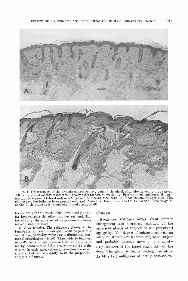
FIG. 1. Enlargement of the prepuberal sebaceous glands of the cheek of an eleven year old boy given100 milligrams of methyl testosterone orally daily for twelve weeks. A) Pretreatment specimen. Sehace-ous glands are small lateral outpocketings of undifferentiated cells. B) Post-treatment specimen. Theglands and the follicles have greatly enlarged. Note that the corium has thickened too. This magnifi-cation is the same as A (hematoxylin and eosin, X 39).
terone daily for six weeks. One developed glandu-lar hyperplasia; the other did not respond. Un-fortunately, the more sensitive gravimetric assaymethod was not used.
D. Aged females. The sebaceous glands of thefemale are thought to undergo involution partiallyin old age, probably reflecting a diminished hor-monal stimulation (19, 20). Three elderly females,over 65 years of age, received 100 milligrams ofmethyl testosterone daily orally for six to eightweeks. In each ease, sebum production increasedslightly, but not as rapidly as in the prepuberalsubjects (Figure 3).
Comment
Exogenous androgen brings about promptenlargement and increased secretion of thesebaceous glands of subj eets in the prepuberalage group. The degree of enlargement with anadequate stimulus varies from subject to subjectand probably depends more on the geneticresponsiveness of the target organ than on thedose. The gland is highly androgen-sensitive.As little as 5 milligrams of methyl testosterone
2.0
1,5
I.0
daily causes enlargement which becomes evidentin two to three weeks.
Male sex hormone is not a single compoundbut a family of substances derived primarily fromthe adrenal glands and the testes (21). Individual
androgens vary greatly in potency. Even thedetermination of total androgen secretion posesgreat technical problems. However, Fukushima,et al., basing their estimates on studies withradioactive testosterone, suggest that the daily
142 THE .TOIJRNAL OF INVESTIGATIVE DERMATOLOGY
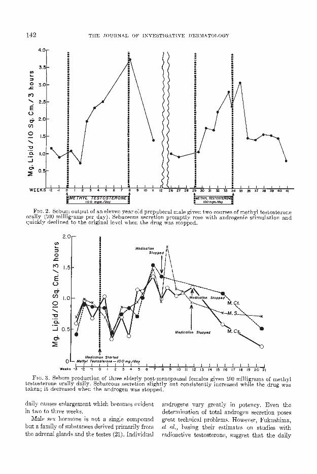
FIG. 2. Sebum output of an eleven year old prepuberal male given two courses of methyl testosteroneorally (100 milligrams per day). Sebaceous secretion promptly rose with androgenic stimulation andquickly declined to the original level when the drug was stopped.
MedicotlonStopped
U,
0II')-S...
EC-)
&U)0-S..
0._i 0.5a'
Medication Started0 - Me/hy/ Tes/oslerone — /00 mg/day
I I I I I I I I I I IWeeks 3 -2 -l 0 I 2 3 4 5 6 7 8 9 10 II 12 3 14 15 16 17 8 19 20 21
CS•S.%%-S••%
Fia. 3. Sebum production of three elderly post-menopausal females given 100 milligrams of methyltestosterone orally daily. Sebaceous secretion slightly but consistently increased while the drug wastaken; it decreased when the androgen was stopped.
EFFECT OF ANDROGENS AND ESTROGENS ON HUMAN SEBACEOUS GLANDS 143
endogenous production of androgen in the humanadult male, measured as testosterone, is about 17milligrams per day and certainly no more than36 milligrams per day (22). Since oral methyltestosterone has one-third the potency of circu-lating testosterone (23), our routine 100 milligramdose falls in the upper portion of the normalrange, while the minimally effective dose of 5milligrams is well below the average level. Pre-sumably, the endogenous secretion of androgenin the adult male is probably in excess of thatrequired to maintain maximal sebaceous glandfunction.
The extraordinary androgen sensitivity of theglands has been demonstrated in still anotherway. 17-alpha-ethynyl-19-nor-testosterone (Nor-lutin®) is a synthetic progestin with no significantandrogenic activity in animal assays utilizingweight increases of the seminal vesicles andventral prostate gland of immature castratedrats (24). Yet many female infants, born ofmothers receiving this compound during preg-nancy, have varying degrees of pseudohermaph-rodism (25, 26). We have found that 20 milligramsof this compound causes enlargement of theprepuberal sebaceous glands (27, 28). Accord-ingly, we have suggested that the human pre-puberal sebaceous gland provides a delicatebiological test system for detecting androgenicity(28).
Though we have focused on the sebaceousgland, androgens have far reaching effects onprepuberal skin. The effect is one of generalstimulation; the corium thickens greatly, thevasculature increases, the follicles lengthen, etc.(Figure 1).
At first we thought it surprising that the glandsof adult males did not enlarge after strongandrogenic stimulation. One need only realize,however, that in the post-adolescent male theglands of any given subject are already respond-ing to endogenous androgens which are ordinarilyin excess of that required for maximal effect.
The situation is somewhat different in thepost-puberal female. Despite some controversy,females on the average secrete less sebum thanmales (3, 29—31). We have confirmed this in ourown gravimetric studies (unpublished data).Acne is less severe and less frequent in females(32). Also, the endogenous secretion of androgenby the female is about two-thirds that of themales. The fact that exogenous testosterone
causes slight enlargement in some post-adolescentfemales probably indicates that the endogenousandrogen output is somewhat less than that whichwould enable the glands to achieve their fullresponsiveness.
2) Local Application of Testosterone
The question with which we are concerned hereis whether testosterone exerts a direct action onthe target organ. Topical application, by pre-sumably concentrating the drug locally in theskin, permits one to decide whether this is thecase or not. One has to be aware, however, thatthe changes produced by such a potent, readilyabsorbed hormone might be part of a generalizedresponse following absorption of the drug throughthe skin. To prove that testosterone acts directlyon the target organ, one must show that glandularenlargement occurs earlier and to a greaterdegree at the local site. Therefore, a series ofobservations have to be made, all of which arecontrolled by determining any changes of thecontralateral untreated side.
An alternate method would consist of super-ficially injecting the testosterone in a repositoryform. We abandoned this approach when wefound that injection of depot testosterone causeddermal inflammation and partial involution ofthe glands. Dermal inflammation has been foundto cause atrophy of the glands invariably (33).
ExperimentalThis study utilized prepuberal subjects ex-
clusively because of the high androgenic sensi-tivity of their glands. The scalp is another areawhere the glands reach great size in adults. There-fore, after determining that the glands of the faceand scalp respond in a parallel and proportionatefashion to androgen, we utilized the scalp in somecases.
In four prepuberal boys, 9 to 11 years old, 10per cent testosterone propionate in HydrophylicOintment, DSP., was applied once daily to oneside of the scalp. A control biopsy specimen wasremoved beforehand; biopsy specimens were ob-tained from the treated and contralateral controlsites after the application had been continued forthree weeks. Although there was modest enlarge-ment in the control areas indicating systemiceffects through absorption, the glands of the
Testosterone propionate supplied by G.Kenneth Hawkins, M.D., Schering Corporation,Bloomfield, New Jersey, and by Porter F. Craw-ford, M.D., Hubert C. Peltier, M.D., and HaroldL. Upjohn, M.D., The Upjohn Company, Kala-mazoo, Michigan.

144 THE JOURNAL OF INVESTIGATIVE DERMATOLOGY
treated area underwent a strikiag increase in sizein each case (Figure 4).
In two other prepuberal males, 10 per centtestosterone ointment was applied to one cheekonce daily for two months. There was great and
FIG. 4. Sebaceous gland enlargement producedby daily inunction of approximately 0.1 gram of10 per cent testosterone propionate ointment forthree weeks on the scalp of a prepuberal boy. A)Control specimen with small glands. B) Glandshave enlarged at the site of application. C) Slightor questionable enlargement of glands of un-treated control site. Biopsy specimen taken atsame time as B. (Hematoxylin and eosin, A, B,& C x 55).
equal glandular enlargement on both sides showingthat percutaneous absorption is sufficient toachieve maximal effects throughout the skin wheninunctions of androgen in a high concentration arecarried out for many weeks.
Comment
The evidence is compelling that testosteronehas a direct local action on the sebaccous gland.Enlargement occurs to a far greater degree atthe treated site (three-week experiment) and isfollowed by a generalized uniform response dueto systemic absorption (two-month experiment).The inunction consisted of approximately 0.1to 0.2 grams of ointment daily, which wouldtherefore contain 10 to 20 milligrams of testos-terone propionate. Even if only 10 per cent (1 to 2milligrams) of testosterone propionate wereabsorbed, androgenic effects could be expectedaccording to the data previously given. Perhapsby progressively reducing the concentration ofhormone, one could find a threshold amountwhich would limit the enlargement to the site ofapplication.
The present findings correlate well with otherstudies which show a direct effect of androgcns onthe pilosebaceous apparatus of man (34—36).When applied locally, testosterone causes in-creased hair growth on the abdomen (34), inthe axilla (35, 36), and in the pubic area (36). Toextend these observations, 10 per cent testos-terone propionate ointment was applied to oneaxilla of four male prepuberal subjects, five,seven, ten, and eleven years of age. In the twooldest boys, the first hair growth occurred at thesite of application. After three months, terminalhairs had developed both in the untreated axillaand in the pubes, indicating systemic absorption.The other two boys did not respond to the topicalandrogen.
Enlargement of the cock's comb is commonlyused as a test of androgenicity (37). Morato-Manaro and Albrieux applied an androgen toone half of a comb which had been completelydivided surgically (38). Greater enlargementoccurred in the treated half of the comb, thusfurther authenticating the direct action of testos-terone.
QUANTITATIVE EFFECT OF ESTROGEN ON THESEBACEOU5 GLANn5
1) Systemic Administration of Estrogens
It is commonly accepted that estrogens have asuppressive effect on human sebaceous secretion.

EFFECT OF ANDROGENS AND ESTROGENS ON HUMAN SEBACEOUS GLANDS 145
The evidence usually cited is the reported thera-peutic value of estrogens in acne vulgaris. Never-theless, with the exception of the few easesstudied by Jarrett (39), one searches in vain forobjective, controlled clinical studies whichunequivocally demonstrate inhibition of sebace-ous secretion. As a matter of fact, it will becomeall too clear that the improvement of acne usuallyclaimed for estrogens, whether topical or oral, hasbeen achieved with amounts too small to inhibitsebaceous output. Indeed acne is far too variableand fluctuating a disease to be used casually as abiological measurement of sebaceous glandphysiology. The time is overripe for directquantitative assessment, leaving aside the thornyproblem of acne.
In animals, on the other hand, the issue hasbeen settled beyond doubt. Ebling obtainedwhat he describes as "limited reduction" of thesebaceous glands in rats weighing 40 to 60 gramswith the subcutaneous injection of 1.0 gamma ofestradiol benzoate daily for thirty days. However,when 100 gamma of estradiol benzoate were givenunder the same conditions, the decrease in thesebaceous glands was marked (11). Others haveconfirmed the depression of sebaceous glands inanimals with large amounts of estrogen (15,40—43). On a comparative weight basis, even thesmallest dose that Ebling used, which would beequivalent to a daily subcutaneous dose of 1.5milligrams of estradiol benzoate for a 70 kilogramhuman, is more than twice as large as the maxi-mum therapeutic replacement dose for humans(44, 45); Ebling's higher dosage is obviouslyexcessive.
ExperimentalOf necessity all of the subjects were post-
puberal males and females with good glandulardevelopment. The highly potent synthetic estro-gen, ethynyl estradiol, was used in most of thestudies. In the experiments to be described, whenthe dose was to exceed 1.0 milligram of ethynylestradiol, it was approached slowly over a periodof seven to ten days to avoid nausea and vomiting.
Fifteen milligrams of ethynyl estradiol orallyper day, the largest dose used, were given to threeadult males for six weeks. Histologically, theglands became extremely atrophic and usuallywere reduced to small nests of undifferentiatedcells. This shrinkage and involuting effect seemedto be more or less restricted to the glands, ex-empting the follicle and corium. Under the impact
of estrogen, the entire skin does not revert to itsprepuberal condition.
Four more males, between sixteen and twentyyears of age received 10 milligrams of ethynylestradiol daily for eight weeks with effects identi-cal to that of the higher dose.
Thereafter, twenty young adult subjects (six-teen males, four females) received 5 milligrams ofethynyl estradiol daily for periods up to twomonths. Again, the glands in every case becamealmost completely atrophic, reverting to the prc-puberal state in those with the greatest change(Figure 5). There were individual differences inthe final degree of involution, but these could notbe correlated with original gland size. In all of thesubjects, the skin was subjectively and objectivelyless oily.
Five milligrams of ethynyl estradiol greatlysuppressed sebaceous secretion in three subjects(Figure 6). The decrease was detected in two tothree weeks, although the maximum effect oc-curred later. This was a reversible change; sebumoutput returned to normal eight to twelve weeksafter the drug was stopped. Similarly by histologicexamination of biopsy specimens from the cheekof five adult males who received 5 milligrams ofethynyl cstradiol daily, there was beginninginvolution at three weeks and by four weeks thedecrease was unmistakable. Five weeks after thedrug was stopped, restoration was well under wayand by nine weeks the glands had regeneratedfully.
Seven young adult subjects (four males, threefemales) received 2.5 milligrams of ethynyl cstra-diol daily for six weeks. Here the histologic de-crease in gland size was less. Moreover individualresponses were more variable. One female did notrespond at all.
Sebum output was gravimetrically estimatedin tbree males and two females who received 1.0milligram of ethynyl estradiol daily. In each case,sebum production was reduced significantly(Figure 7). It should be emphasized that 1.0 milli-gram is still many times the physiological re-placement dose.
At this point, the greater sensitivity of gravi-metric assay became apparent. By histologicexamination, doses of 1.0 and 0.5 milligrams failedto cause atrophy of the glands in two groups offive males each. By contrast, in six adult males0.5 milligram of ethynyl estradiol definitely sup-pressed sebaceous secretion (Figure 8). The lowestdose evaluated was 0.25 milligram which gave aquestionable slight reduction in sebum production.This is probably the threshold dose as measuredby sebum production. The disparity betweenhistologic and gravimetric suppression by estro-gens is an exception to the well-founded principlethat sebaceous secretion is proportionate to glandsize. We tentatively hold that threshold doses ofestrogen may have the special effect of inhibitingthe proliferation of sebaceous cells without acorresponding shrinkage of glandular size.
Three young adult males received 50 milligramsEthynyl cstradiol (Estinylit) supplied by G.
Kenneth Hawkins, M.D., Schering Corporation,Bloomfield, New Jersey.

r '
I
146 THE JOURNAL OF INVESTIGATIVE DERMATOLOGY
Fin. 5. Depression of sebaceous glands of adult male by 5 milligrams of ethynyl estradiol orally dailyfor six weeks. A) Pre-treatment specimen with lipid-laden, large sebaceous glands. B) After treatment,the glands, which have been almost completely wiped out, are reduced to nubbins of undifferentiatedcells. (Hematoxylin and eosin, both A and B. X 32).
of diethylstilbestrol5 orally daily for six to eight milligrams, and two males received 40 milligramsweeks. In each case there was moderate histologic of conjugated estrogenic substances (Premarin®)6reduction in gland size, orally daily for six weeks. The 40 milligram dose
Finally, two young adult males received 20 caused a slight decrease in gland size while thelower dose had little or no effect.
Diethylstilbestrol supplied, by Hubert C. _______________________________________________Peltier, M.D., Harold L. Upjohn, M.D., and 6 Conjugated estrogenic substances (Pre-Porter F. Crawford, M.D., The Upjohn Corn- marinR) supplied by Ayerst Laboratories, Newpany, Kalamazoo, Michigan. York, New York.

EFFECT OF ANDROGENS AND ESTROGENS ON HUMAN SEBACEOUS GLANDS 147
30
-S
EaU)
0-S
a.-Ja
Estradlot5mg/day
Fm. 6. Suppressive effect of 5 milligrams ofethynyl estradiol administered daily on thesebaceous glands of an adult male. Sebum outputhas declined greatly after three weeks, reaching amaximum in four to six weeks. It takes 9 to 12weeks for the secretory activity to recover fullyafter stopping the drug. The changes in two otheradult males were similar.
Comment
It is quite evident that extremely high un-physiologic amounts of estrogen are necessaryto cause the glands to revert to the prepuberalrudimentary state. For purposes of comparativeanalysis, the replacement dose of ethynyl estra-diol, which is a rough estimate of the amountsecreted endogenously, is usually less than 0.05milligram per day (44, 45). Five milligrams ofethynyl estradiol invariably produced almostcomplete obliteration of the glands. Histologi-cally, 2.5 milligrams daily (approximately fiftytimes the physiologic amount) was close to thethreshold while 1.0 milligram daily had no effect.With the more sensitive gravimetric assay, 0.25milligram of ethynyl estradiol appears to ap-proach the threshold. Even this dose is at leastfive times the physiologic amount.
It is a fair estimate that ethynyl estradiol istwenty times more potent than diethylstilbestroland thirty times more active than conjugatedestrogenic substances (44, 45); hence, 50 milli-grams of diethylstilbestrol, approximately equiv-alent to 2.5 milligrams of ethynyl estradiol,
U,S..
0
5-.
E0&
Cl)
0-Da.
2.0
do
tTh,,,I E.t,eeeI— p ..ç/dsy
Fio. 7. Suppression of sebum output by 1.0 milli-gram of ethynyl estradiol administered daily toan adult male and female. Similar changes oc-curred in two other males and one other female.
could be expected to suppress the glands mod-erately by histologic criteria. The 20 milligramdose of Premarin®, corresponding to less then 1.0milligram of ethynyl estradiol, was without effecthistologically in agreement with expectations.Forty milligrams of this substance constituted ahistologic threshold dose. It should be notedthat these agents were not evaluated by themore sensitive gravimetric method of assay. Atthe doses used these drugs undoubtedly wouldhave caused some decrease of sebaceous secre-tion. In any case, the amount required to producealmost complete histologic involution is so hugeas to represent a pharmacologic, not a physio-logic, effect.
A further qualification is necessary. All ofthese studies were of relatively short duration (lessthan two months). What effect small doses ofestrogen administered over much longer periodsof time would have is not known.
With these high doses, it is not surprising that
4.0
3.0
2.0 -
E. bY. — Adult Mole
0-Weeks
4.0 -
JO
0-Weeks

148 THE JOURNAL OF INVESTIGATIVE DERMATOLOGY
en
30
If)
EC-)
ci-C/)
C
0.4&
Estradiol05 m9./da
Fio. 8. Suppression of sebum output by 0.5milligram of ethynyl estradiol administered dailyto an adult male. The drop is similar to that oc-curring with higher doses of ethynyl estradiol.
ExperimentalWe first determined the threshold dose which
would just cause glandular suppression at thetreated site. Approximately 0.1 to 0.2 gram of 1.0per cent ethynyl estradiol in Hydrophylic Oint-ment, 1J.S.P., was applied to the forehead of fouradult males once daily for seven to ten weeks.Gravimetrically, sebum output definitely de-creased in three subjects. This decrease wasalready evident in two to three weeks. In thefourth subject, there was no significant sup-pression of sebum output.
A 0.5 per cent ethynyl estradiol ointment wasapplied daily to the forehead of five more adultmales. In only one of these did sebaceous secretiondecrease. It would appear, then, that the minimaleffective topical concentration of cthynyl estra-diol lies between 0.5 and 1.0 per cent. It should benoted, however, that all subjects, including thosewhose sebum output was unaffected, experiencedthe usual galaxy of estrogenic side effects.
To determine whether estrogen acts directly onthe target organ, the sebaccous gland, the experi-mental design of the topical androgen study wasfollowed. The estrogen ointment was appliedlocally to one cheek, and the changes in thesebaceous glands of both cheeks were followedhistologically. A control biopsy specimen wasremoved beforehand.
Six post-puberal males, between the ages of 15and 20 served as subjects. Four received 10 percent ethynyl estradiol ointment once daily; tworeceived 10 per cent beta-estradiol ointment oncedaily. The biopsies taken at six weeks revealed amarked and equal decrease in systemic action.Feminization was evident in all six subjects,obviously reflecting percutaneous absorption.
Since histologic examination is not suitable toserial follow-up, the more sensitive gravimetrictechnique was used in the remaining studies. Theestrogen ointment was applied to one side of theforehead and sebum output was determined onboth sides simultaneously at various intervals.
Ten per cent ethynyl estradiol in HydrophylicOintment, U.S.P., was applied to one side of theforehead of two adult males once daily. Threemore received 5 per cent ethynyl estradiol oint-ment. These concentrations greatly exceed thethreshold dose. In all of these subjects, there wasan equivalent decrease in sebaccous secretion onboth sides (Figure 9).
A threshold concentration of 1.0 per centethynyl estradiol ointment was applied to one sideof the forehead of three adult males daily. In twosubjects, sebum production decreased on thetreated side. However, an equally great sup-pression of sebum output occurred on the un-treated control side (Figure 10).
Comment
With neither the high (5 to 10 per cent) northe threshold (1 per cent) concentration of
4.0
35
3.0
2.5 -
20 -
1.5 -
1.0
0.5
0Weeks
the characteristic systemic effects of estrogenwere displayed in every subject. In the male,these consisted of softening of the testicles, lossof libido, breast enlargement and increasedarcolar pigmentation. It is noteworthy that thelarger dosages of 5 to 15 milligrams had feminiz-ing effects which were no greater than with 1.0milligram. Every female developed arcolarpigmentation and amenorrhea followed by with-drawal bleeding when the drug was stopped.These effects were reversible since the drug wasgenerally not given for more than two months.
2) Local Application of Estrogen
As with testosterone we sought to discoverwhether cthynyl cstradiol acted directly on theeffcctor organ or via a central mechanism. Here,too, if one is to prove a direct inhibitory effect atthe target organ, it will be necessary to show a sup-pressive effect at the application site which isclearly greater tban that occurring in the control,untreated site. Obviously adults must be used assubjects.

0)I-0
S.-
EC-,
S.Cl,
0S.-•0a.-J
Period ofApplication
FIG. 9. Decreased sebum output after daily inunction of approximately 0.1gramof 10 per cent ethynylestradiol ointment to one side of the forehead of an adult male. Sebum production was assayed simul-taneously on both treated and untreated sides. Scbaceous secretion decreased sharply but equally onboth sides.
ethynyl estradiol ointment was there any evi-dence of earlier or greater suppression of scba-ccous secretion at the treated site. This resultindicates that the effects obtained requiredsystemic absorption prior to producing the ob-served results on sebum secretion.
ANTAGONISM BETWEEN ANDROGENS
AND ESTROGENS
It is more of a social than a biological truththat maleness and femaleness arc opposites.Endocrinologically, human beings may be con-sidered ambosexual since both adrogcns andestrogens arc present in all normal males andfemales, although there are marked differencesin the proportions of the various sex steroids.The gonads arc not the only source of sex hor-mones. For example, in the male, orchiectomyonly reduces urinary androgens by one-third;adrcnaleetomy essentially eliminates the remain-ing source of androgens (46). Ketosteroid excre-tion in the female is two-thirds of that of a male(47). Since the adrenals are the predominantsource of androgens in both males and females,
it is not surprising that urinary 17-ketosteroidexcretion is not that much greater in the male.
It is customary in clinical medicine to think ofandrogens and estrogens as counteracting orantagonizing each other. For instance, highdosages of testosterone are masculinizing andsuppress menstruation in females (48). Con-versely, high doses of estrogen feminize the maleand ameliorate prostatic cancer, an androgendependent malignancy (49, 50). It should berealized however that these arc biologicallycomplex situations. Such antithetic responsesprobably involve indirect interactions of severalsystems. There is no clear understanding of howsuch effects are brought about.
The issue before us now is to define experi-mentally wherein lies the so-called "antagonism"between estrogens and androgens. The literatureoften implies that circulating estrogens andandrogens are struggling or competing witheach other for a target organ such as the uterusor the sebaceous gland. If the "antagonism" is atthe target organ, there are several theoreticalways in which this might occur. For example,
—10% Ethynyl Estradlol Cream doily
)t—X Untreated Control side
EFFECT OF ANDROGENS AND ESTROGENS ON HUMAN SEBACEOUS GLANDS 149
6.0 -
5.0
4.0
3.0
2.0
1.0
0-Weeks

150 THE JOURNAL OF INVESTIGATIVE DERMATOLOGY
3.5
Fin. 10. Decreased sebum output after dailyinunetion of approximately 0.1 gram of 1.0 percent ethynyl estradiol ointment to one side of theforehead of an adult male. Sebum production wasassayed simultaneously on both treated and un-treated sides. Sebaceous secretion decreasedsharply but equally on both sides.
estrogens could block androgens in the same waythat atropine pharmacologically inhibits acetyl-choline at the end-organ; or the struggle mightbe compared to the pharmacologically opposingactions of methyl nicotinate and nor-epinephrineon the cutaneous capillaries. Nicotinate-inducedvasodilation is quickly transformed into vaso-constriction by the injection of norepinephrine(51).
The following experiments test whetherestrogen has a direct "neutralizing" or "blocking"effect on testosterone at the target organ. First,however, it will be helpful to summarize thefacts established to this point: 1) testosteronehas a direct local effect on the sebaceous glands;quite small amounts are effective; 2) estrogensdo not appear to act locally; very large un-physiological amounts are necessary for sup-pression.
Experimental
Our reasoning in designing these experimentswas that if estrogens counteract androgens at thetarget organ, for every dose of androgen, there
30Cw)'SE•0a•005'
0.-jWI
Week
should be an approximately corresponding dose ofestrogen which would cancel its effect. Higheramounts of androgen would require proportionateincreases in estrogen.
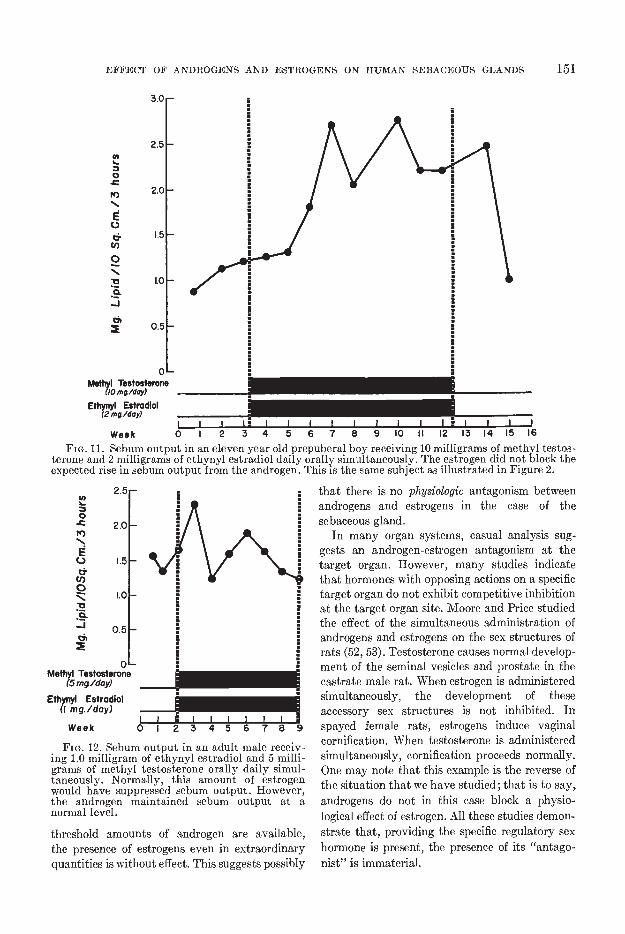
Three different experiments were done. First,10 milligrams of methyl testosterone and 2 milli-grams of ethynyl estradiol were simultaneouslyadministered orally daily to a prepuberal boy fornine weeks. There was a prompt marked rise insebaceous secretion (Figure 11). If one comparesthe rise in sebum output when this same subjectwas given androgens alone (Figure 2), it will beseen that the response was identical. The estrogendid not prevent the androgen from exerting itsgrowth stimulating action on the gland.
Next, three normal adult males received con-comitantly 1.0 milligram of ethynyl estradiol and5 milligrams of methyl testosterone. There wasno suppression in sebaceous secretion (Figure 12).This small amount of methyl testosterone, whichis less than the endogenous secretion of androgenmaintained sebaeeous secretion in the face of asuppressive dose of estrogen.
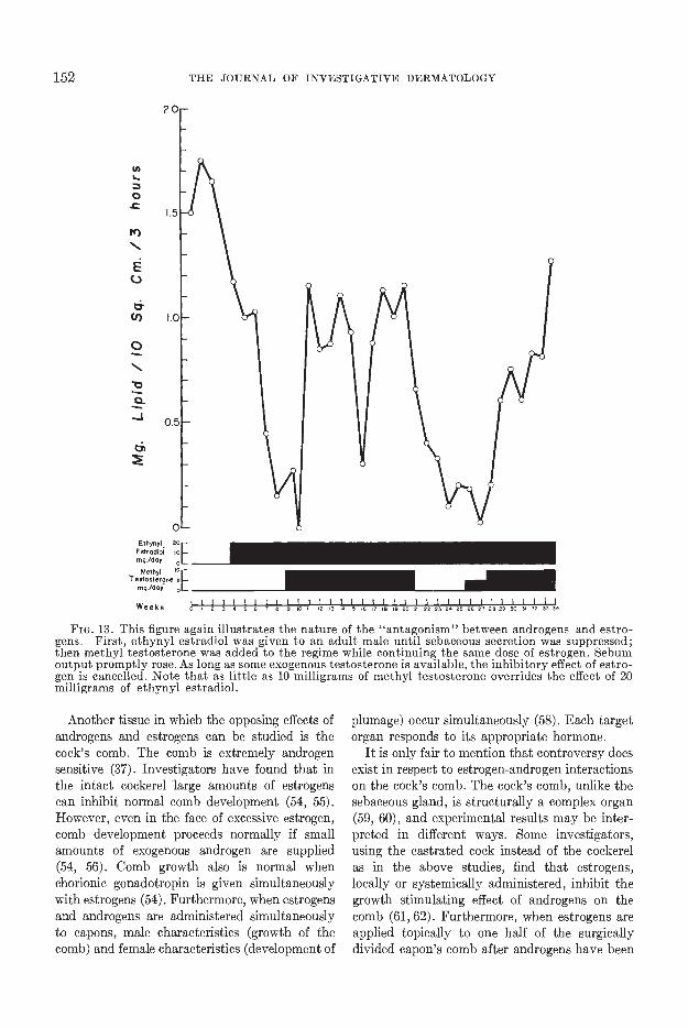
In the final experiment, sebaceous secretion wasfirst maximally suppressed by large amounts ofestrogen (1 to 20 milligrams of ethynyl estradiol).At that point, a small amount of methyl testos-terone (5 to 10 milligrams) was added so that theprepared subjects received both agents con-comitantly daily. The result was clear cut. He-gardless of the amount of estrogen given, theandrogen promptly increased sebum output in allfive subjects. The most dramatic illustration ofthe ability of a small amount of androgen to over-come the suppressive effect of excessive estrogenis given in Figure 13. While this man was receivingthe massive dose of 20 milligrams of ethynylestradiol daily, sebum production rose signifi-cantly during two courses of 10 milligrams ofmethyl testosterone orally.
Comment
These studies rule out the possibility thatestrogens antagonize androgens at the level ofthe target organ. In review, the three separatefindings are: 1) the scbaceous glands of theprepuberal child will respond to exogenousandrogen even in the presence of excess estrogen;2) if a small quantity of androgen is given to anormal adult, even large amounts of estrogenhave no inhibiting effect; and 3) after the glandshave been suppressed by estrogens, the introduc-tion of small amounts of androgen exogenouslywill cause prompt increase in sebum outputdespite continuing excessive estrogen.
These findings possibly seem to indicatethat the effect of large, quite unphysiologicaldosages of estrogen is to block the supply ofandrogen available to the sebaceous gland. When
1% Ethynyl Eetrodlol Cream dolly
X————X LMheeted Control Side
3.0
as -
2.0
0.5
IC
0Methyl Testosterone
(5mg/day)
Ethynyl Estradlol(I mg/day)
Week
FIG. 12. Sebum output in an adult male receiv-ing 1.0 milligram of ethynyl estradiol and 5 milli-grams of methyl testosterone orally daily simul-taneously. Normally, this amount of estrogenwould have suppressed sebum output. However,the androgen maintained sebum output at anormal level.
threshold amounts of androgen are available,the presence of estrogens even in extraordinaryquantities is without effect. This suggests possibly
that there is no physiologic antagonism betweenandrogens and estrogens in the case of thesebaceous gland.
In many organ systems, casual analysis sug-gests an androgen-estrogen antagonism at thetarget organ. However, many studies indicatethat hormones with opposing actions on a specifictarget organ do not exhibit competitive inhibitionat the target organ site. Moore and Price studiedthe effect of the simultaneous administration ofandrogens and estrogens on the sex structures ofrats (52, 53). Testosterone causes normal develop-ment of the seminal vesicles and prostate in thecastrate male rat. When estrogen is administeredsimultaneously, the development of theseaccessory sex structures is not inhibited. Inspayed female rats, estrogens induce vaginalcornification. When testosterone is administeredsimultaneously, cornification proceeds normally.One may note that this example is the reverse ofthe situation that we have studied; that is to sayandrogens do not in this ease block a physio-logical effect of estrogen. All these studies demon-
strate that, providing the specific regulatory sexhormone is present, the presence of its "antago-nist" is immaterial.
EFFECT OF ANDROGENS AND ESTROGENS ON HUMAN SEBACEOUS GLANDS 151
3.0 -
2.5 -
02.0-
EC.)
0•U)0
0.-J
0.5
0-Methyl Testosterone
(10 mg_/day)
Ethynyl Estradlol(2mg/day)
Week 0 I 2 3 4 5 6 7 8 9 10 II 2 3 14FIG. 11. Sebum output in an eleven year old prepuberal boy receiving 10 milligrams of methyl testos-
terone and 2 milligrams of ethynyl estradiol daily orally simultaneously. The estrogen did not block theexpected rise in sebum output from the androgen. This is the same subject as illustrated in Figure 2.
U)
0• 2.0N)
EC.) .5-&Cl)0Z .0-0.
0.5-0
V
0_____ I I I3456789
152 THE JOURNAL OF INVESTIGATIVE DERMATOLOGY
FIG. 13. This figure again illnstrates the nature of the "antagonism" between androgens and estro-gens. First, etbynyl estradiol was given to an adult male until sebaceous secretion was suppressed;then methyl testosterone was added to the regime while continuing the same dose of estrogen. Sebumoutput promptly rose. As long as some exogenous testosterone is available, the inhibitory effect of estro-gen is cancelled. Note that as little as 10 milligrams of methyl testosterone overrides the effect of 20milligrams of ethynyl estradiol.
Another tissue in which the opposing effects ofandrogens and estrogens can be studied is thecock's comb. The comb is extremely androgensensitive (37). Investigators have found that inthe intact cockerel large amounts of estrogenscan inhibit normal comb development (54, 55).However, even in the face of excessive estrogen,comb development proceeds normally if smallamounts of exogenous androgen are supplied(54, 56). Comb growth also is normal whenchorionic gonadotropin is given simultaneouslywith estrogens (54). Furthermore, when estrogensand androgens are administered simultaneouslyto capons, male characteristics (growth of thecomb) and female characteristics (development of
plumage) occur simultaneously (58). Each targetorgan responds to its appropriate hormone.
It is only fair to mention that controversy doesexist in respect to estrogen-androgen interactionson the cock's comb. The cock's comb, unlike thesebaceous gland, is structurally a complex organ(59, 60), and experimental results may be inter-preted in different ways. Some investigators,using the castrated cock instead of the eoekerelas in the above studies, find that estrogens,locally or systemically administered, inhibit thegrowth stimulating effect of androgens on thecomb (61, 62). Furthermore, when estrogens areapplied topically to one half of the surgicallydivided capon's comb after androgens have been
U)
a-C
K)NEU
&U)
CN
0.-j
EIhynyI_ 20Esrodiyl IDmg/day 0
Methyl '°vestos,e,one ,
thg./doI 0
Weeks 1111111 1111111111 III 111111111 II012345672910 111213141516171e19232122232425262722203031323334
EFFECT OF ANDROGENS AND ESTROGENS ON HUMAN SEBACEOUS GLANDS 153
applied to both halves, much greater atrophyoccurs on the estrogen treated portion of thecomb (38, 63, 64). This seems to be unassailableevidence for a direct local effect of estrogen.Closer scrutiny raises some doubts. One of the tworeports involved the use of only two birds (38).The other contains a gross inconsistency. Theauthors applied varying amounts of estrone toone half of the divided comb of capons (63, 64).The combs had been previously stimulated bythe topical application of testosterone to bothhalves of the comb. While they noted significantsuppression of comb growth when 100 and 200gamma of estrone were applied, there was nodepression with 160 gamma. If 100 and 200gamma are effective, how can 160 gamma beineffective?
Assuming as we have, that estrogens exerttheir effect systemically, how is this broughtabout? There is considerable data supporting theconcept that estrogens inhibit the pituitaryproduction of gonadotropins; this in turn de-creases the production of androgens by thetesticle. Payne has demonstrated marked histo-logic changes in the pituitary glands of fowltreated with estrogens (65). This is indirectanatomical evidence. Moore and Price (52, 53)found that estrogens caused atrophy of the ac-cessory male sex organs, an effect which wasovercome by administering gonadotropins ortransplanting isogenetic pituitary tissue. Inaddition, Boas and Ludwig, in a well controlledstudy, found that the administration of chorionicgonadotropin prevented the estrogen inducedatrophy of the cock's comb (54).
In human beings, too, the administration ofestrogens to males decreases androgen produc-tion (57, 66—68). This probably reflects decreasedpituitary output of gonadotropins (69), as in theanimal experiments. Indeed, the reduction of thehigh gonadotropin levels in the post-menopausalfemale by exogenous estrogens is great enough tohave been used as an assay for estrogens (70).
If the gonads were the chief source of andro-gens, the explanations given to this point wouldbe sufficient. However, even in the male, it is theadrenal which produces the major amount ofandrogen. It is necessary to postulate that estro-gens, in some way, suppress the synthesis ofandrogen by the adrenal. This could be a directeffect, or an indirect one operating through thepituitary. Unfortunately, experimental medicineprovides no clear cut answer one way or the
other. There is, however, recent evidence thatestrogens cause a further depression of androgensecretion in castrated human males (68, 71).
SUMMARY AND CONCLUSIONS
The quantitative effect of estrogens andandrogens upon human sebaceous glands hasbeen investigated using histologic and gravi-metric methods.
Androgens, even in less than physiologicdoses, cause great enlargement of the sebaceousglands of the prepuberal male and female in twoto three weeks.
Androgens, even in high doses, cause no furtherenlargement of adult male's sebaceous glands. Bycontrast, the glands of some adult females in-crease moderately as do the partially involutedglands of aged females.
Topical application of testosterone propionatehas a greater local growth stimulating effect onthe prepuberal gland at the treated site as com-pared to the untreated side. Testosterone actsdirectly on the sebaceous glands.
Estrogens suppress sebaceous secretion onlywith unusually high, pharmacologic rather thanphysiologic, doses. Maximal histologic suppres-sion is attained by about 2.5 milligrams of ethynylestradiol, approximately fifty times the replace-ment dose. The threshold suppressive dosemeasured by gravimetric technics is about fourtimes the physiological dose.
A 1.0 per cent ethynyl estradiol ointment,applied once daily, approaches the topicalthreshold level. Lesser concentrations are ineffec-tive in suppression of sebum production.
The topical application of a 1.0 to 10 per centethynyl estradiol ointment is equally suppressiveon the treated and control sides.
The inhibiting effects of large dosages ofestrogen are not manifested when a small amountof androgen is given concomitantly. Estrogendoes not antagonize or block the action of andro-gen peripherally at the target organ. The regula-tion of the sebaceous gland is solely by means ofandrogens. The suppressive effect of extraordi-narily high doses of estrogen is probably due tothe reduction in the endogenous synthesis ofandrogen.
REFERENCES1. MONTAGNA, W.: The Structure and Function
of Skin, Chap. 6, The Sebaceous Glands,pp. 255—293. New York, Academic Press Inc.,1956.

154 THE JOURNAL OF INVESTIGATIVE DERMATOLOGY
2. ROTHMAN, S.: Physiology and Biochemistryof the Skin, Chap. 12, Sebaeeous Gland Ex-cretion, pp. 284—308. Chicago, The Univ.Chicago Press, 1954.
3. EMANUEL, S.: Quantitative determinations ofthe sebaceous glands' function, with particu-lar mention of the method employed. ActaDermat. vener., 17: 444—456, 1936.
4. PROSE, P. H., BARE, R. L. AND HEEEMANN,F.: Studies of the ether-soluble substanceson the human skin. II. Quantitative studiesof the ether-soluble substances on the skinsurface of patients with acne vulgaris. J.Invest. Derm., 19: 227—235, 1952.
5. Hosor-Jos, I. S., MACKENNA, R. M. B.AND WHEATLEY, V. R.: The study of humansebaceous activity. Acta Dermat. vener.,32: Suppl. 29: 155—161, 1952.
6. KLIGMAN, A. M. AND SHELLEY, W. B.: Aninvestigation of the biology of the humansebaceous gland. J. Invest. Derm., 30: 99—125, 1958.
7. MIEscHEE, G. AND ScHONBEEG, A.: Unter-suchungen Uber die Funktion der Talgd-rüsen. Bull. Schweiz. Akad. Med. Wiss., 1:101—114, 1944.
8. Smuss, J. S. AND Pocm, P. E.: The quanti-tative gravimetric determination of sebumproduction. J. Invest. Derm., 36: 293—298,1961.
9. DEGEAAF, H. J.: Endocrine influences on se-baceous glands. Acta Brev. Neerl., 12: 67—68,1942.
10. DEGEAAF, H. J.: Dc invloed van Geslachts-hormonen op de smeerklieren. Ned. T.Geneesk., 87: 1450, 1943.
11. EBLING, F. J.: Sebaceous glands. I. The effectof sex hormones on the sebaceous glands ofthe female albino rat. J. Endoer., 5: 297—302,1948.
12. MONTAGNA, W. AND KENTON, P.: Growthpotentials and mitotic division in thesebaceous glands of the rabbit. Anat. Rec.,103: 365—380, 1949.
13. HAMILTON, J. B. AND MONTAGNA, W.: Thesebaceous glands of the hamster. I. Morpho-
logical effects of androgens on integumen-tary structures. Amer. J. Anat., 86: 191—234,1950.
14. HA5KIN, D., LASHER, N. AND ROTNMAN, S.:Some effects of ACTH, cortisone, proges-terone and testosterone on sebaceous glandsin the white rat. J. Invest. Derm., 20: 207—212, 1953.
15. EBLING, F. J.: The action of testosterone andoestradiol on the sebaceous glands and epi-dermis of the rat. J. Embryol. Exp. Morph.,5: 74—82, 1957.
16. EBLING, F. J.: The action of testosterone onthe sebaceous glands and epidermis incastrated and hypophysectomized malerats. J. Endocr., 15: 297—306, 1957.
17. HAMILTON, J. B.: Male hormone substance:A prime factor in acne. J. Clin. Endocr., 1:570—592, 1941.
18. RONY, H. R. ANO ZAKON, S. J. : Effect of andro-gen on the sebaceous glands of human skin.A.M.A. Arch. Derm., 48: 601—604, 1943.
19. KIRK, E.: Quantitative determinations of theskin lipid secretion in middle-aged and oldindividuals. J. Geront., 3: 251—266, 1948.
20. SMITH, J. G., Ja.: The aged human sebaceous
gland. A. M. A. Arch. Derm., 80: 663—671,1959.
21. DOEFMAN, R. I. AND SHIPLEY, R. A.: Andro-
gens. Biochemistry, Physiology, and Clini-cal Significance. New York, John Wiley &Sons, Inc., 1956.
22. FUKUSHIMA, D. K., BEADLOw, H. L.,DOBEJNEE, K. AND GALLAGHEa, T. F.: Thefate of testosterone infused intravenouslyin man. J. Biol. Chem., 206: 863—874, 1954.
23. DOETMAN, R. I. AND SHIPLEY, R. A.: Andro-gens. Biochemistry, Physiology, and Clini-cal Significance, Chap. 19, Androgen Prep-arations and Methods of Administration,pp. 376-388. New York, John Wiley & Sons,Inc., 1956.
24. McGINTY, G. A. AND DJEEA55I, C.: Somechemical and biological properties of 19-Nor-17-Alpha-Ethynyltestosterone. Ann. N. Y.Acad. Sci., 71: 500—515, 1958.
25. GEUMBAcH, M. M., DUcHAEME, J. R. ANDMoLosHoK, R. E.: On the fetal masculiniz-ing action of certain oral progestins. J. Clin.Endocr., 19: 1369—1380, 1959.
26. WILKINS, L.: Masculinization of female fetusdue to use of orally given progestins. J. A.M. A., 172: 1028—1032, 1960.
27. STEAU55, J. S. AND KLIGMAN, A. M.: The effectof progesterone and progesterone-like com-pounds on the human sebaceous gland. J.Invest.Derm., 36: 309—319, 1961.
28. STEAU55, J. S. AND KLIGMAN, A. M.: Andro-genie effects of a progestational compound,17 - Alpha - Ethynyl - 19 - Nortestosterone(Norlutin), on the human sebaceous gland.J. Clin. Endocr., 21: 215—219, 1961.
29. SUzUKI, S.: Zur Physiologic und Pathologieder Talgsekration, besooders bei Lues. Jap.J. Derm. & Urol., 20: 203—213, 1936.
30. KvOENING, S. A.: Investigations into thepharmacology of the skin fats and of oint-ments: II. On the occurrence and replenish-ment of fat on the skin in normal indi-viduals. Acta Pharmacol., 5: 262—269, 1949.
31. BEUN, R., ENDEELIN, K. AND nEWEcK, A.:Variations de la couche sebacée de l'avantsuivant l'age et Ic sexe. Acta Dermat.vener., 35: 311—317, 1955.
32. BLOCH, B.: Metabolism, endocrine glands andskin diseases, with special reference to acnevulgaris and xanthoma. Brit. J. Derm.,43: 61—87, 1931.
33. Sm&uss, J. S. AND KLIGMAN, A. M.: Path-ologic patterns of the sebaceous gland. J.Invest. Derm., 30: 51—61, 1958.
34. WHITAKEE, W. L.: The stimulation of human
hair production by the topical applicationof testosterone. Univ. Bull., 8: 46—47, 1942.
35. HUEXTNAL, L. M.: Sublingual use of testos-terone in seven cases of hypogonadism:Report of three congenital eunuchoidsoccuring in one family. J. Clin. Endocr.,3: 551—556, 1943.
36. ALBEIEUX, A. S. AND Musslo FOUENIEE, J. C.:The local action of testosterone propionateon the development of axillary hair in man.J. Clin. Endoer., 9: 1434—1436, 1949.
37. DOEFMAN, R. I. AND SHIPLEY, R. A.: Andro-gens. Biochemistry, Physiology, and Clin-ical significance, Appendix C. AndrogenBioassay Methods, pp. 543—555. New York,John Wiley & Sons, Inc., 1956.
EFFECT OF ANDROGENS AND ESTROGENS ON HUMAN SEBACEOUS GLANDS 155
38. MORATO-MANARO, J. AND ALBRIEUX, A.: Theeffect of sex hormones on the combs ofcastrated and normal cocks. Endocrinology,24: 518—522, 1939.
39. JARRRTT, A.: The effects of stilboestrol on thesurface sebum and upon acne vulgaris.Brit. J. Derm., 67: 165—179, 1955.
40. HooKER, C. W. AND PFEIFFER, C. A.: Effectsof sex hormones upon body growth, skin,hair and sebaceous glands in the rat. En-docrinology, 32: 69—76, 1943.
41. EBLING, F. J.: Sebaceous glands. II. Changesin the sebaceous glands following the in-plantation of oestradiol bensoate in thefemale albino rat. J. Endocr., 7: 288—298,1951.
42. EBLING, F. J.: Changes in the sebaceous glandsand epidermis during the oestrous cycle ofthe albino rat. J. Endocr., 10: 147—154, 1954.
43. BULLOL-GH, W. S. AND LAURENcE, E. B.:Experimental sebaceous gland suppressionin the adult male mouse. J. Invest. Derm.,35: 37—42, 1960.
44. BENEDIcT, P. H.: Commercial Preparationsof Endocrine Products Used in Gynecology;in Progress in Gynecology, Vol. II, pp.793—801. edited by J. V. Meigs and S. H.Sturgis, New York, Grune and Stratton,1950.
45. GOODMAN, L. S. AND GiLMAN, A.: The Phar-macological Basis of Therapeutics, SecondEdition, Chap. 68, Estrogens and Proges-togens, pp. 1589—1608. New York, The Mac-millan Company, 1956.
46. HARRISON, J. H., LEMAN, C., MuNsoN, P. L.AND LAIDLAw, J. C.: Hormone excretionbefore and after castration and adrenal-ectomy. New Engl. J. Med., 252: 425—428,1955.
47. DORFMAN, R. I. AND SHIPLEY, H. A.: Andro-gens. Biochemistry, Physiology, and Clin-ical Significance, Chap. 20, The Excretionof Androgens and 17-Ketosteroids in VariousClinical Conditions, pp. 389—450. New York,John Wiley & Sons, Inc., 1956.
48. HERTZ, H.: Physiologic effects of androgensand estrogens in man. Amer. J. Med., 21:671—678, 1956.
49. Hunniics, C. AND HODGES, C. V.: Studies onprostatic cancer. I. The effect of castration,of estrogen and of androgen injection onserum phosphatases in metastic carcinomaof the prostate. Cancer Res., 1: 293—297,1941.
50. WHITMORE, W. F.: Hormone therapy in pros-tatie cancer. Amer. J. Med., 21: 697—713,1956.
51. STOUGHTON, H. B., DEOREO, G. AND CLEN-DENNING, W.: Effects of intradermal in-jection of vasopressors in normal anddiseased human skin. A. M. A. Arch. Derm.,82: 400—407, 1960.
52. MOORE, C. H. AND PRICE, D.: The question ofsex hormone antagonism. Proc. Soc. Exp.Biol. Med., 28: 38—40, 1930.
53. MOORE, C. H. AND PRICE, D.: Gonad hormonefunctions, and the reciprocal influencebetween gonads and hypophysis with itshearing on the problem of sex hormoneantagonism. Amer. J. Anat., 50: 13—71, 1932.
54. BoAs, N. F. AND LUDWIG, A. W.: The mech-anism of estrogen inhibition of comb growth
in the cockerel with histologic observations.Endocrinology, 46: 299—306, 1950.
55. PARKES, A. S. AND EMMEN5, C. W.: Effect ofandrogens and estrogens on birds. VitaminsHormones, 2: 361—408, 1944.
56. KosiN, I. L. AND MUNRO, S. S.: Effect of sexhormones, separately and combined, on theproliferation and hydration of combs andcloacae of male chicks. Endocrinology, 30:102—106, 1942.
57. BIRKE, G., FRANK55ON, C. AND PLANTIN,L. 0.: Carcinoma of the prostate: A clinicaland steroid metabolic study. Acta Chir.Scand., 109: 129—149, 1955.
58. JUHN, M., D'AMouB, F. AND WOMACK, E. B.:The effects of simultaneous injections ofthe female and male hormones in capons.Amer. J. Physiol., 95: 641—649, 1930.
59. HARDE5TY, M.: The structural basis for theresponse of the comb of the brown leghornfowl to the sex hormones. Amer. J. Anat.,47: 277—324, 1931.
60. LUDWIG, A. W. AND BoAs, N. F.: The effectsof testosterone on the connective tissue ofthe comb of the cockerel. Endocrinology,46: 291—298, 1950.
61. MUHLBOCK, 0.: A difference of effect betweenthe oestrogenic hormones and diethylstil-bestrol. Nature, 143: 160—161, 1939.
62. HosKiNs, W. H. ANn Kocn, F. C.: The in-hibition of comb growth in cockerels andcapons by estrone. Endocrinology, 25: 266—274, 1939.
63. MARTIN, J. E., GRAVES, J. H. AND DOHAN,F. C.: Studies of steroid antagonism. I.Local inhibition by estrone of the combstimulating action of testosterone pro-pionate. Amer. J. Med. Sei., 223: 695—696,1952.
64. MARTIN, J. E., GRAVEs, J. H. AND DORAN,F. C.: Local inhibition by estrone of testos-terone-induced growth of the capon's comb.Amer. J. Vet. Res., 14: 141—146, 1955.
65. PAYNE, F.: The cytology of the anteriorpituitary of the fowl. Biol. Bull., 82: 79—111, 1942.
66. HAMBURGER, C. AND SPRECHLER, M.: Theinfluence of steroid hormones on the hor-monal activity of the adenohypophysis inman. Acta Endocr., 7: 167—195, 1951.
67. BIERE, G., FRANKS5ON, C. AND PLANTIN,L. 0.: On the excretion of androgens incarcinoma of the prostate. Acta Endocr.,15: suppl. 17, 1—33, 1954.
68. O'CONOR, V. J., JR., DE5AUTEL5, H. E.,PRYOR, J. W., MuIcsor,i, P. L. AND HAR-BISON, J. H.: Studies of hormonal changesin relation to cancer of the prostate: Aprogress report. J. Urol., 81: 468—473, 1959.
69. MuNsoN, P. L. AND PARLOW, A. F.: Quotedby O'Conor, V. J., Jr., Desautels, H. E.,Pryor, J. W., Munson, P. L. and Harrison,J. H.: Studies of hormonal changes in rela-tion to cancer of the prostate: A progressreport, J. Urol., 81: 468—473, 1959.
70. TOKUTAMA, I., LEACH, H. B., SHRINFELD, S.AND MAnnocK, W. 0.: Depression of gonad-otropin excretion as a method for assay ofestrogens in human subjects. J. Clin. En-doer., 14: 509—521, 1954.
71. BURT, F. B., FINNEY, H. P. AND ScoTT, W. W.:Steroid response to therapy in prostaticcancer. J. Urol., 77: 485—491, 1957.