the effect of 2-methoxyethanol and methoxyacetic acid on sertoli cell lactate production and
TRANSCRIPT

TOXICOLOGY AND APPLIED PHARMACOLOGY 76, 56-61 (1984)
The Effect of 2-Methoxyethanol and Methoxyacetic Acid on Sertoli Cell Lactate Production and Protein Synthesis in Vitro
P. J. BEATTIE,’ M. J. WELSH,* AND M. J. BRABEC
Toxicology Program, Department of Environmental and Industrial Health, and *Department of Anatomy and Cell Biology, The University of Michigan, Ann Arbor, Michigan 48109
Received February 18, 1984; accepted May 23. 1984
The Effect of 2-Methoxyethanol and Methoxyacetic Acid on Sertoli Cell Lactate Production and Protein Synthesis in Vitro. BEATTIE, P. J., WELSH, M. J., AND BRABEC, M. J. (1984).
Toxicol. Appl. Pharmacol. 76, 56-61. Exposure to 2-methoxyethanol (ME) or its major metabolite, methoxyacetic acid (MA), results in spermatocyte depletion and testicular atrophy in experimental animals. The site of spermatogenesis is within the seminiferous tubule. Sertoli cells support spermatogenesis, synthesizing and secreting proteins, and metabolic substrates for utilization by differentiating germ cells in the seminiferous tubule lumen. One of these substrates, lactate, is preferentially metabolized by spermatocytes. Therefore, because germ cells are dependent upon the metabolic products of Sertoli cells, the effect of ME and MA on production of lactate and protein synthesis was measured in cultured rat Sertoli cells. Cell cultures were incubated with ME or MA at 0, 3, or 10 mM for up to 12 hr. No significant difference was seen in total protein synthesis as measured by [3H]leucine incorporation. ME and MA had no apparent effect on cell viability. However, lactate concentrations and rates of lactate accumulation were significantly decreased by MA, but not ME, at both 3 and 10 mM following incubation for 6, 9, and 12 hr. The results suggest that inhibition of Sertoli cell lactate production resulting from ME or MA exposure could account for the inhibitory action of these compounds on spermatogenesis. 0 1984 Academic presg I~C.
2-Methoxyethanol (ME; ethylene glycol monomethyl ether; CAS No. 109-86-4) is a glycol ether, which, because of its unique solvent characteristics, has found numerous applications as a component in surface coat- ings, inks, dyes, hydraulic brake fluids, and water-based cleaners.
Exposure to ME causes testicular damage in experimental animals. Inhalation studies by Miller et al. (198 1, 1983a) with ME at concentrations up to 1000 ppm for periods from 9 days to 13 weeks revealed testicular changes as well as adverse effects on bone marrow and lymphoid tissues in rats, mice, and rabbits. The changes in the testicular
’ To whom correspondence should be sent: General Motors Corp., 3044 W. Grand Blvd., Detroit, Mich. 48202.
germinal epithelium of rats after 9 days of inhalation exposure to 1000 ppm ME were diffuse and severe, with degeneration and necrosis of all spermatogenic elements and formation of spermatidic giant cells. Doe et al. (1983) observed degeneration of primary spermatocytes and spermatids, with sper- matogonia, and Sertoli and Leydig cells ap- parently unaffected in rats exposed to 300 ppm ME for 10 days. In rats exposed for 4 days to 150 mg/kg ME, spermatocytes and round spermatids were necrotic and often missing (Chapin, 1983). Foster et al. (1983) reported that, 24 hr aher a single dose in rats of 100 mg/kg ME, the initial testicular lesion appeared to be a depletion of primary sper- matocytes. In addition, 16 hr after a single dose of 500 mg/kg, spermatocyte mitochon- drial swelling and disruption, cytoplasmic
0041-008X/84 $3.00 Copyright 0 1984 by Academic Press, Inc. All ri&ta of rcpmdwtion in any form reserved.
56

EFFECT OF ME AND MA ON SERTOLI CELLS 57
vacuolization, and early condensation of nu- clear chromatin were observed (Foster et al., 1983). Samuels et al. (1983) found ME to a&ct primary and secondary spermatocytes within 24 hr after exposure to 1000 ppm. Some fragmentation of Sertoli cells was also found.
Methoxyacetic acid (MA; CAS No. 625- 45-6) has been identified as the primary metabolite of ME in rats (Miller et af., 1983b). Miller et al. (1982) demonstrated that the toxicological properties of MA are remarkably similar to ME, and concluded that the toxicity of ME is probably due to its metabolite, MA. Thus, spermatogenesis also appears to be a very sensitive target following MA exposure.
Spermatogenesis occurs in the seminiferous tubules of the testes. Within the seminiferous tubule, the Sertoli cells function, in part, as nurse cells, synthesizing and secreting proteins and metabolic substrates into the lumen of the tubule for the differentiating germ cells (Skinner and Griswold, 1983; Kissinger et al., 1982; Waites and Gladwell, 1982). Sertoli cells metabolize glucose primarily to lactate (Robinson and Fritz, 1981), which may be the prime metabolic substrate of cells early in the spermatogenic cycle (Jutte et al., 198 1; 1982). Thus, alterations in Sertoli cell func- tion induced by ME or MA could be critical for spermatocyte viability and maintenance of spermatogenesis. For these reasons, the effects of ME and MA on lactate production and protein synthesis by cultured Sertoli cells were investigated.
METHODS
Test materials. ME and MA, approximately 99% pure, were purchased from Aldrich Chemical Company, Mil- waukee, Wisconsin. [3H]Leucine, 1 Ci/mmol, was pur- chased from Amersham Corporation, Arlington Heights, Illinois. All other chemicals were purchased from Sigma Chemical Company, St. Louis, Missouri.
Animals. Male Sprague-Dawley CD rats, 21 days old, were purchased from Charles River Breeding Laboratories, Inc., Wilmington, Massachusetts. The animals were killed upon arrival.
Sertoli celt preparation. Sertoli cells were isolated essentially by the method of Welsh and Wiebe (1975).
All glassware and surgical equipment were sterilized prior to use. Testes from 10 to 12 rats were removed and placed in ice-cold, magnesium-free, phosphate-buffered saline consisting of 97.6 mM NaCl, 25 mM KCI, 3.7 mM NarHPO,, 8.3 mM glucose, 50 U/ml penicillin, 50 pgl ml streptomycin sulfate, and 0.008 rnM phenol red in deionized, distilled water, pH 7.4 (adjusted with KHrPO,). The tissues were rinsed in saline solution, detunicatcd, placed in a 50-ml stoppered flask, and rinsed twice with buffered saline. The testes were placed in a sterile 100- mm-diameter plastic Petri dish and minced into pieces about 2 mm square with a stainless-steel razor blade. The minced tissue was placed in a stoppered flask and washed twice by adding 30 ml of saline solution. The flask was placed on ice, and the tissue mince was allowed to sediment. The supematant fraction was discarded, and the tissue mince was divided into two 50-ml stoppered flasks with approximately 1 g tissue, 20 ml saline solution, and 2 mg collagenase (Sigma, type IV, 160 U/mg) per tlask. These were incubated at 32°C in a shaker bath at 135 oscillations/min for approximately 55 min. At the end of this time, a maggmgated mass of tissue, composed primarily of interstitial cells, was removed, leaving tubule fragments in suspension. The tissue mass was washed twice in saline solution to harvest more tubule fragments, and was then discarded. The washed tubule fragments were combined, allowed to sediment, and were washed twice as described above. The tubule fragments were divided into two 50-ml stoppemd flasks with approxi- mately 0.25 g tissue, 20 ml saline solution, and 2 mg pancmatin (Sigma, grade VI, porcine, activity: 4 X NF grade) per flask. After 15 to 20 min. agitation as before, a mass of aggregated tissue, composed primarily of peritubular cells, was removed, washed twice to obtain more tubule fragments, and was discarded. The tubule fragments were combined into one flask and allowed to sediment. The supematant fraction was then decanted, and an additional 30 ml saline solution was added to the flask. This suspension of tubule fragments and clusters of cells was filtered through a sterile, fine-mesh, stainless- steel screen into a 50-ml sterile plastic centrifuge tube and placed on ice to settle. The supematant fraction was discarded, and the cells were poured into a 15-ml sterile plastic centrifuge tube. The cells were washed twice by centrifuging at about 1OOg for 5 min. The packed cell volume was estimated, and the cells were suspended in an appropriate volume of culture medium so that ap- proximately 106 cells were plated in each culture dish (60 mm diameter, Falcon). The Sertoli cells were cultured for 3 days in serum-free medium based on a 50~50 F12/ DMEM formula with added insulin, epidermal growth factor, and transferrin (Mather, 1980) as well as 1 &ml FSH [ovine, NIH-FSH-Sl2, National Pituitary Agency (NIAMDD)], and incubated at 32°C in 5% COz-95% air atmosphere. Microscopic examination of the plates confirmed that the cultures consisted almost exclusively of Sertoli cells (Fig. 1).

58 BEATTIE, WELSH, AND BRABEC
FIG. 1. Sertoli cells were cultured for 3 days (32°C 5% CO*-95% air atmosphere) in serum-free medium based on a 50150 FIZ/DMEM formula with added insulin, epidermal growth factor, and tmnsferrin (Mather, 1980), as well as FSH (1 &ml) to maximally stimulate cellular lactate production and protein synthesis. (X800).
Incubation of Sertoli cell cultures with ME or MA. without lactate, pyruvate, or leucine). The incub Prior to addition of ME or MA, Sertoh cells were was initiated by replacement with fresh modified met incubated for 2 hr in modified culture medium (medium containing [3H]leucine, 25 &i/plate. ME or MA
dium

EFFECT OF ME AND MA ON SERTOLI CELLS 59
adjusted to 7.4) was then added at 0, 3, or IO mM (five plates per dose). Cycloheximide, 50 &ml, was added to at least one plate per experiment as a positive control for protein synthesis. At 0, I, 3, 6, 9, and 12 hr, 0. l-ml aliquots of incubation medium were removed from each plate for lactate determinations.
Lactate determinations. Lactate concentrations in ah- quots of culture medium were measured in duplicate by spectrophotometric measurement (340 nm) of the sto- chiometric conversion of lactate to pyruvate with con- comitant reduction of NAD by lactate dehydrogenase (Hohorst, 1965, as modified by Brabec et al.. 1984).
Protein synthesis determinations. Protein synthesis was measured aa the incorporation of [‘Hlleucine into acid-insoluble material essentially as described (Tash et al., 1981). At the end of the 12-hr incubation, the remaining radioactive medium was aspirated into vacu- tamer tubes. The plates were gently rinsed twice with the standard culture medium (containing leucine, lactate, and pyruvate). The cells were then removed from each plate with a rubber policeman and suspended in 2 ml medium. A 0.5-ml aliquot of this cell suspension was removed for protein determination. The remainder was acidified with 1.5 ml ice-cold 10% tricbloroacetic acid (TCA) solution. This material was then rinsed onto a Whatman filter (2.4 cm GF/A) in a BioRad vacuum manifold, and washed with ice-cold 5% TCA solution. The tilter paper was placed in a scintillation vial with 10 ml of OCS liquid scintillation mixture (Amersham), and radioactivity was determined in a Packard liquid scintil- lation spectrometer.
Protein determinations. Protein determinations were made by the method of Bradford (1976).
Analysis of data. One-way analysis of variance was
TABLE 1
LACTATE LE~EU IN SERTOLI CELr CULTURE MEDIUM
FGLLOWlNG INCUBATION WITH %METIiOXYETHANOL
lactate levels (&ml)’
Incubation time COOtdS 3mM 10 rnM
ON (n = 5) (n = 5) (n = 5)
0 17.94 + 0.62 18.63 f 0.72 23.00 f 2.37 1 28.13 k 0.84 29.67 + 2.61 24.85 + 2.63 3 33.53 + 2.82 33.72 k 2.28 34.49 k 2.03 6 50.86 + 0.92 49.90 + 1.00 50.67 -c 1.65 9 69.55 + 1.26 65.12 + 3.14 67.05 f 1.67
12 86.50 + 2.05 76.79 + 3.47 87.08 + 5.05
Accumulation
(pg ml-’ hr-‘) 5.57 * 0.21 4.72 f 0.27 5.39 f 0.44 Coefficient of
correlation (r) 0.992 0.984 0.986
* Mean + SE.
TABLE 2
LACTATE LEVELS IN SERTOLI CELL CULTURE MEDIUM
FOLLOWING INCUBATION WITH METHOXYACETIC ACID
Lactate levels (&ml)’
Incubation time Controls 3mM 10 mM
(W (n = 5) (n = 5) (n = 5)
0 13.79 f 1.16 15.03 _t 1.49 16.75 2 1.88 1 21.96 f 1.75 19.66 + 1.04 21.77 2 1.27 3 33.91 f 1.91 32.95 2 3.55 25.82 f 4.28 6 45.08 3~ 1.35 37.95 i 2.19’ 32.75 k 0.27*** 9 56.64 f 1.53 48.94 + 1.50’ 46.62 + 1.69*’
12 68.97 k 2.65 57.41 + 1.76** 51.25 f 0.84***
Accumulation (pg ml-’ hf’) 4.42 f 0.16 3.44 + 0.15** 2.91 + 0.16***
Coefficient of correlation (r) 0.994 0.986 0.99 1
@ Mean + SE. * p < 0.05.
l * p < 0.01. l * * p < 0.001.
performed on the sample means. Significant differences (p < 0.05) were assigned by application of a computerized Scheffe-Bonferroni comparison of means (Neter and Wasserman, 1974).
RESULTS
Lactate Levels
Lactate concentrations were measured in the medium at 0, 1, 3, 6, 9, and 12 hr for each plate of Sertoh cells. No difference from controls was seen in lactate concentrations at either 3 or 10 mM ME at any time point. The rates of accumulation were also similar (Table 1). However, lactate concentration in the medium, as well as rate of lactate accu- mulation, were significantly decreased by both 3 and 10 mM MA at 6, 9, and 12 hr of incubation (Table 2).
Protein Synthesis
No significant difference from controls was seen in protein synthesis, as determined by incorporation of radioactive leucine, after 12
60 BEATTIE, WELSH, AND BRABEC
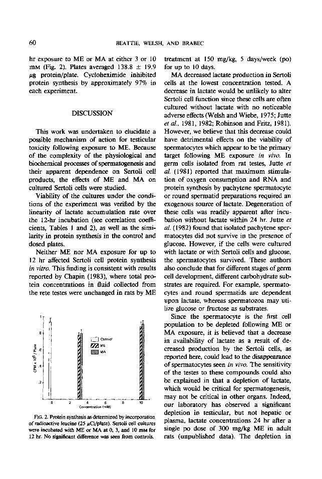
hr exposure to ME or MA at either 3 or 10 mM (Fig. 2). Plates averaged 138.8 +- 19.9 pg protein/plate. Cycloheximide inhibited protein synthesis by approximately 97% in each experiment.
DISCUSSION
This work was undertaken to elucidate a possible mechanism of action for testicular toxicity following exposure to ME. Because of the complexity of the physiological and biochemical processes of spermatogenesis and their apparent dependence on Sertoli cell products, the effects of ME and MA on cultured Sertoli cells were studied.
Viability of the cultures under the condi- tions of the experiment was verified by the linearity of lactate accumulation rate over the 12-hr incubation (see correlation coeffi- cients, Tables 1 and 2), as well as the simi- larity in protein synthesis in the control and dosed plates.
Neither ME nor MA exposure for up to 12 hr affected Sertoli cell protein synthesis in vitro. This finding is consistent with results reported by Chapin (1983), where total pro- tein concentrations in fluid collected from the rete testes were unchanged in rats by ME
” L 4 b L( Concentration (mMI
FIG. 2. Protein synthesis as detemked by incorporation of radioactive leucine (25 &i/plate). Sertoli cell cultures were incubated with ME or MA at 0, 3, and 10 mM for 12 hr. No significant difference was seen from controls.
treatment at 150 mg/kg, 5 days/week (PO) for up to 10 days.
MA decreased lactate production in Sertoli cells at the lowest concentration tested. A decrease in lactate would be unlikely to alter Sertoli cell function since these cells are often cultured without lactate with no noticeable adverse effects (Welsh and Wiebe, 1975; Jutte et al., 198 1, 1982; Robinson and Fritz, 198 1). However, we believe that this decrease could have detrimental effects on the viability of spermatocytes which appear to be the primary target following ME exposure in vivo. In germ cells isolated from rat testes, Jutte et al. (198 1) reported that maximum stimula- tion of oxygen consumption and RNA and protein synthesis by pachytene spermatocyte or round spermatid preparations required an exogenous source of lactate. Degeneration of these cells was readily apparent after incu- bation without lactate within 24 hr. Jutte et al. (1982) found that isolated pachytene sper- matocytes did not survive in the presence of glucose. However, if the cells were cultured with lactate or with Sertoli cells and glucose, the spermatocytes survived. These authors also conclude that for different stages of germ cell development, different carbohydrate sub- strates are required. For example, spermato- cytes and round spermatids are dependent upon lactate, whereas spermatozoa may uti- lize glucose or fructose as substrates.
Since the spermatocyte is the first cell population to be depleted following ME or MA exposure, it is believed that a decrease in availability of lactate as a result of de- creased production by the Sertoli cells, as reported here, could lead to the disappearance of spermatocytes seen in vivo. The sensitivity of the testes to these compounds could also be explained in that a depletion of lactate, which would be critical for spermatogenesis, may not be critical in other organs. Indeed, our laboratory has observed a significant depletion in testicular, but not hepatic or plasma, lactate concentrations 24 hr after a single po dose of 300 mg/kg ME in adult rats (unpublished data). The depletion in

EFFECT OF ME AND MA ON SERTOLI CELLS 61
lactate observed following incubation of Ser- toli cells with MA, but not ME, is also consistent with the hypothesis that MA is the toxic metabolite following ME exposure in vivo.
Our results suggest that a decrease in sertoli cell lactate production caused by the metab- olite, MA, may be a physiologically signihcant mechanism of toxicity in the testis following ME exposure. However, because other tissues, such as bone marrow and lymphoid tissues, are also affected, other mechanisms of action may contribute to the overall toxicity of ME.
ACKNOWLEDGMENTS
The authors thank Mr. Don Ayer for his laboratory assistance, and Ms. Patricia Houtteman for her assistance in preparation of the manuscript. This work was sup ported by NIH Grants HD15246 and HD17121 (MJW).
REFERENCES
BRABE, M. J., BEDO~S, E., DAVIDSON, B., ANDKNIGHT, P. R. (1984). Effect of general anesthetics and pressure on aerobic metabolism of monkey kidney cells. Anes- thesiology 61, 4348.
BRADFORD, M. (1976). A rapid and sensitive method for the quantitation of microgram quantities of protein utilizing principle of protein-dye binding. Anal. Biochem. 12, 248-254.
CHAPIN, R. E. (1983). Effects of various glycol ethers on testicular morphology, protein secretion and reproduc- tive performance. In Toxic Effects of Glycol Ethers, NIOSH Symposium, Cincinnati, OH.
DOE, J. E., SAMUELS, D. M., TINSTON, D. J., AND DE SILVA WICKRAMARATNE, G. A. (1983). Comparative aspects of the reproductive toxicology by inhalation in rats of ethylene glycol monomethyl ether and propylene glycol monomethyl ether. Toxicol. Appl. Pharmacol. 69,43-47.
FOSTER, P. M. D., CREASY, D. M., FOSTER, J. R., THOMAS, L. V., COOK, M. W., AND GANGOLLI, S. D. (1983). Testicular toxicity of ethylene glycol mono- methyl and monoethyl ethers in the rat. Toxicol. Appl. Pharmacol. 69, 385-399.
HOHORST, H. J. (1965). L(+)-Lactate: Determination with lactic dehydrogenase and DPN. In Methods of Enzymatic Analysis (H. U. Bergmeyer, ed.), pp. 266- 270. Verlag Chemie, Weinheim.
Ju-rru, N. H. P. M., GROOTEGOED, J. A., ROMMERTS, F. F. G., AND VAN DER MOLESTS, H. J. (1981). Exoge- nous lactate is essential for metabolic activities in
isolated rat spermatocytes and spermatids. J. Reprod. Fert. 62,399-405.
JUTTE, N. H. P. M., JANSEN, R., GROOTEGO ED, J. A., ROMMERTS, F. F. G., CLAUSEN, 0. P. F., AND VAN DER MOLEN, H. J. (1982). Regulation of survival of rat pachytene spermatocytes by lactate supply from Sertoli cells. J. Reprod. Fert. 65,431-438.
KISSINGER, C., SKINNER, M. K., AND GRISWOLD, M. D. (1982). Analysis of Sertoli cell-secreted proteins by two-dimensional gel electrophoresis. Biol. Reprod. 27,233-240.
MATHER, J. P. (1980). Establishment and chara&rimtion of two distinct mouse testicular epithelial cell lines. Biol. Reprod. 23, 243-252.
MILLER, R. R., AYRES, J. A., CALHOUN, L. L., YOUNG, J. T., AND MCKENNA, M. J. (198 1). Comparative short-term inhalation toxicity of ethylene glycol monomethyl ether and propylene glycol monomethyl ether in rats and mice. Toxicol. Appl. Pharmacol. 61, 368-377.
MIUER, R. R., CARREON, R. E., YOUNG, J. T., AND MCKENNA, M. J. (1982). Toxicity of methoxyacetic acid in rats. Fundam. Appl. Toxicol. 2, 158-160.
MILLER, R. R., AYRES, J. A., YOUNG, J. T., AND MCKENNA, M. J. (1983a). Ethylene glycol monomethyl ether. I. Subchronic vapor inhalation study with rats and rabbits. Fundam. Appl. Toxicol. 3,49-54.
MILLER, R. R., HERMANN, E. A., LANGVARDT, P. W., MCKENNA, M. J., AND SCHWETZ, B. A. (1983b). Comparative metabolism and disposition of ethylene glycol monomethyl ether and propylene glycol mono- methyl ether in male rats. Toxicoi. Appl. Pharmacol. 67,229-237.
NETER, J., AND WASSERMAN, W. (I 974). Applied Linear Statistical Models. Regression, Analysis of Variance, and Experimental Design, pp. 477-482. Richard D. Irwin, Homewood, Illinois.
ROBINSON, R., AND FRITZ, I. B. (1981). Metabolism of glucose by Sertoli cells in culture. Biol. Reprod. 24, 1032-1041.
SAMUEL& D. M., DOE, J. E., ANDTINSTON, D. J. (1983). The effects on rat testis of single inhalation exposures to ethylene glycol alkyl ethers, in particular ethylene glycol monomethyl ether (EGME). In Presentation, European Society of Toxicology, Rome, Italy.
SKINNER, M. K., AND GRISWOLD, M. D. (1983). Sertoh cells synthesize and secrete a ceruloplasmin-like protein. Biol. Reprod. 28, 1225-1230.
TASH, J. S., WELSH, M. J., ANDMEANS, A. R. (1981). Regulation of protein kinase inhibitor by follicle- stimulating hormone in Sertoli cells in vitro. Endocri- nology 108,427-434.
WAITES, G. M. H., AND GLADWELL, R. T. (1982). Physiological significance of fluid secretion in the testis and blood-testis barrier. Physiol. Rev. 62, 624-67 1.
WELSH, M. J., AND WIEBE, J. P. (1975). Rat Sertoli cells: A rapid method for obtaining viable cells. Endocrinol- ogy %,6 18-624.