the diagnostic challenge of amelanotic melanoma – case
TRANSCRIPT

OPEN ACCESSHuman & Veterinary MedicineInternational Journal of the Bioflux Society Case Report
Volume 7 | Issue 1 Page 23 HVM Bioflux
http://www.hvm.bioflux.com.ro/
The diagnostic challenge of amelanotic melanoma – case reports and short review of the literature
¹Loredana Ungureanu, 1Rodica Cosgarea, 2Liliana Rogojan, 1Simona C. Șenilă ¹ Department of Dermatology, “Iuliu Hațieganu” University of Medicine and Pharmacy, Cluj-Napoca, Romania; 2 Department of Histopathology, Cluj-Napoca Emergency County Hospital, Romania.
IntroductionCutaneous melanoma represents a very aggressive type of can-cer that has one of the fastest growing incidences worldwide, with over 150000 new cases estimated in developing countries in 2010 (Jemal et al 2011). Although it represents only 10% of the total cutaneous malignant tumours, melanoma is responsible for over 90% of the deaths caused by these tumours (Jemal et al 2011). Amelanotic melanoma (AM) accounts for 2-8% of all melanomas (Jaimes et al 2012) and represent an atypical form of presentation that may not be easily recognized as malignant melanoma (McClain et al 2012). Studies show that survival after diagnosis of amelanotic melanoma is poorer than after pigmented melanoma, probably because the diagnosis is diffi-cult and is made in more advanced stage (Thomas et al 2014). Clinically, the term of AM reffers to any melanoma lacking pig-ment, however there are also melanomas that produce low-levels of melanin (hypomelanotic melanoma - HM) and may appear to have no pigment (Jaimes et al 2012). Because of their lack of pigment, such lesions may be misdiagnosed as other benign or malignant skin tumors or even as inflamatory disorders, and the treatment can be delayed until advanced stages when the le-sion becames nodular, vascular or ulcerated (Bono et al 2001). Dermatoscopy is an in vivo, non-invasive technique that allows a 10x magnification of the skin which enables the clinician to analyse the morphological structures within pigmented lesions that are not visible with the naked eye, structures with a well-defined histological correspondent. Various studies have dem-onstrated the improved capacity of dermatoscopy in differenti-ating benign lesions from malignant ones, bringing a valuable contribution to the early diagnosis of melanoma (Kittler 2008;
Argenziano et al 2012; Argenziano et al 1997). Although der-moscopic evaluation has been shown to improve the accuracy of pigmented melanoma diagnosis compared with naked eye examination, less literature is found regarding melanomas lack-ing significant pigment. Still, dermoscopic evaluation has been shown to be superior to naked eye examination for the diagno-sis of amelanotic or hypomelanotic melanoma (Pizzichetta et al 2004; de Giorgi et al 2006)We report two unusual presentations of amelanotic melanoma resembling squamous cell carcinoma and basal cell carcinoma, two types of malignant skin tumours with completely different prognosis from AM. Both patients signed an informed consent for their data and pictures to be used for scientific purposes.
Case 1A 75-year-old Caucasian women presented with a relatively rap-idly growing solitary lesion on the right cheek. The lesion was first noted by the patient two years before the presentation as a red, slowly enlarging plaque. The lesion was diagnosed by the general practitioner as a dermatitis and treated with corticos-teroids for three weeks. One year after these treatment and four month prior to the presentation, the lesion started to grow rap-idly. On physical examination, there was a firm, dome-shaped, pink, 1.5 cm nodule, with a central ulceration covered by a yel-low fine crust and fine scales (Fig. 1a). The clinical examina-tion of the lymph nodes revealed firm, painless submandibular and laterocervical lymphadenopathy. Dermoscopic examination revealed white-yellow circles, white-yellow structureless areas, milky red areas, reticular white lines and a polymorphous vascu-lar pattern (Fig. 1b). The clinical picture supported the diagnosis
Abstract. Amelanotic melanoma is a type of cutaneous melanoma characterised by the absence of pigment. The clinical diagnosis of amelanotic melanoma represents a challenge for the practitioner because it can mimic benign or malignant skin tumours and even inflammatory skin dis-orders. Although the dermoscopic criteria for amelanotic melanoma are not so well established, dermoscopy proves to be a useful tool for the diagnosis. We present two cases that illustrate the challenge in the diagnosis of amelanotic melanoma.
Key Words: amelanotic melanoma, dermoscopy.
Copyright: This is an open-access article distributed under the terms of the Creative Commons Attribution License, which permits unrestricted use, distribution, and reproduction in any medium, provided the original author and source are credited.
Corresponding Authors: L. Ungureanu, e-mail: [email protected]

Ungureanu et al 2015
Volume 7 | Issue 1 Page 24 HVM Bioflux
http://www.hvm.bioflux.com.ro/
of squamos cell carcinoma but the presence of milky red areas, reticular white lines and polymorphous vascular pattern on der-moscopy raised the suspicion of amelanotic melanoma. The le-sion was excised and the histopathological evaluation revealed a 5.8-mm thick (according to Breslow), Clark level IV, ulcerated non-pigmented melanoma without regression structures, with a high mitotic rate of 22 mitosis/mm2 (Fig. 1c).
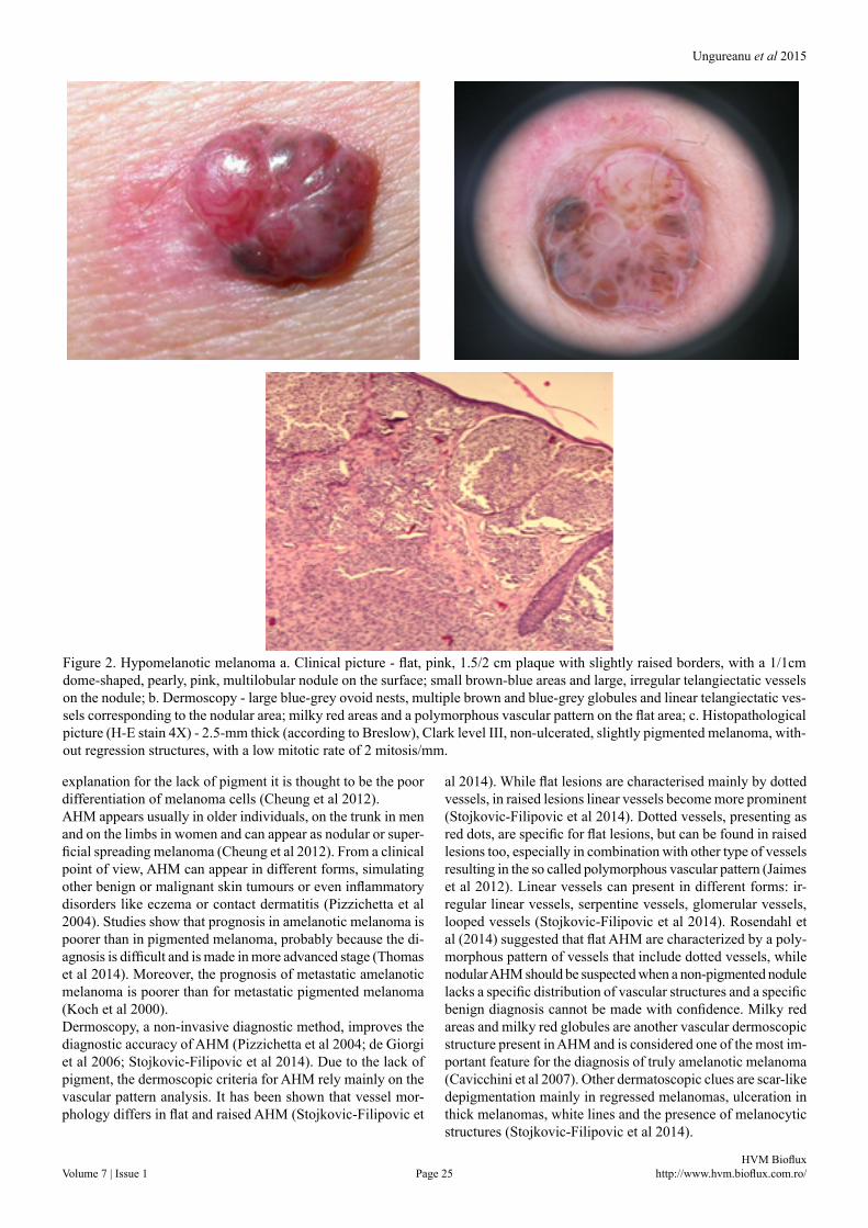
Case 2A 60-year-old Caucasian men presented with a slowly grow-ing solitary lesion on the posterior thoracic region. The lesion was first noted by the patient one year before the presentation and it grew slowly during the mentioned period of time. On physical examination, there was a flat, pink, 1.5/2 cm plaque with slightly raised borders, with a 1/1cm dome-shaped, pearly, pink, multilobular nodule on the surface; small brown-blue ar-eas and large, irregular telangiectatic vessels were visible on the surface (Fig. 2a). Dermoscopic examination revealed large blue-grey ovoid nests, multiple brown and blue-grey globules and linear telangiectatic vessels corresponding to the nodular area, while on the flat part of the lesion it showed milky red
areas and a polymorphous vascular pattern (Fig 2b). The clini-cal picture supported the diagnosis of pigmented basal cell car-cinoma, the only clues for an amelanotic melanoma being the presence of milky red areas, brown globules and polymorphous vascular pattern on dermoscopy. The lesion was excised and the histopathologic evaluation revealed a 2.5-mm thick (according to Breslow), Clark level III, non-ulcerated, slightly pigmented melanoma, without regression structures, with a low mitotic rate of 2 mitosis/mm2 (Fig. 2c). The lesion was re-excised according to the guidelines and a subsequent sentinel lymph node biopsy was negative for regional nodal metastases.
DiscussionsAmelanotic / hypomelanotic melanoma (AHM) is a subtype of cutaneous melanoma characterised by little or no pigment on clinical examination (Pizzichetta et al 2004). Hypomelanotic melanomas are more frequent than completely amelanotic forms, the last being very rare (Pizzichetta et al 2004; Moloney et al 2011). Although the incidence of AHM is appreciated to be 2-8% of all melanomas, the real incidence is difficult to esti-mate due to misdiagnosis (Stojkovic-Filipovic et al 2014). The
Figure 1. Amelanotic melanoma a. Clinical picture - firm, dome-shaped, pink, 1.5 cm nodule, with a central ulceration covered by a yellow fine crust and fine; b. Dermoscopy - white-yellow circles, white-yellow structureless areas, milky red areas, retic-ular white lines and a polymorphous vascular pattern; c,d. Histopathological picture (H-E stain 4X, 10X ) - 5.8-mm thick (ac-cording to Breslow), Clark level IV, ulcerated non-pigmented melanoma without regression structures, with a high mitotic rate of 22 mitosis/mm2.
Ungureanu et al 2015
Volume 7 | Issue 1 Page 25 HVM Bioflux
http://www.hvm.bioflux.com.ro/
explanation for the lack of pigment it is thought to be the poor differentiation of melanoma cells (Cheung et al 2012). AHM appears usually in older individuals, on the trunk in men and on the limbs in women and can appear as nodular or super-ficial spreading melanoma (Cheung et al 2012). From a clinical point of view, AHM can appear in different forms, simulating other benign or malignant skin tumours or even inflammatory disorders like eczema or contact dermatitis (Pizzichetta et al 2004). Studies show that prognosis in amelanotic melanoma is poorer than in pigmented melanoma, probably because the di-agnosis is difficult and is made in more advanced stage (Thomas et al 2014). Moreover, the prognosis of metastatic amelanotic melanoma is poorer than for metastatic pigmented melanoma (Koch et al 2000). Dermoscopy, a non-invasive diagnostic method, improves the diagnostic accuracy of AHM (Pizzichetta et al 2004; de Giorgi et al 2006; Stojkovic-Filipovic et al 2014). Due to the lack of pigment, the dermoscopic criteria for AHM rely mainly on the vascular pattern analysis. It has been shown that vessel mor-phology differs in flat and raised AHM (Stojkovic-Filipovic et
al 2014). While flat lesions are characterised mainly by dotted vessels, in raised lesions linear vessels become more prominent (Stojkovic-Filipovic et al 2014). Dotted vessels, presenting as red dots, are specific for flat lesions, but can be found in raised lesions too, especially in combination with other type of vessels resulting in the so called polymorphous vascular pattern (Jaimes et al 2012). Linear vessels can present in different forms: ir-regular linear vessels, serpentine vessels, glomerular vessels, looped vessels (Stojkovic-Filipovic et al 2014). Rosendahl et al (2014) suggested that flat AHM are characterized by a poly-morphous pattern of vessels that include dotted vessels, while nodular AHM should be suspected when a non-pigmented nodule lacks a specific distribution of vascular structures and a specific benign diagnosis cannot be made with confidence. Milky red areas and milky red globules are another vascular dermoscopic structure present in AHM and is considered one of the most im-portant feature for the diagnosis of truly amelanotic melanoma (Cavicchini et al 2007). Other dermatoscopic clues are scar-like depigmentation mainly in regressed melanomas, ulceration in thick melanomas, white lines and the presence of melanocytic structures (Stojkovic-Filipovic et al 2014).
Figure 2. Hypomelanotic melanoma a. Clinical picture - flat, pink, 1.5/2 cm plaque with slightly raised borders, with a 1/1cm dome-shaped, pearly, pink, multilobular nodule on the surface; small brown-blue areas and large, irregular telangiectatic vessels on the nodule; b. Dermoscopy - large blue-grey ovoid nests, multiple brown and blue-grey globules and linear telangiectatic ves-sels corresponding to the nodular area; milky red areas and a polymorphous vascular pattern on the flat area; c. Histopathological picture (H-E stain 4X) - 2.5-mm thick (according to Breslow), Clark level III, non-ulcerated, slightly pigmented melanoma, with-out regression structures, with a low mitotic rate of 2 mitosis/mm.

Ungureanu et al 2015
Volume 7 | Issue 1 Page 26 HVM Bioflux
http://www.hvm.bioflux.com.ro/
McClain SE, Mayo KB, Shada AL, et al. Amelanotic melanomas pre-senting as red skin lesions: a diagnostic challenge with potentially lethal consequences. Int J Dermatol 2012;51:420-426.
Moloney FJ, Menzies SW. Key points in the dermoscopic diagnosis of hypomelanotic melanoma and nodular melanoma. J Dermatol 2011;38:10–5.
Pizzichetta MA, Talamini R, Stanganelli I, et al. Amelanotic/hy-pomelanotic melanoma:clinical and dermoscopic features. Br J Dermatol 2004;150(6):1117-1124.
Rosendahl C, Cameron A, Tschandl P, et al. Prediction without Pigment: a decision algorithm for non-pigmented skin malignancy. Dermatol Pract Concept 2014;4:9
Stojkovic-Filipovic J, Kittler H. Dermatoscopy of amelanotic and hy-pomelanotic melanoma. J Dtsch Dermatol Ges. 2014 Jun;12(6):467-72. doi: 10.1111/ddg.12368.
Thomas NE, Kricker A, Waxweiler WT, et al. Comparison of Clinicopathologic Features and Survival of Histopathologically Amelanotic and Pigmented Melanomas: A Population-Based Study. JAMA Dermatol. 2014 Aug 27.doi:10.1001/ jamadermatol.2014.1348.
Authors•Loredana Ungureanu, Department of Dermatology “Iuliu Hațieganu” University of Medicine and Pharmacy, 3-6 Clinicilor Street, 400006, Cluj-Napoca, Cluj, Romania, EU, email: [email protected]
•Rodica Cosgarea, Department of Dermatology “Iuliu Hațieganu” University of Medicine and Pharmacy, 3-6 Clinicilor Street, 400006, Cluj-Napoca, Cluj, Romania, EU, email: [email protected]
• Liliana Rogojan, Department of Histopathology “Iuliu Hațieganu” University of Medicine and Pharmacy, 3-6 Clinicilor Street, 400006, Cluj-Napoca, Cluj, Romania, EU, email: [email protected]
•Simona C. Șenilă, Department of Dermatology “Iuliu Hațieganu” University of Medicine and Pharmacy, 3-6 Clinicilor Street, 400006, Cluj-Napoca, Cluj, Romania, EU, email: [email protected]
Our cases illustrate the challenge in the diagnosis of AHM and the usually advanced stage in the moment of diagnosis. Both patients were misdiagnosed as having other skin tumours based on the clinical aspect, although dermoscopy offered clues for the correct diagnosis. Consequently, dermoscopy proves to be a useful tool to improve AHM detection and should be routinely used in the evaluation of non-pigmented skin tumours.
AcknowledgementThis paper was published under the frame of European Social Fund, Human Resources Development Operational Programme 2007-2013, project no. POSDRU/159/1.5/S/138776.
ReferencesArgenziano G, Cerroni L, Zalaudek I, et al. Accuracy in melanoma
detection: A 10-year multicenter survey. J Am Acad Dermatol 2012;67(1):54-59.
Argenziano G, Fabbrocini G, Carli P, et al. Epiluminescence micros-copy: Criteria for cutaneous melanoma progression, J Am Acad Dermatol 1997;37(1):68-74.
Bono A, Maurichi A, Moglia D, et al. Clinical and dermatoscopic diagno-sis of early amelanotic melanoma. Melanoma Res 2001;11:491–494.
Cavicchini S, Tourlaki A, Bottini S. Dermoscopic vascular patterns in nodular “pure” amelanotic melanoma. Arch Dermatol 2007;143:556.
Cheung WL, Patel RR, Leonard A, et al. Amelanotic melanoma: a de-tailed morphologic analysis with clinicopathologic correlation of 75 cases. J Cutan Pathol 2012;39:33–9.
de Giorgi V, Sestini S, Massi D, et al. Dermoscopy for “true” amelanotic melanoma:a clinical dermoscopic-pathologic case study. J Am Acad Dermatol 2006;54(2):341-344.
Jaimes N, Braun RP, Thomas L, Marghoob A.A. Clinical and dermo-scopic characteristics of amelanotic melanomas that are not of the nodular subtype. J Eur Acad Dermatol Venereol 2012;26:591-596.
Jemal A, Bray F, Center MM, et al. Global cancer statistics. CA Cancer J Clin 2011;61(2):69-90.
Kittler H. Early recognition at last. Arch Dermatol 2008;144(4):533-534.
CitationUngureanu L, Cosgarea R, Rogojan L, Senila SC. The diagnostic challenge of amelanotic melanoma – case reports and short review of the literature. HVM Bioflux 2015;7(1):23-26.
Editor Stefan C. VesaReceived 29 October 2014Accepted 6 January 2015
Published Online 8 January 2015
Funding European Social Fund, Human Resources Development Operational Programme 2007-2013, project no. POSDRU/159/1.5/S/138776
Conflicts/ Competing
InterestsNone reported