the diabetic foot - clinical establishmentsclinicalestablishments.gov.in/writereaddata/5381.pdf ·...
TRANSCRIPT
TheDiabeticFoot–FullBackgroundDocumentVer.3.0(Draft)
STANDARD TREATMENT GUIDELINES
The Diabetic foot
Prevention and management in India
Full Background document (Draft) January 2016
Ministry of Health & Family Welfare Government of India

The Diabetic Foot: Full background document
National Standard Treatment Guidelines 1
TABLE OF CONTENTS pp
Section 1 Introduction
1.1 Definition and burden of disease 2
1.2 Morbidity and mortality 2
Section 2 Financial burden and costs, Current practices in India 3
Section 3 Need for a Standard Treatment Guideline 3
Section 4 Recommendations (Moves from primary to tertiary)
4.0 Key terms used 4
4.1 Prevention 5
4.2 Assessment, Classification, Referral 6
4.3 Infection 7
4.4 Wound care 8
4.5 Footwear 9
4.6 Diabetic foot with Osteomyelitis 9
4.7 Charcot’s foot 10
4.8 Surgical intervention and revascularization 11
Section 5 Way forward for India 13
Section 6 How this guideline has been developed 13
Section 7 References 24
Annexure 1 Glossary of terms 26
Annexure 2 Risk grading tables for decision making 30
Annexure 3 Adapt/ Adopt Tables 38

The Diabetic Foot: Full background document
National Standard Treatment Guidelines 2
Section 1. INTRODUCTION 1.1.a Definition Diabetic foot as defined by the World Health Organization is, “The foot of a diabetic patient that has the potential risk of pathologic consequences, including infection, ulceration, and/or destruction of deep tissues associated with neurologic abnormalities, various degrees of peripheral vascular disease, and/or metabolic complications of diabetes in the lower limb”. 1, 2 1.1.b Burden of disease India is set to become the diabetes capital of the world with a projected 109 million individuals with diabetes by 2035.3 India ranks second (after China) with more than 66.8 million diabetics in the age group of 20-70. The prevalence of Diabetes in India is 8.6% 4 and, as of 2013, more than 1 million Indians die each year due to diabetes related causes.5 Diabetic Foot (DF) is one of the most common complications for admissions imposing tremendous medical and financial burden 6 on our healthcare system. 7 The lifetime risk of a person with diabetes having a foot ulcer could be as high as 25%8 and is the commonest reason for hospitalization of diabetic patients (about 30%) and absorbs about 20% of the total health-care costs, more than all other diabetic complications.9, 10 The prevalence of foot ulcers in diabetics attending a centre managing diabetic foot (both indoor and outdoor setup) in India is 3%.11, 12 Foot ulcers among outpatient and inpatient diabetics attending hospitals in rural India was found to be 10.4%.13 Peripheral vascular disease (PVD) occurs in about 3.2% diabetics below 50 years of age and rises to 55% in those above 80 years of age. 14 15% of those with diabetes for a decade suffer from diabetic foot, where as it increases to almost 50% by another decade.15 1.2: Morbidity and mortality Approximately, 85% of non-traumatic lower limb amputations are seen in patients with prior history of diabetic foot ulcer.16,17 Each year, more than 1 million people with diabetes lose at least a part of their leg due to diabetic foot. It shows that every 20 seconds a limb is lost in the world somewhere. 18 In India, though recent population based data is not available, it is estimated that approximately 45,000 legs are amputated every year in India. The vast majority (75%) of these are probably preventable because the amputation often results from an infected neuropathic foot.19 More than half of all foot ulcers become infected, requiring hospitalization, while 20% of infections result in amputation.20 After a major amputation, 50% of people will have the other limb amputated within two years’ time. People with a history of diabetic foot ulcer have a 40% greater 10-year death rate than people with diabetes alone. 21 Section 2: 2.1 Financial burden and costs For treating a simple and complex diabetic foot ulcer in low Income countries like India can be equivalent to 5.7 years of average annual income 22. However, pain and suffering, loss of mobility, even life-long dependence on others and limitations imposed by the change of role is incalculable 23.

The Diabetic Foot: Full background document
National Standard Treatment Guidelines 3
The management of diabetic foot requires a holistic and rigorous approach without which there will be high levels of relapse, morbidity and even mortality. The expenditure on care for amputation has tow fold burden on patient as well as health care system with requirement for surgery, postoperative care followed by rehabilitation and the need for adaptations to home and prostheses fitting. However, prevention is clearly more cost effective than cure in diabetic foot management. 2.2 Current practice in India Diabetic foot care is one of the most ignored aspects of diabetes care in India. 24 Due to social, religious, and economic compulsions, many people walk barefoot. Poverty and lack of education lead to usage of inappropriate footwear and late presentation of foot lesions. Many non-medically qualified persons are interfering in the treatment of diseases, including diabetes. Patients also try home remedies before visiting their physicians. 24 It estimated that 90% of diabetic patients in India do not see a specialist in their lifetime.25
Problem is further worsened by a delay in accessing healthcare due to patient approaching informal care providers and alternative medicine prescribers. Section 3 3.1 Need for a Standard Treatment Guideline There is a lack of a good evidence-based standard guideline on Diabetic foot are in India. Currently, diabetic feet are treated by individual practitioners. Physicians, General surgeons, orthopaedic surgeons, primary care physicians, endocrinologists and podiatrists all look after the diabetic feet. But neither their roles, responsibilities nor the protocols are clearly defined in the public domain. Moreover, in the Indian context, due to the pronounced variability in the health care system, a common national guidance for providing curative as well as preventive methods to curb the growth of diabetic foot in the future is essential. Hence, it is a public health imperative to create an integrated framework for comprehensive management of diabetic foot. 3.2 The purpose of the guideline This standard treatment guideline aims to provide evidence-informed guidance on the key components of care of people with diabetic foot from community care to hospital admission both in the public and the private sector. 3.3 Who this guideline is for This guideline is intended to be relevant to hospital staff caring for patients with diabetic foot problems in referral centres, non-specialized carers who provide secondary and primary care, prevention podiatrists and patient and their care givers.

The Diabetic Foot: Full background document
National Standard Treatment Guidelines 4
SECTION 4: RECOMMENDATIONS

The Diabetic Foot: Full background document
National Standard Treatment Guidelines 5
Divided into subsections: 1. Prevention 2. Assessment 3. Infection 4. Wound care 5. Footwear and follow up 6. Charcot’s foot 7. Surgical intervention and Revascularization
Key terms used:
Peripheral Neuropathy: The presence of symptoms or signs of peripheral nerve dysfunction in people with diabetes, after exclusion of other causes. Peripheral Artery Disease (PAD): Obstructive atherosclerotic vascular disease with clinical symptoms, signs or abnormalities on non-invasive vascular assessment, resulting in disturbed or impaired circulation in one or more extremities.
4. 1: PREVENTION Overview: 1. Identification of the at-risk foot 2. Regular inspection and examination of the at-risk foot 3. Education of patient, family and healthcare providers 4. Routine wearing of appropriate footwear 5. Treatment of pre-ulcerative signs 4.1.1 Prevention of Diabetic foot problems 4.1.1 To identify a person with diabetes at risk for foot ulceration, examine the feet annually / six monthly / quarterly / monthly (depending on patient’s risk category) to seek evidence for signs or symptoms of peripheral neuropathy and peripheral artery disease. IWGDF 2015 (Adapted) Risk Classification System and preventive screening frequency
Category Characteristic Frequency
0 No peripheral neuropathy Once a year
1 Peripheral neuropathy Once every 6 months
2 Peripheral neuropathy with peripheral artery disease and/or a foot deformity
Once every 3-6 months

The Diabetic Foot: Full background document
National Standard Treatment Guidelines 6
3 Peripheral neuropathy and a history of foot ulcer or lower-extremity amputation
Once every 1-3 months
source : The IWGDF guidelines 2015 4.1.2 In a person with diabetes who has peripheral neuropathy, screen for: a history of foot ulceration or lower-extremity amputation; peripheral artery disease; foot deformity; pre-ulcerative signs on the foot; poor foot hygiene; and ill-fitting or inadequate footwear. (Strong; Low) IWGDF 2015 (Adopted) 4.1.3 Treat any pre-ulcerative sign on the foot of a patient with diabetes. This includes: removing callus; protecting blisters and draining when necessary; treating ingrown or thickened toe nails; treating haemorrhage when necessary; and prescribing antifungal treatment for fungal infections. (Strong; Low) IWGDF 2015 (Adopted) 4.1.4 To protect their feet, instruct an at-risk patient with diabetes not to walk barefoot, in socks, or in thin-soled standard slippers, whether at home or when outside. (Strong; Low) IWGDF 2015 (Adopted) 4.1.5 Instruct an at-risk patient with diabetes to: daily inspect their feet and the inside of their shoes; daily wash their feet (with careful drying particularly between the toes); avoid using chemical agents or plasters to remove callus or corns; Adopted use emollients to lubricate dry skin; and cut toe nails straight across. (Weak; Low) IWGDF 2015 (Adopted) 4.1.6 Instruct an at-risk patient with diabetes to wear properly fitting footwear to prevent a first foot ulcer, either plantar or non-plantar, or a recurrent non-plantar foot ulcer. When a foot deformity or a pre-ulcerative sign is present, consider prescribing therapeutic shoes, custom-made insoles, or toe orthosis. (Strong; Low) IWGDF 2015 (Adopted) 4.1.7 Instruct a high-risk patient with diabetes to monitor foot skin temperature at home to prevent a first or recurrent plantar foot ulcer. This aims at identifying the early signs of inflammation, followed by action taken by the patient and care provider to resolve the cause of inflammation. (Weak; Moderate) IWGDF 2015 (Adopted) 4.1.8 To prevent a first foot ulcer in an at-risk patient with diabetes, provide education aimed at improving foot care knowledge and behaviour, as well as encouraging the patient to adhere to this foot care advice. (Weak; Low) IWGDF 2015 (Adopted) 4.1.9 To prevent a recurrent plantar foot ulcer in an at-risk patient with diabetes, prescribe therapeutic footwear that has a demonstrated plantar pressure relieving effect during walking and encourage the patient to wear this footwear. (IWGDF 2015 (Adapted) 4.1.10 To prevent a recurrent foot ulcer in an at-risk patient with diabetes, provide integrated foot care, which includes professional foot treatment, adequate footwear and education. This should be repeated or re-evaluated once every one to three months as necessary. (Strong; Low) IWGDF 2015 (Adopted) 4.2.1: Assessing the risk of developing a diabetic foot problem

The Diabetic Foot: Full background document
National Standard Treatment Guidelines 7
4.2.1a Evaluate a diabetic patient presenting with a foot wound at 3 levels: the patient as a whole, the affected foot or limb, and the infected wound. IDSA 2012 guidelines (Adapted) 4.2.1b Assess the affected limb and foot for arterial ischemia (strong, moderate), venous insufficiency, presence of protective sensation, and biomechanical problems. IDSA 2012 guidelines (Adapted) Explanatory note: Biomechanical problems means anatomical and physiological disturbances of the foot. i.e., structural changes which happen in the bones, joints and muscles of the foot of diabetics and the changes in the blood circulation and nerve sensation of the foot of diabetics. 4.2.2. Classification of Diabetic foot 4.2.2a Assess the severity of any diabetic foot infection using the Infectious Diseases Society of America/ International Working Group on the Diabetic Foot Classification system. (Strong; Moderate) IWGDF 2015 (adopted) (Refer to Annexure below for IDSA/IWGDF classification system -Table 1) 4.2.2b Do not use the Wagner classification system to assess the severity of a diabetic foot ulcer. (Adopted) (NICE 2015 Guidelines on Diabetic foot problems: prevention and management.) 4.2.3 Referral for Diabetic foot problems 4.2.3a Initially hospitalize all patients with a severe infection, selected patients with a moderate infection with complicating features (eg, severe peripheral arterial disease [PAD] or lack of home support), and any patient unable to comply with the required outpatient treatment regimen for psychological or social reasons. Also hospitalize patients who do not meet any of these criteria, but are failing to improve with outpatient therapy. IDSA 2012 guidelines (Adapted) (Also refer to Table-3 and 4 in the Annexure below, for explanatory notes.) 4.2.3b Prior to being discharged, make sure that a patient with a DFI (Diabetic Foot Infection) is clinically stable; has had any urgently needed surgery performed; has achieved acceptable glycemic control; is able to manage (on his/her own or with help) at the designated discharge location; and has a well defined plan that includes an appropriate antibiotic regimen to which he/she will adhere, an off-loading scheme (if needed), specific wound care instructions, and appropriate outpatient follow-up. IDSA 2012 guidelines (Adapted) 4.3: DIABETIC FOOT INFECTIONS 4.3.1 Consider the possibility of infection occurring in any foot wound in a patient with diabetes. Evidence of infection generally includes classic signs of inflammation (redness, warmth, swelling, tenderness, or pain) or purulent secretions, but may also include additional or secondary signs (e.g., non-purulent secretions, friable or discoloured granulation tissue, undermining of wound edges, foul odour). IDSA 2012 Guidelines (Adapted) 4.3.2 Be aware of factors that increase the risk for diabetic foot infections (DFI) and especially consider infection when these factors are present; these include a wound for which the probe-to-bone (PTB) test is positive; an ulceration present for >30 days; a history of recurrent foot ulcers; a traumatic foot wound; the presence of peripheral vascular disease in the affected limb; a previous lower extremity amputation; loss of
The Diabetic Foot: Full background document
National Standard Treatment Guidelines 8
protective sensation; the presence of renal insufficiency; or a history of walking barefoot. IDSA 2012 guidelines (Adapted) 4.3.3 Take plain radiographs of the affected foot of all patients presenting with a new Diabetic Foot Infection to look for bony abnormalities (deformity, destruction) as well as for soft tissue gas and radio-opaque foreign bodies. IDSA 2012 Guidelines (Adapted) 4.3.4 When and how to obtain culture from Diabetic foot patients? 4.3.4a For clinically uninfected wounds, do not collect a specimen for culture. IDSA 2012 guidelines (Adapted) 4.3.4b Send a specimen for culture that is from deep tissue, obtained by biopsy or curettage and after the wound has been cleansed and debrided. Avoid swab specimens, especially of inadequately debrided wounds, as they provide less accurate results. IDSA 2012 guidelines (Adapted) Explanatory note: Wash the wound with saline and the surrounding skin with antiseptic solution before taking culture to avoid contamination of the specimen obtained for culture. 4.3.4c Do not obtain repeat cultures unless the patient is not clinically responding to treatment. IWDGF 2015 Guidance Document (Adapted) Explanatory note - Expert consensus says that if the signs of inflammation do not subside even after 72 hours of starting treatment, then it should be considered that patient is not responding. 4.3.4d For infected wounds, send appropriately obtained specimens for culture prior to starting empiric antibiotic therapy, if possible. Cultures may be unnecessary for a mild infection in a patient who has not recently received antibiotic therapy. IDSA 2012 guidelines (Adapted) 4.3.5. Selection of Antibiotic and when should it be modified? 4.3.5a Do not treat clinically uninfected wounds with antibiotic therapy. IDSA 2012 Guidelines (Adapted) 4.3.5b Prescribe antibiotic therapy for all infected wounds but caution that this is often insufficient unless combined with appropriate wound care. IDSA 2012 guidelines (Adapted) 4.3.5c Base the route of therapy largely on infection severity. Prefer parenteral therapy for all severe, and some moderate DFls, at least initially, with a switch to oral agents when the patient is systemically well and culture results are available. Use highly bioavailable oral antibiotics alone in most mild, and in many moderate, infections and topical therapy for selected mild superficial infections. IDSA 2012 guidelines (Adapted) 4.3.5d Select an empiric antibiotic regimen on the basis of the severity of the infection and the likely etiologic agent(s). a. For mild to moderate infections in patients who have not recently received antibiotic treatment, therapy just targeting aerobic gram-positive cocci (GPC) is sufficient. b. For most severe infections, start broad-spectrum empiric antibiotic therapy, pending culture results and antibiotic susceptibility data. c. Empiric therapy directed at P. aeruginosa is usually unnecessary except for patients with risk factors* for true infection with this organism.
The Diabetic Foot: Full background document
National Standard Treatment Guidelines 9
d. Consider providing empiric therapy directed against MRSA in a patient with a prior history of MRSA infection; when the local prevalence** of MRSA colonization or infection is high; or if the infection is clinically severe. IDSA 2012 guidelines (Adapted) Explanatory notes: * Risk factors for true infection with Pseudomonas aeruginosa include Immunocompromised status, Chronic Kidney Disease, warm climate and frequent exposure of foot to water. **The local prevalence of MRSA (i.e., percentage of all Staph. aureus clinical isolates in that locale that are methicillin resistant) is high enough (perhaps 50% for a mild and 30% for a moderate soft tissue infection) that there is a reasonable probability of MRSA infection. 4.3.5e Give an initial antibiotic course for a soft tissue infection of about 1–2 weeks for mild infections and 2–3 weeks for moderate to severe infections. IDSA 2012 guidelines (Adapted) 4.3.5f Continue antibiotic therapy until, but not beyond, resolution of findings of infection, but not through complete healing of the wound. IDSA 2012 guidelines (Adapted) 4.3.5g Administer parenteral therapy initially for most severe infections and some moderate infections, with a switch to oral therapy when the infection is responding. (Strong; Low) IDSA 2012 guidelines (Adopted) 4.4: WOUND CARE 4.4.1 Clean ulcers regularly with clean water or saline*, debride them when possible in order to remove debris from the wound surface and dress them with a sterile, inert dressing in order to control excessive exudate and maintain a warm, moist environment in order to promote healing**. (Strong; Low)IWGDF 2015 (Adopted) Explanatory note: *Clean water is boiled cooled water (distilled water) ** Do not use hydrogen peroxide, EUSOL (Edinburgh University Solution), povidone iodine, chlorhexidine, etc 4.4.2 Select dressings principally on the basis of exudate control, comfort and cost. (Strong; Low) IWGDF 2015 (Adopted) 4.4.3 Do not use antimicrobial dressings with the goal of improving wound healing or preventing secondary infection. (Strong; Moderate) IWGDF 2015 (Adopted) 4.4.4 Do not offer the following to treat diabetic foot ulcers, unless as part of a clinical trial: · Electrical stimulation therapy, autologous platelet-rich plasma gel, regenerative wound matrices and dalteparin. · Growth factors (granulocyte colony-stimulating factor [G-CSF], platelet-derived growth factor [PDGF], epidermal growth factor [EGF] and transforming growth factor beta [TGF-β]). · Hyperbaric oxygen therapy. NICE clinical guideline NG19 (Adopted)
The Diabetic Foot: Full background document
National Standard Treatment Guidelines 10
4.4.5 Consider dermal or skin substitutes as an adjunct to standard care when treating diabetic foot ulcers, only when healing has not progressed and on the advice of the multidisciplinary foot care service. NICE clinical guideline NG19 (Adopted) 4.4.6 1f Consider negative pressure wound therapy after surgical debridement for diabetic foot ulcers, on the advice of the multidisciplinary foot care service. NICE clinical guideline NG19 (Adopted) 4.4.7 Do not select agents reported to improve wound healing by altering the biology of the wound, including growth factors, bioengineered skin products and gases, in preference to accepted standards of good quality care. (Strong; Low) IWGDF 2015 (Adopted) 4.4.8 Do not select agents reported to have an impact on wound healing through alteration of the physical environment, including through the use of electricity, magnetism, ultrasound and shockwaves, in preference to accepted standards of good quality care. (Strong; Low) IWGDF 2015 (Adopted) 4.4.9 Do not select systemic treatments reported to improve wound healing, including drugs and herbal therapies, in preference to accepted standards of good quality care. (Strong; Low) IWGDF 2015 (Adopted) 4.4.10 Redistribution of pressure off the wound to the entire weight-bearing surface of the foot (“off-loading”). While particularly important for plantar wounds, this is also necessary to relieve pressure caused by dressings, footwear, or ambulation to any surface of the wound. (Strong; High) IDSA 2012 guidelines (Adopted) 4.4.11 When deciding about wound dressings and offloading when treating diabetic foot ulcers, take into account the clinical assessment of the wound and the person’s preference, and use devices and dressings with the lowest acquisition cost appropriate to the clinical circumstances. NICE clinical guideline NG19 (Adopted) 4.5 : FOOTWEAR 4.5.1 To heal a neuropathic plantar forefoot ulcer without ischemia or uncontrolled infection in a patient with diabetes, offload with a non-removable knee-high device with an appropriate foot-device interface. (Strong; High) IWGDF 2015 (Adopted) Non-removable (cast) walker: Same as removable (cast) boot/walker but then with a layer(s) of fibreglass cast material circumferentially wrapped around it rendering it irremovable (also known as "instant total contact cast") 4.5.2 When a non-removable knee-high device is contraindicated or not tolerated by the patient, consider offloading with a removable knee-high walker with an appropriate foot-device interface to heal a neuropathic plantar forefoot ulcer in a patient with diabetes, but only when the patient can be expected to be adherent to wearing the device. (Weak; Moderate) IWGDF 2015 (Adopted) Removable (cast) boot/walker: Prefabricated removable knee-high boot with a rocker or roller outsole configuration, padded interior, and an insertable and adjustable insole which may be total contact. 4.5.3 When a knee-high device is contraindicated or cannot be tolerated by the patient, consider offloading with a forefoot offloading shoe, cast shoe, or custom-made temporary shoe to heal a neuropathic plantar forefoot ulcer in a patient with diabetes,
The Diabetic Foot: Full background document
National Standard Treatment Guidelines 11
but only and when the patient can be expected to be adherent to wearing the shoes. (Weak; Low) IWGDF 2015 (Adopted) 4.5.4 Instruct an at-risk patient with diabetes to wear properly fitting footwear to prevent a first foot ulcer, either plantar or non-plantar, or a recurrent non-plantar ulcer. When a foot deformity or a pre-ulcerative sign is present, consider prescribing therapeutic shoes, custom-made insoles, or toe orthosis*. (Strong; Low) IWGDF 2015 (Adopted) *Toe orthosis:- An in-shoe orthosis to achieve some alteration in the function of the toe. 4.5.5 To prevent a recurrent plantar foot ulcer in an at-risk patient with diabetes, prescribe therapeutic footwear that has a demonstrated plantar pressure relieving effect during walking and encourage the patient to wear this footwear. IWGDF 2015 (Adapted) 4.5.6 Instruct a patient with diabetes not to use conventional or standard therapeutic footwear to heal a plantar foot ulcer. IWGDF 2015 (Adapted) Explanatory note: Use footwear with following features - Sandals: should have adjustable straps, insole, full heel counter and rigid outsole. Shoes: should have wide toe box extra depth and without laces. 4.5.7 Consider using shoe modifications, temporary footwear, toe spacers or orthoses to offload and heal a non-plantar foot ulcer without ischemia or uncontrolled infection in a patient with diabetes. The specific modality will depend on the type and location of the foot ulcer. (Weak; Low) IWGDF 2015 (Adopted) 4.5.8 If other forms of biomechanical relief are not available, consider using felted foam* in combination with appropriate footwear to offload and heal a neuropathic foot ulcer without ischemia or uncontrolled infection in a patient with diabetes. (Weak; Low) IWGDF 2015 (Adopted) Felted foam - A fibrous, unwoven material backed by foam with absorbing and cushioning characteristics. The foam is generally ‘rubber foam’ or ‘PU foam’ which is formed by either a polyester or polyether polyol resin, in conjunction with water and Toluene di Isocyanate, along with various catalysts and blowing agents and colouring pigments to give the desired compression. 4.6: Diabetic foot with osteomyelitis and CHARCOT’S FOOT 4.6 Treatment of Diabetic foot with osteomyelitis 4.6.1 For an infected open wound, perform a probe-to-bone test; in a patient at low risk for osteomyelitis a negative test largely rules out the diagnosis, while in a high risk patient a positive test is largely diagnostic. (Strong; High)IDSA 2012 guidelines (Adopted) 4.6.2 Markedly elevated ESR is suggestive of osteomyelitis in suspected cases. IWGDF 2015 (Adapted) Explanatory note: Tests for serum inflammatory markers are costly and not widely available, except ESR. Also these tests are not diagnostic of DFO. 4.6.3If osteomyelitis is suspected in a person with diabetes but is not confirmed by initial X-ray, consider an MRI to confirm the diagnosis. In places where MRI is unavailable, diagnose osteomyelitis by the PTB test (clinically) and/or taking a Bone biopsy and culture. (Adapted from NICE clinical guideline NG19 and Expert group discussion) Explanatory note: Expert Consensus says that as availability of MRI is limited across the country, it is recommended to use MRI wherever available. At the primary and secondary health centre levels, the PTB test and bone biopsy and culture are more feasible and economical and reasonably accurate.
The Diabetic Foot: Full background document
National Standard Treatment Guidelines 12
4.6.4 A definite diagnosis of bone infection usually requires positive results on both histological and microbiological examinations of an aseptically obtained bone sample, but this is usually required only when the diagnosis is in doubt or determining the causative pathogen’s antibiotic susceptibility is crucial. (Strong; Moderate) IDSA 2012 guidelines (Adopted) 4.6.5 Avoid using results of soft tissue or sinus tract specimens for selecting antibiotic therapy for osteomyelitis as they do not accurately reflect bone culture results. (Strong; Moderate) IDSA 2012 guidelines (Adopted) 4.6.6 When a radical resection leaves no remaining infected tissue*, we suggest prescribing antibiotic therapy for only a short duration (2–5 days). When there is persistent infected or necrotic bone, we suggest prolonged (≥4 weeks) antibiotic treatment. (weak, low) IDSA 2012 guidelines (Adapted) Explanatory note: *A proximal bone histopath to be done if available to get a clear margin and confirm that no infected bone remains. 4.6.7 For specifically treating Diabetic foot osteomyelitis, we do not currently support using adjunctive treatments such as hyperbaric oxygen therapy, growth factors (including granulocyte colony stimulating factor), maggots (larvae), or topical negative pressure therapy (eg, vacuum-assisted closure). (weak, low) IDSA 2012 guidelines (Adapted) Also refer to Table 4 and 5 in the Annexure below, for explanatory notes. 4.7. Charcot’s Foot 4.7.1 Suspect acute Charcot arthropathy if there is redness, warmth, swelling or deformity (in particular, when the skin is intact), especially in the presence of peripheral neuropathy or renal failure. Think about acute Charcot arthropathy even when deformity is not present or pain is not reported. NICE clinical guideline NG19 (Adopted) 4.7.2 Refer the person with suspected charcot’s foot early (within one week) to the - Diabetic Foot care centre to confirm the diagnosis, and offer non-weight-bearing treatment until definitive treatment can be started. NICE clinical guideline NG19(Adapted) Diabetic Foot care centre:- InIndia,sincetherearenominimumstandardsofservicesofferedtothediabeticfootpatients,inourrecommendationswehaveusedthistermtodenotethisfacility,whichmayexistattheGeneralPractitioner’soffice,Primaryhealthcentre,Secondarycarecentreoratatertiarycarecentre.Preferably,thediabeticfootcarecentreshouldconsistofatleastasurgeon,aphysician,andanorthotist. 4.7.3 If acute Charcot arthropathy is suspected, X-ray the affected foot. Consider an MRI if the X-ray is normal but clinical suspicion still remains. NICE clinical guideline NG19 (Adopted) 4.7.4 Distinguishing the bony changes of osteomyelitis from those of the less common entity of diabetic neuro-osteoarthropathy (Charcot foot) may be particularly challenging and requires considering clinical information in conjunction with imaging. IDSA 2012 guidelines (Adopted) 4.7.5 Clinical clues supporting neuro-osteoarthropathy in this context include midfoot location and absence of a soft tissue wound, whereas those favoring osteomyelitis include presence of an overlying ulcer (especially of the forefoot or heel), either alone or superimposed on Charcot changes. IDSA 2012 guidelines (Adopted)
The Diabetic Foot: Full background document
National Standard Treatment Guidelines 13
4.7.6 If the films show classic changes suggestive of osteomyelitis (cortical erosion, periosteal reaction, mixed lucency, and sclerosis), and if there is little likelihood of neuro-osteoarthropathy, it is reasonable to initiate treatment for presumptive osteomyelitis, preferably after obtaining appropriate specimens for culture. IDSA 2012 guidelines (Adopted) 4.7.7 If the Diabetic Foot care centre suspects acute Charcot arthropathy, offer treatment with a non-removable off-loading device. Only consider treatment with a removable off-loading device if a non-removable device is not advisable because of the clinical or the person’s circumstances. NICE clinical guideline NG19 (Adapted) 4.7.8 Do not offer bisphosphonates to treat acute Charcot arthropathy, unless as part of a clinical trial. NICE clinical guideline NG19 (Adopted) 4.7.9 Monitor the treatment of acute Charcot arthropathy using clinical assessment. This should include measuring foot–skin temperature difference and taking serial X-rays until the acute Charcot arthropathy resolves. Acute Charcot arthropathy is likely to resolve when there is a sustained temperature difference of less than two degrees centigrade between both feet and when X-ray changes show no further progression. NICE clinical guideline NG19 (Adapted) 4.7.10 People who have a foot deformity that may be the result of a previous Charcot arthropathy are at high risk of ulceration and should be cared for by the. Diabetic Foot care centre. NICE clinical guideline NG19 (Adopted) 4.7.10 The Diabetic Foot care centre should care for people who have a foot deformity resulting from a previous Charcot’s arthropathy as they are at high risk of ulceration. NICE clinical guideline NG19 (Adapted) 4.8: SURGICAL INTERVENTIONS AND REVASCULARIZATION 4.8.1 Consult a surgical specialist in all cases of diabetic foot infections that are moderate or severe. IWGDF 2015 (Adapted) 4.8.2 Perform urgent surgical intervention in cases of deep abscesses, compartment syndrome and necrotizing soft tissue infections. IWGDF 2015 (Adapted) 4.8.3 Debride any wound that has necrotic tissue or surrounding callus; the required procedure may range from minor to extensive. IWGDF 2015 (Adapted) 4.8.4 Perform urgent surgical intervention for most foot infections accompanied by gas in the deeper tissues, an abscess, or necrotizing fasciitis, and less urgent surgery for wounds with substantial nonviable tissue or extensive bone or joint involvement. IDSA 2012 guidelines (Adapted) Additional note: In those with a non-severe infection, carefully observing the effectiveness of medical therapy and the demarcation line between necrotic and viable tissue before operating may be prudent.
The Diabetic Foot: Full background document
National Standard Treatment Guidelines 14
4.8.5 Consider surgical intervention in cases of osteomyelitis accompanied by: spreading soft tissue infection; destroyed soft tissue envelope; progressive bone destruction on X-ray; or, bone protruding through the ulcer. IWGDF 2015 (Adapted) 4.8.6 Remove slough, necrotic tissue & surrounding callus with sharp debridement in preference to other methods, taking relative contraindications such as severe ischemia into account. IWGDF 2015 (Adapted) 4.8.7 Consider digital flexor tenotomy to prevent a toe ulcer when conservative treatment fails in a high-risk patient with diabetes, hammertoes and either a pre-ulcerative sign or an ulcer on the toe. (weak; low) IWGDF 2015 (Adopted) 4.8.8 Consider Achilles tendon lengthening, joint arthroplasty, single or pan metatarsal head resection or osteotomy to prevent a recurrent foot ulcer when conservative treatment fails in a high-risk patient with diabetes and a plantar foot ulcer. (weak; low) IWGDF 2015 (Adopted) Management of Peripheral Artery Disease in patients with Diabetic foot problems 4.8.9 Examine a patient with diabetes annually for the presence of peripheral artery disease (PAD); this should include, at a minimum, taking a history and palpating foot pulses. (Strong; low) IWGDF 2015 (Adopted) 4.8.10 Evaluate a patient with diabetes and a foot ulcer for the presence of PAD. Determine, as part of this examination, ankle or pedal Doppler arterial waveforms; measure both ankle systolic pressure and systolic ankle brachial index (ABI). (Strong; Low) IWGDF 2015 (Adopted) 4.8.11 Use bedside non-invasive tests to exclude PAD. No single modality has been shown to be optimal. Measuring ABI (with <0.9 considered abnormal) is useful for the detection of PAD. Tests that largely exclude PAD are the presence of ABI 0.9-1.3, toe brachial index (TBI) ≥0.75 and the presence of triphasic pedal Doppler arterial waveforms. IWGDF 2015 (Adapted) 4.8.12 In patients with a non-healing ulcer with either an ankle pressure <50mmHg or ABI <0.5 consider urgent vascular imaging and revascularisation. (Strong; Low) IWGDF 2015 (Adopted) 4.8.13 Consider vascular imaging and revascularisation in all patients with a foot ulcer in diabetes and PAD, irrespective of the results of bedside tests, when the ulcer does not improve within 6 weeks despite optimal management. (Strong; Low) IWGDF 2015 (Adopted) 4.8.14 Do not consider Diabetic microangiopathy to be the cause of poor wound healing in patients with a foot ulcer. IWGDF 2015 (Adapted) 4.8.15 To obtain anatomical information when revascularisation is being considered, use one of these tests - Colour Doppler ultrasound, CT-angiography, MR-angiography or intra-arterial digital subtraction angiography. Evaluate the entire lower extremity arterial circulation, with detailed visualization of below-the-knee and pedal arteries. IWGDF 2015 (Adapted)

The Diabetic Foot: Full background document
National Standard Treatment Guidelines 15
4.8.16 Offer duplex ultrasound as first-line imaging to all people with peripheral arterial disease for whom revascularization is being considered. Take the decision of revascularisation on the basis of colour doppler findings and use DSA for defining the vascular anatomy prior to the procedure. (Adapted from NICE 2012 PAD Guidelines, last modified August 2015) 4.8.17 The aim of revascularisation is to restore direct flow to at least one of the foot arteries, preferably the artery that supplies the anatomical region of the wound, and adequate revascularization should be assessed post-operatively with a colour Doppler wave-fronts (preferable) or a hand held Doppler probe used bedside. IWGDF 2015. (Adapted) 4.8.18 A centre treating patients with a foot ulcer in diabetes should have liaison / association with a centre having the expertise necessary to diagnose and treat PAD; both endovascular techniques and bypass surgery should be available. (Adapted) IWGDF 2015 4.8.19 The multidisciplinary team should treat the patient after a revascularisation procedure for a foot ulcer in diabetes, as part of a comprehensive care plan. (Strong; Low) IWGDF 2015 (Adapted). 4.8.20 There is inadequate evidence to establish which revascularisation technique is superior and a multidisciplinary team should decide the technique of revascularization for a patient based on a number of individual factors, such as morphological distribution of PAD, availability of autogenous vein, patient co-morbidities and local expertise. (Strong; Low) IWGDF 2015 (Adapted) 4.8.21 Give emergency treatment to patients with signs of PAD and a foot infection as they are at particularly high risk for major limb amputation. IWGDF 2015 (Adapted) 4.8.22 Avoid revascularisation in patients in whom, from the patient perspective, the risk-benefit ratio for the probability of success is unfavourable*. (Strong;Low) IWGDF 2015 (Adopted) Explanatory note: Unfavourable risk benefit ratio would indicate those patients who are frail, elderly, bed ridden, having low life expectancy, multiple co-morbidities imposing high risk for surgical intervention, etc 4.8.23 All patients with diabetes and an ischemic foot ulcer should receive aggressive cardiovascular risk management including support for cessation of smoking, treatment of hypertension and prescription of a statin as well as low-dose aspirin or clopidogrel. (Strong;Low) IWGDF 2015 (Adopted) 4.8.24 Do not offer major amputation to people with critical limb ischaemia unless all options for revascularisation have been considered by a vascular multidisciplinary team. Major amputation without giving a chance for revascularization is indicated only in lifesaving situations like foot causing septicemia, wet gangrene, or completely destroyed foot (post charcot’s or osteomyelitis etc). (Adapted) NICE 2012 PAD guidelines, last modified August 2015 THE WAY FORWARD FOR INDIA

The Diabetic Foot: Full background document
National Standard Treatment Guidelines 16
In keeping with the rising burden, India will need to plan for a foot protection service at the primary and community level, to implement the proven cost-effective preventive interventions which are learnings from other countries. The lesser referrals to the hospital hopefully will in the future be treated by a multidisciplinary foot care service at the hospital level, using a more robust protocolized care pathway, built on Indian audited data and cost-impact analysis research. Professionals for this interdisciplinary foot care service will include, Diabetes physicians and nurses, podiatrists, orthotists, microbiologists, interventional radiologists, vascular surgeons, wound care nurses, plastic surgeons and disability specialists.

The Diabetic Foot: Full background document
National Standard Treatment Guidelines 17
Section 6. How This STG was Developed

The Diabetic Foot: Full background document
National Standard Treatment Guidelines 18
Background A Task Force was constituted in December 2014 to guide the development of Standard Treatment Guidelines (STG) in India. The Task Force subsequently approved the draft STG development manual of India (Part 1) for development of adapted guidelines. In addition, it approved a list of 14 topics recommended by a subgroup of the task force appointed to select prioritized topics for STG development. These 14 topics are from 10 clinical specialties for which the first set of STGs will be developed. The topic diabetic foot is included in this first list and was the dealt with by the General Surgery clinical subgroup. Overview
The STG on Diabetic foot management, was developed by a team of experts and relevant stakeholders. The recommendations in the STG were adopted/ adapted from four source guidelines which are the IWGDF (2015), IDSA (2012) and NICE guideline (26th August, 2015) on diabetic foot, and the NICE guidelines on PAD (Peripheral Arterial Disease) November 2014. Available from and full reference below: http://www.iwgdf.org/files/2015/ http://www.idsociety.org https://www.nice.org.uk/guidance/NG19 https://www.nice.org.uk/guidance/cg147 1. 2012 Infectious Diseases Society of America Clinical Practice Guideline for the Diagnosis and Treatment of Diabetic Foot Infections 26
2. The National Institute for Health and Care Excellence (NICE) guidelines- Diabetic foot problems: prevention and management (NG19) (Published: 26 August 2015) 27
3. International Working Group on the Diabetic Foot (IWGDF) 2015 - Prevention and Management of Foot Problems in Diabetes Guidance Documents and Recommendations 28
4. NICE guideline- Lower limb peripheral arterial disease: diagnosis and management (NICE clinical guideline 147) (Issued: August 2012 last modified: August 2015) 29
The processes and methods used in developing this STG draw on those outlined in the STG development manual of India (Part 1) for development of adapted guidelines and summarized in

The Diabetic Foot: Full background document
National Standard Treatment Guidelines 19
the Stepwise guide on STG development. The figure below contains a schematic of the process followed and each of the steps are detailed in subsequent sections below.
The NHSRC with technical support from NICE international carried out a training workshop in May 2015 to guide the STG group members and chairs on the methodology to follow in developing adapted STGs suitable for the Indian context. This workshop was conducted on 29th & 30th May, 2015 and two members (NR, MK) of the surgery STG team attended. Subsequently, NHSRC facilitated the STG development process by providing resources approved by the Ministry of Health & family welfare to the expert group. To assist widespread implementation of the diabetic foot STG, three implementation tools have been developed in addition to the STG document. They include:
● The Quick Reference Guide to help the clinical practitioner (Clinical pathways) ● An information document for the public to create patient awareness about the disease
and ● the quality standards developed from key priority recommendations.
Steps followed during the development of the STG on diabetic foot are as follows: 1) Diabetic foot STG Subgroup established A multi-disciplinary group consisting of health professionals, subject matter experts in various fields and a patient representative undertook the development of this evidence-based STG on diabetic foot. Official letters of invitation were sent from the NHSRC head office. The members of the task force who worked for the Government experienced delays and difficulties in procuring permissions from their respective departments for this work. Subsequently, there were drop outs

The Diabetic Foot: Full background document
National Standard Treatment Guidelines 20
and new experts invited. The names of the 14 group members in the STG sub-group on diabetic foot, their specialities and organization affiliation are listed here: Task Force: The STG Subgroup on Diabetic foot None of the members report any conflict of interest in the development of this guideline and have all signed their declarations. Facilitator Nobhojit Roy, Prof & Head, Dept of Surgery, BARC hospital
(HBNI University), Mumbai
Rapporteurs/Writing team Monty Khajanchi, Assistant Prof, Dept of Surgery, KEM Hospital and G S Medical college, Mumbai Satish Mishra, Associate Prof, Dept of Surgery, BARC Hospital, (HBNI University), Mumbai Kunal Chhatbar, Senior registrar, JRH Hospital, Mumbai Dikpal Singh Jadhav, BARC Hospital, (HBNI University), Mumbai Aditi S Kashikar, MGIMS Wardha (Research assistant)
Expert Arun Bal, President, Diabetic Foot Association of India and Raheja Hospital for Diabetes.
Physician Usha Menon, Prof of Clinical Endocrinology, Amrita Institute of Medical Sciences, Kochi-682041, Kerala, India.
Private Practitioner Sanjay Vaidya, Plastic surgeon, Hinduja Hospital DFSI Exec committee member
Paramedic /Nurse/Rehabilitation Experts
Gautham Gopalakrishna,Principal senior Scientist & Head, Footwear Design and Development, Central Leather Institute, Chennai
Primary Care Practitioners Trupti Patil, Chembur CHSS dispensary, Chembur, Mumbai
Patient Right Group/NGO Raman Kataria, Surgery, Jan Swasthya Sahyog, Ganiyari Village, Bilaspur District, Chhattisgarh Nandakumar M, Senior secondary care surgeon, Ashwini, Gudalur, Tamil Nadu Sushil Patil, JSS, Ganyari, Chattisgarh
Expert Adviser: Dr. Raghuram Sekhar, Vascular surgeon, Kokilaben Hospital, Mumbai Dr Abha Mehndiratta was the technical person providing oversight and guidelines in the meetings. Satish Mishra and Sushil Patil were the patient right representatives in the group.
The STG Subgroup met twice face-to-face and all meetings (including the smaller weekly ones were quorate (50%=7 members). The working group met every tuesday evening, before and after each of these meetings over a period of two months. Some of the members joined the small group meetings via video-conference. In the induction and orientation session held on 21st July 2015, the facilitator (Chair) welcomed all the members of the subgroup, and set up the rules of operation based on the STG development manual, on the consistent use of terminology and definitions, using the structured powerpoint presentation provided by NHSRC/NICE.

The Diabetic Foot: Full background document
National Standard Treatment Guidelines 21
The induction and orientation session was held on 21st July 2015 in which the facilitator (Chair) welcomed all the members of the subgroup, and set up the rules of operation based on the STG development manual, on the consistent use of terminology and definitions, using the structured power-point presentation provided by NHSRC/NICE. The STG Subgroup met face-to-face twice between July 2015 to November 2015. The writing team met every week, on Tuesday evening during the same time period. All these meetings were quorate (50% = 7 members). Those who could not attend physically joined in via video conferencing (Skype). Also, the individual members in the writing team kept in touch via e-mails and Whatsapp. In the initial few meetings the recommendations were drafted, and in the subsequent meetings, the recommendations were edited as per the peer review comments from the NHSRC/NICE. 2) Scope the STG To develop the scope of this STG we followed the principle of what the scope will include and exclude. It also identifies only the key aspects of care that “must” be included, set the “boundaries” of the development work and provide a clear “framework” to enable work to stay within the agreed priorities, inform the development of the clinical questions and search strategy, inform “professionals” and the “public” about expected content of the guideline, keep the STG to a “reasonable size” to ensure that its development can be carried out within the “allocated period”. Based on these principles, the scope of the STG underwent multiple revisions. In the 28th July 2015 meeting, the first draft of scope of the diabetic foot guideline was discussed. The scope of the STG on diabetic foot was initially drafted by the working group described above in step 1 and was subsequently reviewed and approved by the STG sub group on diabetic foot. It was decided that this guideline will not include cost-effectiveness analysis and cost impact analysis. The costing task force instituted by the Ministry of Health & Family Welfare may carry out a cost impact analysis of the STG subsequent to its finalization. The main deliberation was whether to expand the scope beyond diabetic foot, and include prevention, glycaemic control, footwear, prosthesis and regional anaesthesia techniques for diabetic foot. The group were in favour of including all these topics, though other international guidelines have not included it in their scope. The final version of the SCOPE of this guideline is as follows: What will be covered: Adult (18 years and older) diabetics with foot at risk or with a foot ulcer. This STG addresses primary, secondary and tertiary care of diabetic foot patients and includes prevention in the community. Ensuring relevance to India, this guideline provides clear, appropriate and relevant clinical pathways to the healthcare provider and in plain language explain diabetic foot to the patients, who are the target audience. Prevention and Footwear are included. This STG remains brief and precise and it will be updated every three years. What this guideline does not include: Children and young people (younger than 18 years) will not be covered. Pregnant women will not be included.

The Diabetic Foot: Full background document
National Standard Treatment Guidelines 22
Glycaemic control will be briefly included in the form of an annexure. Regional Anaesthesia techniques and Prosthesis are not covered 3) Search & select guidelines The STG working group searched for existing evidence based guidelines on diabetic foot. They searched the National Guideline Clearinghouse (NGC), NICE website, WHO website, Uptodate and also performed a Google search to obtain the available guidelines which were not on the guideline.gov website. These included the Australian guidelines on diabetic foot and the Indian guidelines by the Diabetic foot society of India. In the 28th July 2015 meeting, the writing group expressed discontent about finding only two guidelines on the guideline.gov site and felt they were not enough. After the preliminary search, more guidelines were found in literature, but the evidence was not graded as per the AGREE2 method (Appraisal of Guidelines Research and Evaluation). Both situations were not fully acceptable. The group deliberated that instead of reinventing the wheel, they would exhaust the two available evidence-based guidelines completely, and only thereafter seek answers to unanswered questions through other guidelines. 4) Compare & sift guidelines After sifting through all the available guidelines, the group selected four guidelines as the primary source guidelines viz. IWGDF (2015), IDSA (2012) and NICE guideline (26th August, 2015) on diabetic foot, and the NICE guidelines on PAD (Peripheral Arterial Disease) November 2014. Following a review of all the selected guidelines against international criteria. In addition, the selected guidelines were compared in terms of relevance to the topic and key clinical issues listed in the scope, evidence ratings, target population and also their applicability or relevance to the Indian context. Currency of the selected guideline was ensured by including only guidelines published/ updated in the last 5 years. The selected four guidelines were subsequently approved as source guidelines by the full STG sub group on diabetic foot. Before the first face-to-face meeting of the STG sub-group, the working group prepared a draft scope for the STG (refer to step 2), performed background research on available evidence based source guidelines (refer to step 3), compared & sifted the guidelines to select evidence based guidelines developed according to the international methodology for guideline development (refer to step 4) and subsequently created a draft table with proposed recommendations (adopted/ adapted) selected from the selected source guideline (refer to step 5). The first draft of guidelines adopted or adapted from the source guidelines were ready and the group met on 4th August 2015, and went through each of the recommendations, presented by members of the group who were writing editors of sub-sections within the Diabetic foot guideline.
Guidelines available Selected /Not Selected
Rationale
Frykberg RG, Zgonis T, Armstrong DG, Driver VR, Giurini MSJM, Kravitz SR, et al. Surgery DIABETIC FOOT DISORDERS : A CLINICAL PRACTICE GUIDELINE ( 2006 revision ) DIABETIC FOOT DISORDERS : The Journal of foot and ankle surgery. 2006;45(5):1–66
Not selected, though this guideline was well constructed, since it was about a decade old.
International Working Group on the Diabetic Foot (IWGDF) 2015 - Prevention and Management of Foot
Selected GRADE compatible and very well structured guideline available on the

The Diabetic Foot: Full background document
National Standard Treatment Guidelines 23
Problems in Diabetes Guidance Documents and Recommendation
guideline.gov website. Easy to adapt to the Indian context.
International consensus on the Diabetic Foot, by the International working group on the Diabetic Foot, 1999.
Not selected, as this guideline was old, though informative.
2012 Infectious Diseases Society of America Clinical Practice Guideline for the Diagnosis and Treatment of Diabetic Foot Infections
Selected
The National Institute for Health and Care Excellence (NICE) guidelines- Diabetic foot problems: prevention and management (NG19) (Published: 26 August 2015)
Selected. This guideline was available to us, after we had nearly completed our first draft. The previous version of the NICE DF guideline was not very useful in the India context, but this new one was extensively used by our group.
The don’ts are firmly stated in these guidelines, and these NICE clearly delineates the roles and responsibilities at each provider level, including the patient.
NICE guideline- Lower limb peripheral arterial disease (PAD): diagnosis and management (NICE clinical guideline 147) (Issued: August 2012 last modified: August 2015) https://www.nice.org.uk/guidance/cg147
Referenced for specific sections
Only used for PAD section
Lipsky B a, Berendt AR, Cornia PB, et al (2012) 2012 Infectious Diseases Society of America clinical practice guideline for the diagnosis and treatment of diabetic foot infections. Clin Infect Dis. doi: 10.1093/cid/cis346
Selected This guideline was GRADE compatible, and was excellent for Diabetic foot infections, which is a predominant problem in India.
Diabetic foot society of India (DFSI) National Guidelines for the Management of Diabetic foot (2007)
This was our first starting point, as this was developed in India, by expert consensus. However, modifying these guidelines, adding evidence in keeping with the NICE and GRADE standards proved to be very difficult. It was more
The Diabetic Foot: Full background document
National Standard Treatment Guidelines 24
feasible to contextualize to India, from guidelines which were already GRADE compatible.
5) Search & select recommendations Each key clinical issue defined in the scope of the STG was reviewed and relevant recommendations were searched for in each of the 4 selected guidelines. It was found that none of the 4 guidelines individually covered the whole STG scope, so recommendations from all 4 guidelines were used. They were chosen according to their applicability to the Indian context (for example available expertise and resources for implementing them in practice). Each recommendation listed in the draft was circulated 3 days before the meeting and subsequently discussed with the full sub-group during the meeting of the STG sub-group on diabetic foot surgery on 14th August 2015 from 11.30 am to 4.00 pm in BARC Hospital Conference Room, Anushakti Nagar, Mumbai. All group members declared and signed conflict of interest forms, before the meeting. 6 Adopt/ adopt recommendations Recommendations selected from various source guidelines are considered adaptations of existing guidelines and should maintain the standards (evidence) used in the original guideline. A systematic approach was followed to ensure high quality of the process. The STG clinical sub group was required to make a series of judgments on the new STG recommendations. The meetings of 25th August and 1st September (with Dr Abha over Skype) were completely dedicated to these discussions. In between these meetings, the subsection editors contacted external experts and advisers for matters arising, which could not be resolved within the group. There was clear documentation of the deliberations and the reasons for each recommendation being adapted in the process in order to maintain quality assurance. This led to several options: a) Adopted recommendation - this entailed transferring a recommendation verbatim to the new STG. b) Adapted recommendation – This ranged from a minor edit in order to ensure local compatibility with India, or adding precisions to the wording to clarify the recommendation. It is important that when adapting a recommendation the evidence underpinning the recommendation remains intact. Implementation challenges were considered when decisions were made to adopt or adapt recommendations. Factors considered included public/ private health infrastructure available and affordability and primary, secondary and tertiary care. We started with tertiary care recommendations in the initial discussions (July and early August) and then handed them over to the secondary care members of the group and then to the primary care member/patient representatives (late August - early September) for feasibility in the primary care setting. At the end, before submission we collated all the recommendations and chose the top six recommendations for implementation, the quick reference guide and the patient information sheet.
The Diabetic Foot: Full background document
National Standard Treatment Guidelines 25
The working group compiled a list of the proposed recommendations which was reviewed by the STG sub-group. Each proposed recommendation was discussed and debated before a decision was taken on whether it can be adopted or needed adapting to the Indian context. Few recommendations were excluded as they were considered inappropriate in view of the required resources/cost and/or feasibility. There was much debate about the use of MRI, and the consequences of making it the standard of care in India, triggering widespread transfers of all diabetic foots from primary to tertiary care institutions. The details of adopted and adapted recommendations and the rationale for adaptation are available in the Annexure named “Adopt/Adapt guidelines”. Note on STG sub group meetings: The First Face-to-face Meeting - The STG sub-group on diabetic foot met on 14th August 2015 from 11.30 am to 4.00 pm in BARC Hospital Conference Room, Anushakti Nagar, Mumbai. The members who attended the meeting were: Dr. Arun Bal Dr.Sanjay Vaidya Dr. Abha Mehndiratta Dr. Monty Khajanchi Dr. Satish Mishra Dr. Kunal Chhatbar Dr. Raman Kataria (via Skype) Dr. Sushil Patil (via Skype) Dr. Nandakumar M (via Skype) Mr. Santosh Tirlotkar Aditi S Kashikar Dr. Nobhojit Roy (Facilitator) Apologies were sent by: Dr. Usha Menon (draft submitted by email) Dr. Dikpal Jadhav Dr. Trupti Patil Mr. Gautham Gopalakrishna The agenda of the meeting & details of the key issues discussed are included in an annexure. The quorum (50%) of members was maintained during the meeting and all the members of the STG sub group declared whether they had any conflict of interest/ financial interest in the development of the STG on diabetic foot. Subsequently, all the members of the group were oriented by Dr Abha (NICE-India) on the methodology defined in the STG development manual of India (Part 1) for development of adapted guidelines and summarized in the Stepwise guide on STG development. The sub-group reviewed and approved the draft scope of the diabetic foot STG compiled by the smaller group. Subsequently the sub-group had a brief discussion on the source guidelines selected by the smaller group and arrived at a consensus agreement of approval of all the 4 selected guidelines since they conformed to the international criteria for guideline development and were either published/ updated in the last 5 years. The sub group subsequently reviewed individual recommendations on prevention, diagnosis, management and rehabilitation of diabetic foot patients collated as a draft by the smaller group in preparation for the meeting. Each recommendation was discussed and debated

The Diabetic Foot: Full background document
National Standard Treatment Guidelines 26
before a decision was taken on whether it can be adopted (verbatim without any changes) or can be included after adaptation to the Indian context. Few recommendations were not included at all since they were considered absolutely inappropriate in view of the required resources/cost and/or feasibility. Discussions over the electronic media: Subsequent to the STG sub group meeting the recommendations in the STG document were revised based on the decisions made by the expert group. The document was shared with all the members by utilizing technology like Google Docs and personal e-mails wherever required. Any changes made in the document were shared with all the group members. In the event of any areas of uncertainty requiring a discussion, opinion was sought over electronic media and all issues were addressed after developing mutual consensus. A group was also created on Whatsapp to enable organization of meetings and discussion of issues. The working group of the sub group members entrusted with the responsibility of writing the STG continued to meet weekly. The Second Face-to-Face STG Subgroup meeting was held on 8th September from 5pm to 12 AM in the BARC Hospital, Anushakti Nagar, Mumbai to review the final list of recommendations. Some of the members were available in person and some joined via skype. The implementation tools were developed using Google doc, by a consensus process and finalized in a Face-to-face meeting on 6th of October 2015. During the entire development of the STG, the diabetic foot sub group has deliberately chosen the most up-to-date and robust evidence base available. In addition, to ensure a patient and carer focus, the concerns of patients and carers regarding health and social care have been considered in at all subgroup meetings and throughout the development of the guideline. 7. Writing the STG: Compiling all recommendations (adopted/ adapted) and developing the implementation tools
This was the final step of the ‘adaptation process’ and involved compiling and writing the adopted, adapted, updated in the final STG (Standard Treatment Guideline). The STG document was written by a smaller group comprising members of the STG group and 1 research assistant.
The STG comprises of the following documents:
I. The Background Document
II. Implementation tools a) The Quick Reference Guide (QRG) b) The Patient’s Information Document c) The Quality Standard
The Diabetic Foot: Full background document
National Standard Treatment Guidelines 27
The background document contains details about how the STG was developed i.e the process by which key recommendations were chosen either by adoption or adaptation from recommendations in selected guidelines (developed according to the international guideline development criteria). It is aimed at peer reviewers and all those who are interested to understand how the STG was developed, including who was involved, the evidence (guidelines) underpinning the recommendations, how decisions were made. This document provides information to demonstrate that the STG has been developed according to international criteria and is therefore of good quality. The implementation tools included here are:
a. The Quick Reference Guide (QRG) that summarizes the recommendations in an easy-to-use format aimed at healthcare providers (doctors, nurses) for use in practice. The QRG is a practical resource to use on a day-to-day basis and includes all the recommendations. It presents the STG recommendations in a concise, easy-to-use format and utilizes a chart of a care pathway (represented in the form on an algorithm) containing the clinical decisions (recommendations) described in the STG. We expect that this will be the most read part of the DF STG guideline.
b. The Patients Information Document summarises the recommendations in the STG in everyday language, and is aimed at patients, their families and carers, and the wider public. It does not describe the condition or interventions in detail. We have done this in Hindi and Marathi too.
c. We have developed seven Quality Standards, which are aimed at all those involved in the care of patients:
● Payers (governments, health insurers, and often patients themselves) ● Regulatory bodies ● Provider organizations ● Healthcare professionals (doctors, nurses, pharmacists, allied health
professionals)
They were derived from key priority for implementation. They aim to enhance implementation of STGs and maximize impact in terms of efficiency, effectiveness and ethical considerations, prioritizing quality improvement in areas where existing clinical practice is likely to be poor, highly variable, or disadvantages particular populations (such as tribal groups, or other groups who may not have geographical access to specialist care at central-level hospitals). The Quality Standards were developed as per the methodology listed in the STG development manual for India (Part 1), which is aligned to the principles listed in the Guide on developing quality standards in Low and Middle Income Countries developed by NICE International.
1. Review. Review 1: The STG was reviewed by Francoise Cluzeau (NICE UK) and Abha Mehndiratta (NICE India) to ensure the process followed by the STG sub group on diabetic foot was as per the methods and processes listed in the Stepwise guide on STG development and the Manual on STG development for India (part 1). After ensuring the robustness of the process followed, the STG was sent for peer review. 63 comments were received, mainly about methodology, referencing and each of the comments were addressed by five STG group members on 11th, 12th and 13th of September 2015 and resolved by consensus.

The Diabetic Foot: Full background document
National Standard Treatment Guidelines 28
Review 2: Peer review received on 16th October 2015, from NHSRC internal peer-review committee: Recommendations-
1. Scope of the guidelines not provided in the guidelines. “Scope of STG” at Page 19 refers to final version of scope is given in an annexure. But no such document is included in annexure. This was an inadvertent error in attachment. At any rate, now the scope is a part of this main document, as advised and not an annexure (as advised by the previous peer-review)
2. Priority recommendation may be shifted after listing all recommendations (Page 4) – Priority recommendations have been deleted altogether, as the previous NICE peer review by Dr. Cluzeau and Dr.Mehndiratta advised that these need to metamorphose into Quality Recommendation guidelines (QRGs). The priority recommendations are now QRGs in this document, in keeping with the QRG template provided by NICE.
3. Photos of the group should not be included in the guidelines. Later on we can include these in process document for STG taskforce. – These have been removed, as advised.
4. Rationale for including or excluding clinical questions while defining scope should be should be given in section 2 (Page 19, Scope) rather than giving details of meetings. – The details of the meetings was in the template provided to us previously, but this has been modified this section to include the discussion about the scope; and including or excluding clinical questions while defining scope.
5. Rationale of selecting the two guidelines should be given in objective manner. It may be illustrated in a comparison table with three columns – first listing of all available guidelines, second column with selected guidelines and third with rationale for selecting guidelines for adoption/adaptation (Search & Select Guidelines) – The three column format has been created to describe why a particular guideline was selected over the other.
6. Level of evidence should be mentioned against each recommendation based on the level in source guidelines. (STG Adopt-Adapt Annexure) – We had removed the Level of Evidence in the previous submitted version, as advised by the previous NICE peer review. Only two guidelines are GRADE compatible, and the NICE guideline (Aug 2015) does not have Level of Evidence (LOE) included with each recommendation. Also recommendations 4.6.2, 4.7.4, 4.7.5, 4.7.6 are without grades and levels even though they are from the GRADEd IDSA 2012 guidelines. As per the advice of this peer-review committee, we have reinserted the level of evidence in each recommendation. However, since not all adopted/adapted recommendations have a level of evidence, please find that these are missing in places, giving rise to an inconsistent way of writing the recommendations in this version.
7. Risk categories should be defined with a matrix (Page 4)This matrix was in the annexure, and has been brought under the relevant recommendation, as advised. We must admit, that this looks a bit odd, as all the recommendations are clean, and all explanations and grading being available only in Annexures. But here it is.
8. Definitions for key terms should be given at the beginning of guidelines. List Acronyms and abbreviations with explanation should be given in annexure. Acronym/ Abbreviation should be expanded while it is used for the first time in the text.The key termshave been given in the beginning of the guideline. We have revised this point 8 in this version, however, there may be a need for more shuffling terms from annexures to main document.
9. Semantics – Recommendations should start with an active verb. – All recommendations now start with an active verb. However, this violates the “ad verbatim”
The Diabetic Foot: Full background document
National Standard Treatment Guidelines 29
principle of not changing the original guideline recommendation and the words. In this version, in the Adapt-adopt table, we have mentioned that we have changed the recommendation to start with an active verb.
10. Avoid phrases like “We Recommend”Names of all contributors can be written at one place. –Similar to point 9, ‘we recommend’ was the writing style of one of the guidelines, and we had retained it. But for the sake of consistency, we have removed ‘we recommend’ in this version.
Review 3: Peer review comments on the Full Background Document and the Adopt-Adapt Tables received on 19th October 2015 from the NHSRC internal harmonization group. The comments on the Quick Reference Guide (clinical pathways) and the Quality Standards received on 31st October 2015. The STG team members met twice (via Skype on 6th November 2015 and face-to-face on 17th Novermber 2015) to discuss these comments. All these comments were in track changes mode in the Microsoft word document. The suggestions have been carried out and appropriate changes made in the relevant places in the documents.
The Diabetic Foot: Full background document
National Standard Treatment Guidelines 30
References 1. Frykberg Rg, Zgonis T, Armstrong Dg, Driver Vr, Giurini Msjm, Kravitz Sr, Et Al.
Surgery Diabetic Foot Disorders: A Clinical Practice Guideline (2006 Revision ) Diabetic Foot Disorders : The Journal of Foot and Ankle Surgery. 2006;45(5):1–66.
2. International Working Group on the Diabetic Foot (2015) In: International consensus on the diabetic foot. International Working Group on the Diabetic Foot, The Netherlands, pp 20–96 Available from:http://iwgdf.org/guidelines/definitions-criteria-2015/
3. Indian Heart Association Why South Asians Facts Web. 30 April 2015. <http://indianheartassociation.org/why-indians-why-south-asians/overview/>
4. International Diabetes Federation Regional Fact Sheet 6th edition 2014. http://www.idf.org/sites/default/files/DA-regional-factsheets-2014_FINAL.pdf
5. WHO Global Health Observatory Data Repository, eff date; 2014-09-05 6. Reiber G, Lipsky B, Gibbons G. The burden of diabetic foot ulcers. Am J Surg.
1998;176(2A Suppl):5S–10. doi: 10.1016/S0002-9610(98)00181-0 7. Wu SC, Driver VR, Wrobel JS, Armstrong DG. Foot ulcers in the diabetic patient,
prevention and treatment. Vascular health and risk management . 2007 Jan;3(1):65–7 8. Singh N, Armstrong DG, Lipsky BA. Preventing foot ulcers in patients with diabetes.
JAMA [Internet]. 2005 [cited 2015 Feb 12];293:217–28. Available from: http://www.ncbi.nlm.nih.gov/pubmed/15644549
9. Thomson FJ, et al. A team approach to diabetic foot care: the Manchester experience. Foot 1991;1:75-82.
10. Williams R, Airey M. The size of the problem: economic aspects of foot problems in diabetes. In: Boulton AJM, Connor H, Cavanagh PR, eds. The Foot in Diabetes, 3rd edn. Chichester: Wiley, 2000:3-17
11. Pendsey SP, Epidemiological aspects of Diabetic Foot. Int. J Diab. Dev Countries 1994; 14:37-38. Available from: http://diabetes.org.in/journal/1994_april-june/article1.pdf
12. International consensus on the Diabetic Foot, by the International working group on the Diabetic Foot, 1999.
13. Evaluation of foot problems among diabetics in rural population. Mehra BR, Thawait AP, Karandikar SS, Gupta DO, Narang RR. Indian J Surg. 2008 Aug; 70(4):175-80.[PubMed] [Ref list]
14. Palumbo PJ, Melton LJ. Peripheral vascular disease and diabetes in M. I. Harris and R. F. Hamman (Eds), Diabetes in America, NIH 1985; publ no.85 – 1468. Washington:US Government Printing Office, 1985;XVI-21.
15. Janka H U, StandI E and Mehnert H. Peripheral vascular disease in diabetes mellitus and its relation to cardiovascular risk factor : screening with Doppler ultrasonic technique. Diabetes Care 1980; 3: 207.
16. Larsson J, Apelqvist J. Towards less amputations in diabetic patients: Incidence, causes, cost, treatment, and prevention—a review. Informa UK Ltd UK; 2009 [cited 2015 Aug 31]; Available from: http://informahealthcare.com/doi/abs/10.3109/17453679508995520
17. Reiber G, Lipsky B, Gibbons G. The burden of diabetic foot ulcers. Am. J. Surg. [Internet]. 1998 [cited 2015 Aug 4];176:5S – 10S. Available from: http://www.sciencedirect.com/science/article/pii/S0002961098001810[PubMed] [Cross Ref]

The Diabetic Foot: Full background document
National Standard Treatment Guidelines 31
18. Boulton AJ, Vileikyte L, Ragnarson-Tennvall G, Apelqvist J. The global burden of diabetic foot disease. Lancet 2005 Nov 12;366(9498): 1719-1724 http://www.iwgdf.org/files/2015/website_development.pdf
19. Pendsey S. Clinical Profile of Diabetic Foot in India. The international journal of lower extremity wounds9. 2010;9(4):180–4.
20. Pendsey S. Reducing diabetic foot problems and limb amputation: An Experience from India. In: Dinh T, editor. Global Perspective on Diabetic Foot Ulcerations. Croatia: InTech; 2011. p. 15-24
21. Diabetic foot ulcers double death rate: Study. http://timesofindia.indiatimes.com/city/ahmedabad/Diabetic-foot-ulcers-double-death-rate-Study/articleshow/25783943.cms
22. Cavanagh P, Attinger C, Abbas Z, Bal A, Rojas N, Xu Z-R. Cost of treating diabetic foot ulcers in five different countries. Diabetes. Metab. Res. Rev. [Internet]. 2012 [cited 2015 Aug 31];28 Suppl 1:107–11. Available from: http://www.ncbi.nlm.nih.gov/pubmed/22271734
23. Meaney B. Diabetic foot care: prevention is better than cure. Journal of renal care. 2012 Feb;38 Suppl 1:90–8.
24. Shankhdhar K, Shankhdhar LK, Shankhdhar U, Shankhdhar S. Diabetic foot problems in India: an overview and potential simple approaches in a developing country. Current diabetes reports. 2008 Dec;8(6):452–7.
25. Pendsey S. Clinical Profile of Diabetic Foot in India. The international journal of lower extremity wounds9. 2010;9(4):180–4.
26. Lipsky B a, Berendt AR, Cornia PB, et al (2012) 2012 Infectious Diseases Society of America clinical practice guideline for the diagnosis and treatment of diabetic foot infections. Clin Infect Dis. doi: 10.1093/cid/cis346
27. Diabetic foot problems: prevention and management | Guidance and guidelines | NICE.
28. The 2015 IWGDF Guidance documents on prevention and management of foot problems in diabetes: development of an evidence-based global consensus. http://www.iwgdf.org/files/2015/website_development.pdf. Accessed 13 Sep 2015
29. Lower limb peripheral arterial disease: diagnosis and management | Guidance and guidelines | NICE.
30. Nalini Singh, David Armstrong et al. JAMA. 2005;293(2):217-228. doi:10.1001/jama.293.2.217. Preventing Foot Ulcers in Patients. http://jama.jamanetwork.com/article.aspx?articleid=200119
The Diabetic Foot: Full background document
National Standard Treatment Guidelines 32
Glossary of terms and definitions used in this guideline
NICE National Institute for Health and Clinical Excellence – a special
health authority set up within the NHS to develop appropriate and consistent advice on health care technologies, and to commission evidence-based guidelines.
DFI Diabetic Foot Infection
PAD Peripheral Artery Disease
PTB test Probe to Bone test
IWGDF International Working Group on the Diabetic Foot- was founded in 1996, and consists of experts from almost all disciplines involved in the care of patients with diabetes and foot problems.
IDSA Infectious Disease Society of America
Critical Limb Ischaemia
Persistent rest pain requiring regular analgesia for more than 2 weeks; ulceration or gangrene attributable to objectively proven peripheral artery disease.
Claudication Pain in a foot, thigh or calf that occurs during walking and is relieved by rest, due to peripheral artery disease.
Rest pain Severe and persistent pain localized to the foot due to peripheral artery disease, that can, at least partially, be relieved by putting the foot in a dependent position.
Diabetic neuropathy
The presence of symptoms or signs of peripheral nerve dysfunction in people with diabetes, after exclusion of other causes.
Loss of Protective Sensation
Inability to sense light pressure, e.g. as applied with a 10 gram Semmes-Weinstein monofilament.
Offloading Redistribution of pressure off the wound to the entire weight-bearing surface of the foot.
Foot deformity Structural abnormalities of the foot such as hammer toes, mallet toes, claw-toes, hallux valgus, prominent metatarsal heads, residuals of neuro-osteoarthropathy, amputations or other foot surgery.
Total contact cast
A well-moulded, minimally padded, below-the-knee non-removable fibreglass or plaster cast that maintains total contact with the entire plantar surface and lower leg. The cast is often worn with an attachable sole to facilitate walking.

The Diabetic Foot: Full background document
National Standard Treatment Guidelines 33
Non removable cast walker
Same as removable cast boot/walker but then with a layer(s) of fibreglass cast material circumferentially wrapped around it rendering it irremovable (also known as “instant total contact cast”).
Necrosis Devitalized (dead) tissue.
Gangrene Death of tissue in all tissue layers (cutis, tendon, fascia, muscle) due to insufficient blood supply. Without infection this generally results in dry and black tissue, frequently called dry gangrene; when the tissue is infected, with accompanying putrefaction and surround cellulitis, it is often called wet gangrene.
Callus Hyperkeratosis caused by excessive mechanical loading.
Foot protection service
The foot protection service is usually based outside the hospital, for example, in a health centre or GP clinic. The foot protection service specialises in providing foot care for people with diabetes, preventing diabetic foot problems and dealing with foot problems that don't need to be treated in hospital. The foot protection service should be led by a podiatrist (someone trained to look after your feet; sometimes called a chiropodist) with special training in dealing with diabetic foot problems.
PAD Obstructive atherosclerotic vascular disease with clinical symptoms, signs or abnormalities on non-invasive vascular assessment, resulting in disturbed or impaired circulation in one or more extremities.
Therapeutic Footwear
Some form of customization to the patient’s foot regarding insole, shoe, and/or orthosis.
Diabetic Foot ulcer
A localised injury to the skin and/or underlying tissue, below the ankle, in a person with diabetes. (NICE 2015 Guidelines)
Monofilament test
Fig: showing the monofilament test and the most sensitive sites (90%-blue, other recommended sites-white) JAMA2005;293(2)30
Done with 10 g monofilament.First touch the fiber on palm and make him familiar with the kind of sensation. Then make the patient in a position that he does not see his sole. Then tell the patient that you are going to touch the bottom of his feet at different points and ask him to count. The monofilament is pressed at a point till it buckles and the contact should be for one second.
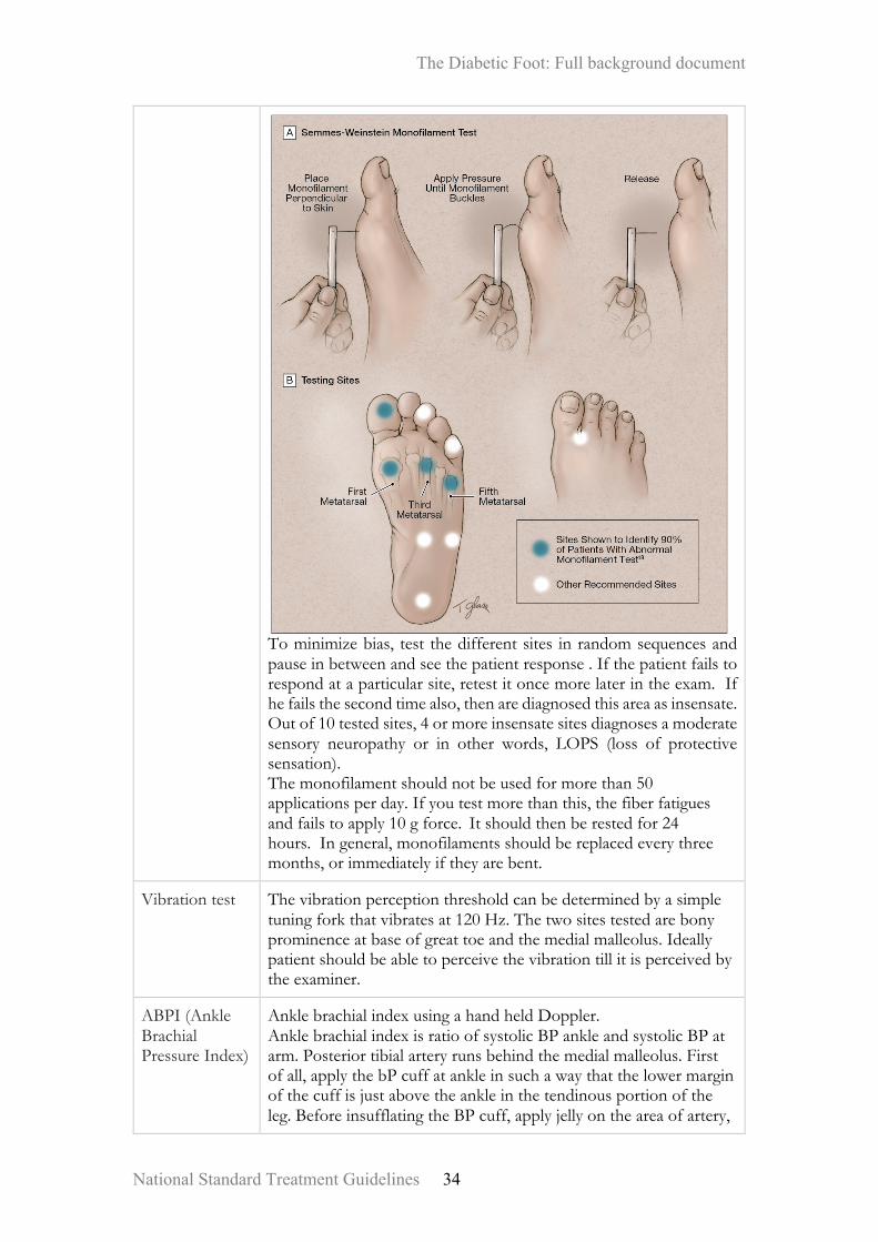
The Diabetic Foot: Full background document
National Standard Treatment Guidelines 34
To minimize bias, test the different sites in random sequences and pause in between and see the patient response . If the patient fails to respond at a particular site, retest it once more later in the exam. If he fails the second time also, then are diagnosed this area as insensate. Out of 10 tested sites, 4 or more insensate sites diagnoses a moderate sensory neuropathy or in other words, LOPS (loss of protective sensation). The monofilament should not be used for more than 50 applications per day. If you test more than this, the fiber fatigues and fails to apply 10 g force. It should then be rested for 24 hours. In general, monofilaments should be replaced every three months, or immediately if they are bent.
Vibration test The vibration perception threshold can be determined by a simple tuning fork that vibrates at 120 Hz. The two sites tested are bony prominence at base of great toe and the medial malleolus. Ideally patient should be able to perceive the vibration till it is perceived by the examiner.
ABPI (Ankle Brachial Pressure Index)
Ankle brachial index using a hand held Doppler. Ankle brachial index is ratio of systolic BP ankle and systolic BP at arm. Posterior tibial artery runs behind the medial malleolus. First of all, apply the bP cuff at ankle in such a way that the lower margin of the cuff is just above the ankle in the tendinous portion of the leg. Before insufflating the BP cuff, apply jelly on the area of artery,

The Diabetic Foot: Full background document
National Standard Treatment Guidelines 35
put the Doppler probe and move it around till you hear a clear sound. Then with other hand insufflate the cuff. As the cuff pressure exceeds the systolic pressure, the sound will disappear. Then start releasing the insufflated air slowly. At one point, you will start getting the sound on Doppler. This is systolic BP. Now deflate the BP cuff completely and apply jelly over the area of dorsalis pedis artery, and similarly measure the systolic pressure. In a similar way you can measure the systolic brachial in both the forearms. The highest recorded pedal pressure in one lower limb, regardless of the vessel, is divided by the highest of the two extremity pressures. This is called ABPI.
Probe to bone (PTB) test
When there is high clinical suspicion of osteomyelitis , any sterile blunt instrument can be used as probe and inserted into the wound, if bone can be sound then it is considered as positive test. negative test largely rules out osteomyelitis
Ipswich Touch Test
Done at home by a nonprofessional (friend, relative, or caregiver) following written instructions. The test comprised light touching of the tips of the patient's 1st, 3rd, and 5th toes of each foot with the tip of the tester's index finger for 1 to 2 seconds while the patient's eyes were closed. The patient indicated whether they felt the touch by responding right or left, and results were recorded on a foot diagram.

The Diabetic Foot: Full background document
National Standard Treatment Guidelines 36
ANNEXURE 2: Risk grading tables for decision making Table 1. Infectious Diseases Society of America and International Working Group on the Diabetic Foot Classifications of Diabetic Foot Infection
Clinical Manifestation of Infection PEDIS Grade
IDSA Infection
Uninfected: No symptoms or signs of infection
1 Uninfected
Infected: At least two of the following items are present: • Local swelling or induration • Erythema >0.5cm around the wound • Local tenderness or pain • Local warmth • Purulent discharge (thick, opaque to white or sanguineous secretion) Exclude other causes of an inflammatory response of the skin (eg, trauma, gout, acute Charcot neuro-osteoarthropathy, fracture, thrombosis, venous stasis). Local infection involving only the skin and the subcutaneous tissue (without involvement of deeper tissues and without systemic signs as described below). An erythema, must be </=2 cm around the ulcer.
2 Mild
Local infection (as described above) with erythema > 2 cm, or involving structures deeper than skin and subcutaneous tissues (eg, abscess, osteomyelitis, septic arthritis, fasciitis), and No systemic inflammatory response signs (as described below)
3 Moderate
Local infection (as described above) with the signs of SIRS, as manifested by ≥2 of the following: • Temperature >38°C or <36°C • Heart rate >90 beats/min • Respiratory rate >20 breaths/min or PaCO2 <32 mm Hg • White blood cell count >12,000 or <4000 cells/µL or ≥10% immature (band) forms
3 Severea
Abbreviations: IDSA, Infectious Diseases Society of America; PaCO2, partial pressure of arterial carbon dioxide; PEDIS, perfusion, extent/size, depth/tissue loss, infection, and sensation; SIRS, systemic inflammatory response syndrome. a.Ischemia may increase the severity of any infection, and the presence of critical ischemia often makes the infection severe. Systemic infection may sometimes manifest with other clinical findings, such as hypotension, confusion, vomiting, or evidence of metabolic disturbances, such as acidosis, severe hyperglycemia, and new-onset azotemia. Source: IWGDF 2015
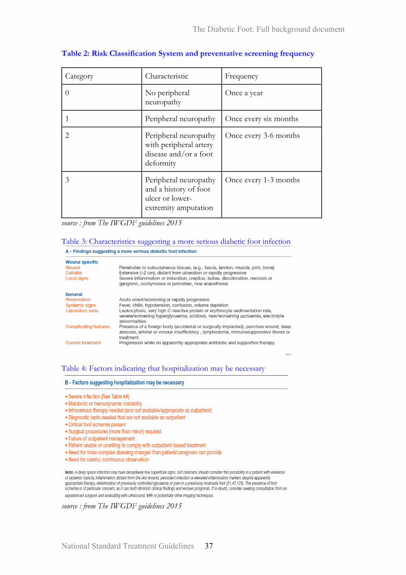
The Diabetic Foot: Full background document
National Standard Treatment Guidelines 37
Table 2: Risk Classification System and preventative screening frequency
Category Characteristic Frequency
0 No peripheral neuropathy
Once a year
1 Peripheral neuropathy Once every six months
2 Peripheral neuropathy with peripheral artery disease and/or a foot deformity
Once every 3-6 months
3 Peripheral neuropathy and a history of foot ulcer or lower-extremity amputation
Once every 1-3 months
source : from The IWGDF guidelines 2015 Table 3: Characteristics suggesting a more serious diabetic foot infection
Table 4: Factors indicating that hospitalization may be necessary
source : from The IWGDF guidelines 2015

The Diabetic Foot: Full background document
National Standard Treatment Guidelines 38
Annexure 1
Adapt / Adopt Tables

The Diabetic Foot: Full background document
National Standard Treatment Guidelines 39
ADAPT–ADOPTTABLES
4.1PreventionofDiabeticFootproblems
MoHFWguideline Adopted/Adapted
OriginalGuideline Remarks
4.1.1Toidentifyapersonwithdiabetesatriskforfootulceration,examinethefeetannually/sixmonthly/quarterly/monthly(dependingonpatient’sriskcategory*)toseekevidenceforsignsorsymptomsofperipheralneuropathyandperipheralarterydisease.
Adapted IWGDF2015GuidanceDocumentsays-Toidentifyapersonwithdiabetesatriskforfootulceration,examinethefeetannuallytoseekevidenceforsignsorsymptomsofperipheralneuropathyandperipheralarterydisease.(Strong;Low)
ExpertconsensusrecommendsmorefrequentfollowupsforhighriskpatientsofDF.RiskstratificationandfrequencyoffollowuphasbeenmentionedintheAnnexuretable-2.
4.1.2Inapersonwithdiabeteswhohasperipheralneuropathy,screenfor:ahistoryoffootulcerationorlower-extremityamputation;peripheralarterydisease;footdeformity;pre-ulcerativesignsonthefoot;poorfoothygiene;andill-fittingorinadequatefootwear.(Strong;Low)
Adopted IWGDF2015Guidancedocument
4.1.3Treatanypre-ulcerativesignonthefootofapatientwithdiabetes.Thisincludes:removingcallus;protectingblistersanddrainingwhennecessary;treatingingrownorthickened
Adopted IWGDF2015Guidancedocument
The Diabetic Foot: Full background document
National Standard Treatment Guidelines 40
toenails;treatinghaemorrhagewhennecessary;andprescribingantifungaltreatmentforfungalinfections.(Strong;Low)
4.1.4Toprotecttheirfeet,instructanat-riskpatientwithdiabetesnottowalkbarefoot,insocks,orinthin-soledstandardslippers,whetherathomeorwhenoutside.(Strong;Low)
Adopted IWGDF2015Guidancedocument
4.1.5Instructanat-riskpatientwithdiabetesto:dailyinspecttheirfeetandtheinsideoftheirshoes;dailywashtheirfeet(withcarefuldryingparticularlybetweenthetoes);avoidusingchemicalagentsorplasterstoremovecallusorcorns;useemollientstolubricatedryskin;andcuttoenailsstraightacross.(Weak;Low)
Adopted IWGDF2015Guidancedocument
4.1.6Instructanat-riskpatientwithdiabetestowearproperlyfittingfootweartopreventafirstfootulcer,eitherplantarornon-plantar,orarecurrentnon-plantarfootulcer.Whenafootdeformityorapre-ulcerativesignispresent,considerprescribing
Adopted IWGDF2015Guidancedocument

The Diabetic Foot: Full background document
National Standard Treatment Guidelines 41
therapeuticshoes,custom-madeinsoles,ortoeorthosis.(Strong;Low)
4.1.7Instructahigh-riskpatientwithdiabetestomonitorfootskintemperatureathometopreventafirstorrecurrentplantarfootulcer.Thisaimsatidentifyingtheearlysignsofinflammation,followedbyactiontakenbythepatientandcareprovidertoresolvethecauseofinflammation.(Weak;Moderate)
Adopted IWGDF2015Guidancedocument
4.1.8Topreventafirstfootulcerinanat-riskpatientwithdiabetes,provideeducationaimedatimprovingfootcareknowledgeandbehaviour,aswellasencouragingthepatienttoadheretothisfootcareadvice.(Weak;Low)
Adopted IWGDF2015Guidancedocument
4.1.9Topreventarecurrentplantarfootulcerinanat-riskpatientwithdiabetes,prescribetherapeuticfootwearthathasademonstratedplantarpressurerelievingeffectduringwalkingandencouragethepatienttowearthisfootwear.
Adapted IWGDF2015guidancedocumentsays-Topreventarecurrentplantarfootulcerinanat-riskpatientwithdiabetes,prescribetherapeuticfootwearthathasademonstratedplantarpressurerelievingeffectduringwalking(i.e.30%relief
ExpertConsensussaysthatnofootwearmanufacturecanmentionthequantityofpressurereductionasitdependsontheheightandweightofthepatientaswellasthepatternoffootstrike.

The Diabetic Foot: Full background document
National Standard Treatment Guidelines 42
comparedtoplantarpressureinstandardofcaretherapeuticfootwear)andencouragethepatienttowearthisfootwear.(Strong;Moderate)
4.1.10Topreventarecurrentfootulcerinanat-riskpatientwithdiabetes,provideintegratedfootcare,whichincludesprofessionalfoottreatment,adequatefootwearandeducation.Thisshouldberepeatedorre-evaluatedonceeveryonetothreemonthsasnecessary.(Strong;Low)
Adopted IWGDF2015Guidancedocument
*Riskcategoryandfrequencyoffollowupisgivenintheannexuredocument.
4.2.1:Assessingtheriskofdevelopingadiabeticfootproblem
MoHFWguideline Adopted/Adapted
OriginalGuideline Remarks
4.2.1aEvaluateadiabeticpatientpresentingwithafootwoundat3levels:thepatientasawhole,theaffectedfootorlimb,andtheinfectedwound.
Adapted IDSA2012Guidelinessays–Cliniciansshouldevaluateadiabeticpatientpresentingwithafootwoundat3levels:thepatientasawhole,theaffectedfootorlimb,andtheinfectedwound.(Strong;Low)
Rephrasingdoneforusingactiveverb.
4.2.1bAssesstheaffectedlimbandfootforarterialischemia,venousinsufficiency,presenceof
Adapted IDSA2012Guidelinessays-Werecommendassessingtheaffectedlimbandfootfor
Rephrasingdoneforusingactiveverb.

The Diabetic Foot: Full background document
National Standard Treatment Guidelines 43
protectivesensation,andbiomechanicalproblems*.
arterialischemia(strong,moderate),venousinsufficiency,presenceofprotectivesensation,andbiomechanicalproblems.(Strong;Low)
*Biomechanicalproblemsmeansanatomicalandphysiologicaldisturbancesofthefoot, i.e.,structuralchangeswhichhappeninthebones,jointsandmusclesofthefootofdiabeticsandthechangesinthebloodcirculationandnervesensationofthefootofdiabetics.
4.2.2ClassificationofDiabeticfoot
MoHFWguideline Adopted/Adapted
OriginalGuideline
Remarks
4.2.2aAssesstheseverityofanydiabeticfootinfectionusingtheInfectiousDiseasesSocietyofAmerica/InternationalWorkingGroupontheDiabeticFootClassificationsystem.(Strong;Moderate)
Adopted IWGDF2015GuidanceDocument
ClassificationhasbeenprovidedintheAnnexureTable-1.
4.2.2bDonotusetheWagnerclassificationsystemtoassesstheseverityofadiabeticfootulcer.
Adopted NICE2015GuidelinesonDiabeticfootproblems:preventionandmanagement
Wagnerclassificationdoesnottakeintoaccountthepresenceofneuropathyandsizeofthelesion.Thesefactorscanhaveapronouncedeffectonthetreatmentandpotentialoutcome.(Reference–PodiatryToday–AGuidetoUnderstandingtheVariousWoundClassificationSystems–byKathleenSatterfield–vol.19–Issue6–June2006)
4.2.3ReferralforDiabeticfootproblems(AdmissioncriteriaandDischargecriteria)
The Diabetic Foot: Full background document
National Standard Treatment Guidelines 44
MoHFWguideline Adopted/Adapted
OriginalGuideline Remarks
4.2.3aInitiallyhospitalizeallpatientswithasevereinfection,selectedpatientswithamoderateinfectionwithcomplicatingfeatures(eg,severeperipheralarterialdisease[PAD]orlackofhomesupport),andanypatientunabletocomplywiththerequiredoutpatienttreatmentregimenforpsychologicalorsocialreasons.Alsohospitalizepatientswhodonotmeetanyofthesecriteria,butarefailingtoimprovewithoutpatienttherapy.
Adapted IDSA2012Guidelinessays-Werecommendthatallpatientswithasevereinfection,selectedpatientswithamoderateinfectionwithcomplicatingfeatures(e.g.,severePADorlackofhomesupport),andanypatientunabletocomplywithanappropriateoutpatienttreatmentregimenforpsychologicalorsocialreasonsbehospitalizedinitially.Patientswhodonotmeetanyofthesecriteriabutarefailingtoimprovewithoutpatienttherapymayalsoneedtobehospitalized(Strong,low).
Rephrasingdoneforusingactiveverb.
4.2.3bPriortobeingdischarged,makesurethatapatientwithaDiabeticFootInfectionisclinicallystable;hashadanyurgentlyneededsurgeryperformed;hasachievedacceptableglycemiccontrol;isabletomanage(onhis/herownorwithhelp)atthedesignateddischargelocation;andhasawelldefinedplanthatincludesanappropriateantibioticregimentowhichhe/shewilladhere,an
Adapted IDSA2012Guidelinessays-Werecommendthatpriortobeingdischarged,apatientwithaDFIshouldbeclinicallystable;havehadanyurgentlyneededsurgeryperformed;haveachievedacceptableglycemiccontrol;beabletomanage(onhis/herownorwithhelp)atthedesignateddischargelocation;andhaveawelldefinedplanthatincludesanappropriateantibiotic
Rephrasingdoneforusingactiveverb.
The Diabetic Foot: Full background document
National Standard Treatment Guidelines 45
off-loadingscheme(ifneeded),specificwoundcareinstructions,andappropriateoutpatientfollow-up.
regimentowhichhe/shewilladhere,anoff-loadingscheme(ifneeded),specificwoundcareinstructions,andappropriateoutpatientfollow-up.(Strong,low).
4.3DiabeticFootInfections
MoHFWguideline Adopted/Adapted
OriginalGuideline Remarks
4.3.1Considerthepossibilityofinfectionoccurringinanyfootwoundinapatientwithdiabetes.Evidenceofinfectiongenerallyincludesclassicsignsofinflammation(redness,warmth,swelling,tenderness,orpain)orpurulentsecretions,butmayalsoincludeadditionalorsecondarysigns(e.g.,nonpurulentsecretions,friableordiscoloredgranulationtissue,underminingofwoundedges,foulodour).
Adapted IDSA2012Guidelinessays–Cliniciansshouldconsiderthepossibilityofinfectionoccurringinanyfootwoundinapatientwithdiabetes.Evidenceofinfectiongenerallyincludesclassicsignsofinflammation(redness,warmth,swelling,tenderness,orpain)orpurulentsecretions,butmayalsoincludeadditionalorsecondarysigns(e.g.,non-purulentsecretions,friableordiscolouredgranulationtissue,underminingofwoundedges,foulodour).(Strong;Low)
Rephrasingdoneforusingactiveverb.
4.3.2Beawareoffactorsthatincreasetheriskfordiabeticfootinfections(DFI)andespeciallyconsiderinfectionwhenthesefactorsarepresent;theseincludeawound
Adapted IDSA2012Guidelinessays–Cliniciansshouldbeawareoffactorsthatincreasetheriskfordiabeticfootinfections(DFI)andespeciallyconsiderinfection
Rephrasingdoneforusingactiveverb.
The Diabetic Foot: Full background document
National Standard Treatment Guidelines 46
forwhichtheprobe-to-bone(PTB)testispositive;anulcerationpresentfor>30days;ahistoryofrecurrentfootulcers;atraumaticfootwound;thepresenceofperipheralvasculardiseaseintheaffectedlimb;apreviouslowerextremityamputation;lossofprotectivesensation;thepresenceofrenalinsufficiency;orahistoryofwalkingbarefoot.
whenthesefactorsarepresent;theseincludeawoundforwhichtheprobe-to-bone(PTB)testispositive;anulcerationpresentfor>30days;ahistoryofrecurrentfootulcers;atraumaticfootwound;thepresenceofperipheralvasculardiseaseintheaffectedlimb;apreviouslowerextremityamputation;lossofprotectivesensation;thepresenceofrenalinsufficiency;orahistoryofwalkingbarefoot.(Strong;Low)
4.3.3TakeplainradiographsoftheaffectedfootofallpatientspresentingwithanewDiabeticFootInfectiontolookforbonyabnormalities(deformity,destruction)aswellasforsofttissuegasandradio-opaqueforeignbodies.
Adapted IDSA2012Guidelinessays–WerecommendthatallpatientspresentingwithanewDFIhaveplainradiographsoftheaffectedfoottolookforbonyabnormalities(deformity,destruction)aswellasforsofttissuegasandradio-opaqueforeignbodies(strong,moderate).
Rephrasingdoneforusingactiveverb.
4.3.4WhenandhowtoobtainculturefromDiabeticfootpatients
MoHFWguideline Adopted/Adapted
OriginalGuideline Remarks
4.3.4aForclinicallyuninfectedwounds,donotcollectaspecimenforculture.
Adapted IDSA2012Guidelinessays–Forclinicallyuninfectedwounds,we
Rephrasingdoneforusingactiveverb.

The Diabetic Foot: Full background document
National Standard Treatment Guidelines 47
recommendnotcollectingaspecimenforculture(strong,low).
4.3.4bSendaspecimenforculturethatisfromdeeptissue,obtainedbybiopsyorcurettageandafterthewoundhasbeencleansedanddebrided.Avoidswabspecimens,especiallyofinadequatelydebridedwounds,astheyprovidelessaccurateresults.*
Adapted IDSA2012Guidelinessays–Werecommendsendingaspecimenforculturethatisfromdeeptissue,obtainedbybiopsyorcurettageafterthewoundhasbeencleansedanddebrided.Wesuggestavoidingswabspecimens,especiallyofinadequatelydebridedwounds,astheyprovidelessaccurateresults(strong,moderate).
Rephrasingdoneforusingactiveverb.
4.3.4cDonotobtainrepeatculturesunlessthepatientisnotclinicallyrespondingtotreatment.
Adapted IWDGF2015says–Donotobtainrepeatculturesunlessthepatientisnotclinicallyrespondingtotreatment,oroccasionallyforinfectioncontrolsurveillanceofresistantpathogens(Strong;Low)
Infectioncontrolsurveillanceofresistantpathogensmaynotbepossibleatalllevels.
4.3.4dForinfectedwounds,sendappropriatelyobtainedspecimensforculturepriortostartingempiricantibiotictherapy,ifpossible.Culturesmaybeunnecessaryforamildinfectioninapatientwhohasnotrecentlyreceivedantibiotictherapy.
Adapted IDSA2012GuidelinesForinfectedwounds,werecommendthatclinicianssendappropriatelyobtainedspecimensforculturepriortostartingempiricantibiotictherapy,ifpossible.Culturesmaybeunnecessaryforamildinfectioninapatientwhohasnotrecently
Rephrasingdoneforusingactiveverb.
The Diabetic Foot: Full background document
National Standard Treatment Guidelines 48
receivedantibiotictherapy(strong,low).
*Washthewoundwithsalineandthesurroundingskinwithantisepticsolutionbeforetakingculturetoavoidcontaminationofthespecimenobtainedforculture.
4.3.5SelectionofAntibioticandwhenshoulditbemodified
MoHFWguideline Adopted/Adapted
OriginalGuideline
Remarks
4.3.5aDonottreatclinicallyuninfectedwoundswithantibiotictherapy.
Adapted IDSA2012Guidelinessays–Werecommendthatclinicallyuninfectedwoundsnotbetreatedwithantibiotictherapy(strong,low).
Rephrasingdoneforusingactiveverb.
4.3.5bPrescribeantibiotictherapyforallinfectedwoundsbutcautionthatthisisofteninsufficientunlesscombinedwithappropriatewoundcare.
Adapted IDSA2012Guidelinessays–Werecommendprescribingantibiotictherapyforallinfectedwoundsbutcautionthatthisisofteninsufficientunlesscombinedwithappropriatewoundcare(strong,low).
Rephrasingdoneforusingactiveverb.
4.3.5cBasetherouteoftherapylargelyoninfectionseverity.Preferparenteraltherapyforallsevere,andsomemoderateDFls,atleastinitially,withaswitchtooralagentswhenthepatientissystemicallywell
Adapted IDSA2012guidelinessays–Wesuggestbasingtherouteoftherapylargelyoninfectionseverity.Wepreferparenteraltherapyforallsevere,andsomemoderateDFIs,atleastinitially(weak,low),withaswitch
Rephrasingdoneforusingactiveverb.

The Diabetic Foot: Full background document
National Standard Treatment Guidelines 49
andcultureresultsareavailable.Clinicianscanprobablyusehighlybioavailableoralantibioticsaloneinmostmild,andinmanymoderate,infectionsandtopicaltherapyforselectedmildsuperficialinfections.
tooralagentswhenthepatientissystemicallywellandcultureresultsareavailable.Clinicianscanprobablyusehighlybioavailableoralantibioticsaloneinmostmild,andinmanymoderate,infectionsandtopicaltherapyforselectedmildsuperficialinfections(strong,moderate).
4.3.5dCliniciansshouldselectanempiricantibioticregimenonthebasisoftheseverityoftheinfectionandthelikelyetiologicagent(s).a. Formildtomoderateinfectionsinpatientswhohavenotrecentlyreceivedantibiotictreatment,therapyjusttargetingaerobicgram-positivecocci(GPC)issufficient.b. Formostsevereinfections,startbroad-spectrumempiricantibiotictherapy,pendingcultureresultsandantibioticsusceptibilitydata.
Adapted IDSA2012Guidelinessaysthat–Werecommendthatcliniciansselectanempiricantibioticregimenonthebasisoftheseverityoftheinfectionandthelikelyetiologicagent(s).(Strong;Low)a. Formildtomoderateinfectionsinpatientswhohavenotrecentlyreceivedantibiotictreatment,wesuggestthattherapyjusttargetingaerobicgram-positivecocci(GPC)issufficient.(Weak;Low)b. Formostsevereinfections,
Rephrasingdoneforusingactiveverb.
The Diabetic Foot: Full background document
National Standard Treatment Guidelines 50
c. EmpirictherapydirectedatP.aeruginosaisusuallyunnecessaryexceptforpatientswithriskfactors*fortrueinfectionwiththisorganism.d. ConsiderprovidingempirictherapydirectedagainstMRSAinapatientwithapriorhistoryofMRSAinfection;whenthelocalprevalence**ofMRSAcolonizationorinfectionishigh;oriftheinfectionisclinicallysevere.
werecommendstartingbroad-spectrumempiricantibiotictherapy,pendingcultureresultsandantibioticsusceptibilitydata.(Strong;Low)c. EmpirictherapydirectedatP.aeruginosaisusuallyunnecessaryexceptforpatientswithriskfactors*fortrueinfectionwiththisorganism.(Strong;Low)d. ConsiderprovidingempirictherapydirectedagainstMRSAinapatientwithapriorhistoryofMRSAinfection;whenthelocalprevalence**ofMRSAcolonizationorinfectionishigh;oriftheinfectionisclinicallysevere.(Weak;Low).
4.3.5eGiveaninitialantibioticcourseforasofttissueinfectionofabout1–2weeksformildinfectionsand2–3weeksformoderatetosevereinfections.
Adapted IDSA2012Guidelinessays–Wesuggestcontinuingantibiotictherapyuntil,butnotbeyond,resolutionoffindingsofinfection,butnotthroughcompletehealingofthewound(weak,low).Wesuggestaninitialantibiotic
Boththesentencesofthisoriginalguidelinecanthemselvesbeanindividualrecommendationandhencewehavesplitthemforeaseofunderstandingwithoutchangingthemeaningorthemessagetobeconveyed.
The Diabetic Foot: Full background document
National Standard Treatment Guidelines 51
courseforasofttissueinfectionofabout1-2weeksformildinfectionsand2-3weeksformoderatetosevereinfections(weak,low).
Also,rephrasingdoneforusingactiveverb.
4.3.5fContinueantibiotictherapyuntil,butnotbeyond,resolutionoffindingsofinfection,butnotthroughcompletehealingofthewound.
Adapted IDSA2012Guidelinessays–Wesuggestcontinuingantibiotictherapyuntil,butnotbeyond,resolutionoffindingsofinfection,butnotthroughcompletehealingofthewound(weak,low).Wesuggestaninitialantibioticcourseforasofttissueinfectionofabout1-2weeksformildinfectionsand2-3weeksformoderatetosevereinfections(weak,low).
Boththesentencesofthisoriginalguidelinecanthemselvesbeanindividualrecommendationandhencewehavesplitthemforeaseofunderstandingwithoutchangingthemeaningorthemessagetobeconveyed.Also,rephrasingdoneforusingactiveverb.
4.3.5gAdministerparenteraltherapyinitiallyformostsevereinfectionsandsomemoderateinfections,withaswitchtooraltherapywhentheinfectionisresponding.(Strong;Low)
Adopted IDSA2012Guidelines
*RiskfactorsfortrueinfectionwithPseudomonasaeruginosaincludeImmunocompromisedstatus,ChronicKidneyDisease,warmclimateandfrequentexposureoffoottowater.

The Diabetic Foot: Full background document
National Standard Treatment Guidelines 52
**ThelocalprevalenceofMRSA(i.e.,percentageofallS.aureusclinicalisolatesinthatlocalethataremethicillinresistant)ishighenough(perhaps50%foramildand30%foramoderatesofttissueinfection)thatthereisareasonableprobabilityofMRSAinfection.
4.4Woundcaretechniquesanddressings
MoHFWguideline Adopted/Adapted
OriginalGuideline
Remarks
4.4.1Cleanulcersregularlywithcleanwater*orsaline,debridethemwhenpossibleinordertoremovedebrisfromthewoundsurfaceanddressthemwithasterile,inertdressinginordertocontrolexcessiveexudateandmaintainawarm,moistenvironmentinordertopromotehealing**.(Strong;Low)
Adopted IWGDF2015Guidancedocument
4.4.2Selectdressingsprincipallyonthebasisofexudatecontrol,comfortandcost.(Strong;Low)
Adopted IWGDF2015Guidancedocument
4.4.3Donotuseantimicrobialdressingswiththegoalofimprovingwoundhealingorpreventingsecondaryinfection.(Strong;Moderate)
Adopted IWGDF2015Guidancedocument
4.4.4Donotofferthefollowingtotreatdiabeticfootulcers,unlessaspartofaclinicaltrial:· Electricalstimulationtherapy,autologousplatelet-richplasmagel,regenerativewoundmatricesanddalteparin.· Growthfactors(granulocytecolony-stimulatingfactor[G-CSF],platelet-derivedgrowthfactor[PDGF],epidermalgrowthfactor[EGF]andtransforminggrowthfactorbeta[TGF-β]).·Hyperbaricoxygentherapy.
Adopted NICE2015GuidelinesonDiabeticFoot(NG19)

The Diabetic Foot: Full background document
National Standard Treatment Guidelines 53
4.4.5Considerdermalorskinsubstitutesasanadjuncttostandardcarewhentreatingdiabeticfootulcers,onlywhenhealinghasnotprogressedandontheadviceofthemultidisciplinaryfootcareservice.
Adopted NICE2015GuidelinesonDiabeticFoot(NG19)
4.4.6Considernegativepressurewoundtherapyaftersurgicaldebridementfordiabeticfootulcers,ontheadviceofthemultidisciplinaryfootcareservice.
Adopted NICE2015GuidelinesonDiabeticFoot(NG19)
4.4.7Donotselectagentsreportedtoimprovewoundhealingbyalteringthebiologyofthewound,includinggrowthfactors,bioengineeredskinproductsandgases,inpreferencetoacceptedstandardsofgoodqualitycare.(Strong;Low)
Adopted IWGDF2015Guidancedocument
4.4.8Donotselectagentsreportedtohaveanimpactonwoundhealingthroughalterationofthephysicalenvironment,includingthroughtheuseofelectricity,magnetism,ultrasoundandshockwaves,inpreferencetoacceptedstandardsofgoodqualitycare.(Strong;Low)
Adopted IWGDF2015Guidancedocument
4.4.9Donotselectsystemictreatmentsreportedtoimprovewoundhealing,includingdrugsandherbaltherapies,inpreferencetoacceptedstandardsofgoodqualitycare.(Strong;Low)
Adopted IWGDF2015Guidancedocument
4.4.10Redistributethepressureoffthewoundtotheentireweight-bearingsurfaceofthefoot(“off-loading”).Whileparticularly
Adopted IDSA2012Guidelines
Rephrasingdoneforusingactiveverb

The Diabetic Foot: Full background document
National Standard Treatment Guidelines 54
importantforplantarwounds,thisisalsonecessarytorelievepressurecausedbydressings,footwear,orambulationtoanysurfaceofthewound.(Strong;High)
4.4.11Whendecidingaboutwounddressingsandoffloadingwhentreatingdiabeticfootulcers,takeintoaccounttheclinicalassessmentofthewoundandtheperson’spreference,andusedevicesanddressingswiththelowestacquisitioncostappropriatetotheclinicalcircumstances.
Adopted NICE2015Guidelines
*Cleanwaterisboiledcooledwater(distilledwater)**DonotuseH2O2,EUSOL,etc
4.5Footwear
MoHFWguideline Adopted/Adapted
OriginalGuideline
Remarks
4.5.1Tohealaneuropathicplantarforefootulcerwithoutischemiaoruncontrolledinfectioninapatientwithdiabetes,offloadwithanon-removableknee-highdevicewithanappropriatefoot-deviceinterface.(Strong;High)
Adopted IWGDF2015GuidanceDocument
Non-removable(cast)walker:Sameasremovable(cast)boot/walkerbutthenwithalayer(s)offibreglasscastmaterialcircumferentiallywrappedarounditrenderingitirremovable(alsoknownas"instanttotalcontactcast")
4.5.2Whenanon-removableknee-highdeviceiscontraindicatedornottoleratedbythepatient,consider
Adopted IWGDF2015GuidanceDocument
Removable(cast)boot/walker:Prefabricatedremovableknee-highbootwitharockerorrolleroutsole

The Diabetic Foot: Full background document
National Standard Treatment Guidelines 55
offloadingwitharemovableknee-highwalkerwithanappropriatefoot-deviceinterfacetohealaneuropathicplantarforefootulcerinapatientwithdiabetes,butonlywhenthepatientcanbeexpectedtobeadherenttowearingthedevice.(Weak;Moderate)
configuration,paddedinterior,andaninsertableandadjustableinsolewhichmaybetotalcontact.
4.5.3Whenaknee-highdeviceiscontraindicatedorcannotbetoleratedbythepatient,consideroffloadingwithaforefootoffloadingshoe,castshoe,orcustom-madetemporaryshoetohealaneuropathicplantarforefootulcerinapatientwithdiabetes,butonlyandwhenthepatientcanbeexpectedtobeadherenttowearingtheshoes.(Weak;Low)
Adopted IWGDF2015GuidanceDocument
4.5.4Instructanat-riskpatientwithdiabetestowearproperlyfittingfootweartopreventafirstfootulcer,eitherplantarornon-plantar,orarecurrentnon-plantarulcer.When
Adopted IWGDF2015GuidanceDocument

The Diabetic Foot: Full background document
National Standard Treatment Guidelines 56
afootdeformityorapre-ulcerativesignispresent,considerprescribingtherapeuticshoes,custom-madeinsoles,ortoeorthosis.(Strong;Low)
4.5.5Topreventarecurrentplantarfootulcerinanat-riskpatientwithdiabetes,prescribetherapeuticfootwearthathasademonstratedplantarpressurerelievingeffectduringwalkingandencouragethepatienttowearthisfootwear.
Adapted IWGDF2015guidancedocumentsays-Topreventarecurrentplantarfootulcerinanat-riskpatientwithdiabetes,prescribetherapeuticfootwearthathasademonstratedplantarpressurerelievingeffectduringwalking(i.e.30%reliefcomparedtoplantarpressureinstandardofcaretherapeuticfootwear)andencouragethepatienttowearthisfootwear.(Strong;Moderate)
ExpertConsensussaysthatnofootwearmanufacturecanmentionthequantityofpressurereductionasitdependsontheheightandweightofthepatientaswellasthepatternoffootstrike.
4.5.6Instructapatientwithdiabetesnottouseconventionalorstandardtherapeuticfootweartohealaplantarfootulcer.*
Adapted IWGDF2015GuidanceDocumentsays–Donotprescribe,andinstructapatientwithdiabetesnottouse,conventionalorstandardtherapeuticshoes
Rephrasingdoneforbetterunderstandingoftherecommendation.Also,theExpertconsensusfelttheword“shoes”shouldbereplacedwith“footwear”asmanypeopleinIndiawear

The Diabetic Foot: Full background document
National Standard Treatment Guidelines 57
tohealaplantarfootulcer.(Strong;Low)
chappalsandsandals,insteadofshoes.Also,itmeans–Usefootwearwithfollowingfeatures–Sandals:shouldhaveadjustablestraps,insole,fullheelcounterandrigidoutsole.Shoes:shouldhavewidetoeboxextradepthandwithoutlaces
4.5.7Considerusingshoemodifications,temporaryfootwear,toespacersororthosestooffloadandhealanon-plantarfootulcerwithoutischemiaoruncontrolledinfectioninapatientwithdiabetes.Thespecificmodalitywilldependonthetypeandlocationofthefootulcer.(Weak;Low)
Adopted IWGDF2015GuidanceDocument
4.5.8Ifotherformsofbiomechanicalreliefarenotavailable,considerusingfeltedfoam**incombinationwithappropriatefootweartooffloadandhealaneuropathicfootulcerwithoutischemiaoruncontrolledinfectioninapatientwith
Adopted IWGDF2015GuidanceDocument
Felted foam - A fibrous, unwoven material backed by foam with absorbing and cushioning characteristics. The foam is generally ‘rubber foam’ or ‘PU foam’ which is formed by either a polyester or polyether polyol resin, in conjunction with water and Toluene di Isocyanate, along with various catalysts and blowing agents and colouring pigments to give the desired compression.

The Diabetic Foot: Full background document
National Standard Treatment Guidelines 58
diabetes.(Weak;Low)
*Usefootwearwithfollowingfeatures-Sandals:shouldhaveadjustablestraps,insole,fullheelcounterandrigidoutsole.Shoes:shouldhavewidetoeboxextradepthandwithoutlaces.** Felted foam - A fibrous, unwoven material backed by foam with absorbing and cushioning characteristics. The foam is generally ‘rubber foam’ or ‘PU foam’ which is formed by either a polyester or polyether polyol resin, in conjunction with water and Toluene di Isocyanate, along with various catalysts and blowing agents and colouring pigments to give the desired compression.
4.6Diabeticfootwithosteomyelitis&Charcot’sFoot
MoHFWguideline Adopted/Adapted
OriginalGuideline
Remarks
4.6.1Foraninfectedopenwound,performaprobe-to-bonetest;inapatientatlowriskforosteomyelitisanegativetestlargelyrulesoutthediagnosis,whileinahighriskpatientapositivetestislargelydiagnostic.(Strong;High)
Adopted IDSA2012Guidelines
4.6.2MarkedlyelevatedESRissuggestiveofosteomyelitisinsuspectedcases.
Adapted IWDGF2015says-Markedlyelevatedseruminflammatorymarkers,especiallyerythrocytesedimentationrate,aresuggestiveofosteomyelitisinsuspectedcases.
Testsforseruminflammatorymarkersarecostlyandnotwidelyavailable,exceptESR.AlsothesetestsarenotdiagnosticofDFO.
4.6.3IfosteomyelitisissuspectedinapersonwithdiabetesbutisnotconfirmedbyinitialX-ray,consideranMRIto
Adapted NICE2015GuidelinesonDiabeticFoot(NG19)saysthat–Ifosteomyelitisissuspectedina
ExpertConsensussaysthatasavailabilityofMRIislimitedacrossthecountry,itisrecommendedto
The Diabetic Foot: Full background document
National Standard Treatment Guidelines 59
confirmthediagnosis.InplaceswhereMRIisunavailable,diagnoseosteomyelitisbythePTBtest(clinically)and/ortakingaBonebiopsyandculture.
personwithdiabetesbutisnotconfirmedbyinitialX-ray,consideranMRItoconfirmthediagnosis.
useMRIwhereveravailable.Attheprimaryandsecondaryhealthcentrelevels,thePTBtestandbonebiopsyandculturearemorefeasibleandeconomicalandreasonablyaccurate.
4.6.4Adefinitediagnosisofboneinfectionusuallyrequirespositiveresultsonbothhistologicalandmicrobiologicalexaminationsofanasepticallyobtainedbonesample,butthisisusuallyrequiredonlywhenthediagnosisisindoubtordeterminingthecausativepathogen’santibioticsusceptibilityiscrucial.(Strong;Moderate)
Adopted IDSA2012Guidelines
4.6.5Avoidusingresultsofsofttissueorsinustractspecimensforselectingantibiotictherapyforosteomyelitisastheydonotaccuratelyreflectbonecultureresults.(Strong;Moderate)
Adopted IDSA2012Guidelines
4.6.6Whenaradicalresectionleavesnoremaininginfected
Adapted IDSA2012Guidelinessays–
Aproximalbonehistopathtobedoneifavailableto

The Diabetic Foot: Full background document
National Standard Treatment Guidelines 60
tissue*,prescribeantibiotictherapyforonlyashortduration(2–5days).Whenthereispersistentinfectedornecroticbone,giveprolonged(≥4weeks)antibiotictreatment.
Whenaradicalresectionleavesnoremaininginfectedtissue,wesuggestprescribingantibiotictherapyforonlyashortduration(2-5days)(weak,low').Whenthereispersistentinfectedornecroticbone,wesuggestprolonged(2:4weeks)antibiotictreatment(weak,low).
getaclearmarginandconfirmthatnoinfectedboneremains.Also,rephrasingdoneforusingactiveverb.
4.6.7ForspecificallytreatingDiabeticfootosteomyelitis,donotuseadjunctivetreatmentssuchashyperbaricoxygentherapy,growthfactors(includinggranulocytecolonystimulatingfactor),maggots(larvae),ortopicalnegativepressuretherapy(eg,vacuum-assistedclosure).(weak,low)
Adapted IDSA2012Guidelinessaysthat–ForspecificallytreatingDFO,wedonotcurrentlysupportusingadjunctivetreatmentssuchashyperbaricoxygentherapy,growthfactors(includinggranulocytecolonystimulatingfactor),maggots(larvae),ortopicalnegativepressuretherapy(eg,vacuum-assistedclosure).(weak,low)
Rephrasingdoneforusingactiveverb.AlsorefertoTable4and5intheAnnexurebelow,forexplanatorynotes.
*Aproximalbonehistopathtobedoneifavailabletogetaclearmarginandconfirmthatnoinfectedboneremains.
4.7CHARCOT’SFOOT:-
MoHFWguideline Adopted/Adapted
OriginalGuideline Remarks
The Diabetic Foot: Full background document
National Standard Treatment Guidelines 61
4.7.1SuspectacuteCharcotarthropathyifthereisredness,warmth,swellingordeformity(inparticular,whentheskinisintact),especiallyinthepresenceofperipheralneuropathyorrenalfailure.ThinkaboutacuteCharcotarthropathyevenwhendeformityisnotpresentorpainisnotreported.
Adopted NICE2015guidelinesonDiabeticFoot(NG19)
4.7.2ReferthepersonwithsuspectedCharcot’sfootearly(withinoneweek)tothemultidisciplinaryfootcareservice Diabetic Foot care centretoconfirmthediagnosis,andoffernon-weight-bearingtreatmentuntildefinitivetreatmentcanbestarted.
Adapted NICE2015GuidelinesonDiabeticFoot(NG19)says-Referthepersonurgently(within24hours)tothemultidisciplinaryfootcareservicetoconfirmthediagnosis,andoffernon-weight-bearingtreatmentuntildefinitivetreatmentcanbestarted.
Urgently(within24hours)maynotbepossibleinIndiancontext.Sothewordearly(withinoneweek)isused.Diabetic Foot care centre:- InIndia,sincetherearenominimumstandardsofservicesofferedtothediabeticfootpatients,inourrecommendationswehaveusedthistermtodenotethisfacility,whichmayexistattheGeneralPractitioner’soffice,Primaryhealthcentre,Secondarycarecentreoratatertiarycarecentre.Preferably,thediabeticfootcarecentreshouldconsistofatleastasurgeon,aphysician,andanorthotist.
4.7.3IfacuteCharcotarthropathyissuspected,X-raytheaffectedfoot.ConsideranMRIiftheX-rayisnormalbutclinical
Adopted NICE2015guidelinesonDiabeticFoot(NG19)
The Diabetic Foot: Full background document
National Standard Treatment Guidelines 62
suspicionstillremains.
4.7.4Distinguishingthebonychangesofosteomyelitisfromthoseofthelesscommonentityofdiabeticneuro-osteoarthropathy(Charcotfoot)maybeparticularlychallengingandrequiresconsideringclinicalinformationinconjunctionwithimaging.
Adopted IDSA2012Guidelines
4.7.5Clinicalcluessupportingneuro-osteoarthropathyinthiscontextincludemidfootlocationandabsenceofasofttissuewound,whereasthosefavoringosteomyelitisincludepresenceofanoverlyingulcer(especiallyoftheforefootorheel),eitheraloneorsuperimposedonCharcotchanges.
Adopted IDSA2012Guidelines
4.7.6Ifthefilmsshowclassicchangessuggestiveofosteomyelitis(corticalerosion,periostealreaction,mixedlucency,andsclerosis),andifthereislittlelikelihoodofneuro-osteoarthropathy,it
Adopted IDSA2012Guidelines
The Diabetic Foot: Full background document
National Standard Treatment Guidelines 63
isreasonabletoinitiatetreatmentforpresumptiveosteomyelitis,preferablyafterobtainingappropriatespecimensforculture.
4.7.7IftheDiabeticfootcarecentresuspectsacuteCharcotarthropathy,offertreatmentwithanon-removableoff-loadingdevice.Onlyconsidertreatmentwitharemovableoff-loadingdeviceifanon-removabledeviceisnotadvisablebecauseoftheclinicalortheperson’scircumstances.
Adapted NICE2015guidelinesonDiabeticFoot(NG19)saysthat–IfthemultidisciplinaryfootcareservicesuspectsacuteCharcotarthropathy,offertreatmentwithanon-removableoff-loadingdevice.Onlyconsidertreatmentwitharemovableoff-loadingdeviceifanon-removabledeviceisnotadvisablebecauseoftheclinicalortheperson’scircumstances.
Asmultidisciplinaryfootcareserviceisnotavailableatpresent,wesuggest“Diabeticfootcarecentre”asthealternative.Ithasbeendescribedaboveinrecommendationno.4.7.2
4.7.8DonotofferbisphosphonatestotreatacuteCharcotarthropathy,unlessaspartofaclinicaltrial.
Adopted NICE2015guidelinesonDiabeticFoot(NG19)
4.7.9MonitorthetreatmentofacuteCharcotarthropathyusingclinicalassessment.Thisshouldincludemeasuringfoot–skin
Adapted NICE2015GuidelinesonDiabeticFoot(NG19)says-MonitorthetreatmentofacuteCharcot
UnitofmeasuringtemperatureisspecifiedasCentigradeasperexpertopinion.

The Diabetic Foot: Full background document
National Standard Treatment Guidelines 64
temperaturedifferenceandtakingserialX-raysuntiltheacuteCharcotarthropathyresolves.AcuteCharcotarthropathyislikelytoresolvewhenthereisasustainedtemperaturedifferenceoflessthantwodegreescentigradebetweenbothfeetandwhenX-raychangesshownofurtherprogression.
arthropathyusingclinicalassessment.Thisshouldincludemeasuringfoot–skintemperaturedifferenceandtakingserialX-raysuntiltheacuteCharcotarthropathyresolves.AcuteCharcotarthropathyislikelytoresolvewhenthereisasustainedtemperaturedifferenceoflessthan2degreescentigradebetweenbothfeetandwhenX-raychangesshownofurtherprogression.
4.7.10ThediabeticfootcarecentreshouldcareforpeoplewhohaveafootdeformityresultingfromapreviousCharcot’sarthropathyastheyareathighriskofulceration.
Adapted NICE2015guidelinesonDiabeticFoot(NG19)saysthat–PeoplewhohaveafootdeformitythatmaybetheresultofapreviousCharcotarthropathyareathighriskofulcerationandshouldbecaredforbythefootprotectionservice
Rephrasingdoneforusingactiveverb.Also,asadedicatedfootprotectionserviceisnotavailableinthecountryatpresent,hencethediabeticfootcarecentreisusedasanalternative.
4.8Surgicalinterventionandrevascularisation
MoHFWguideline Adopted/Adapted
OriginalGuideline Remarks

The Diabetic Foot: Full background document
National Standard Treatment Guidelines 65
4.8.1Consultasurgicalspecialistinallcasesofdiabeticfootinfectionsthataremoderateorsevere.
Adapted IWGDF2015Guidancedocumentsaysthat:-Consultasurgicalspecialistinselectedcasesofdiabeticfootinfectionsthataremoderate,andinallcasesthataresevere.(weak;low)
Expertconsensuspreferredtoreplacetheword“selected”with“all”soasavoidconfusionastowhichcaseswouldfallunderthecategory“selected”.
4.8.2Performurgentsurgicalinterventionincasesofdeepabscesses,compartmentsyndromeandnecrotizingsofttissueinfections.
Adapted IWGDF2015Guidancedocumentsays–Performurgentsurgicalinterventionisnecessaryinmostcasesofdeepabscesses,compartmentsyndromeandvirtuallyallnecrotizingsofttissueinfections(Strong;Low)
Expertconsensussaysthatallcasesofabscesses,compartmentsyndromeandnecrotizingsofttissueinfectionswillrequiresurgicalintervention.Assuchthewords“mostcases”and“virtuallyall”usedintheoriginalguidelinearelikelytocreateconfusion,andhencebetterdroppedoffinrephrasing.
4.8.3Debrideanywoundthathasnecrotictissueorsurroundingcallus;therequiredproceduremayrangefromminortoextensive.
Adapted IWGDF2015says–Cliniciansshoulddebrideanywoundthathasnecrotictissueorsurroundingcallus;therequiredproceduremayrangefromminortoextensive.(strong,low)
Rephrasingdoneforusingactiveverb.
4.8.4Performurgentsurgical
Adapted IDSA2012Guidelinessays–
In those with a non-severe

The Diabetic Foot: Full background document
National Standard Treatment Guidelines 66
interventionformostfootinfectionsaccompaniedbygasinthedeepertissues,anabscess,ornecrotizingfasciitis,andlessurgentsurgeryforwoundswithsubstantialnonviabletissueorextensiveboneorjointinvolvement.*
Werecommendurgentsurgicalinterventionformostfootinfectionsaccompaniedbygasinthedeepertissues,anabscess,ornecrotizingfasciitis,andlessurgentsurgeryforwoundswithsubstantialnonviabletissueorextensiveboneorjointinvolvement(strong,low).
infection, carefully observing the effectiveness of medical therapy and the demarcation line between necrotic and viable tissue before operating may be prudent. Also,rephrasingdoneforusingactiveverb.
4.8.5Considersurgicalinterventionincasesofosteomyelitisaccompaniedby:spreadingsofttissueinfection;destroyedsofttissueenvelope;progressivebonedestructiononX-ray;or,boneprotrudingthroughtheulcer.
Adapted IWGDF2015says–Considersurgicalinterventionisusuallyadvisableincasesofosteomyelitisaccompaniedby:spreadingsofttissueinfection;destroyedsofttissueenvelope;progressivebonedestructiononX-ray;or,boneprotrudingthroughtheulcer(Strong;Low)
Rephrasingdoneforusingactiveverb.
4.8.6Removeslough,necrotictissue&surroundingcalluswithsharpdebridementinpreferencetoothermethods,takingrelativecontraindicationssuchassevereischemiaintoaccount.
Adapted IWGDF2015says–Ingeneralremoveslough,necrotictissue&surroundingcalluswithsharpdebridementinpreferencetoothermethods,takingrelativecontraindicationssuchassevereischemiaintoaccount.(Strong;Low)
Rephrasingdoneforusingactiveverb.
4.8.7Considerdigitalflexor
Adopted IWGDF2015GuidanceDocument

The Diabetic Foot: Full background document
National Standard Treatment Guidelines 67
tenotomytopreventatoeulcerwhenconservativetreatmentfailsinahigh-riskpatientwithdiabetes,hammertoesandeitherapre-ulcerativesignoranulceronthetoe.(weak;low)
4.8.8ConsiderAchillestendonlengthening,jointarthroplasty,singleorpanmetatarsalheadresectionorosteotomytopreventarecurrentfootulcerwhenconservativetreatmentfailsinahigh-riskpatientwithdiabetesandaplantarfootulcer.(weal;low)
Adopted IWGDF2015GuidanceDocument
*Inthosewithanon-severeinfection,carefullyobservingtheeffectivenessofmedicaltherapyandthedemarcationlinebetweennecroticandviabletissuebeforeoperatingmaybeprudent.
ManagementofPADindiabeticfootpatients:-
MoHFWguideline Adopted/Adapted
OriginalGuideline Remarks
4.8.9Examineapatientwithdiabetesannuallyforthepresenceofperipheralarterydisease(PAD);thisshouldinclude,ataminimum,takingahistoryandpalpatingfoot
Adopted IWGDF2015Guidancedocument
The Diabetic Foot: Full background document
National Standard Treatment Guidelines 68
pulses.(Strong;low)
4.8.10EvaluateapatientwithdiabetesandafootulcerforthepresenceofPAD.Determine,aspartofthisexamination,ankleorpedalDopplerarterialwaveforms;measurebothanklesystolicpressureandsystolicanklebrachialindex(ABI).(Strong;Low)
Adopted IWGDF2015Guidancedocument
4.8.11Usebedsidenon-invasiveteststoexcludePAD.Nosinglemodalityhasbeenshowntobeoptimal.MeasuringABI(with<0.9consideredabnormal)isusefulforthedetectionofPAD.TeststhatlargelyexcludePADarethepresenceofABI0.9-1.3,toebrachialindex(TBI)≥0.75andthepresenceoftriphasicpedalDopplerarterialwaveforms.
Adapted IWGDF2015says–Werecommendtheuseof bedsidenon-invasiveteststoexcludePAD.Nosinglemodalityhasbeenshowntobeoptimal.MeasuringABI(with<0.9consideredabnormal)isusefulforthedetectionofPAD.TeststhatlargelyexcludePADarethepresenceofABI0.9-1.3,toebrachialindex(TBI)≥0.75andthepresenceoftriphasicpedalDopplerarterialwaveforms.(Strong;Low)
Rephrasingdoneforusingactiveverb.
4.8.12Inpatientswithanon-healingulcerwitheitherananklepressure<50mmHgorABI
Adopted IWGDF2015Guidancedocument

The Diabetic Foot: Full background document
National Standard Treatment Guidelines 69
<0.5considerurgentvascularimagingandrevascularisation.(Strong;low)
4.8.13ConsidervascularimagingandrevascularisationinallpatientswithafootulcerindiabetesandPAD,irrespectiveoftheresultsofbedsidetests,whentheulcerdoesnotimprovewithin6weeksdespiteoptimalmanagement.(strong;low)
Adopted IWGDF2015Guidancedocument
4.8.14DonotconsiderDiabeticmicroangiopathytobethecauseofpoorwoundhealinginpatientswithafootulcer.
Adapted IWGDF2015says–Diabeticmicroangiopathyshouldnotbeconsideredtobethecauseofpoorwoundhealinginpatientswithafootulcer.(Strong;Low)
Rephrasingdoneforusingactiveverb.
4.8.15Toobtainanatomicalinformationwhenrevascularisationisbeingconsidered,useoneofthesetests-ColourDopplerultrasound,CT-angiography,MR-angiographyorintra-arterialdigitalsubtractionangiography.Evaluatetheentirelowerextremity
Adapted IWGDF2015says–ColourDopplerultrasound,CT-angiography,MR-angiographyorintra-arterialdigitalsubtractionangiographycaneachbeusedtoobtainanatomicalinformationwhenrevascularisationisbeingconsidered.Theentirelowerextremityarterial
Rephrasingdoneforusingactiveverb.
The Diabetic Foot: Full background document
National Standard Treatment Guidelines 70
arterialcirculation,withdetailedvisualizationofbelow-the-kneeandpedalarteries.
circulationshouldbeevaluated,withdetailedvisualizationofbelow-the-kneeandpedalarteries.(Strong;Low)
4.8.16Offerduplexultrasoundasfirst-lineimagingtoallpeoplewithperipheralarterialdiseaseforwhomrevascularizationisbeingconsidered.TakethedecisionofrevascularisationonthebasisofcolourdopplerfindingsanduseDSAfordefiningthevascularanatomypriortotheprocedure.
Adapted NICE2012PADGuidelines,lastmodifiedAugust2015saysthat–Offerduplexultrasoundasfirst-lineimagingtoallpeoplewithperipheralarterialdiseaseforwhomrevascularizationisbeingconsidered.Offercontrast-enhancedmagneticresonanceangiographytopeoplewithperipheralarterialdiseasewhoneedfurtherimaging(afterduplexultrasound)beforeconsideringrevascularization.Offercomputedtomographyangiographytopeoplewithperipheralarterialdiseasewhoneedfurtherimaging(afterduplexultrasound)ifcontrastenhancedmagneticresonanceangiographyiscontraindicatedornottolerated.
AsperExpertConsensus,inmostvascularcentresMRIorCTisnotdoneroutinelyforplanninginterventionasMRIiscostlyandthequalityandreportingarehighlymachineandoperatorandradiologistdependent.AnaveragequalityMRdoesnotcontribute.CTischeaperbutagainasIndianshavehighlycalcifiedvessels,CThaslimitationthatitmaynotbeabletodifferentiateacalcificationandcontrast.

The Diabetic Foot: Full background document
National Standard Treatment Guidelines 71
4.8.17Theaimofrevascularisationistorestoredirectflowtoatleastoneofthefootarteries,preferablythearterythatsuppliestheanatomicalregionofthewound,andassessforadequaterevascularizationpost-operativelywithacolourDopplerwave-fronts(preferable)orahandheldDopplerprobeusedbedside.
Adapted IWGDF2015Guidancedocumentsaysthat–Theaimofrevascularisationistorestoredirectflowtoatleastoneofthefootarteries,preferablythearterythatsuppliestheanatomicalregionofthewound,withtheaimofachievingaminimumskinperfusionpressure≥40mmHg;atoepressure≥30mmHg;or,aTcPO2≥25mmHg.(Strong;Low)
Teststomeasureskinperfusionpressure,toepressureandTcPO2areverycostlyandnoteasilyavailableeveninmetrocities.AhandheldDopplerusedbedsideislessaccurateasitcanpickupsignalsfromanadjacentarterialbranchalso,hencegivingafalsereassurance.
4.8.18Acentretreatingpatientswithafootulcerindiabetesshouldhaveliaison/associationwithacentrehavingtheexpertisenecessarytodiagnoseandtreatPAD;bothendovasculartechniquesandbypasssurgeryshouldbeavailable.
Adapted IWGDF2015Guidancedocumentsaysthat-AcentretreatingpatientswithafootulcerindiabetesshouldhavetheexpertiseinandrapidaccesstofacilitiesnecessarytodiagnoseandtreatPAD;bothendovasculartechniquesandbypasssurgeryshouldbeavailable.(Strong;Low)
MostofthesecondaryandsometertiarycentresinIndiadon’tthemselveshavetheinfrastructurenecessaryforrevacularisationandhenceacollaborationisrecommended.
4.8.19Themultidisciplinaryteamshouldtreatthepatientafterarevascularisationprocedureforafootulcerindiabetes,aspartofa
Adapted IWGDF2015says–Afterarevascularisationprocedureforafootulcerindiabetes,thepatientshouldbetreatedbyamultidisciplinary
Rephrasingdoneforusingactiveverb.

The Diabetic Foot: Full background document
National Standard Treatment Guidelines 72
comprehensivecareplan.
teamaspartofacomprehensivecareplan.(Strong;Low)
4.8.20Thereisinadequateevidencetoestablishwhichrevascularisationtechniqueissuperiorandamultidisciplinaryteamshoulddecidethetechniqueofrevascularizationforapatientbasedonanumberofindividualfactors,suchasmorphologicaldistributionofPAD,availabilityofautogenousvein,patientco-morbiditiesandlocalexpertise.
Adapted IWGDF2015says–Thereisinadequateevidencetoestablishwhichrevascularisationtechniqueissuperioranddecisionsshouldbemadeinamultidisciplinaryteamonanumberofindividualfactors,suchasmorphologicaldistributionofPAD,availabilityofautogenousvein,patientco-morbiditiesandlocalexpertise.(Strong;Low)
Rephrasingdoneforusingactiveverb.
4.8.21GiveemergencytreatmenttopatientswithsignsofPADandafootinfectionastheyareatparticularlyhighriskformajorlimbamputation.
Adapted IWGDF2015says–PatientswithsignsofPADandafootinfectionareatparticularlyhighriskformajorlimbamputationandrequireemergencytreatment.(Strong;Moderate)
Rephrasingdoneforusingactiveverb.
4.8.22Avoidrevascularisationinpatientsinwhom,fromthepatientperspective,therisk-benefitratiofortheprobabilityofsuccessisunfavourable.*(Strong;Low)
Adopted IWGDF2015Guidancedocument
Unfavourableriskbenefitratiowouldindicatethosepatientswhoarefrail,elderly,bedridden,havinglowlifeexpectancy,multipleco-morbidities

The Diabetic Foot: Full background document
National Standard Treatment Guidelines 73
imposinghighriskforsurgicalintervention,etc
4.8.23Allpatientswithdiabetesandanischemicfootulcershouldreceiveaggressivecardiovascularriskmanagementincludingsupportforcessationofsmoking,treatmentofhypertensionandprescriptionofastatinaswellaslow-doseaspirinorclopidogrel.(Strong;Low)
Adopted IWGDF2015Guidancedocument
4.8.24Donotoffermajoramputationtopeoplewithcriticallimbischaemiaunlessalloptionsforrevascularisationhavebeenconsideredbyavascularmultidisciplinaryteam.Majoramputationwithoutgivingachanceforrevascularizationisindicatedonlyinlifesavingsituationslikefootcausingsepticemia,wetgangrene,orcompletelydestroyedfoot(postCharcot’sorosteomyelitisetc).
Adapted NICE2012PADguidelines,lastmodifiedAugust2015saysthat-Donotoffermajoramputationtopeoplewithcriticallimbischaemiaunlessalloptionsforrevascularisationhavebeenconsideredbyavascularmultidisciplinaryteam.
Expertconsensusrecommendsthatthereismeritinconsideringmajoramputationinlifesavingsituationsanddestroyedfoot.
The Diabetic Foot: Full background document
National Standard Treatment Guidelines 74
*unfavourableriskbenefitratiowouldindicatethosepatientswhoarefrail,elderly,bedridden,havinglowlifeexpectancy,multipleco-morbiditiesimposinghighriskforsurgicalintervention,etc