the concept of visual competence as seen from the perspective of the psychological and brain...
TRANSCRIPT
This article was downloaded by: [University of California, Riverside Libraries]On: 08 October 2014, At: 19:59Publisher: RoutledgeInforma Ltd Registered in England and Wales Registered Number: 1072954 Registered office: MortimerHouse, 37-41 Mortimer Street, London W1T 3JH, UK
Visual StudiesPublication details, including instructions for authors and subscription information:http://www.tandfonline.com/loi/rvst20
The concept of visual competence as seen from theperspective of the psychological and brain sciencesArvid Kappas & Bettina OlkPublished online: 21 Aug 2008.
To cite this article: Arvid Kappas & Bettina Olk (2008) The concept of visual competence as seen from the perspective ofthe psychological and brain sciences, Visual Studies, 23:2, 162-173, DOI: 10.1080/14725860802276313
To link to this article: http://dx.doi.org/10.1080/14725860802276313
PLEASE SCROLL DOWN FOR ARTICLE
Taylor & Francis makes every effort to ensure the accuracy of all the information (the “Content”) containedin the publications on our platform. However, Taylor & Francis, our agents, and our licensors make norepresentations or warranties whatsoever as to the accuracy, completeness, or suitability for any purpose ofthe Content. Any opinions and views expressed in this publication are the opinions and views of the authors,and are not the views of or endorsed by Taylor & Francis. The accuracy of the Content should not be reliedupon and should be independently verified with primary sources of information. Taylor and Francis shallnot be liable for any losses, actions, claims, proceedings, demands, costs, expenses, damages, and otherliabilities whatsoever or howsoever caused arising directly or indirectly in connection with, in relation to orarising out of the use of the Content.
This article may be used for research, teaching, and private study purposes. Any substantial or systematicreproduction, redistribution, reselling, loan, sub-licensing, systematic supply, or distribution in anyform to anyone is expressly forbidden. Terms & Conditions of access and use can be found at http://www.tandfonline.com/page/terms-and-conditions
The concept of visual competence as seen from theperspective of the psychological and brain sciences
ARVID KAPPAS AND BETTINA OLK
Visual competence in its most basic form relates to
fundamental processes of visual perception. The analysis of
neuropsychological impairments and recent advances in
the brain sciences have led to considerable changes in how
vision is understood in recent years. In this sense, vision is
a mix of general and specific processes (e.g. face perception,
motion perception). It is influenced by personal experience
and thus cultural influences within the given biological
constraints. Furthermore, there is evidence for an early
interaction of vision with other perceptual modalities,
particularly sound. The recent discovery of a neural
‘mirror system’ suggests a strong coupling between
perception and action. Visual exploration is driven by
volitional and automatic processes. The authors argue that
applied concepts of visual competence are likely to benefit
from considering basic research in the psychological and
brain sciences.
Whether we are watching a soap opera, browsing
through a catalogue, admiring a sculpture at an
exhibition or glancing at the face of a colleague for
signs of approval, a complex set of processes in our
brain related to vision is involved in making sense of
the stream of information that our eyes provide. Vision
is a highly complex interaction with our environment
that relies on learned information and is shaped by
biological constraints of our brain. The importance of
vision is reflected by the fact that over half of our
cortex is involved in visual processing in one way or
another.
Vision is closely linked with other sense information.
For example, upon entering a train station, visual
information is integrated with sounds and smells. Even
the feeling of concrete under our feet, or the cold air that
brushes against our face, become part of a holistic
perception of a situation or place that allows us to act
and pursue our goals, such as finding a particular
platform to continue our journey.
We are used to considering the world as a stationary
entity through which objects move and in which we
move as well. In reality, there is nothing stationary about
the information our eyes provide to the brain. Our eyes
tend to fixate only very briefly – typically fractions of a
second – on objects or locations in space and then tend
to move on. These jumps from fixation point to fixation
point are called saccades. In consequence, our subjective
experience of the world and of objects in there involves a
lot of processing by our brains. The outcome of this
processing is shaped by biological and environmental
factors. Understanding the complexity of this process is
useful and perhaps even necessary for the development
of a concept such as visual competence.
It is important, in the context of this special issue, to
underline that perceiving mediated visuals or other
forms of visual communication recruits a variety of
more general processes, including various types of
knowledge and expertise. The present contribution is an
attempt to give a very brief overview over the complexity
of vision. We try to highlight that an understanding of
visual competence involves both fundamental and
domain-specific processes that are studied in the
psychological and brain sciences. In particular, we would
like to argue that visual competence is not static but can
change. In the following we will provide examples of
how it can increase (e.g. with gaining expertise), and
decrease (e.g. due to injuries). Applications of basic
psychological research will be briefly discussed.
THE COMPLEXITY OF SEEING AND PERCEIVING
Our visual world is undoubtedly very complex. It is
filled with moving or stationary objects that differ in
shape, size, colour, brightness and texture. Rays of light
travel through our eyes and it is those flat patches of
light on the retina that eventually have to be detected as
objects or people in the world. To make this possible,
Bettina Olk is Assistant Professor of Psychology at Jacobs University Bremen, Germany. Current projects, funded by the Royal Society and the German Academic
Exchange Service, investigate the integration of reflexive and volitional visual attention and eye movements, and the control of reaching behaviour and eye
movements in healthy persons as well as patients with visual attention deficits following brain injuries. Further studies examine brain mechanisms underlying the
control of visual attention using Transcranial Magnetic Stimulation (TMS).
Arvid Kappas is Professor of Psychology at Jacobs University Bremen, Germany. His work focuses on what causes/modulates physiological and expressive
reactions associated with emotions as well as interpersonal affective processes, such as nonverbal communication. He is associate editor of the journals Biological
Psychology and Emotion, member of the editorial boards of Cognition and Emotion, Journal of Nonverbal Behavior and British Journal of Social Psychology, and has
authored numerous articles and chapters on human emotions.
Visual Studies, Vol. 23, No. 2, September 2008
ISSN 1472–586X printed/ISSN 1472–5878 online/08/020162-12 # 2008 International Visual Sociology Association
DOI: 10.1080/14725860802276313
Dow
nloa
ded
by [
Uni
vers
ity o
f C
alif
orni
a, R
iver
side
Lib
rari
es]
at 1
9:59
08
Oct
ober
201
4
the visual information makes its journey via the visual
pathways through the brain, undergoing several stages of
processing.
Basic research has discovered that our visual system is an
expert at detecting features such as colour, brightness,
orientation, length and curvature of visual stimuli, and to
group what belongs together. In the early twentieth
century, the Gestalt psychologists (e.g. Wertheimer,
Koffka, Kohler; see Sahakian 1968) investigated how parts
are organised into a ‘whole’, the ‘Gestalt’. Their laws of
perception include, among others, that if elements are
close to one another (‘law of proximity’), are similar (‘law
of similarity’) or follow in the same direction (‘law of
good continuation’), we tend to perceive them as
belonging together (see Coren, Ward, and Enns 1999).
And importantly, our knowledge of the world needs to be
combined with what we see to give it meaning. Seeing an
object that consists of, say, a long line with a narrow
rectangle at one end, made up of many short lines, is not
sufficient to recognise it as a toothbrush and to know
what it is used for. This small example illustrates that
perceiving is not a one-way road and that it is more than
the projection of the outside world onto the retina of the
eye and the transfer of information via visual pathways to
visual areas in the brain. Our knowledge and our
expectations influence what we perceive. Perception is in
fact a very active endeavour. Psychological research on
perception is interested in how our visual system
processes ‘stimuli’ and how the brain integrates visual
information with input from the other senses, such as
auditory, tactile, somatosensory and olfactory, and allows
the organism to respond with appropriate behaviour. The
psychology of perception thus seeks to understand how
the human visual system and brain tackles the non-trivial
challenge of perception.
Similarly, imagining an object recruits areas of the brain
that are involved in seeing (e.g. Kosslyn et al. 1995).
Kosslyn and his colleagues argue that ‘visual mental
imagery involves ‘‘depictive’’ representations, not solely
language-like descriptions’ (496). In other words, the
relationship between perceiving and thinking is a two-
way road. Typically, one requires the other. More recent
evidence suggests that such findings can provide
objective means of measuring the vividness of mental
imagery even in the absence of people reporting their
subjective experience (Cui et al. 2007)!
EXPLORING OUR VISUAL WORLD
In order to interact with our stimulus-rich world, it is
imperative that we explore it and orient towards sources
of information in our environment. On the one hand,
our attention is attracted by suddenly appearing, visually
salient and new stimuli (e.g. Theeuwes et al. 1998). On
the other hand, we are able to disregard unimportant
pieces of information and direct our attention in a
controlled, goal-directed manner to relevant
information (Yarbus 1967; Serences and Yantis 2006).
That both ways of orienting affect where (or to what) we
attend, and that in fact a selection is crucial, is illustrated
by the following example. When driving on a busy city
street, our goal will be to focus on the road ahead while
many potentially distracting events will be present in the
periphery. Some of those peripheral events, such as
advertisements or people walking on the pavement, may
be largely disregarded, but others are attended to
because they are of importance and relevant for the
required driving behaviour (e.g. hitting the brake for a
pedestrian who is suddenly starting crossing the street or
looking at a sign that indicates where we need to turn).
Selection is important because allowing our attention
and our eyes to wander to each peripheral event and to
dwell on them would surely result in an accident. The
example thus illustrates the link between attending,
understanding a situation and behaving appropriately.
A very fast way of exploring our visual world is to move
our eyes. Moving our eyes is a behaviour that we engage
in more than 170,000 times each day, making about
three saccadic eye movements each second. The function
of the saccades is to bring areas of interest into our
fovea, the part of our retina where we can see with the
highest spatial resolution. Because the acuity of vision
decreases outside the fovea, making eye movements is
essential. And as we can only fixate one small area at a
time and move our eyes in one direction at a time, our
brain needs to decide where the eyes should be moved to
and for how long they should dwell on a given area. Eye
movement research is thus concerned with investigating
which factors are considered by the brain in this decision
process and which brain areas are involved.
An elegant way to learn about visual exploration is to
record the rapid sequences of saccades and fixations and
to determine where saccades are directed and for how
long a given area is looked at under different
experimental conditions. Technological developments of
modern eye-tracking equipment allow the recording of
such information in great temporal and spatial detail.
Modern eye trackers are able to record the fixation
location of the eyes up to twice every millisecond! There
are different types of eye-tracking devices on the market.
Some consist of headsets with small cameras attached to
them that are placed on the head of the participants;
Visual competence 163
Dow
nloa
ded
by [
Uni
vers
ity o
f C
alif
orni
a, R
iver
side
Lib
rari
es]
at 1
9:59
08
Oct
ober
201
4

others are positioned on the table some 30 cm away
from the eyes of the participant (remote eye trackers),
while participants sit at a table and view visual stimuli
on a computer monitor. Mobile systems allow the
participant to move around, usually wearing on their
head the eye tracker and a camera that records the
environment, and further necessary equipment in a
backpack. The obtained data provide objective
measurements from which the scanpath can be
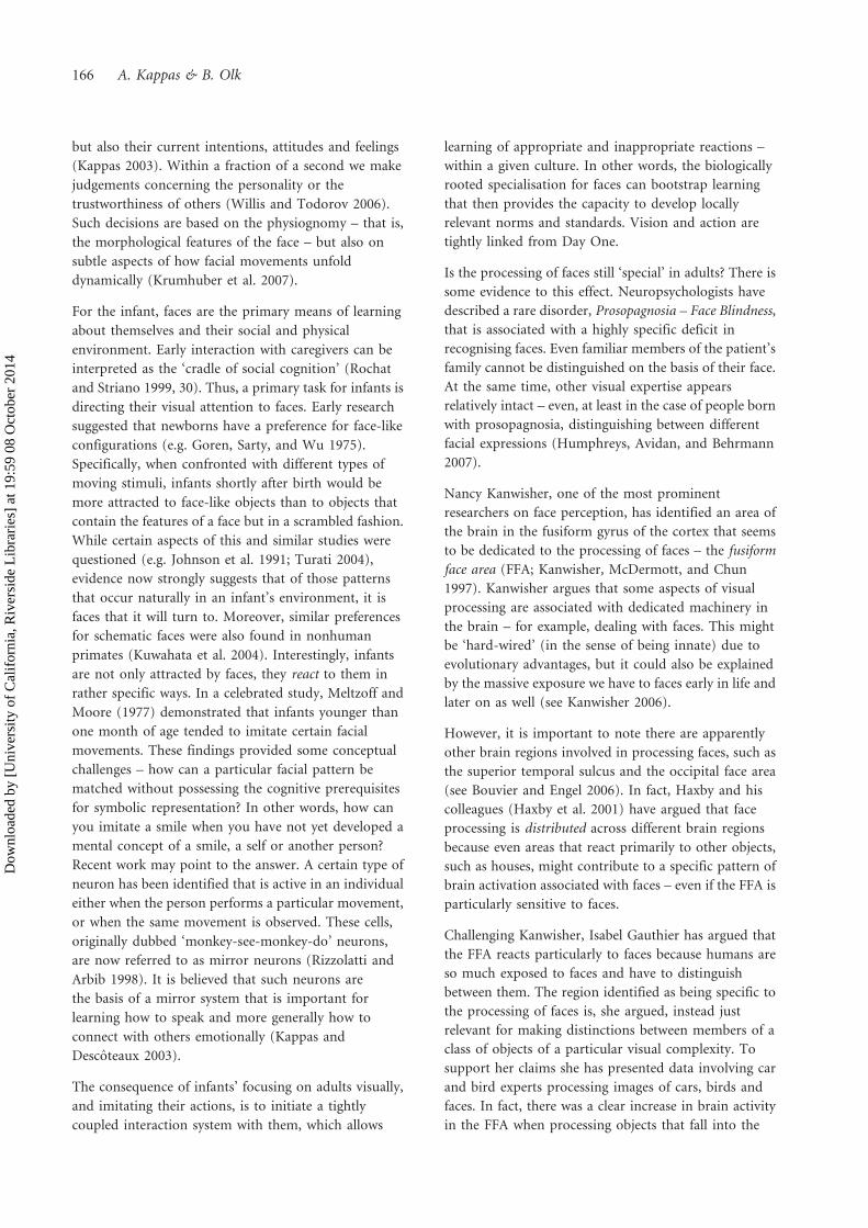
reconstructed. It can be inferred, for example where,
when and for how long someone looked at a certain
location. An example is given in Figure 1. The data are
also available in numeric format, allowing statistical
analyses.
Neurophysiological and neuropsychological studies
have contributed much knowledge about the network
of brain areas involved in the generation of eye
movements and how these brain areas work. Cells
that are active when we fixate and cells that are active
when we move our eyes have been identified. The
network of brain areas consists of structures lying
under the cortex (subcortical), such as the superior
colliculus (SC), basal ganglia and brain stem, cortical
areas in the front of the brain such as the frontal eye
field (FEF), the supplementary eye field (SEF) and
the dorsolateral prefrontal cortex (dlPFC), and areas
located more towards the back of the brain, such as
the parietal eye field (Schiller 1984; Guitton, Buchtel,
and Douglas 1985; Henik, Rafal, and Rhodes 1994;
Schlag-Rey et al. 1997; Everling, Dorris, and Munoz
1998; Gaymard et al. 1998; Conolly et al. 2000; Rafal
et al. 2000; Olk et al. 2006). Understanding the exact
role of these areas as part of the network, and which
rules and mechanisms underlie their processing, is
one of the major challenges of the interdisciplinary
field of studying visual attention and eye
movements.
Recent eye-movement studies have shown that where we
look is influenced by an array of factors (see Henderson
and Hollingworth 1999 and Henderson 2003 for
reviews). Low-level characteristics, such as contrast,
colour, texture and luminance of the visual stimuli,
affect which areas are fixated – that is, areas of higher
contrast are fixated more (Reinagel and Zador 1999;
Parkhurst and Niebur 2003). And as mentioned above,
the mere appearance of a stimulus draws our eyes
towards it. The ‘urge to look’ at something that appears
is so strong that at times we may not be able to prevent
it. This is illustrated by studies investigating how well we
can control where we look. Even in a seemingly easy task
in which participants are requested to refrain from
looking at a suddenly appearing stimulus on the
computer screen, such as a simple dot, and to look in
the opposite direction instead, erroneous saccades
towards the stimulus occur (Godijn and Kramer 2006;
Olk and Kingstone 2003; Reuter et al. 2006).
At a more intermediate level, shape and spatial relation
guide our eyes (Henderson and Hollingworth 1999) –
that is, a stimulus of a different shape than the
surrounding stimuli will be particularly visually salient
and attract attention and gaze (Duncan and Humphreys
1989; Harvey et al. 2002). And last but not least, eye
movements are also controlled by high-level factors,
such as information about previously fixated areas in a
current scene stored in short-term memory and spatial
FIGURE 1. Example of a scanpath, with circles representing fixations and arrows representing saccades.
164 A. Kappas & B. Olk
Dow
nloa
ded
by [
Uni
vers
ity o
f C
alif
orni
a, R
iver
side
Lib
rari
es]
at 1
9:59
08
Oct
ober
201
4
and semantic knowledge about similar types of scenes
stored in long-term memory – for example, when
viewing a beach scene, we know that the beach is likely
to be found at the bottom of the picture whereas the sky
is likely at the top of the picture (Henderson 2003). Such
knowledge-guided exploration was, for example,
observed in a study by Shinoda, Hayhoe, and
Shrivastava (2001), who tracked eye movements while
participants looked at dynamic street scenes. The results
showed that the likelihood of fixating a traffic sign was
higher if this sign was shown at an intersection than
mid-block. Knowledge-driven eye-movement control
seems to increase over time with scene viewing as more
knowledge is accumulated about the identity of the
objects and their relationship to each other and to the
scene (Henderson, Weeks, and Hollingworth 1999).
Recent research also indicates that emotional content
affects where we look (Nummenmaa, Hyona, and Calvo
2006). Participants were presented with two pictures at a
time, one picture with either an unpleasant, neutral or
pleasant content, along with a neutral picture.
Importantly, arousal values and also low-level features,
such as colour saturation and contrast and luminance
levels, were kept constant. Participants freely looked at
the pictures and estimated whether the affective valence
of the two pictures was similar or not. The results
showed that participants tended to direct their first eye
movement to the emotional pictures and fixated those
for longer than the neutral pictures, irrespective of
valence.
There is consensus that pictures are viewed in an active
manner and observers search for relevant information,
in line with their goals. People, faces and informative
regions are fixated more (Mackworth and Morandi
1967; Henderson and Hollingworth 1999). That the
intentions of the observer play a very important role was
already shown in the seminal study by Yarbus (1967). In
his experiment, the participants looked at the picture
‘They did not Expect Him’ by the painter Repin. On this
picture a family is shown in a living room, and a person
enters the room. Yarbus recorded where his participants
were looking. In the different conditions of the
experiment the participants either engaged in free
viewing or they were asked, for example, to estimate the
social status of the persons, their age, to memorise the
clothing of the persons or to estimate for how long the
person entering the room had been away from his
family. The impressive finding was that the fixation
patterns differed depending on the task. When
participants were asked about the economic
standing of the persons on the picture, they looked
predominantly on the clothes of a person shown in the
foreground of the picture and on the furniture in the
room. When they were asked about the age of the
persons, they tended to look at the faces. When asked to
memorise the clothing, they mostly looked at the clothes
and when asked to estimate for how long the person
entering the room had been away, their gaze moved very
frequently back and forth between the faces of the
persons. Thus, the distribution of fixations and saccades
changed depending on the type of information required,
showing that participants actively sought pieces of
information that were relevant for the task they had to
complete. Based on these findings, Yarbus concluded
that observers look at elements that are considered by
them to convey information relevant for the perception
of the picture. Yarbus went so far as to suggest that eye
movements reflect the thought processes of the
observers, and therefore allow us to make, to a certain
degree, inferences about the thoughts of the observers.
More recent studies that recorded saccades while
participants engaged in everyday life tasks, such as
making tea and driving (Land and Lee 1994; Land,
Mennie, and Rusted 1999; Shinoda, Hayhoe, and
Shrivastava 2001) or that have addressed the effects of
expectations of actions and events on fixation patterns
(Jin and Olk 2007) confirm that observers look at areas
that are significant for the task at hand and that
observers look at a given object or a location just before
it becomes relevant for an action or an event to occur.
To study how people explore their visual world is of
great importance to research on visual competence. Not
only are the studies informative with respect to what
attracts our attention and our eyes, but one could claim
that making an appropriate selection of the visual
stimuli that are focused on is an indicator for a visually
competent person. Following this thought, one could
make the prediction that a more visually competent
person may select different stimuli than a less visually
competent person, and as a result behave differently
from them. Individual differences (see below) support
this view.
FACE PERCEPTION
Humans are a social species. Thus, the perception of
faces is one of the earliest and most important visual
tasks. In consequence, the study of face perception has a
long history and the related findings have shaped our
understanding of vision.
For the adult, a face is a source of information about the
identity of other human beings, their age and gender,
Visual competence 165
Dow
nloa
ded
by [
Uni
vers
ity o
f C
alif
orni
a, R
iver
side
Lib
rari
es]
at 1
9:59
08
Oct
ober
201
4
but also their current intentions, attitudes and feelings
(Kappas 2003). Within a fraction of a second we make
judgements concerning the personality or the
trustworthiness of others (Willis and Todorov 2006).
Such decisions are based on the physiognomy – that is,
the morphological features of the face – but also on
subtle aspects of how facial movements unfold
dynamically (Krumhuber et al. 2007).
For the infant, faces are the primary means of learning
about themselves and their social and physical
environment. Early interaction with caregivers can be
interpreted as the ‘cradle of social cognition’ (Rochat
and Striano 1999, 30). Thus, a primary task for infants is
directing their visual attention to faces. Early research
suggested that newborns have a preference for face-like
configurations (e.g. Goren, Sarty, and Wu 1975).
Specifically, when confronted with different types of
moving stimuli, infants shortly after birth would be
more attracted to face-like objects than to objects that
contain the features of a face but in a scrambled fashion.
While certain aspects of this and similar studies were
questioned (e.g. Johnson et al. 1991; Turati 2004),
evidence now strongly suggests that of those patterns
that occur naturally in an infant’s environment, it is
faces that it will turn to. Moreover, similar preferences
for schematic faces were also found in nonhuman
primates (Kuwahata et al. 2004). Interestingly, infants
are not only attracted by faces, they react to them in
rather specific ways. In a celebrated study, Meltzoff and
Moore (1977) demonstrated that infants younger than
one month of age tended to imitate certain facial
movements. These findings provided some conceptual
challenges – how can a particular facial pattern be
matched without possessing the cognitive prerequisites
for symbolic representation? In other words, how can
you imitate a smile when you have not yet developed a
mental concept of a smile, a self or another person?
Recent work may point to the answer. A certain type of
neuron has been identified that is active in an individual
either when the person performs a particular movement,
or when the same movement is observed. These cells,
originally dubbed ‘monkey-see-monkey-do’ neurons,
are now referred to as mirror neurons (Rizzolatti and
Arbib 1998). It is believed that such neurons are
the basis of a mirror system that is important for
learning how to speak and more generally how to
connect with others emotionally (Kappas and
Descoteaux 2003).
The consequence of infants’ focusing on adults visually,
and imitating their actions, is to initiate a tightly
coupled interaction system with them, which allows
learning of appropriate and inappropriate reactions –
within a given culture. In other words, the biologically
rooted specialisation for faces can bootstrap learning
that then provides the capacity to develop locally
relevant norms and standards. Vision and action are
tightly linked from Day One.
Is the processing of faces still ‘special’ in adults? There is
some evidence to this effect. Neuropsychologists have
described a rare disorder, Prosopagnosia – Face Blindness,
that is associated with a highly specific deficit in
recognising faces. Even familiar members of the patient’s
family cannot be distinguished on the basis of their face.
At the same time, other visual expertise appears
relatively intact – even, at least in the case of people born
with prosopagnosia, distinguishing between different
facial expressions (Humphreys, Avidan, and Behrmann
2007).
Nancy Kanwisher, one of the most prominent
researchers on face perception, has identified an area of
the brain in the fusiform gyrus of the cortex that seems
to be dedicated to the processing of faces – the fusiform
face area (FFA; Kanwisher, McDermott, and Chun
1997). Kanwisher argues that some aspects of visual
processing are associated with dedicated machinery in
the brain – for example, dealing with faces. This might
be ‘hard-wired’ (in the sense of being innate) due to
evolutionary advantages, but it could also be explained
by the massive exposure we have to faces early in life and
later on as well (see Kanwisher 2006).
However, it is important to note there are apparently
other brain regions involved in processing faces, such as
the superior temporal sulcus and the occipital face area
(see Bouvier and Engel 2006). In fact, Haxby and his
colleagues (Haxby et al. 2001) have argued that face
processing is distributed across different brain regions
because even areas that react primarily to other objects,
such as houses, might contribute to a specific pattern of
brain activation associated with faces – even if the FFA is
particularly sensitive to faces.
Challenging Kanwisher, Isabel Gauthier has argued that
the FFA reacts particularly to faces because humans are
so much exposed to faces and have to distinguish
between them. The region identified as being specific to
the processing of faces is, she argued, instead just
relevant for making distinctions between members of a
class of objects of a particular visual complexity. To
support her claims she has presented data involving car
and bird experts processing images of cars, birds and
faces. In fact, there was a clear increase in brain activity
in the FFA when processing objects that fall into the
166 A. Kappas & B. Olk
Dow
nloa
ded
by [
Uni
vers
ity o
f C
alif
orni
a, R
iver
side
Lib
rari
es]
at 1
9:59
08
Oct
ober
201
4
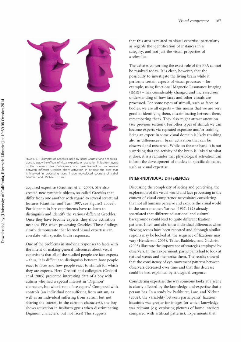
acquired expertise (Gauthier et al. 2000). She also
created new synthetic objects, so-called Greebles that
differ from one another with regard to several structural
features (Gauthier and Tarr 1997, see Figure 2 above).
Participants in her experiments have to learn to
distinguish and identify the various different Greebles.
Once they have become experts, they show activation
near the FFA when processing Greebles. These findings
clearly demonstrate that learned visual expertise can
correlate with specific brain responses.
One of the problems in studying responses to faces with
the intent of making general inferences about visual
expertise is that all of the studied people are face experts
– thus, it is difficult to distinguish between how people
react to faces and how people react to stimuli for which
they are experts. Here Grelotti and colleagues (Grelotti
et al. 2005) presented interesting data of a boy with
autism who had a special interest in ‘Digimon’
characters, but who is not a face expert.1 Compared with
controls (an individual not suffering from autism, as
well as an individual suffering from autism but not
sharing the interest in the cartoon characters), the boy
shows activation in fusiform gyrus when discriminating
Digimon characters, but not faces! This suggests
that this area is related to visual expertise, particularly
as regards the identification of instances in a
category, and not just the visual properties of
a stimulus.
The debates concerning the exact role of the FFA cannot
be resolved today. It is clear, however, that the
possibility to investigate the living brain while it
performs certain aspects of visual processes – for
example, using functional Magnetic Resonance Imaging
(fMRI) – has considerably changed and increased our
understanding of how faces and other visuals are
processed. For some types of stimuli, such as faces or
bodies, we are all experts – this means that we are very
good at identifying them, discriminating between them,
remembering them. They also might attract attention
(see previous section). For other types of stimuli we can
become experts via repeated exposure and/or training.
Being an expert in some visual domain is likely resulting
also in differences in brain activation that can be
observed and measured. While on the one hand it is not
surprising that the activity of the brain is linked to what
it does, it is a reminder that physiological activation can
inform the development of models in specific domains,
such as visual expertise.
INTER-INDIVIDUAL DIFFERENCES
Discussing the complexity of seeing and perceiving, the
exploration of the visual world and face processing in the
context of visual competence necessitates considering
that not all humans perceive and explore the visual world
in the same manner. Yarbus (1967, 192) already
speculated that different educational and cultural
backgrounds could lead to quite different fixation
patterns. Inter- and also intra-individual differences when
viewing scenes have been reported and although similar
regions may be looked at, the sequence of fixations may
vary (Henderson 2003). Tatler, Baddeley, and Gilchrist
(2005) illustrate the importance of strategies employed by
observers. In their experiment, participants had to look at
natural scenes and memorise them. The results showed
that the consistency of eye-movement patterns between
observers decreased over time and that this decrease
could be best explained by strategic divergence.
Considering expertise, the way someone looks at a scene
is clearly affected by the knowledge and expertise that a
person has. In a study by Parkhurst, Law, and Niebur
(2002), the variability between participants’ fixation
locations was greater for images for which knowledge
was relevant (e.g. exploring pictures of home interiors
compared with artificial patterns). Experiments that
FIGURE 2. Examples of ‘Greebles’ used by Isabel Gauthier and her collea-gues to study the effects of visual expertise on activation in fusiform gyrusof the human cortex. Participants who have learned to discriminatebetween different Greebles show activation in or near the area thatis involved in processing faces. Image reproduced courtesy of IsabelGauthier and Michael J. Tarr.
Visual competence 167
Dow
nloa
ded
by [
Uni
vers
ity o
f C
alif
orni
a, R
iver
side
Lib
rari
es]
at 1
9:59
08
Oct
ober
201
4
compare eye movements of experts and novices, for
example in sports (e.g. Kato, and Fukuda 2002; Nagano,
Kato, and Fukuda 2004; Naito, Kato, and Fukuda 2004)
or chess (Charness et al. 2001), showed that the visual
search strategies of experts and novices differed
significantly. Soccer experts fixated more often on the
knee and the hip regions of opponents than novices did,
suggesting that information gained from these areas was
important in anticipating an opponent’s next move
(Nagano, Kato, and Fukuda 2004). In another study, eye
movements of expert and intermediate chess players were
monitored while they were required to choose the best
move in five chess positions. Naturally, the experts were
faster and more accurate than intermediates in choosing
the best move. When the spatial distribution of the first
five fixations for each position were examined, the eye-
movement patterns of the experts showed that they made
more fixations on empty squares than did intermediates
and when they looked at pieces, they looked more
frequently on relevant pieces than did intermediates.
According to the authors, these results may indicate that
expert chess players encode chess configurations instead
of individual pieces and that their exploration is guided
by peripheral vision, allowing them to select the target of
their next fixation by piece saliency (Charness et al. 2001).
Similarly, Vogt and Magnussen (2007) demonstrated
expertise effects in artists compared with non-trained
observers in an eye-tracking study.
For the concept of visual competence, such work may
suggest that it depends on the knowledge and expertise
that a person has and, importantly, that it is not static
but may increase – for example, with acquiring expertise.
As hardly any person will be an expert for everything, it
may well be that visual competence may also vary within
a person. How far competence transfers from one
domain of expertise to another requires further
empirical investigation.
Visual competence can also decrease. This could be due to
peripheral factors – for example, by injuries to the eyes
that cannot be remedied or by impairments shown by
patients with brain injuries (e.g. after a stroke). Injuries to
the visual pathway can lead to deficits, such as
hemianopia, which is the loss of vision in half of the visual
field. Such patients tend to compensate for their deficit,
though, by performing more saccades to the hemianopic
side (Ishiai, Furukawa, and Tsukagoshi 1987) and
spending more time exploring on that side (Behrmann
et al. 1997).
Other patients may suffer from visual agnosia, the loss of
the ability to identify objects despite being able to see
them, or of visuospatial neglect, shown in deficits in
exploring the visual world, for example due to
difficulties in allocating visual attention (Olk and
Harvey 2006). Spatial neglect has been defined as the
failure ‘to respond or orient to novel or meaningful
stimuli presented to the side opposite a brain lesion’
(Heilman, Valenstein, and Watson 1985). Although
visual field deficits such as hemianopia often
accompany neglect, neglect can also be seen in cases
without such impairments (Halligan, Marshall, and
Wade 1990) and is believed to arise at higher levels of
processing (Bisiach and Luzatti 1978; Riddoch and
Humphreys 1987; Milner and Goodale 2006; Harvey
1998). It can be demonstrated after injuries to the right
or to the left side of the brain, but seems to be much
more frequent and severe after right-sided lesions
(Weintraub and Mesulam 1987; Vallar 1993), leading to
impaired exploration and perception of the left side of
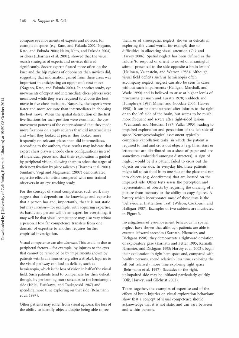
space. Neuropsychological assessment typically
comprises cancellation tasks, in which the patient is
required to find and cross out objects (e.g. lines, stars or
letters that are distributed on a sheet of paper and are
sometimes embedded amongst distracters). A sign of
neglect would be if a patient failed to cross out the
objects on one side. In everyday life, these patients
might fail to eat food from one side of the plate and run
into objects (e.g. doorframes) that are located on the
impaired side. Other tests assess the perception and
representation of objects by requiring the drawing of a
picture from memory or the ability to copy figures. A
battery which incorporates most of these tests is the
‘Behavioural Inattention Test’ (Wilson, Cockburn, and
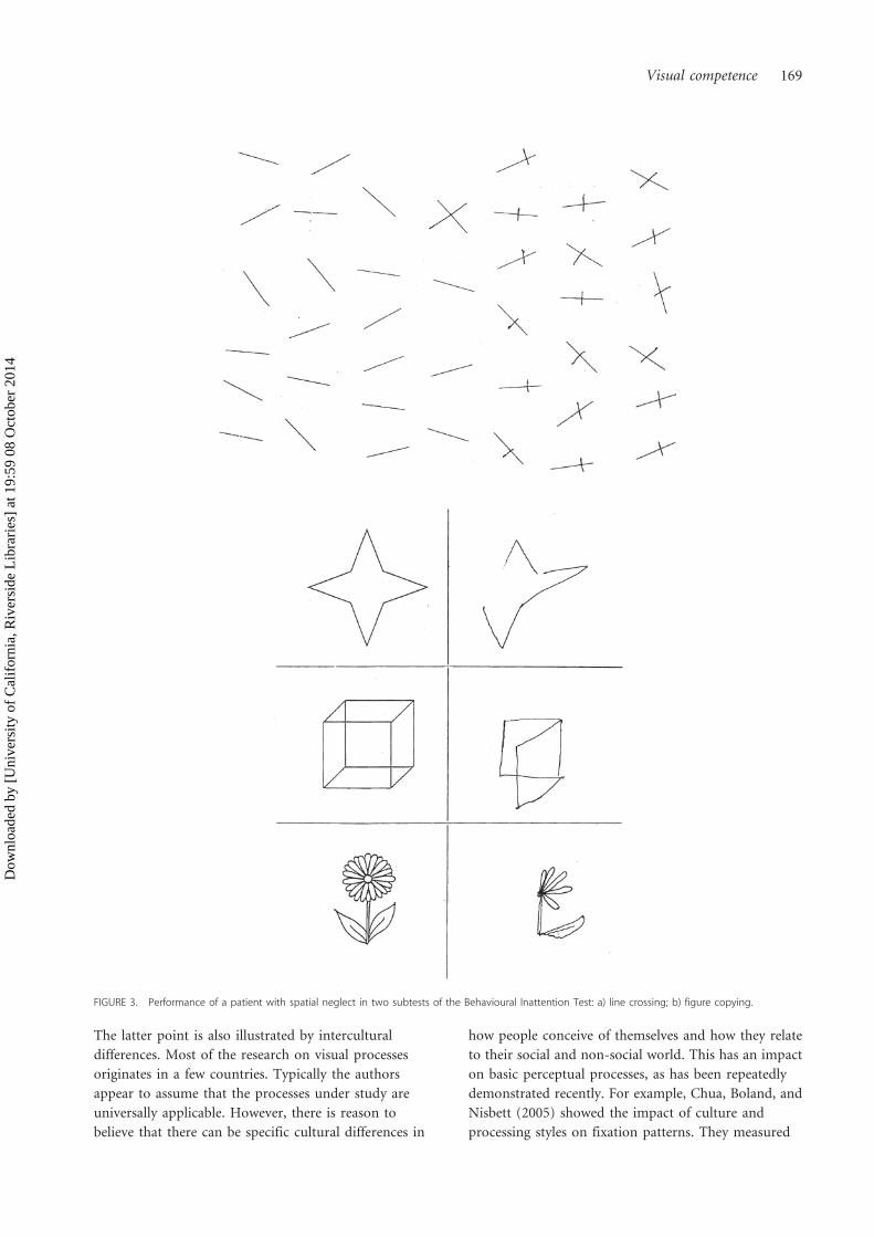
Halligan 1987). Examples of two subtests are illustrated
in Figure 3.
Investigations of eye-movement behaviour in spatial
neglect have shown that although patients are able to
execute leftward saccades (Karnath, Niemeier, and
Dichgans 1998), they demonstrate a rightward deviation
of exploratory gaze (Karnath and Fetter 1995; Karnath,
Niemeier, and Dichgans 1998; Harvey et al. 2002), begin
their exploration in right hemispace and, compared with
healthy persons, spend relatively less time exploring the
left but relatively more time exploring right space
(Behrmann et al. 1997). Saccades to the right,
unimpaired side may be initiated particularly quickly
(Olk, Harvey, and Gilchrist 2002).
Taken together, the examples of expertise and of the
effects of brain injuries on visual exploration behaviour
show that a concept of visual competence should
acknowledge that it is not static and can vary between
and within persons.
168 A. Kappas & B. Olk
Dow
nloa
ded
by [
Uni
vers
ity o
f C
alif
orni
a, R
iver
side
Lib
rari
es]
at 1
9:59
08
Oct
ober
201
4
The latter point is also illustrated by intercultural
differences. Most of the research on visual processes
originates in a few countries. Typically the authors
appear to assume that the processes under study are
universally applicable. However, there is reason to
believe that there can be specific cultural differences in
how people conceive of themselves and how they relate
to their social and non-social world. This has an impact
on basic perceptual processes, as has been repeatedly
demonstrated recently. For example, Chua, Boland, and
Nisbett (2005) showed the impact of culture and
processing styles on fixation patterns. They measured
FIGURE 3. Performance of a patient with spatial neglect in two subtests of the Behavioural Inattention Test: a) line crossing; b) figure copying.
Visual competence 169
Dow
nloa
ded
by [
Uni
vers
ity o
f C
alif
orni
a, R
iver
side
Lib
rari
es]
at 1
9:59
08
Oct
ober
201
4
eye movements of American and Chinese participants
while they explored photographs of natural scenes. The
results showed that the Americans fixated foreground
objects sooner and longer than the Chinese participants,
while the Chinese participants made more saccades to
the background than did the Americans, a result in line
with possible differences in expertise, socialisation and
experience of the two groups. Such differences may
influence the allocation of attention, reflected in the eye-
movement patterns. Understanding these differences is
not only of theoretical interest, but, in the context of
visual communication, of concrete relevance for
applications. For example, layouts of press media, or the
structure of images used on television or the Internet,
cannot be assumed to be globally appropriate. In
addition, there are content issues that relate to symbols
and associations that may differ considerably from
culture to culture and might lead to strife and conflict
(e.g. Muller and Ozcan 2007).
APPLICATIONS
Much of the research described in this contribution is
targeted at the illumination of basic processes and not
applications in the real world. Nevertheless, we would
like to claim that this basic research has large potential
for applied use. The findings regarding the activation in
specific brain regions linked to visual expertise (see
section on face processing) could indicate that brain
research might have a diagnostic value for expertise.
Similarly, studies using eye-tracking methodology have
shown differences between novices and experts and
highlight difficulties of persons with brain injuries. A
very interesting application of such methods might be to
assess how training changes the way that certain types of
visuals are perceived and processed, or, in other words,
how visual competence can be enhanced.
Basic research has also shown which properties of visuals
– such as contrast, complexity, distribution of objects,
symbols used – affect where we attend and look, not to
forget profound inter- and intra-individual differences
within and between cultures. The consideration of such
findings has the potential to greatly enhance the choices
of media-producers that are typically driven by personal
experience and intuition. In turn, the methods
developed in the context of psychological research and
neuroscience can be useful tools in the evaluation of
effectiveness or, in the negative, in the potential for
communication failure.
Certain stimuli, such as faces, may be processed in a
specific way – they appear to attract attention and
require few cognitive resources. For example, the
communication of emotions is rapid. Faces are often
interpreted as expressing an emotion, and if certain
stereotypical patterns of facial activation are shown, this
process can take place in a few milliseconds (e.g. Kirouac
and Dore 1984). However, this does not mean that the
perceived emotion is necessarily corresponding to the
emotional state of the person depicted. Thus, a smile
might be recognised as a smile but this does not mean
the smiling person is indeed happy (Kappas 2003)!
In the case of emotional displays, perception involves
the activation not only of knowledge (‘this person is
happy!’), but also of structures that are associated with
the experience of emotions in the individual. There is
much evidence for such an embodied communication
of affect (Kappas and Descoteaux 2003). This could
mean that showing suffering victims of violence in a
news programme might have a very physical impact
on the observers due to very basic processes that
link perception and experience of emotions. Basic
research from this area is relevant for the decision
process of an editor regarding what should be shown
and how.
The processing of human movement is also associated
with specific brain regions, such as the superior
temporal sulcus (e.g. Michels, Lappe, and Vaina 2005),
as well as the aforementioned mirror system. Calvo-
Merino and his colleagues (2005, 2006) could
demonstrate that expertise in classical dance or Capoeira
influences the activation of the mirror system when
watching the types of movements for which the
individual was, or was not, an expert. Again, this offers
the potential for diagnostic uses. However, it also
underlines the fact that different observers will perceive
certain types of media very differently. Taking such
findings into account might allow tailoring contents and
form to maximise the success and impact of specific
mediate messages.
CONCLUSIONS
The availability of new methods of studying ongoing
activity of the brain in recent years, systematic study of
patients with neuropsychological methods and
behavioural methodologies with a variety of participant
groups, has greatly increased our understanding of how
visual information is processed. Sometimes
neuroscientific findings are criticised because ‘just
finding a location where the brain is activated during a
particular process does not help understanding the
associated mental processes’. This is true. However, the
170 A. Kappas & B. Olk
Dow
nloa
ded
by [
Uni
vers
ity o
f C
alif
orni
a, R
iver
side
Lib
rari
es]
at 1
9:59
08
Oct
ober
201
4
body of research addressing the issue of vision is diverse
and mostly driven by theories. And it is important to
consider that no method alone can unravel all the
mysteries of vision, but that converging findings using
different methodologies within psychology and
neuroscience should be considered.
In fact, by now, the body of knowledge and the research
methods are so solid that it is time for an exchange with
experts in visual research from different disciplines. This
exchange requires patience, because the level of control
that laboratory research requires cannot be easily
translated into a naturalistic situation. Similarly,
studying the perception of dots, lines and arrows is not
the same as watching a newscast of a terrorist attack as it
unfolds in real time. But in our view, the reward of such
collaborations with other disciplines should be
significant new insights, making the effort worthwhile.
The concept of visual competence that is the focus of this
special issue has the potential to serve as a nexus of basic
and applied interests. Thus, we have provided a cursory
description of the state of knowledge with regard to some
selected aspects of vision as seen by the psychological and
brain sciences. While a detailed review is well beyond the
scope of this manuscript, we hope that we succeeded in
creating interest in the type of basic research that is
currently being conducted in psychology and
neuroscience laboratories in those readers who might not
be familiar with it or who thought that it is unrelated to
their interests. If such interest is created, then one
important prerequisite for exchange is taken care of.
We believe that the field of psychology has much to gain
from an inter-disciplinary exchange in the context of
‘visual competence’. First, psychology can expect to be
inspired with respect to specific research questions to
ask and to test whether psychological models and
theories hold true in the complex real world outside the
laboratory. Second, tackling research questions from
different angles and learning about methods used in
other disciplines should be exceptionally fruitful. We are
currently in the process of dealing with such an
interdisciplinary endeavour and we are convinced that it
is possible to bridge the complex divides between the
disciplines that are linked to differences regarding not
only the type of visual material studied, but also the
paradigms and models, and even the usage of the most
basic terms, such as ‘information’ or ‘knowledge’. We
are sensitised to these issues. And it is precisely for these
reasons that we want to communicate what we consider
essential: a discussion of the concept of visual
competence would not be complete without a
consideration of basic mental/neural processes within
the individual.
NOTE
[1] It has been argued that autistic individuals do not develop
a theory of mind, due to them not understanding or
recognising mental states in others (Klein and Kihlstrom
1998). It is believed that there are deficits in perceiving
and interpreting nonverbal social behaviours displayed by
others, though performance regarding the identification of
faces is unclear, but there is evidence pointing towards
deficits as well (McGee and Morrier 2003).
REFERENCES
Behrmann, M., S. Watt, S. E. Black, and J. J. S. Barton. 1997.
Impaired visual search in patients with unilateral neglect:
An oculographic analysis. Neuropsychologia 35(11):
1445–58.
Bisiach, E., and C. Luzatti. 1978. Unilateral neglect of
representational space. Cortex 14(1): 129–33.
Bouvier, S. E., and S. A. Engel. 2006. Behavioral deficits and
cortical damage loci in cerebral achromatopsia. Cerebral
Cortex 16(2): 183–91.
Calvo-Merino, B., D. E. Glaser, J. Grezes, R. E. Passingham,
and P. Haggard. 2005. Action observation and acquired
motor skills: An fMRI study with expert dancers. Cerebral
Cortex 15(8): 1243–49.
Calvo-Merino, B., J. Grezes, D. E. Glaser, R. E. Passingham,
and P. Haggard. 2006. Seeing or doing? Influence of visual
and motor familiarity in action observation. Current
Biology 16(19): 1905–10.
Charness, N., E. M. Reingold, M. Pomplun, and D. M. Stampe.
2001. The perceptual aspect of skilled performance in
chess: Evidence from eye movements. Memory &
Cognition 29(8): 1146–52.
Chua, H. F., J. E. Boland, and R. E. Nisbett. 2005. Cultural
variation in eye movements during scene perception.
Proceedings of the National Academy of Sciences 102(35):
12629–33.
Connolly, J. D., M. A. Goodale, J. F. X. DeSouza, R. S. Menon,
and T. Vilis. 2000. A comparison of frontoparietal fMRI
activation during anti-saccades and anti-pointing. Journal
of Neurophysiology 84(3): 1645–55.
Coren, S., L. M. Ward, and J. T. Enns. 1999. Sensation and
perception. 5th ed. New York: Harcourt Brace.
Cui, X., C. B. Jeter, D. Yang, P. R. Montague, and D. M.
Eagleman. 2007. Vividness of mental imagery: Individual
variability can be measured objectively. Vision Research
47(4): 474–78.
Duncan, J., and G. W. Humphreys. 1989. Visual search and
stimulus similarity. Psychological Review 96(3): 433–58.
Everling, S., M. C. Dorris, and D. P. Munoz. 1998. Reflex
suppression in the anti-saccade task is dependent on
prestimulus neural processes. Journal of Neurophysiology
80(3): 1584–89.
Visual competence 171
Dow
nloa
ded
by [
Uni
vers
ity o
f C
alif
orni
a, R
iver
side
Lib
rari
es]
at 1
9:59
08
Oct
ober
201
4

Gauthier, I., P. Skudlarski, J. C. Gore, and A. W. Anderson.
2000. Expertise for cars and birds recruits brain areas
involved in face recognition. Nature Neuroscience 3(2):
568–73.
Gauthier, I., and M. J. Tarr. 1997. Becoming a
‘‘Greeble’’expert: Exploring mechanisms for face
recognition. Vision Research 37(12): 1673–82.
Gaymard, B., C. J. Ploner, S. Rivaud, A. I. Vermersch, and C.
Pierrot-Deseilligny. 1998. Cortical control of saccades.
Experimental Brain Research 123(1–2): 159–63.
Godijn, R., and A. F. Kramer. 2006. Prosaccades and
antisaccades to onsets and color singletons: Evidence that
erroneous prosaccades are not reflexive. Experimental
Brain Research 172(4): 439–48.
Goren, C. C., M. Sarty, and P. Y. Wu. 1975. Visual following
and pattern discrimination of face-like stimuli by
newborn infants. Pediatrics 56(4): 544–49.
Grelotti, D. J., A. J. Klin, I. Gauthier, P. Skudlarski, D. J.
Cohen, J. C. Gore, F. R. Volkmar, and R. T. Schultz. 2005.
fMRI activation of the fusiform gyrus and amygdala to
cartoon characters but not to faces in a boy with autism.
Neuropsychologia 43(3): 373–85.
Guitton, D., H. A. Buchtel, and R. M. Douglas. 1985. Frontal
lobe lesions in man cause difficulties in suppressing
reflexive glances and in generating goal-directed saccades.
Experimental Brain Research 58(3): 455–72.
Halligan, P. W., J. C. Marshall, and D. T. Wade. 1990. Do
visual field deficits exacerbate visuo-spatial neglect?
Journal of Neurology, Neurosurgery and Psychiatry 53(6):
487–91.
Harvey, Monika. 1998. Perspectives on visuospatial neglect. In
Comparative Neuropsychology, edited by A. D. Milner.
Oxford: Oxford University Press.
Harvey, M., B. Olk, K. Muir, and I. D. Gilchrist. 2002. Manual
responses and saccades in chronic and recovered
hemispatial neglect: A study using visual search.
Neuropsychologia 40(7): 705–17.
Haxby, J. V., M. I. Gobbini, M. L. Furey, A. Ishai, J. L.
Schouten, and P. Pietrini. 2001. Distributed and
overlapping representations of faces and objects in ventral
temporal cortex. Science 293(5539): 2425–30.
Heilman, K. M., E. Valenstein, and R. T. Watson. 1985. The
neglect syndrome. In Handbook of clinical neurology:
Clinical neuropsychology, edited by J. A. M. Frederiks.
Amsterdam: Elsevier.
Henderson, J. M. 2003. Human gaze control during real-world
scene perception. Trends in Cognitive Sciences 7(11):
498–504.
Henderson, J. M., and A. Hollingworth. 1999. High-level scene
perception. Annual Review of Psychology 50(1): 243–71.
Henderson, J. M., P. A. Weeks, Jr., and A. Hollingworth. 1999.
The effects of semantic consistency on eye movements
during complex scene viewing. Journal of Experimental
Psychology: Human Perception and Performance 25(1):
210–28.
Henik, A., R. Rafal, and D. Rhodes. 1994. Endogenously
generated and visually guided saccades after lesions of the
human frontal eye fields. Journal of Cognitive Neuroscience
6(4): 400–11.
Humphreys, K., G. Avidan, and M. Behrmann. 2007. A detailed
investigation of facial expression processing in congenital
prosopagnosia as compared to acquired prosopagnosia.
Experimental Brain Research 176(2): 356–73.
Ishiai, S., T. Furukawa, and H. Tsukagoshi. 1987. Eye-fixation
patterns in homonymous hemianopia and unilateral
spatial neglect. Neuropsychologia 25(4): 675–79.
Jin, Y., and B. Olk. 2007. Measuring expectations: An
application of eye movement tracking. Poster presented at
the 14th European Conference on Eye Movements,
August 19–23, in Potsdam, Germany.
Johnson, M. H., S. Dziurawiec, H. D. Ellis, and J. Morton.
1991. Newborns’ preferential tracking of faces and its
subsequent decline. Cognition 40(1–2): 1–19.
Kanwisher, N. 2006. What’s in a face? Science 311(5761): 617–18.
Kanwisher, N., J. McDermott, and M. M. Chun. 1997. The
fusiform face area: A module in human extrastriate cortex
specialized for face perception. Journal of Neuroscience
17(11): 4302–11.
Kappas, Arvid. 2003. What facial activity can and cannot tell us
about emotions. In The human face: Measurement and
meaning, edited by M. Katsikitis. Dordrecht: Kluwer
Academic Publishers.
Kappas, A., and J. Descoteaux. 2003. Of butterflies and roaring
thunder: Nonverbal communication in interaction and
regulation of emotion. In Nonverbal behavior in clinical
settings, edited by P. Philippot, E. J. Coats and R. S.
Feldman. New York: Oxford University Press.
Karnath, H.-O., and M. Fetter. 1995. Ocular space exploration
in the dark and its relationship to subjective and objective
body orientation in neglect patients with parietal lesions.
Neuropsychologia 33(3): 371–77.
Karnath, H.-O., M. Niemeier, and J. Dichgans. 1998. Space
exploration in neglect. Brain 121(12): 2357–67.
Kato, T., and T. Fukuda. 2002. Visual search strategies of
baseball batters: Eye movements during the preparatory
phase of batting. Perceptual and Motor Skills 94(2): 380–86.
Kirouac, G., and F. Y. Dore. 1984. Judgment of facial
expressions of emotion as a function of exposure time.
Perceptual and Motor Skills 59(1): 147–50.
Klein, S. B., and J. F. Kihlstrom. 1998. On bridging the gap
between social-personality psychology and
neuropsychology. Personality and Social Psychology Review
2(4): 228–42.
Kosslyn, S. M., W. L. Thompson, I. J. Kim, and N. M. Alpert.
1995. Topographical representations of mental images in
primary visual cortex. Nature 378(6556): 496–98.
Krumhuber, E., A. S. R. Manstead, D. Cosker, D. Marshall, P.
Rosin, and A. Kappas. 2007. Facial dynamics as indicators
of trustworthiness and cooperative behavior. Emotion
7(3): 730–35.
Kuwahata, H., I. Adachi, K. Fujita, M. Tomonaga, and T.
Matsuzawa. 2004. Development of schematic face
preference in macaque monkeys. Behavioural Processes
66(1): 17–21.
172 A. Kappas & B. Olk
Dow
nloa
ded
by [
Uni
vers
ity o
f C
alif
orni
a, R
iver
side
Lib
rari
es]
at 1
9:59
08
Oct
ober
201
4
Land, M. F., and D. N. Lee. 1994. Where we look when we
steer. Nature 369(6483): 742–44.
Land, M. F., N. Mennie, and J. Rusted. 1999. The roles of
vision and eye movements in the control of activities of
daily living. Perception 28(11): 1311–28.
Mackworth, N. H., and A. J. Morandi. 1967. The gaze selects
information details within pictures. Perception and
Psychophysics 2(11): 547–51.
McGee, G., and M. Morrier. 2003. Clinical implications of
research in nonverbal behavior of children with autism. In
Nonverbal behavior in clinical settings, edited by P.
Philippot, E. J. Coats and R. S. Feldman. New York:
Oxford University Press.
Meltzoff, A. N., and M. K. Moore. 1977. Imitation of facial and
manual gestures by human neonates. Science 198(4312): 75–78.
Michels, L., M. Lappe, and L. M. Vaina. 2005. Visual areas
involved in the perception of human movement from
dynamic form analysis. Brain Imaging 16(10): 1037–41.
Milner, A. D., and M. A. Goodale. 2006. The visual brain in
action. 2nd ed. Oxford: Oxford Psychology Series.
Muller, M. G., and A. E. Ozcan. 2007. The political
iconography of Muhammad cartoons: Understanding
cultural conflict and political action. Political Science &
Politics 40(2): 287–91.
Nagano, T., T. Kato, and T. Fukuda. Visual search strategies of
soccer players in one-on-one defensive situations on the
field. Perceptual and Motor Skills 99(3): 968–74.
Naito, K., T. Kato, and T. Fukuda. 2004. Expertise and position
of line of sight in golf putting. Perceptual and Motor Skills
99(1): 163–70.
Nummenmaa, L., J. Hyona, and M. G. Calvo. 2006. Eye
movement assessment of selective attentional capture by
emotional pictures. Emotion 6(2): 257–68.
Olk, B., and M. Harvey. 2006. Characterizing exploration
behaviour in spatial neglect: Omissions and repetitive
search. Brain Research 1118(1): 106–15.
Olk, B., and A. Kingstone. 2003. Why are antisaccades slower
than prosaccades? A novel finding using a new paradigm.
NeuroReport 14(1): 151–55.
Olk, B., E. Chang, A. Kingstone, and T. Ro. 2006. Modulation
of antisaccades by transcranial magnetic stimulation over
the human frontal eye field. Cerebral Cortex 16(1): 76–82.
Olk, B., E. Chang, and I. D. Gilchrist. 2002. First saccades
reveal biases in recovered neglect. Neurocase 8(4): 306–13.
Parkhurst, D., K. Law, and E. Niebur. 2002. Modeling the role
of salience in the allocation of overt visual attention.
Vision Research 42(1): 107–23.
Parkhurst, D. J., and E. Niebur. 2003. Scene content selected by
active vision. Spatial Vision 16(2): 125–54.
Rafal, Robert D., Liana Machado, Tony Ro, and Harris Ingle.
2000. Looking forward to looking: Saccade preparation
and the control of midbrain visuomotor reflexes. In
Attention & Performance XVIII, edited by S. Monsell and
J. Driver. Cambridge, MA: MIT Press.
Reinagel, P., and A. M. Zador. 1999. Natural scene statistics at
the centre of gaze. Network-Computation in Neural
Systems 10(4): 341–50.
Reuter, B., A. M. Philipp, I. Koch, and N. Kathmann. 2006.
Effects of switching between leftward and rightward pro-
and antisaccades. Biological Psychology 72(11): 88–95.
Riddoch, M. Jane, and Glyn W. Humphreys. 1987. Perceptual
and action systems in unilateral visual neglect. In
Neurophysiological and neuropsychological aspects of spatial
neglect, edited by M. Jeannerod. Amsterdam: Elsevier.
Rizzolatti, G., and M. A. Arbib. 1998. Language within our
grasp. Trends in Neurosciences 21(5): 188–94.
Rochat, P., and T. Striano. 1999. Social-cognitive development
in the first year. In Early social cognition: Understanding
others in the first months of life, edited by P. Rochat.
Mahwah, NJ: Lawrence Erlbaum Associates.
Sahakian, W. S. 1968. History of psychology. Itasca, IL: Peacock.
Schiller, Peter H. 1984. The neural control of visually guided
eye movements. In Cognitive neuroscience of attention,
edited by J. Richards. Mahwah, NJ: Lawrence Erlbaum
Associates.
Schlag-Rey, M., N. Amador, H. Sanchez, and J. Schlag. 1997.
Antisaccade performance predicted by neuronal activity
in the supplementary eye field. Nature 390(6658):
398–401.
Serences, J. T., and S. Yantis. 2006. Selective visual attention
and perceptual coherence. Trends in Cognitive Sciences
10(1): 38–45.
Shinoda, H., M. M. Hayhoe, and A. Shrivastava. 2001. What
controls attention in natural environments? Vision
Research 41(25): 3535–45.
Tatler, B. W., R. J. Baddeley, and I. D. Gilchrist. 2005. Visual
correlates of fixation selection: Effects of scale and time.
Vision Research 45(5): 643–59.
Theeuwes, J., A. F. Kramer, S. Hahn, and D. E. Irwin. 1998.
Our eyes do not always go where we want them to go:
Capture of the eyes by new objects. Psychological Science
9(5): 379–85.
Turati, C. 2004. Why faces are not special to newborns: An
alternative account of the face preference. Current
Directions in Psychological Science 13(1): 5–8.
Vallar, Giuseppe. 1993. The anatomical basis of spatial
hemineglect in humans. In Unilateral neglect: Clinical and
experimental studies, edited by I. H. Robertson and J. C.
Marshall. Hove: Lawrence Erlbaum Associates.
Vogt, S., and S. Magnussen. 2007. Expertise in pictorial
perception: Eye-movement patterns and visual memory
in artists and laymen. Perception 36(1): 91–100.
Weintraub, S., and M.-M. Mesulam. 1987. Right cerebral
dominance in spatial attention: Further evidence based on
ipsilateral neglect. Archives of Neurology 44(6): 621–25.
Willis, J., and A. Todorov. 2006. First impressions: Making up
your mind after a 100-ms exposure to a face. Psychological
Science 17(7): 592–98.
Wilson, B. A., J. Cockburn, and P. Halligan. 1987. Behavioral
inattention test. Titchfield, Hampshire: Thames Valley
Test Company.
Yarbus, Alfred L. 1967. Eye movements and vision. New York:
Plenum Press.
Visual competence 173
Dow
nloa
ded
by [
Uni
vers
ity o
f C
alif
orni
a, R
iver
side
Lib
rari
es]
at 1
9:59
08
Oct
ober
201
4