the complexities of interpreting reversible elevated serum...
TRANSCRIPT

1521-009X/44/9/1498–1509$25.00 http://dx.doi.org/10.1124/dmd.115.067694DRUG METABOLISM AND DISPOSITION Drug Metab Dispos 44:1498–1509, September 2016Copyright ª 2016 by The American Society for Pharmacology and Experimental Therapeutics
Minireview
The Complexities of Interpreting Reversible Elevated SerumCreatinine Levels in Drug Development: Does a Correlation with
Inhibition of Renal Transporters Exist?
Xiaoyan Chu, Kelly Bleasby, Grace Hoyee Chan, Irene Nunes, and Raymond Evers
Department of Pharmacokinetics, Pharmacodynamics and Drug Metabolism (X.C., K.B., G.H.C., R.E.), and Global RegulatoryAffairs, Oncology, Immunology, Biologics & Devices (I.N.), Merck Sharp & Dohme Corporation, Kenilworth, New Jersey
Received October 6, 2015; accepted January 28, 2016
ABSTRACT
In humans, creatinine is formed by a multistep process in liver andmuscle and eliminated via the kidney by a combination of glomerularfiltration and active transport. Based on current evidence, creatininecan be taken up into renal proximal tubule cells by the basolaterallylocalized organic cation transporter 2 (OCT2) and the organic aniontransporter 2, and effluxed into the urine by the apically localizedmultidrug and toxin extrusion protein 1 (MATE1) andMATE2K. Drug-induced elevation of serum creatinine (SCr) and/or reduced creat-inine renal clearance is routinely used as a marker for acute kidneyinjury. Interpretation of elevated SCr can be complex, because suchincreases can be reversible and explained by inhibition of renaltransporters involved in active secretion of creatinine or other
secondary factors, such as diet and disease state. Distinctionbetween these possibilities is important from a drug developmentperspective, as increases in SCr can result in the termination ofotherwise efficacious drug candidates. In this review,we discuss thechallenges associated with using creatinine as a marker for kidneydamage. Furthermore, to evaluate whether reversible changes inSCr can be predicted prospectively based on in vitro transporterinhibition data, an in-depth in vitro–in vivo correlation (IVIVC) anal-ysis was conducted for 16 drugs with in-house and literature in vitrotransporter inhibition data for OCT2, MATE1, and MATE2K, as wellas total and unbound maximum plasma concentration (Cmax andCmax,u) data measured in the clinic.
Introduction
Serum creatinine (SCr), an endogenous cation produced mainly bymuscle metabolism, is the most widely used marker to assess renalinjury (Tschuppert et al., 2007). Traditional monitoring for nephrotox-icity relies upon SCr measurements (Waikar et al., 2012). Creatinine isprimarily filtered through the kidney through the glomeruli, butdepending on a number of factors, ;10–40% is actively secreted bythe proximal tubule cells through transporter-mediated active uptakeand efflux (Levey et al., 1988; Breyer and Qi, 2010). Therefore,alterations in glomerular filtration rate (GFR) and/or proximal tubularsecretion of creatinine can lead to increases in SCr and decreases in theestimated creatinine clearance. Elevation of SCr often results inreduction of drug dose (Arya et al., 2013, 2014) and may lead todiscontinuation of the development of potentially promising drugcandidates. Therefore, it is critical to distinguish clinically relevantincreases in SCr due to renal toxicity from the nonpathologic increase inSCr attributed to the inhibition of renal transporters.Mild tomoderate andreversible elevation of SCr and decrease in creatinine renal clearance
(CLcr) has been reported, which can be attributed to inhibition of creat-inine transporters without affecting renal function per se (Arya et al.,2013, 2014). This is supported by the clinical observation that severaldrugs, such as cobicistat (Lepist et al., 2014), pyrimethamine (Opravilet al., 1993), cimetidine (Dubb et al., 1978), and trimethoprim (Berglundet al., 1975), lead to increased levels of SCr without affecting kidneyfunction. Such observations have also been reported for several recentlyapproved drugs, including crizotinib (Brosnan et al., 2014; Camidgeet al., 2014) and dolutegravir (Koteff et al., 2013). Understanding themechanism of active secretion of SCr and how drugs may interfere withthis process is therefore important from both a drug development andclinical practice perspective, where SCr is used as a marker of kidneyinjury.Acute kidney injury (AKI) is a common condition that complicates
up to 7% of all hospital admissions and 25% of intensive care unitadmissions (Klevens et al., 2007; Vaidya et al., 2008; Minejima et al.,2011). Although progress has been made in understanding thepathophysiology of AKI and in the clinical care of patients with AKI,mortality rates have remained unchanged at 50–70% over the past 50years (Minejima et al., 2011). Despite routine monitoring of systemicdrug levels and renal function using traditional blood and urinarydx.doi.org/10.1124/dmd.115.067694.
ABBREVIATIONS: AGAT, L-arginine-glycine amidinotransferase; AKI, acute kidney injury; CLcr, creatinine renal clearance; DDI, drug-druginteraction; DX-619, 7-[(3R)-3-(1-aminocyclopropyl)pyrrolidin-1-yl]-1-[(1R,2S)-2-fluorocyclopropyl]-8-methoxy-4-oxoquinoline-3-carboxylic acid;FR, fraction reabsorbed; GFR, glomerular filtration rate; IVIVC, in vitro–in vivo correlation; MATE, multidrug and toxin extrusion protein; NMN,N-methylnicotimide; OAT, organic anion transporter; OCT, organic cation transporter; QD, every day; SCr, serum creatinine; SNP, single nucleotidepolymorphism.
1498
at ASPE
T Journals on M
ay 1, 2019dm
d.aspetjournals.orgD
ownloaded from

markers of kidney injury (e.g., creatinine, blood urea nitrogen, tubularcasts, urinary concentrating ability), 10–20% of patients receiv-ing aminoglycoside therapy, for instance, will develop AKI (Rybaket al., 1999). The lack of sensitive and specific markers of AKI limitsthe ability for early detection and intervention in drug-inducednephrotoxicity.In the kidney, the elimination of drugs and endogenous compounds,
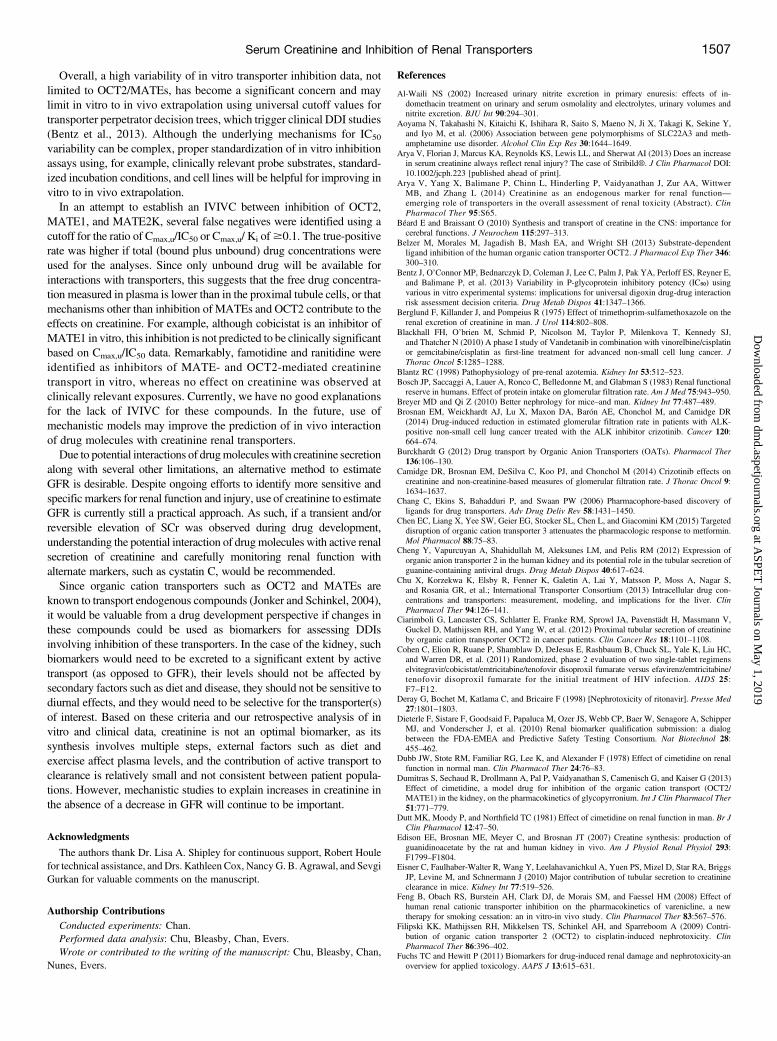
such as creatinine, is the net result of passive glomerular filtration andreabsorption, as well as transporter-mediated active tubular secretionand/or reabsorption. The major transporters in human proximal tubulecells that play a role in the uptake of drugs and endogenous compoundsfrom blood into proximal tubule cells are the organic cation transporter2 (OCT2) and the organic anion transporters 1 and 3 (OAT1 and OAT3;Fig. 1). In the apical membrane, major efflux transporters involved inthe excretion of drugs into the urine are the multidrug and toxin extrusionprotein 1 (MATE1) and MATE2K, and the multidrug-resistance proteinMDR1 P-glycoprotein. Inhibition of these transportersmay alter systemicand tissue exposure of drugs, metabolites, and endogenous compounds,which may subsequently lead to clinically significant drug-drug interac-tions (DDIs). This can be of concern from a drug efficacy or safetyperspective (Giacomini et al., 2010; Hillgren et al., 2013). Othertransporters, such as the breast cancer resistance protein, are alsoexpressed in the proximal tubule (Fig. 1), but their clinical significanceis less well defined (Giacomini et al., 2010; Giacomini and Huang,2013; Hillgren et al., 2013). In general, drug transporters are pro-miscuous in substrate recognition, and in addition to the charge of drugs,other factors, such as polar surface area, molecular weight, and number ofhydrogen bond donors and acceptors, contribute to substrate specificity(Chang et al., 2006). Excellent reviews on renal transporters have beenpublished previously, and the reader is referred to these for further details(Masereeuw and Russel, 2010; Morrissey et al., 2013).In this review we provide: 1) an overview of the biosynthesis and
disposition of creatinine in humans; 2) the current knowledge of
transporters involved in the active renal secretion of creatinine; 3) adiscussion of potential mechanisms that could result in increased levelsof SCr; 4) a retrospective analysis to assess the correlation of elevationof SCr and inhibition of the renal transporters OCT2, MATE1, andMATE2K, and a discussion on the challenges associated with theidentification of reliable biomarkers for AKI; and 5) a discussion onwhether creatinine is a predictive and sensitive biomarker for DDIsattributed to inhibition of OCT2 and MATEs.
Markers of Renal Function
GFR is generally accepted as the best index of renal function in healthand disease (Levey et al., 2015), and it can be accurately assessed bymeasuring the clearance of an exogenous substance, such as inulin,99mTc-diethylenetriamine pentaacetic acid, 125I-othalamate, or 51Cr-EDTA (Korhonen, 2015). However, as these methods are expensiveand inconvenient for use in the clinical setting, GFR is routinelyestimated from the measurement of SCr using a variety of equations,such as those recommended by the Modification of Diet in RenalDisease study (National Kidney Foundation, 2002) and the ChronicKidney Disease Epidemiology Collaboration (Levey et al., 2009),which take into account the impact of age, gender, and race on SCr.The accuracy of the GFR estimate relies heavily upon the laboratory
measurement of SCr. The interlaboratory differences in the measure-ment of SCr have been widely documented (Miller et al., 2005; Seronie-Vivien et al., 2005). Miller et al. compared 50 methods of creatininemeasurements in 5624 laboratories with an isotope-dilution massspectrometry reference method and reported large bias and discrep-ancies between the methods and laboratories (Miller et al., 2005). Forexample, measurements ranged from 0.87 to 1.21 mg/dl for the 0.90-mg/dl creatinine reference sample. To put this into context, using theModification of Diet in Renal Disease equation, a 0.1-mg/dL change increatinine for a 60-year-old woman causes a 10% change in calculatedGFR. More recently, the introduction of calibration standards, whichcan be traced to the “gold standard” isotope-dilution mass spectrometrymethod, has helped resolve these concerns (Korhonen, 2015).During the last decade, there has also been increasing interest in
cystatin C as an additional endogenous marker of renal function.Cystatin C, produced at a constant rate by human nucleated cells, isfreely filtered, not actively secreted, or dependent on muscle mass ordiet (Nyman et al., 2015). Equations combining serum cystatin C andcreatinine have been proposed to provide a more accurate estimate ofGFR (Inker et al., 2012).
Biosynthesis and Disposition of Creatinine
Creatinine is a product of the degradation of creatine, which is anorganic nitrogenous compound that plays an important role in cellularenergy metabolism. Creatine is derived from dietary sources and denovo synthesis. As illustrated in Fig. 2, the biosynthesis of creatine inhumans accounts for ;50% of the daily requirement and is a two-stepprocess: first guanidinoacetate is formed from arginine and glycineprecursors, under the control of L-arginine-glycine amidinotransferase(AGAT), followed by the guanidoacetate methyl transferase–catalyzedtransfer of a methyl group from S-adenosyl-methionine to producecreatine. AGAT and guanidoacetate methyl transferase activities havebeen reported in many tissues. However, they are most highly expressedin the kidney and liver, respectively (Edison et al., 2007; Beard andBraissant, 2010). Creatine synthesis is balancedwith that of dietary intakethrough feedback inhibition of AGAT. On a creatine-free diet, thispathway is fully active. However, when creatine is ingested through thediet, AGAT is partially repressed, and guanidinoacetate synthesis
Fig. 1. Membrane transporters expressed on human renal proximal tubule cells. Thetransporters located in the basolateral plasma membrane include OAT1 (SLC22A6),OAT3 (SLC22A8), OAT2 (SLC22A7), OCT2 (SLC22A2), OCT3 (SLC22A3), andorganic anion transporting polypeptide 4C1 (OATP4C1; SLCO4C1). Transporterslocated in the apical membrane include P-glycoprotein (MDR1, ABCB1), MATE1(SLC47A1), MATE2K (SLC47A2), breast cancer resistance protein (BCRP;ABCG2), multidrug resistance protein 2 (MRP2; ABCC2), MRP4 (ABCC4),OAT4 (SLC22A11), urate transporter 1 (URAT1; SCL22A12), peptide transporter 1(PEPT1; SLC15A1), PEPT2 (SLC15A2), organic cation/carnitine transporter 1(OCTN1; SLC22A4) and OCTN2 (SLC22A5).
Serum Creatinine and Inhibition of Renal Transporters 1499
at ASPE
T Journals on M
ay 1, 2019dm
d.aspetjournals.orgD
ownloaded from

(and thus subsequent creatine synthesis) is reduced (Heymsfieldet al., 1983). Once synthesized, creatine is released into bloodcirculation, where it is taken up into muscle and other tissues by theNa+-Cl2–dependent creatine transporter SLC6A8 (Verhoeven et al.,2005). The majority (98%) of the total body creatine pool is found inskeletal muscle, with small amounts also found in the brain, kidney, andliver (Heymsfield et al., 1983). Approximately 1.7% of the total creatinepool (creatine and phosphocreatine) dehydrates to creatinine per day(Edison et al., 2007) and permeates through the cell plasma membraneinto the blood circulation.As a low-molecular-weight cation (molecular weight = 113),
creatinine is eliminated solely by renal excretion through a combinationof glomerular filtration and tubular secretion, with minimal binding toplasma proteins and metabolism (Fig. 3). Glomerular filtration, thepassive process of ultrafiltration of plasma from blood as it crosses theglomerular capillaries, accounts for the large majority of the renalelimination of creatinine (Levey et al., 2015), whereas the secretorycomponent is estimated to be 10–20% of total creatinine elimination insome reports (Breyer and Qi, 2010) and up to 40% in others, undernormal conditions (Levey et al., 1988). Net tubular reabsorption ofcreatinine is uncommon, but may occur in infants and the elderly (Mussoet al., 2009). During chronic renal failure, the proportion of creatinineexcreted by glomerular filtration decreases, and the fraction undergoingtubular secretion may increase to 50–60%. In addition, under conditionsof greatly reduced GFR, up to 60% of the daily creatinine generated maybe eliminated by extrarenal routes, such as degradation by intestinalmicroflora (Shemesh et al., 1985; Levey et al., 1988).Beyond renal injury or disease, several factors are known to impact
the formation and elimination of creatinine, including exercise, diet,emotional stress, age, fever, and trauma, as well as inhibition of the
secretory component by drugs (as discussed later) (Heymsfield et al.,1983; Levey et al., 1988). For example, creatinine excretion declinesin the elderly, and this is likely the result of several factors, includingreduced muscle mass, decreased dietary protein consumption, and the nettubular reabsorption of creatinine (Heymsfield et al., 1983; Musso et al.,2009).
Mathematical Concepts of Renal Clearance of Creatinine
The renal clearance of creatinine is determined by its glomerularfiltration, tubular secretion, and reabsorption:
CLcr ¼ CLfiltration þ CLsecretion 2CLreabsorption ð1Þ
where CLfiltration, CLsecretion, and CLreabsorption, represent creatinineclearance by renal filtration, tubular secretion, and reabsorption,respectively.CLcr can be described by eq.2 (Shitara et al., 2005):
CLcr¼ð12 FRÞ ×ðfu × GFRþ CLsecretionÞ¼ð12 FRÞ ×
�fu × GFRþ �
QR × fu × CLser;int�QR þ fu × CLser;int
��
ð2Þ
where FR, fu, QR, and CLser,int represent the fraction reabsorbed,protein unbound fraction in the blood, renal blood flow rate, andintrinsic clearance of tubular secretion, respectively.As described later, tubular secretion of creatinine involves
transporter-mediated active uptake and efflux (Fig.3). Therefore,CLser,int is saturable and may be inhibited by drugs that are inhibitorsof these transporters. FR may be in part saturable (Shitara et al., 2005),
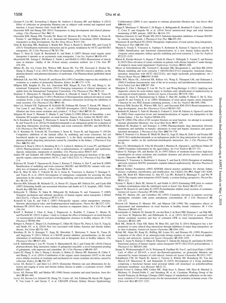
Fig. 2. Schematic representation of the bio-synthesis and disposition of creatine and creat-inine. GAMT, guanidoacetate methyl transferase.
1500 Chu et al.
at ASPE
T Journals on M
ay 1, 2019dm
d.aspetjournals.orgD
ownloaded from

but the mechanism(s) contributing to reabsorption of creatinine, inparticular, the role of transporters, is not well understood.Imamura et al. (2011) established mechanistic models to describe the
renal elimination of creatinine. The model analysis suggested thatactive tubular secretion contributed significantly to the renal elimina-tion of creatinine (30–60%), whereas the significance of reabsorptiondepended on the models used.
Transporters Involved in Active Renal Secretion of Creatinine
Several drugs are reported to impact creatinine secretion, therebycausing transient increase in SCr without altering GFR (Table 2 anddiscussed later). The current hypothesis is that these changes are explainedby the reversible inhibition of transporters involved in tubular secretion ofcreatinine (German et al., 2012). In the following sections, we summarizethe current knowledge of the role of transporters in the uptake of creatinineinto the proximal tubular cells of the kidney and efflux into the urine.
Transporters Involved in Creatinine Uptake in the Kidney
Renal uptake of creatinine has been studied by members of theSLC22A family, such as the organic cation transporters OCT2 andOCT3, and organic anion transporters OAT1, OAT2, and OAT3.Comprehensive reviews on these organic cation and anion transporterscan be found in several publications (Jonker and Schinkel, 2004;Koepsell et al., 2007; Burckhardt, 2012; Nigam et al., 2015).Expression and Function of OCT2, OCT3, OAT1, OAT2, and
OAT3. OCT2 is a renal organic cation uptake transporter primarilylocalized in the basolateral membrane of the whole segment of the renalproximal tubule cells. It plays a major role in renal uptake of mostlycationic compounds, but also transports some anionic and zwitterioniccompounds (Jonker and Schinkel, 2004). On the contrary, OCT3 isrecognized as an extraneuronal monoamine transporter (Jonker andSchinkel, 2004). It is widely expressed in many tissues, such as liver,kidney, skeletal muscle, placenta, and heart, as well as in glial cells andepithelial cells of the choroid plexus, and neurons. OCT3 transports awide range of monoamine neurotransmitters, hormones, and steroids(Wu et al., 1998). OCT3 mRNA was detected in the human kidneycortex; however, its level was much lower compared with OCT2(Motohashi et al., 2002). Therefore, at least based on mRNA analysis,the importance of OCT3 in transport of cationic compounds in thekidney is much less compared with OCT2 (Motohashi et al., 2002).Nevertheless, recent studies in Oct3 knockout mice demonstrate thatdeletion of Oct3 has an impact on the pharmacokinetic and pharma-cological effects of its substrates, such as metformin (Chen et al., 2015).OAT1, OAT2, and OAT3 are renal organic anion uptake transporters
located in the basolateral membrane of proximal tubules (Motohashiet al., 2002). OAT1 and OAT3 have overlapping substrate specificities,and are responsible for the uptake of many anionic drugs, such asantibiotics, antivirals, diuretics, uricosurics, statins, angiotensin-converting enzyme inhibitors, and antineoplastic drugs (Burckhardt,2012). In contrast to OAT1 and OAT3, the role of OAT2 is less wellcharacterized. More studies have emerged this decade focusing onOAT2 expression in the human kidney as well as its role in renal tubularhandling of drugs (Cheng et al., 2012; Lepist et al., 2014; Shen et al.,2015). OAT2 is expressed in the basolateral membrane of renalproximal tubule cells as well as in the sinusoidal membrane ofhepatocytes (Kobayashi et al., 2005; Cheng et al., 2012). One groupshowed that OAT2 was localized in both basolateral and apicalmembranes of human and cynomolgus monkey renal proximal tubules,but only in the apical membrane of rat proximal tubules (Shen et al.,2015). These findings suggest species differences for OAT2/Oat2localization and possibly a role in reabsorption of OAT2 in primates.
Species differences in OAT2/Oat2 localization make rodents a poortranslatable model to predict effects in primates for substrates of thistransporter. OAT2 has many substrates in common with OAT1 andOAT3. However, several antiviral drugs eliminated exclusively in theurine were preferentially transported by OAT2 and not by OAT1 andOAT3 (Cheng et al., 2012). OAT1, OAT2, and OAT3 mRNAs arepresent in the human kidney cortex, with the highest mRNA levelobserved for OAT3, and different mRNA levels for OAT1 and OAT2 intwo separate reports (Motohashi et al., 2002; Cheng et al., 2012).However, it is not known whether these differences in mRNA levelstranslate into different amount of transporter protein.Transporters Involved in Creatinine Uptake. Creatinine has been
reported to be an in vitro substrate for OCT2 (Urakami et al., 2004;Imamura et al., 2011; Ciarimboli et al., 2012; Lepist et al., 2014), with aMichaelis constant (Km) ranging from 2 to 56 mM, suggesting low-affinity transport. Based on the physiologic concentration of creatininein plasma (30–85 mM), OCT2-mediated transport of creatinine will notbe saturable, which is especially important for patients with reducedGFR (Urakami et al., 2004). In vivo studies using Oct1/2 double-knockout mice showed the significance of Oct in creatinine secretion;creatinine clearance and renal accumulation of exogenous creatininewere 35- and 23-fold lower in Oct1/2 knockout mice compared withwild-type mice, respectively (Ciarimboli et al., 2012). One group,however, questioned the role of Oct2 in creatinine transport, as they didnot observe a significant difference in creatinine secretion in Oct1/2knockout mice compared with control mice (Eisner et al., 2010). Thisdiscrepancy may be explained by the use of ketamine by Eisner et al.(2010), which has the potential to interfere with creatinine secretion(Ciarimboli et al., 2012). It should be noted that species differences maycomplicate the translation of the OCT2/Oct2 contribution in creatininetransport from rodents to humans. For instance, in mice, both Oct1 andOct2 are expressed in the kidney, whereas in humans, only OCT2 isexpressed in the kidney and OCT1 is predominantly expressed in theliver (Jonker and Schinkel, 2004). More direct evidence of creatinine asan OCT2 substrate came from genome-wide association studies showingacute elevation of SCr (24% increase) in cancer patients followingtreatment with cisplatin, a known substrate and inhibitor of OCT2. Theeffect of cisplatin on creatinine secretion is attributed primarily tocompetitive inhibition of OCT2 transport (Ciarimboli et al., 2012).In a genetic association study, an intergenic single nucleotide
polymorphism (SNP) (rs2504954; T.G), located on chromosome 6in the region between the OCT2 and OCT3 genes, was significantlyassociated with higher SCr level (Ciarimboli et al., 2012). Anothernoncoding SNP (rs2279463; T.C) in the OCT2 gene was associatedwith creatinine metabolism (Kottgen et al., 2010). On the other hand, acoding SNP in OCT2 (rs316019; S270A) has been associated withreduced cisplatin-induced nephrotoxicity (Filipski et al., 2009) andreduced renal elimination of metformin (Wang et al., 2008), but notwith altered SCr levels. An intronic OCT2 SNP (rs316009; G.A),which highly correlated to rs316019, showed a strong association withtubular creatinine secretion and end-stage renal disease (Reznichenkoet al., 2013). Taken together, both in vitro and in vivo data indicate arole of OCT2 in tubular secretion of creatinine.Creatinine has also been reported to be an in vitro substrate for OCT3
(Imamura et al., 2011; Ciarimboli et al., 2012; Lepist et al., 2014).Similar to OCT2, OCT3 transports creatinine with a Km in themillimolar range (;1.9 mM for OCT2 and ;1.3 mM for OCT3)(Lepist et al., 2014). Clinically significant polymorphisms have beenidentified in OCT3. It is unknown, however, whether or not these SNPshave an impact on creatinine secretion (Aoyama et al., 2006; Sakataet al., 2010). Based on current evidence, OCT3 is likely less importantthan OCT2 for creatinine uptake in the kidney.
Serum Creatinine and Inhibition of Renal Transporters 1501
at ASPE
T Journals on M
ay 1, 2019dm
d.aspetjournals.orgD
ownloaded from

Opposing the traditional view of organic cationic pathways as the solemechanism of creatinine secretion in the kidney, creatinine has beenreported to be an in vitro substrate for OAT2 (Ciarimboli et al., 2012;Lepist et al., 2014; Shen et al., 2015). A comprehensive analysis to identifythe transporters for creatinine was performed, in which each transporter’smRNA and function were measured (Lepist et al., 2014). Creatinineshowed a somewhat higher affinity toward OAT2 (Km = 986 mM) ascompared with OCT2 and OCT3. Other groups also observed higheraffinity transport of creatinine by OAT2 compared with that by othertransporters (Shen et al., 2015). OAT2 might contribute to creatininesecretion, and possibly reabsorption in human renal proximal tubules, butclinical data are needed to support this hypothesis.The role of OAT3 in creatinine secretion is unclear. Contradictory
findings were observed in vitro in OAT3-transfected cell lines(Urakami et al., 2004), and kinetic data have not been reported. Theinvolvement of mouse Oat3 in creatinine secretion is also unclear.Vallon et al. (2012) used Xenopus laevis oocytes to show that creatininewas transported by mouse Oat3, and renal creatinine clearance wassignificantly reduced in Oat3 knockout compared with wild-type mice.However, Ciarimboli et al. (2012) did not observe any creatinine uptakeby mouse Oat3 in a transfected cell line. The contribution of OAT3 torenal creatinine uptake in humans was estimated to be very low basedon a relative activity factor evaluation (Imamura et al., 2011). ForOAT1, several reports showed that creatinine was not a substrate forthis transporter (Urakami et al., 2004; Imamura et al., 2011; Ciarimboliet al., 2012; Lepist et al., 2014).
Transporters Involved in Creatinine Efflux in the Kidney
Creatinine transport has been studied by members of the SLC47Afamily, such as MATE1 and MATE2K, and SLC22A family members,such as organic cation and carnitine transporters OCTN1 and OCTN2,and organic anion transporter OAT4.Expression and Function of MATE1, MATE2K, OCTN1,
OCTN2, and OAT4. MATEs are proton/organic cation antiporters.MATE1 is highly expressed in the kidney, liver, adrenal gland, skeletalmuscle, and several other tissues, whereas MATE2K is specificallyexpressed in the kidney (Masuda et al., 2006). Both MATE1 andMATE2K play a role in the renal tubular secretion of cationic drugs and
endogenous compounds in humans (Yonezawa and Inui, 2011).OCTN1 and OCTN2 are organic cation transporters expressed in manytissues. They are localized at the brush border membrane of theproximal tubules in the kidney and play a role in L-carnitine tissuedistribution and renal reabsorption (Wu et al., 1999; Tamai, 2013).OAT4 is also located at the brush border membrane of proximal tubulesand mediates the bidirectional transport of urate and some organicanions in a substrate-dependent manner (Miyazaki et al., 2005; Hagoset al., 2007).
Transporters Involved in Creatinine Efflux. Creatinine has beenreported to be a substrate for MATE1 and MATE2K (Tanihara et al.,2007). Although MATEs function as efflux transporters in vivo, theyare often evaluated as uptake transporters by manipulating extracellularpH in vitro. In interpreting in vitro data for MATEs, it is assumed thatthe intra- and extracellular binding sites have an equal affinity forsubstrates and inhibitors. In vitro studies suggest that MATE1 andMATE2K are involved in tubular secretion of creatinine (Taniharaet al., 2007; Lepist et al., 2014; Shen et al., 2015). The uptake windowof creatinine by MATEs at extracellular pH 8.4 was relatively low, andonly;2- to 3-fold higher in MATE1-transfected cells and 1.3- to 3-foldhigher in MATE2K-transfected cells compared with control cells(Tanihara et al., 2007; Lepist et al., 2014; Shen et al., 2015).Intracellular acidification by pretreatment with ammonium chlorideenhanced the uptake of creatinine by MATE1 and MATE2K (Taniharaet al., 2007). Kinetic analyses showed that creatinine has low affinitytoward MATE1 and MATE2K, with Km values of ;10 and ;21 mM,respectively (Shen et al., 2015). Orthologs of human MATE1, but notMATE2K, have been identified in rats and mice (Yonezawa and Inui,2011). When studying the nephrotoxicity of cisplatin, a significantincrease in creatinine was observed in cisplatin-treated Mate1 knockoutmice compared with control mice. In addition, the combination ofpyrimethamine, a selective inhibitor of mouse Mate1, with cisplatinsignificantly increased creatinine levels compared with cisplatin alonein wild-type mice. Both studies indirectly suggested a role of Mate1 increatinine transport, at least in mice (Nakamura et al., 2010).Several polymorphisms have been identified in MATE1
(rs111060524-G64D, rs111060526-A310V, rs111060527-D328A,rs111060528-N474S) and MATE2K (rs111060529-K64N and
Fig. 3. Schematic representation of renalelimination of creatinine and the transportersknown to transport creatinine in vitro.
1502 Chu et al.
at ASPE
T Journals on M
ay 1, 2019dm
d.aspetjournals.orgD
ownloaded from

rs111060532-G211V) in Japanese subjects, and these variants wereassociated with loss of transport activity of TEA and metformin in vitro(Kajiwara et al., 2009). Other MATE1 SNPs (rs35646404 -T159M andrs35790011-V338I) have also been described with similar reduction intransport activity of TEA and metformin in other subjects from variousethnic groups (Meyer zu Schwabedissen et al., 2010). The effect ofthese MATE1 andMATE2K variants on creatinine transport remains tobe elucidated.To date, no reports have demonstrated creatinine transport by
OCTN1 and/or OCTN2. It is unclear whether creatinine is reabsorbedby proximal tubular cells through OAT4, as OAT4 functions as abidirectional transporter (Hagos et al., 2007), suggesting that it could beinvolved in excreting substrates into urine and/or reuptake of substratesfrom urine into cells. Using different transfected cell lines, someresearchers observed creatinine uptake by OAT4 (Imamura et al.,2011), but this was not confirmed by others (Lepist et al., 2014).In summary, based on current evidence, OCT2, MATE1, and
MATE2K are the major transporters involved in renal creatininesecretion. OAT2 also could be involved based on in vitro evidence,but its in vivo relevance in humans is not clear yet (Fig. 3).
Inhibition of Renal Transporters and Elevation of SCr: An InVitro–In Vivo Correlation Analysis
In drug development, it is desirable to develop approaches tounderstand underlying mechanisms for interactions of drug candidateswith active renal secretion of creatinine and to subsequently distinguishclinically relevant increases in SCr due to impairment of renal functionfrom nonpathologic increases in SCr caused by inhibition of renaltransporters. We therefore conducted a retrospective analysis toevaluate whether an in vitro–in vivo correlation (IVIVC) exists betweeninhibition of the renal transporters OCT2, MATE1, MATE2K,OAT2, and OCT3 and elevations of SCr and/or decreases in CLcr.In this analysis (Tables 1 and 2), a total of 16 compounds wereidentified that showed 1) $10% reversible elevation of SCr withouta significant change in measured GFR (cimetidine, pyrimethamine,trimethoprim, dronedarone, 7-[(3R)-3-(1-aminocyclopropyl)pyrrolidin-1-yl]-1-[(1R,2S)-2-fluorocyclopropyl]-8-methoxy-4-oxoquinoline-3-carboxylic acid (DX-619), dolutegravir, cobicistat, ritonavir,
ranolazine, rilpivirine, and telaprevir); 2).10% reversible elevation ofSCr, without reported data on changes in GFR (amiodarone, vandetanib);and 3) no significant elevation of SCr and GFR and/or other renaltoxicity markers at clinically relevant exposure as negative in vivocontrols (famotidine, ranitidine, and raltegravir). In vitro inhibition data(IC50 or Ki values) for human OCT2, MATE1, MATE2K, OAT2, andOCT3 were collected for these compounds from the University ofWashington DDI database (https://www.druginteractioninfo.org). Therange of IC50 or Ki values is summarized in Table 1. To betterunderstand the correlation between in vitro inhibition of OCT2 andMATEs and the elevation of SCr, in vitro IC50 values for inhibition ofOCT2, MATE1, and MATE2K for 15 compounds listed in Table 1were measured at Merck Research Laboratories (Kenilworth, NJ) usingmetformin as the probe substrate and the method described by Rizket al. (2014) in CHO-K1-OCT2, CHO-K1-MATE1, and MDCKII-MATE2K cells. Although creatinine is an ideal in vitro probe forIVIVC evaluations, its assay window in OCT2 and MATE uptakeassays is relatively low (our unpublished observations) (Lepist et al.,2014; Shen et al., 2015), and therefore, it is unsuitable for measuringIC50 values.IVIVC analysis in this review will be focused on OCT2 andMATEs.
In vitro inhibition data for OAT2 and OCT3, which were recentlyidentified as renal creatinine transporters, are currently available onlyfor a few compounds (Table 1). These compounds generally showweakinhibition of OAT2 and OCT3 compared with MATEs and/or OCT2,suggesting that inhibition of these transporters might be clinically lessrelevant. Indomethacin is a relatively potent in vitro inhibitor of OAT2(IC50 = 2.1 mM) (Shen et al., 2015). However, the effect ofindomethacin on elevation of SCr in several clinical studies iscontroversial (Prescott et al., 1990; Al-Waili, 2002). In female healthyvolunteers, indomethacin (150 mg daily for 3 days) had no significanteffect on SCr, GFR, or renal blood flow (Prescott et al., 1990).However, indomethacin was reported to increase SCr in neonates (Al-Waili, 2002). As indomethacin is a potent prostaglandin synthesisinhibitor, it is likely that mechanisms other than transporter inhibitioncould result in the observed elevation of blood creatinine (Al-Waili,2002).In vitro inhibition data on OCT2, MATE1, and MATE2K in the
literature showed high variability for several compounds (Table 1). For
TABLE 1
In vitro inhibition (IC50 or Ki) of selected compounds on human OCT2, MATE1, MATE2K, OAT2, and OCT3
Inhibitors
OCT2 IC50 or Ki (mM) MATE1 IC50 or Ki (mM) MATE2K IC50 or Ki (mM) OAT2 IC50 or Ki (mM) OCT3 IC50 or Ki (mM)
Probe Metformin(10 mM)a
Reported inthe Literatureb
ProbeMetformin (5 mM)a
Reported inthe Literatureb
ProbeMetformin (5 mM)a
Reported inthe Literatureb
Reported inthe Literatureb
Reported inthe Literatureb
Cimetidine 2.9 6 0.7 16.6–1650 0.6 6 0.05 0.2–16.3 5.6 6 0.7 2.1–46.6 22–72.8 9.8–111Pyrimethamine 0.61 6 0.04 4.8–23.6 0.02 6 0.002 0.077–0.63 0.045 6 0.003 0.046–0.52 — —
Famotidine 21.6 6 3.4 36.1–1800 0.45 6 0.03 0.6–0.76 6.6 6 0.9 9.7–36.2 — 6.7–14Ranitidine 11.7 6 1.3 30.5–79 8.2 6 0.6 8.3–25.4 21 6 2 25 — 62–290Trimethoprim 19.8 6 1.5 13.2–1327 0.51 6 0.03 3.31–29.1 0.14 6 0.02 0.61–28.9 NI 12.3Dronedarone 1.9 6 0.3 — 0.46 6 0.02 — 8.9 6 1.7 — — —
DX-619 — 0.94–1.29 — 0.82–4.32 — 0.1 — —
Dolutegravir 0.21 6 0.04 0.066–1.93 3.6 6 0.7 4.67 12.5 6 1.6 .100 .100 .100Cobicistat 37.9 6 4.9 8.24–33 0.98 6 0.19 0.99–1.87 20.5 6 3.0 33.5 .100 .100Ritonavir 24.8 6 3.4 20–25 0.28 6 0.05 0.08–15.4 40.1 6 6.5 23.7 .20 300Ranolazine 47 6 9 — 16.8 6 1.5 — 50 6 8 — — —
Rilpivirine 0.38 6 0.05 5.13 0.25 6 0.04 — 0.28 6 0.08 — — —
Amiodarone 4.7 6 1.1 .1000 1.0 6 0.2 — .50 — — .1000Raltegravir .100 .100 .100 .100 .100 .100 — —
Telaprevir .100 6.35 62 6 5 22.98 .100 — — —
Vandetanib 0.4 6 0.05 5.5–73.4 0.06 6 0.01 0.16–1.23 0. 04 6 0.01 0.3–1.26 — —
—, Data are not reported or available; NI, no inhibition.aData generated at Merck & Co. for inhibition of OCT2, MATE1, and MATE2K using metformin as the probe substrate in CHO-K1-OCT2, CHO-K1-MATE1, and MDCKII-MATE2K cells using
the method described by Rizk et al. (2014).bData obtained from the University of Washington DDI database (https://www.druginteractioninfo.org).
Serum Creatinine and Inhibition of Renal Transporters 1503
at ASPE
T Journals on M
ay 1, 2019dm
d.aspetjournals.orgD
ownloaded from

TABLE2
Effectof
selected
compounds
onSCr,CLcr,andGFR
inhumansandthecorrelationwith
invitroinhibitio
nof
OCT2,
MATE1,
andMATE2K
Inhibitors
DosingRegim
enSCr↑
CLcr↓
GFR↓
Markers
forGFR
Cmax
f uCmax/IC50a
Cmax,u/IC50a
References
OCT2
MATE1
MATE2K
OCT2
MATE1
MATE2K
%%
mM
Cim
etidine
400mg(Q
DS)
13–26
20NS
51Cr–EDTA,
inulin
8–12
0.8
4.14
20.00
2.14
3.31
16.00
1.71
Duttet
al.,1981;Hilb
randset
al.,1991
Pyrim
ethamine
50–100mgSD
18–26
25–27
NS
inulin
2.3a
0.13
3.77
115.00
51.11
0.49
14.95
6.64
Opravilet
al.,1993;Kusuharaet
al.,2011
Fam
otidine
40mgQD,7days
NS
NS
——
0.39
0.8
0.02
0.87
0.06
0.01
0.69
0.05
Ishigamiet
al.,1989
Fam
otidine
200mgSD;
160mgq4h
SI
SI
——
1.25
0.8
0.06
2.78
0.19
0.05
2.22
0.15
Hibmaet
al.,2015
Ranitidine
300mgQD
NS
NS
——
3.72
0.85
0.32
0.45
0.18
0.27
0.39
0.15
Motyl,2004
Trimethoprim
20mg/kg/day
(10days);
200mgBID
3116
NS
51Cr–EDTA
iothalam
ate
3.4–
6.9
0.56
0.35
13.53
49.29
0.20
7.58
27.60
Naderer
etal.,1997;Aryaet
al.,2014
Dronedarone
400mgBID
7days
10–15
18NS
Sinistrin,PAH
0.30
0.02
0.16
0.65
0.03
0.003
0.013
0.001
Tschuppertet
al.,2007
DX-619
800mg(Q
D)
(4days)
30–40
26NS
Iohexol
20.5–22
0.29
–0.35
b23.40
26.83
220.0
8.19
9.39
77.0
Sarapaet
al.,2007
Dolutegravir
50mg(Q
Dor
BID
,14
days)
9–17
10–14
NS
Iohexol
Cystatin
C6.7–
13.1
0.01
62.38
3.64
1.05
0.62
0.04
0.01
Koteffet
al.,2013
Cobicistat
150mgQD,
7days,PO
10.5,23
8–14,9–20
NS
Iohexol
1.55
0.03
0.04
1.58
0.08
0.001
0.05
0.002
Cohen
etal.,2011;German
etal.,2012;Aryaet
al.,2014
Rito
navir
100mgQD
—25
NS
Iohexol
2.16
0.015
0.09
7.71
0.05
0.001
0.12
0.001
Deray
etal.,1998;German
etal.,2012;Lepistet
al.,2014
Ranolazine
1000
mgBID
,5days,PO
1210
NS
Sinistrin
6.01
0.38
0.13
0.36
0.12
0.05
0.14
0.05
Aryaet
al.,2014
Rilp
ivirine
25mg(Q
D,
96weeks)
10—
NS
Cystatin
C0.6
0.005
1.58
2.40
2.14
0.01
0.01
0.01
Druglabel;Maggi
etal.,2014
Amiodarone
400mgQD
11—
——
0.8–
2.3
0.04
0.49
2.30
0.05
0.02
0.09
0.002
Pollaket
al.,1993
Raltegravir
400mgBID
NS
NS
——
3.38
0.17
,0.03
,0.03
,0.03
,0.01
,0.01
,0.01
Druglabel;Maggi
etal.,2014;
Rizket
al.,2014
Telaprevir
750mgq8h
SI
—NS
Cystatin
C,
L–FABP,
NAG
5.82
0.04
–0.24
b,0.06
0.09
,0.06
0.01
0.02
,0.01
Suzukiet
al.,2013;Matsuiet
al.,2015
Vandetanib
300mgQD
15—
——
0.33
0.1
0.83
5.50
8.25
0.08
0.55
0.83
Shenet
al.,2013
—,dataarenotreportedor
available;BID
,twicedaily
;L-FABP,liver-typefatty
acid
bindingprotein;
NAG,N
-acetyl-b-D
glucosam
inidase;NS,not
significant(eitherstatistically
orclinically);PAH,para-am
ino-hippurate;PO,bymouth;Q
DS,fourtim
esaday;
q4h,
every4ho
urs;q8h,
every8hours;SD,single
dose;SI,significantly
increasedcomparedwith
baselin
elevel.
aIC
50values
used
weregeneratedat
Merck
&Co.
andshow
nin
Table
1,except
forDX-619,forwhich
thelowestIC
50valueobtained
from
theliteratureisused
(see
Table
1).
bHighestf u
values
areused
toestim
ateCmax,u/IC50as
theworst-casescenario.
1504 Chu et al.
at ASPE
T Journals on M
ay 1, 2019dm
d.aspetjournals.orgD
ownloaded from

instance, the in vitro IC50 or Ki for ritonavir with MATE1 showed a193-fold variability, and inhibition of OCT2 by trimethoprim andcimetidine showed a 101- and 99-fold variability, respectively. Thereasons for this high variability are not understood, but it could becaused by the use of different probe substrates, and differences in invitro systems and assay conditions. For example, remarkable substrate-dependent differences in IC50 values for inhibition of MATE2K bytrimethoprim were reported (47-fold, metformin vs. N-methylnicotina-mide as probes) (Muller et al., 2015) and for OCT2 inhibition byvandetanib (13-fold, N-methyl-4-phenylpyridinium iodide vs. metfor-min as probes) (Shen et al., 2013) when the studies were conducted inthe same laboratory using the same in vitro system. Substrate-dependent inhibition of OCT2, MATE1, and MATE2K has beensystemically evaluated with several prototypic substrates (Belzer et al.,2013; Martinez-Guerrero and Wright, 2013), suggesting that bothOCT2 and MATEs have multiple drug binding sites. In contrast to suchsubstrate-dependent inhibition, several other studies have shownconsistent Ki or IC50 values with selected OCT2/MATE inhibitorsacross different probe substrates. For instance, Ito et al. (2012b)reported no marked substrate dependence in cimetidine Ki values forOCT2, MATE1, and MATE2K with five probe substrates. Likewise,similar IC50 values were obtained with cobicistat for OCT2 andMATE1 using TEA and creatinine as probe substrates (Lepist et al.,2014). Nevertheless, development of predictive DDI models for OCT2and MATEs needs to take into account the potential for substratedependence of ligand interactions with these proteins. Furthermore,different in vitro systems and assay conditions may have a markedeffect on IC50 variability. For example, in studies where metformin wasused as probe substrate, the ritonavir IC50 for MATE1 was 0.08 mMwhen preincubating MATE1-transfected human embryonic kidney 293cells for 30 minutes in a 30 mM NH4Cl buffer to create an artificial pHgradient (Wittwer et al., 2013), whereas the IC50 was 15.4 mM whenusing MATE1-transfected HeLa cells without preincubation withNH4Cl (Meyer zu Schwabedissen et al., 2010).In Table 2, the risk for in vivo inhibition of OCT2, MATE1, and
MATE2K was assessed by comparing total and unbound maximalplasma concentrations (Cmax and Cmax,u) of test compounds with invitro IC50 values (Cmax/IC50 and Cmax, u/IC50). A cutoff of Cmax/IC50$0.1 or Cmax,u/IC50 $ 0.1 was used to predict the risk for in vivoinhibition of respective transporters. As the relative contribution ofthese transporters (fraction transported) and the rate-determining stepfor renal secretion of creatinine are not well known, we assume thatOCT2, MATE1, andMATE2K contribute equally to the renal secretionof creatinine. Therefore, in assessing the existence of an IVIVC,inhibition of any of the aforementioned transporters was considered asan indication of in vivo inhibition of creatinine secretion as the worstcase scenario. As shown in Table 2, using our in-house IC50 data, Cmax/IC50 ($0.1) provided a reasonably good prediction for the elevation ofSCr for this set of compounds, as there were no false-negativepredictions. Use of Cmax,u/IC50 ($0.1) resulted in four false negatives(dronedarone, cobicistat, rilpivirine, and telaprevir). Both Cmax/IC50
and Cmax,u/IC50 resulted in a false-positive prediction for famotidine[40 mg every day (QD) for 7 days] and ranitidine.Considering the variability of IC50 and Ki values reported in the
literature, using the lowest IC50 or Ki values for OCT2, MATE1, andMATE2K available for 11 compounds (Table 1), Cmax/IC50 ($0.1)provided a reasonably good prediction for the elevation of SCr, whereasCmax,u/IC50 ($0.1) resulted in a false-negative prediction for cobicistat(data not shown). Likewise, using the highest IC50 or Ki values reportedfor OCT2, MATE1, and MATE2K, Cmax/IC50 ($0.1) still provided agood prediction of the elevation of SCr, whereas Cmax,u/IC50 ($0.1)resulted in a false-negative prediction for cobicistat, dolutegravir,
ritonavir, and vandetanib. Use of either the lowest or highest IC50
literature values, Cmax/IC50 and Cmax,u/IC50 both resulted in false-positivepredictions for famotidine (40 mg QD for 7 days) and ranitidine (data notshown). However, Hibma et al. (2015) have recently reported an elevationof SCr and a reduction in CLcr by famotidine in humans at a single dose of200mg andmultiple doses of 160mg, whichwere 4- to 5-fold higher thanin a previous report (Ishigami et al., 1989) (Table 2). The reason for thelack of IVIVC for these two compounds at clinically relevant exposure isunclear. As there are no major circulating metabolites for ranitidine andfamotidine, it is less likely for metabolites to cause transporter inhibition.An effect on reabsorption of creatinine cannot be excluded, however.Currently, Cmax,u/IC50 $ 0.1 is being recommended by the Food and
Drug Administration for OCT2 (U.S. Department of Health and HumanServices et al., 2012) and the International Transporter Consortium forOCT2 and MATEs (Hillgren et al., 2013) as the cutoff value to assessthe risk for DDIs with OCT2/MATEs transporters. For prediction oftransporter-related DDIs, it is critical to use relevant inhibitor concen-trations, which are unbound inhibitor concentrations at the site ofinteractions with the transporter of interest. As such, Cmax,u will be therelevant concentration for predicting DDI with OCT2, which is localizedin the basolateral plasma membrane of renal proximal tubule cells,whereas it may not be adequate to predict DDIs for efflux transporters,such as MATEs, as these are localized in the apical plasma membrane.For example, if the inhibitor is actively taken up by the proximal tubulecells, Cmax,u may underestimate the inhibitory effects for effluxtransporters. Thus, unbound intracellular inhibitor concentrations inrelevant tissues would be more relevant for prediction of effluxtransporter–related DDIs. However, the methodologies to measureand/or predict such values are currently still limited (Chu et al., 2013).
Is Creatinine a Sensitive Biomarker for Renal CationicTransporter–Related DDIs?
Determining the impact of perpetrator drugs on plasma concentrationor urinary excretion of suitable endogenous biomarkers is a valuabletool to assess the risk for drug interactions early in drug development(e.g., phase I clinical trials). Recently, some endogenous probes forstudying renal cationic transporter–related DDIs have been identi-fied. Ito et al. (2012a) have found that the endogenous metaboliteN-methylnicotimide (NMN), a substrate forOCT2,MATE1, andMATE2K,could be used as an endogenous probe to study the DDIs related toOCT2/MATE inhibition in humans. Pyrimethamine, a potent inhibitorof MATE1 and MATE2K, almost completely diminished tubularsecretion of NMN (renal clearance 403 vs. 119 ml/min), but had aminimal effect on plasma exposure of NMN. Furthermore, Muller et al.(2015) reported that trimethoprim, another OCT2/MATE inhibitor,decreased NMN renal clearance by 19.9% without a significant impacton NMN plasma area under the curve. The magnitude of trimethoprim-induced renal clearance reduction was positively correlated betweenNMN and metformin in 12 subjects, suggesting the potential use ofNMN as an endogenous probe for DDIs involving OCT2/MATEs.Using untargeted metabolomics analysis of urine specimens fromhealthy subjects and mice treated with or without pyrimethamine, Katoet al. (2014) found that thiamine, a vitamin B1, which is essential forcarbohydrate metabolism and neural function, is also a potentialbiomarker for inhibition of MATE1 and MATE2K.To evaluate if creatinine can be used as a biomarker to assess OCT2/
MATE-related DDIs, we searched the literature for examples whereclinical DDIs can be mechanistically explained by inhibition of OCT2and/or MATEs, and the changes in SCr or CLcr were measured in thesame clinical studies. As shown in Table 3, observed DDIs with severalOCT2/MATE inhibitors (cimetidine, pyrimethamine, trimethoprim,and vandetanib) at the dose indicated correlated with a 10–30% elevation
Serum Creatinine and Inhibition of Renal Transporters 1505
at ASPE
T Journals on M
ay 1, 2019dm
d.aspetjournals.orgD
ownloaded from

in SCr or a decrease inCLcr. Thiswas not the case for ranitidine, however,as it caused DDIs with procainamide and triamterene without affectingSCr levels. Interestingly, famotidine, a recently reported MATE1selective inhibitor, significantly increased SCr (200 mg QD and 160 mgevery 4 hours) without affecting the plasma exposure of metformin(Hibma et al., 2015). The latter is likely explained by the opposing effectsof the famotidine-induced increase in both metformin absorption andrenal clearance. Elevation of SCr by cimetidine was variable and lesssensitive in some DDI studies at the clinically relevant dose of 300–400mg. Considering the weak tomoderate change in SCr associated withOCT/MATE-related DDIs, and that a range of other factors maypotentially impact SCr exposure, as we have discussed elsewhere in thisreview, SCr does not appear to be a biomarker with sufficient sensitivityto assess the risk, either qualitatively or quantitatively, of inhibition ofOCT2 or MATEs in humans. However, follow-up mechanistic studies,such as transporter inhibition experiments, are still useful in cases whereincreases in SCr exposure are observed.
Is Serum Creatinine an Appropriate Marker for Renal Injury?
Traditional monitoring for nephrotoxicity relies upon the measure-ment of SCr. However, SCr retains poor specificity for AKI and isinsensitive to the degree of AKI for three reasons. First, a large amountof nephron loss can occur without significant changes in SCr due toresidual renal reserve. This fact is most clearly evident in kidney donorsin whom no significant change in SCr occurs despite a 50% loss offunctioning renal mass (Bosch et al., 1983). Second, the rate of rise inSCr following a renal insult is delayed due to the kinetics of creatinineproduction from muscle turnover and accumulation secondary toreduced glomerular filtration. At a normal GFR of 120 ml/min, theserum half-life of creatinine is approximately 4 hours; however, at aGFR of 30 ml/min, the half-life extends to 16 hours and will thereforenot reach steady state for nearly 3 days (Waikar and Bonventre, 2009).Third, as previously discussed, SCr is influenced by a number of otherfactors, including inhibition of tubular secretion by drugs, weight,gender, age, muscle metabolism, hydration state, and protein intake(Blantz, 1998). Reduced muscle mass secondary to malnutrition orimmobility is a frequently observed clinical problem that severely limitsthe utility of SCr as a marker of kidney function. Based on thelimitations of SCr, there has been great interest in the identification ofalternate markers of renal function. To date, a number of promisingbiomarker candidates have been identified, characterized, and validated
using models of kidney injury in animals or described for variousclinical settings in humans, such as sepsis, cardiac bypass surgery, andcontrast media exposure (Fuchs and Hewitt, 2011;Waring andMoonie,2011; Vanmassenhove et al., 2013). Importantly, the utility of thesenew biomarkers in clinically detecting drug-induced AKI in either thepatient-care or drug-development setting has not been established.Presently, regulatory agencies have agreed that urine biomarkers shouldbe used for nonclinical phases of drug development, and on a case-by-case basis for clinical drug development research investigation (Dieterleet al., 2010). Clinical qualification of novel AKI urine biomarkers foruse during clinical drug development is currently ongoing.
Conclusions
Based on the in vitro and pharmacogenomic evidence available, OCT2is one of the transporters involved in the uptake of creatinine into kidneyproximal tubule cells, but its quantitative involvement is unknown. Morerecent in vitro data suggest that OAT2 also transports creatinineefficiently, but to what extent this is relevant in humans is not yet clear.Following uptake into the kidney, MATE1 and MATE2K mediate theefflux of creatinine into the urine. Important questions that remain arewhether uptake or efflux is rate-determining in the active secretion ofcreatinine, what the relative contribution is of each transporter in thisprocess, and whether there are yet-unidentified transporters involved increatinine excretion and/or reabsorption. Similar to hepatobiliary trans-port, it is generally hypothesized that uptake is the rate-limiting step foractive tubular secretion, if the luminal efflux is markedly greater than thebasolateral efflux. In this case, the inhibition of the luminal efflux shouldhave less impact on the overall systemic intrinsic clearance. However,this cannot explain the significant elevation of SCr by pyrimethamine, aselective inhibitor of MATEs, relative to OCT2.Currently, the effect of drugs on creatinine transport is measured in
cell lines transfected with individual transporters. Recently, a quintuplein vitro transporter model expressing OAT2/OCT2/OCT3/MATE1/MATE2K was explored to evaluate the impact of test compounds oncreatinine transport (Zhang et al., 2015), but more data are needed toestablish the predictive value of this model. Development and use ofholistic models and integrated systems, such as immortalized cell linesderived from human kidney with preserved activity of transporters anddrug-metabolizing enzymes, may provide more physiologically rele-vant models to study the interaction of drugs with the renal secretion ofcreatinine in the future (Schophuizen et al., 2015).
TABLE 3
Examples of transporter-related DDIs involving OCT2, MATE1, and/or MATE2K and correlation with transient elevation of SCr
Perpetrator Perpetrator Dose Regimen Victim Victim Dose Regimen% Changein AUC
% Changein Renal CL
Elevation of SCr and/or% Decrease of CLcr
References
Cimetidine 400 mg QID (8 days) Gabapentin 1200 mg QD [4 days] 23.7 217.8 Yes; CLcr↓10% Lal et al., 2010Cimetidine 800 mg BID (6 days) Glycopyrronium 100 mg inhalation SD 19.2 222.1 — Dumitras et al., 2013Cimetidine 400 mg BID (6.5 days) Metformin 500 mg SD 54.2 244.6 No; CLcr NS Wang et al., 2008Cimetidine 400 mg BID (5 days) Metformin 250 mg QD (10 days) 46.2 228.3 Noa Somogyi et al., 1987Cimetidine 300 mg TID (5 days) Varenicline 2 mg SD 29.7 225.1 Yes; CLcr↓5-10% Feng et al., 2008Dolutegravir 50 mg BID (7 days) Metformin 500 mg BID (12 days) 145 — Yes; SCr ↑ Zong et al., 2014Pyrimethamine 50 mg SD Metformin 250 mg SD 35.3 235 Yes; CLcr↓20% Kusuhara et al., 2011Ranitidine 150 mg BID Procainamide 1 g SD 13.7 218.5 — Somogyi and Bochner, 1984Ranitidine 150 mg BID (4 days) Triamterene 100 mg/day QD (8 days) 224 251 No; NS Clcr Muirhead et al., 1988Trimethoprim 200 mg TID (6 days) Metformin 500 mg TID (10 days) 37 232 Yes; CLcr↓20%;
SCr ↑23%Grun et al., 2013
Trimethoprim 200 mg TID(5 days) Metformin 850 mg QD (2 doses) 29.5 226.4 Yes; CLcr↓16.9% Muller et al., 2015Vandetanib 100 mg QD (three
21-day cycles)Cisplatin 75 mg/m2 SD (three
21-day cycles)32.7 — — Blackhall et al., 2010
Vandetanib 800 mg SD Metformin 1000 mg SD 73.3 252 Yes; SCr ↑ 8-29% Johansson et al., 2014
—, Data are not reported or available; AUC, area under the curve; BID, twice daily; NS, not significant statistically; QID, four times a day; SD, single dose; TID, three times a day.aA time-dependent variation of SCr was observed.
1506 Chu et al.
at ASPE
T Journals on M
ay 1, 2019dm
d.aspetjournals.orgD
ownloaded from
Overall, a high variability of in vitro transporter inhibition data, notlimited to OCT2/MATEs, has become a significant concern and maylimit in vitro to in vivo extrapolation using universal cutoff values fortransporter perpetrator decision trees, which trigger clinical DDI studies(Bentz et al., 2013). Although the underlying mechanisms for IC50
variability can be complex, proper standardization of in vitro inhibitionassays using, for example, clinically relevant probe substrates, standard-ized incubation conditions, and cell lines will be helpful for improving invitro to in vivo extrapolation.In an attempt to establish an IVIVC between inhibition of OCT2,
MATE1, and MATE2K, several false negatives were identified using acutoff for the ratio of Cmax,u/IC50 or Cmax,u/ Ki of$0.1. The true-positiverate was higher if total (bound plus unbound) drug concentrations wereused for the analyses. Since only unbound drug will be available forinteractions with transporters, this suggests that the free drug concentra-tion measured in plasma is lower than in the proximal tubule cells, or thatmechanisms other than inhibition of MATEs and OCT2 contribute to theeffects on creatinine. For example, although cobicistat is an inhibitor ofMATE1 in vitro, this inhibition is not predicted to be clinically significantbased on Cmax,u/IC50 data. Remarkably, famotidine and ranitidine wereidentified as inhibitors of MATE- and OCT2-mediated creatininetransport in vitro, whereas no effect on creatinine was observed atclinically relevant exposures. Currently, we have no good explanationsfor the lack of IVIVC for these compounds. In the future, use ofmechanistic models may improve the prediction of in vivo interactionof drug molecules with creatinine renal transporters.Due to potential interactions of drugmoleculeswith creatinine secretion
along with several other limitations, an alternative method to estimateGFR is desirable. Despite ongoing efforts to identify more sensitive andspecific markers for renal function and injury, use of creatinine to estimateGFR is currently still a practical approach. As such, if a transient and/orreversible elevation of SCr was observed during drug development,understanding the potential interaction of drugmolecules with active renalsecretion of creatinine and carefully monitoring renal function withalternate markers, such as cystatin C, would be recommended.Since organic cation transporters such as OCT2 and MATEs are
known to transport endogenous compounds (Jonker and Schinkel, 2004),it would be valuable from a drug development perspective if changes inthese compounds could be used as biomarkers for assessing DDIsinvolving inhibition of these transporters. In the case of the kidney, suchbiomarkers would need to be excreted to a significant extent by activetransport (as opposed to GFR), their levels should not be affected bysecondary factors such as diet and disease, they should not be sensitive todiurnal effects, and they would need to be selective for the transporter(s)of interest. Based on these criteria and our retrospective analysis of invitro and clinical data, creatinine is not an optimal biomarker, as itssynthesis involves multiple steps, external factors such as diet andexercise affect plasma levels, and the contribution of active transport toclearance is relatively small and not consistent between patient popula-tions. However, mechanistic studies to explain increases in creatinine inthe absence of a decrease in GFR will continue to be important.
Acknowledgments
The authors thank Dr. Lisa A. Shipley for continuous support, Robert Houlefor technical assistance, and Drs. Kathleen Cox, Nancy G. B. Agrawal, and SevgiGurkan for valuable comments on the manuscript.
Authorship ContributionsConducted experiments: Chan.Performed data analysis: Chu, Bleasby, Chan, Evers.Wrote or contributed to the writing of the manuscript: Chu, Bleasby, Chan,
Nunes, Evers.
References
Al-Waili NS (2002) Increased urinary nitrite excretion in primary enuresis: effects of in-domethacin treatment on urinary and serum osmolality and electrolytes, urinary volumes andnitrite excretion. BJU Int 90:294–301.
Aoyama N, Takahashi N, Kitaichi K, Ishihara R, Saito S, Maeno N, Ji X, Takagi K, Sekine Y,and Iyo M, et al. (2006) Association between gene polymorphisms of SLC22A3 and meth-amphetamine use disorder. Alcohol Clin Exp Res 30:1644–1649.
Arya V, Florian J, Marcus KA, Reynolds KS, Lewis LL, and Sherwat AI (2013) Does an increasein serum creatinine always reflect renal injury? The case of Stribild®. J Clin Pharmacol DOI:10.1002/jcph.223 [published ahead of print].
Arya V, Yang X, Balimane P, Chinn L, Hinderling P, Vaidyanathan J, Zur AA, WittwerMB, and Zhang L (2014) Creatinine as an endogenous marker for renal function—emerging role of transporters in the overall assessment of renal toxicity (Abstract). ClinPharmacol Ther 95:S65.
Béard E and Braissant O (2010) Synthesis and transport of creatine in the CNS: importance forcerebral functions. J Neurochem 115:297–313.
Belzer M, Morales M, Jagadish B, Mash EA, and Wright SH (2013) Substrate-dependentligand inhibition of the human organic cation transporter OCT2. J Pharmacol Exp Ther 346:300–310.
Bentz J, O’Connor MP, Bednarczyk D, Coleman J, Lee C, Palm J, Pak YA, Perloff ES, Reyner E,and Balimane P, et al. (2013) Variability in P-glycoprotein inhibitory potency (IC₅₀) usingvarious in vitro experimental systems: implications for universal digoxin drug-drug interactionrisk assessment decision criteria. Drug Metab Dispos 41:1347–1366.
Berglund F, Killander J, and Pompeius R (1975) Effect of trimethoprim-sulfamethoxazole on therenal excretion of creatinine in man. J Urol 114:802–808.
Blackhall FH, O’brien M, Schmid P, Nicolson M, Taylor P, Milenkova T, Kennedy SJ,and Thatcher N (2010) A phase I study of Vandetanib in combination with vinorelbine/cisplatinor gemcitabine/cisplatin as first-line treatment for advanced non-small cell lung cancer. JThorac Oncol 5:1285–1288.
Blantz RC (1998) Pathophysiology of pre-renal azotemia. Kidney Int 53:512–523.Bosch JP, Saccaggi A, Lauer A, Ronco C, Belledonne M, and Glabman S (1983) Renal functionalreserve in humans. Effect of protein intake on glomerular filtration rate. Am J Med 75:943–950.
Breyer MD and Qi Z (2010) Better nephrology for mice–and man. Kidney Int 77:487–489.Brosnan EM, Weickhardt AJ, Lu X, Maxon DA, Barón AE, Chonchol M, and Camidge DR(2014) Drug-induced reduction in estimated glomerular filtration rate in patients with ALK-positive non-small cell lung cancer treated with the ALK inhibitor crizotinib. Cancer 120:664–674.
Burckhardt G (2012) Drug transport by Organic Anion Transporters (OATs). Pharmacol Ther136:106–130.
Camidge DR, Brosnan EM, DeSilva C, Koo PJ, and Chonchol M (2014) Crizotinib effects oncreatinine and non-creatinine-based measures of glomerular filtration rate. J Thorac Oncol 9:1634–1637.
Chang C, Ekins S, Bahadduri P, and Swaan PW (2006) Pharmacophore-based discovery ofligands for drug transporters. Adv Drug Deliv Rev 58:1431–1450.
Chen EC, Liang X, Yee SW, Geier EG, Stocker SL, Chen L, and Giacomini KM (2015) Targeteddisruption of organic cation transporter 3 attenuates the pharmacologic response to metformin.Mol Pharmacol 88:75–83.
Cheng Y, Vapurcuyan A, Shahidullah M, Aleksunes LM, and Pelis RM (2012) Expression oforganic anion transporter 2 in the human kidney and its potential role in the tubular secretion ofguanine-containing antiviral drugs. Drug Metab Dispos 40:617–624.
Chu X, Korzekwa K, Elsby R, Fenner K, Galetin A, Lai Y, Matsson P, Moss A, Nagar S,and Rosania GR, et al.; International Transporter Consortium (2013) Intracellular drug con-centrations and transporters: measurement, modeling, and implications for the liver. ClinPharmacol Ther 94:126–141.
Ciarimboli G, Lancaster CS, Schlatter E, Franke RM, Sprowl JA, Pavenstädt H, Massmann V,Guckel D, Mathijssen RH, and Yang W, et al. (2012) Proximal tubular secretion of creatinineby organic cation transporter OCT2 in cancer patients. Clin Cancer Res 18:1101–1108.
Cohen C, Elion R, Ruane P, Shamblaw D, DeJesus E, Rashbaum B, Chuck SL, Yale K, Liu HC,and Warren DR, et al. (2011) Randomized, phase 2 evaluation of two single-tablet regimenselvitegravir/cobicistat/emtricitabine/tenofovir disoproxil fumarate versus efavirenz/emtricitabine/tenofovir disoproxil fumarate for the initial treatment of HIV infection. AIDS 25:F7–F12.
Deray G, Bochet M, Katlama C, and Bricaire F (1998) [Nephrotoxicity of ritonavir]. Presse Med27:1801–1803.
Dieterle F, Sistare F, Goodsaid F, Papaluca M, Ozer JS, Webb CP, Baer W, Senagore A, SchipperMJ, and Vonderscher J, et al. (2010) Renal biomarker qualification submission: a dialogbetween the FDA-EMEA and Predictive Safety Testing Consortium. Nat Biotechnol 28:455–462.
Dubb JW, Stote RM, Familiar RG, Lee K, and Alexander F (1978) Effect of cimetidine on renalfunction in normal man. Clin Pharmacol Ther 24:76–83.
Dumitras S, Sechaud R, Drollmann A, Pal P, Vaidyanathan S, Camenisch G, and Kaiser G (2013)Effect of cimetidine, a model drug for inhibition of the organic cation transport (OCT2/MATE1) in the kidney, on the pharmacokinetics of glycopyrronium. Int J Clin Pharmacol Ther51:771–779.
Dutt MK, Moody P, and Northfield TC (1981) Effect of cimetidine on renal function in man. Br JClin Pharmacol 12:47–50.
Edison EE, Brosnan ME, Meyer C, and Brosnan JT (2007) Creatine synthesis: production ofguanidinoacetate by the rat and human kidney in vivo. Am J Physiol Renal Physiol 293:F1799–F1804.
Eisner C, Faulhaber-Walter R, Wang Y, Leelahavanichkul A, Yuen PS, Mizel D, Star RA, BriggsJP, Levine M, and Schnermann J (2010) Major contribution of tubular secretion to creatinineclearance in mice. Kidney Int 77:519–526.
Feng B, Obach RS, Burstein AH, Clark DJ, de Morais SM, and Faessel HM (2008) Effect ofhuman renal cationic transporter inhibition on the pharmacokinetics of varenicline, a newtherapy for smoking cessation: an in vitro-in vivo study. Clin Pharmacol Ther 83:567–576.
Filipski KK, Mathijssen RH, Mikkelsen TS, Schinkel AH, and Sparreboom A (2009) Contri-bution of organic cation transporter 2 (OCT2) to cisplatin-induced nephrotoxicity. ClinPharmacol Ther 86:396–402.
Fuchs TC and Hewitt P (2011) Biomarkers for drug-induced renal damage and nephrotoxicity-anoverview for applied toxicology. AAPS J 13:615–631.
Serum Creatinine and Inhibition of Renal Transporters 1507
at ASPE
T Journals on M
ay 1, 2019dm
d.aspetjournals.orgD
ownloaded from
German P, Liu HC, Szwarcberg J, Hepner M, Andrews J, Kearney BP, and Mathias A (2012)Effect of cobicistat on glomerular filtration rate in subjects with normal and impaired renalfunction. J Acquir Immune Defic Syndr 61:32–40.
Giacomini KM and Huang SM (2013) Transporters in drug development and clinical pharma-cology. Clin Pharmacol Ther 94:3–9.
Giacomini KM, Huang SM, Tweedie DJ, Benet LZ, Brouwer KL, Chu X, Dahlin A, Evers R,Fischer V, and Hillgren KM, et al.; International Transporter Consortium (2010) Membranetransporters in drug development. Nat Rev Drug Discov 9:215–236.
Grün B, Kiessling MK, Burhenne J, Riedel KD, Weiss J, Rauch G, Haefeli WE, and Czock D(2013) Trimethoprim-metformin interaction and its genetic modulation by OCT2 and MATE1transporters. Br J Clin Pharmacol 76:787–796.
Hagos Y, Stein D, Ugele B, Burckhardt G, and Bahn A (2007) Human renal organic aniontransporter 4 operates as an asymmetric urate transporter. J Am Soc Nephrol 18:430–439.
Heymsfield SB, Arteaga C, McManus C, Smith J, and Moffitt S (1983) Measurement of musclemass in humans: validity of the 24-hour urinary creatinine method. Am J Clin Nutr 37:478–494.
Hibma JE, Zur AA, Castro RA, Wittwer MB, Keizer RJ, Yee SW, Goswami S, Stocker SL,Zhang X, Huang Y et al. (2015) The effect of famotidine, a MATE1-selective inhibitor, on thepharmacokinetics and pharmacodynamics of metformin. Clin Pharmacokinet [published aheadof print].
Hilbrands LB, Artz MA, Wetzels JF, and Koene RA (1991) Cimetidine improves the reliability ofcreatinine as a marker of glomerular filtration. Kidney Int 40:1171–1176.
Hillgren KM, Keppler D, Zur AA, Giacomini KM, Stieger B, Cass CE, and Zhang L; In-ternational Transporter Consortium (2013) Emerging transporters of clinical importance: anupdate from the International Transporter Consortium. Clin Pharmacol Ther 94:52–63.
Imamura Y, Murayama N, Okudaira N, Kurihara A, Okazaki O, Izumi T, Inoue K, Yuasa H,Kusuhara H, and Sugiyama Y (2011) Prediction of fluoroquinolone-induced elevation in serumcreatinine levels: a case of drug-endogenous substance interaction involving the inhibition ofrenal secretion. Clin Pharmacol Ther 89:81–88.
Inker LA, Schmid CH, Tighiouart H, Eckfeldt JH, Feldman HI, Greene T, Kusek JW, Manzi J,Van Lente F, and Zhang YL, et al.; CKD-EPI Investigators (2012) Estimating glomerularfiltration rate from serum creatinine and cystatin C. N Engl J Med 367:20–29.
Ishigami M, Sezai Y, Shimada Y, Maeda T, and Yabuki S (1989) Effects of famotidine, a newhistamine H2-receptor antagonist, on renal function. Nippon Jinzo Gakkai Shi 31:687–691.
Ito S, Kusuhara H, Kumagai Y, Moriyama Y, Inoue K, Kondo T, Nakayama H, Horita S, TanabeK, and Yuasa H, et al. (2012a) N-methylnicotinamide is an endogenous probe for evaluation ofdrug-drug interactions involving multidrug and toxin extrusions (MATE1 and MATE2-K).Clin Pharmacol Ther 92:635–641.
Ito S, Kusuhara H, Yokochi M, Toyoshima J, Inoue K, Yuasa H, and Sugiyama Y (2012b)Competitive inhibition of the luminal efflux by multidrug and toxin extrusions, but notbasolateral uptake by organic cation transporter 2, is the likely mechanism underlying thepharmacokinetic drug-drug interactions caused by cimetidine in the kidney. J Pharmacol ExpTher 340:393–403.
Johansson S, Read J, Oliver S, Steinberg M, Li Y, Lisbon E, Mathews D, Leese PT, and Martin P(2014) Pharmacokinetic evaluations of the co-administrations of vandetanib and metformin,digoxin, midazolam, omeprazole or ranitidine. Clin Pharmacokinet 53:837–847.
Jonker JW and Schinkel AH (2004) Pharmacological and physiological functions of the poly-specific organic cation transporters: OCT1, 2, and 3 (SLC22A1-3). J Pharmacol Exp Ther 308:2–9.
Kajiwara M, Terada T, Ogasawara K, Iwano J, Katsura T, Fukatsu A, Doi T, and Inui K (2009)Identification of multidrug and toxin extrusion (MATE1 and MATE2-K) variants with com-plete loss of transport activity. J Hum Genet 54:40–46.
Kato K, Mori H, Kito T, Yokochi M, Ito S, Inoue K, Yonezawa A, Katsura T, Kumagai Y,and Yuasa H, et al. (2014) Investigation of endogenous compounds for assessing the druginteractions in the urinary excretion involving multidrug and toxin extrusion proteins. PharmRes 31:136–147.
Klevens RM, Edwards JR, Richards CL, Jr, Horan TC, Gaynes RP, Pollock DA, and Cardo DM(2007) Estimating health care-associated infections and deaths in U.S. hospitals, 2002. PublicHealth Rep 122:160–166.
Kobayashi Y, Ohshiro N, Sakai R, Ohbayashi M, Kohyama N, and Yamamoto T (2005)Transport mechanism and substrate specificity of human organic anion transporter 2 (hOat2[SLC22A7]). J Pharm Pharmacol 57:573–578.
Koepsell H, Lips K, and Volk C (2007) Polyspecific organic cation transporters: structure,function, physiological roles, and biopharmaceutical implications. Pharm Res 24:1227–1251.
Korhonen PE (2015) How to assess kidney function in outpatient clinics. Int J Clin Pract 69:156–161.
Koteff J, Borland J, Chen S, Song I, Peppercorn A, Koshiba T, Cannon C, Muster H,and Piscitelli SC (2013) A phase 1 study to evaluate the effect of dolutegravir on renal functionvia measurement of iohexol and para-aminohippurate clearance in healthy subjects. Br J ClinPharmacol 75:990–996.
Köttgen A, Pattaro C, Böger CA, Fuchsberger C, Olden M, Glazer NL, Parsa A, Gao X, Yang Q,and Smith AV, et al. (2010) New loci associated with kidney function and chronic kidneydisease. Nat Genet 42:376–384.
Kusuhara H, Ito S, Kumagai Y, Jiang M, Shiroshita T, Moriyama Y, Inoue K, Yuasa H,and Sugiyama Y (2011) Effects of a MATE protein inhibitor, pyrimethamine, on the renalelimination of metformin at oral microdose and at therapeutic dose in healthy subjects. ClinPharmacol Ther 89:837–844.
Lal R, Sukbuntherng J, Luo W, Vicente V, Blumenthal R, Ho J, and Cundy KC (2010) Clinicalpharmacokinetic drug interaction studies of gabapentin enacarbil, a novel transported prodrugof gabapentin, with naproxen and cimetidine. Br J Clin Pharmacol 69:498–507.
Lepist EI, Zhang X, Hao J, Huang J, Kosaka A, Birkus G, Murray BP, Bannister R, Cihlar T,and Huang Y, et al. (2014) Contribution of the organic anion transporter OAT2 to the renalactive tubular secretion of creatinine and mechanism for serum creatinine elevations caused bycobicistat. Kidney Int 86:350–357.
Levey AS, Becker C, and Inker LA (2015) Glomerular filtration rate and albuminuria for de-tection and staging of acute and chronic kidney disease in adults: a systematic review. JAMA313:837–846.
Levey AS, Perrone RD, and Madias NE (1988) Serum creatinine and renal function. Annu RevMed 39:465–490.
Levey AS, Stevens LA, Schmid CH, Zhang YL, Castro AF, 3rd, Feldman HI, Kusek JW, EggersP, Van Lente F, and Greene T, et al. CKD-EPI (Chronic Kidney Disease Epidemiology
Collaboration) (2009) A new equation to estimate glomerular filtration rate. Ann Intern Med150:604–612.
Maggi P, Montinaro V, Mussini C, Di Biagio A, Bellagamba R, Bonfanti P, Calza L, CherubiniC, Corsi P, and Gargiulo M, et al. (2014) Novel antiretroviral drugs and renal functionmonitoring of HIV patients. AIDS Rev 16:144–151.
Martínez-Guerrero LJ and Wright SH (2013) Substrate-dependent inhibition of human MATE1by cationic ionic liquids. J Pharmacol Exp Ther 346:495–503.
Masereeuw R and Russel FG (2010) Therapeutic implications of renal anionic drug transporters.Pharmacol Ther 126:200–216.
Masuda S, Terada T, Yonezawa A, Tanihara Y, Kishimoto K, Katsura T, Ogawa O, and Inui K(2006) Identification and functional characterization of a new human kidney-specific H+/organic cation antiporter, kidney-specific multidrug and toxin extrusion 2. J Am Soc Nephrol17:2127–2135.
Matsui K, Kamijo-Ikemori A, Sugaya T, Ikeda H, Okuse C, Shibagaki Y, Yasuda T, and KimuraK (2015) Does elevation of serum creatinine in patients with chronic hepatitis C under therapyof telaprevir mean renal impairment? Nephrology (Carlton) 20:843–848.
Meyer zu Schwabedissen HE, Verstuyft C, Kroemer HK, Becquemont L, and Kim RB (2010)Human multidrug and toxin extrusion 1 (MATE1/SLC47A1) transporter: functional charac-terization, interaction with OCT2 (SLC22A2), and single nucleotide polymorphisms. Am JPhysiol Renal Physiol 298:F997–F1005.
Miller WG, Myers GL, Ashwood ER, Killeen AA, Wang E, Thienpont LM, and Siekmann L(2005) Creatinine measurement: state of the art in accuracy and interlaboratory harmonization.Arch Pathol Lab Med 129:297–304.
Minejima E, Choi J, Beringer P, Lou M, Tse E, and Wong-Beringer A (2011) Applying newdiagnostic criteria for acute kidney injury to facilitate early identification of nephrotoxicity invancomycin-treated patients. Antimicrob Agents Chemother 55:3278–3283.
Miyazaki H, Anzai N, Ekaratanawong S, Sakata T, Shin HJ, Jutabha P, Hirata T, He X, Non-oguchi H, and Tomita K, et al. (2005) Modulation of renal apical organic anion transporter4 function by two PDZ domain-containing proteins. J Am Soc Nephrol 16:3498–3506.
Morrissey KM, Stocker SL, Wittwer MB, Xu L, and Giacomini KM (2013) Renal transporters indrug development. Annu Rev Pharmacol Toxicol 53:503–529.
Motohashi H, Sakurai Y, Saito H, Masuda S, Urakami Y, Goto M, Fukatsu A, Ogawa O, and InuiK (2002) Gene expression levels and immunolocalization of organic ion transporters in thehuman kidney. J Am Soc Nephrol 13:866–874.
Motyl W (2004) [The effect of H2 receptor blockers on renal function. An attempt to accuratelymeasure glomerular filtration]. Ann Acad Med Stetin 50:97–105.
Muirhead M, Bochner F, and Somogyi A (1988) Pharmacokinetic drug interactions betweentriamterene and ranitidine in humans: alterations in renal and hepatic clearances and gastro-intestinal absorption. J Pharmacol Exp Ther 244:734–739.
Müller F, Pontones CA, Renner B, Mieth M, Hoier E, Auge D, Maas R, Zolk O, and Fromm MF(2015) N(1)-methylnicotinamide as an endogenous probe for drug interactions by renal cationtransporters: studies on the metformin-trimethoprim interaction. Eur J Clin Pharmacol 71:85–94.
Musso CG, Michelángelo H, Vilas M, Reynaldi J, Martinez B, Algranati L, and Macías Núñez JF(2009) Creatinine reabsorption by the aged kidney. Int Urol Nephrol 41:727–731.
Naderer O, Nafziger AN, and Bertino JS, Jr (1997) Effects of moderate-dose versus high-dosetrimethoprim on serum creatinine and creatinine clearance and adverse reactions. AntimicrobAgents Chemother 41:2466–2470.
Nakamura T, Yonezawa A, Hashimoto S, Katsura T, and Inui K (2010) Disruption of multidrugand toxin extrusion MATE1 potentiates cisplatin-induced nephrotoxicity. Biochem Pharmacol80:1762–1767.
National Kidney Foundation (2002) K/DOQI clinical practice guidelines for chronic kidneydisease: evaluation, classification, and stratification. Am J Kidney Dis 39(2, Suppl 1)S1–S266.
Nigam SK, Bush KT, Martovetsky G, Ahn SY, Liu HC, Richard E, Bhatnagar V, and Wu W(2015) The organic anion transporter (OAT) family: a systems biology perspective. Physiol Rev95:83–123.
Nyman U, Bjork J, Back SE, Sterner G, and Grubb A (2015) Estimating GFR prior to contrastmedium examinations-what the radiologist needs to know! Eur Radiol 26:425–435.
Opravil M, Keusch G, and Lüthy R (1993) Pyrimethamine inhibits renal secretion of creatinine.Antimicrob Agents Chemother 37:1056–1060.
Pollak PT, Sharma AD, and Carruthers SG (1993) Creatinine elevation in patients receivingamiodarone correlates with serum amiodarone concentration. Br J Clin Pharmacol 36:125–127.
Prescott LF, Mattison P, Menzies DG, and Manson LM (1990) The comparative effects ofparacetamol and indomethacin on renal function in healthy female volunteers. Br J ClinPharmacol 29:403–412.
Reznichenko A, Sinkeler SJ, Snieder H, van den Born J, de Borst MH, Damman J, van Dijk MC,van Goor H, Hepkema BG, and Hillebrands JL, et al. (2013) SLC22A2 is associated withtubular creatinine secretion and bias of estimated GFR in renal transplantation. PhysiolGenomics 45:201–209.
Rizk ML, Houle R, Chan GH, Hafey M, Rhee EG, and Chu X (2014) Raltegravir has a lowpropensity to cause clinical drug interactions through inhibition of major drug transporters: anin vitro evaluation. Antimicrob Agents Chemother 58:1294–1301.
Rybak MJ, Abate BJ, Kang SL, Ruffing MJ, Lerner SA, and Drusano GL (1999) Prospectiveevaluation of the effect of an aminoglycoside dosing regimen on rates of observed nephro-toxicity and ototoxicity. Antimicrob Agents Chemother 43:1549–1555.
Sakata T, Anzai N, Kimura T, Miura D, Fukutomi T, Takeda M, Sakurai H, and Endou H (2010)Functional analysis of human organic cation transporter OCT3 (SLC22A3) polymorphisms. JPharmacol Sci 113:263–266.
Sarapa N, Wickremasingha P, Ge N, Weitzman R, Fuellhart M, Yen C, and Lloyd-Parks J (2007)Lack of effect of DX-619, a novel des-fluoro(6)-quinolone, on glomerular filtration ratemeasured by serum clearance of cold iohexol. Antimicrob Agents Chemother 51:1912–1917.
Schophuizen CM, De Napoli IE, Jansen J, Teixeira S, Wilmer MJ, Hoenderop JG, Van denHeuvel LP, Masereeuw R, and Stamatialis D (2015) Development of a living membranecomprising a functional human renal proximal tubule cell monolayer on polyethersulfonepolymeric membrane. Acta Biomater 14:22–32.
Séronie-Vivien S, Galteau MM, Carlier MC, Hadj-Aissa A, Hanser AM, Hym B, Marchal A,Michotey O, Pouteil-Noble C, and Sternberg M, et al.; Creatinine Working Group of theSociété Française de Biologie Clinique (2005) Impact of standardized calibration on the inter-assay variation of 14 automated assays for the measurement of creatinine in human serum. ClinChem Lab Med 43:1227–1233.
1508 Chu et al.
at ASPE
T Journals on M
ay 1, 2019dm
d.aspetjournals.orgD
ownloaded from

Shemesh O, Golbetz H, Kriss JP, and Myers BD (1985) Limitations of creatinine as a filtrationmarker in glomerulopathic patients. Kidney Int 28:830–838.
Shen H, Liu T, Morse BL, Zhao Y, Zhang Y, Qiu X, Chen C, Lewin AC, Wang XT, and Liu G,et al. (2015) Characterization of Organic Anion Transporter 2 (SLC22A7): A Highly EfficientTransporter for Creatinine and Species-Dependent Renal Tubular Expression. Drug MetabDispos 43:984–993.
Shen H, Yang Z, Zhao W, Zhang Y, and Rodrigues AD (2013) Assessment of vandetanib as aninhibitor of various human renal transporters: inhibition of multidrug and toxin extrusion as apossible mechanism leading to decreased cisplatin and creatinine clearance. Drug MetabDispos 41:2095–2103.
Shitara Y, Sato H, and Sugiyama Y (2005) Evaluation of drug-drug interaction in the hep-atobiliary and renal transport of drugs. Annu Rev Pharmacol Toxicol 45:689–723.
Somogyi A and Bochner F (1984) Dose and concentration dependent effect of ranitidine onprocainamide disposition and renal clearance in man. Br J Clin Pharmacol 18:175–181.
Somogyi A, Stockley C, Keal J, Rolan P, and Bochner F (1987) Reduction of metformin renaltubular secretion by cimetidine in man. Br J Clin Pharmacol 23:545–551.
Suzuki F, Suzuki Y, Sezaki H, Akuta N, Seko Y, Kawamura Y, Hosaka T, Kobayashi M, Saito S,and Arase Y, et al. (2013) Exploratory study on telaprevir given every 8 h at 500 mg or 750 mgwith peginterferon-alpha-2b and ribavirin in hepatitis C patients. Hepatol Res 43:691–701.
Tamai I (2013) Pharmacological and pathophysiological roles of carnitine/organic cation trans-porters (OCTNs: SLC22A4, SLC22A5 and Slc22a21). Biopharm Drug Dispos 34:29–44.
Tanihara Y, Masuda S, Sato T, Katsura T, Ogawa O, and Inui K (2007) Substrate specificity ofMATE1 and MATE2-K, human multidrug and toxin extrusions/H(+)-organic cation anti-porters. Biochem Pharmacol 74:359–371.
Tschuppert Y, Buclin T, Rothuizen LE, Decosterd LA, Galleyrand J, Gaud C, and Biollaz J(2007) Effect of dronedarone on renal function in healthy subjects. Br J Clin Pharmacol 64:785–791.
Urakami Y, Kimura N, Okuda M, and Inui K (2004) Creatinine transport by basolateral organiccation transporter hOCT2 in the human kidney. Pharm Res 21:976–981.
U.S. Department of Health and Human Services, Food and Drug Administration, and Center forDrug Evaluation and Research (2012) Guidance for industry, drug interaction studies—study design, data analysis, implications for dosing, and labeling recommendations, Foodand Drug Administration, Silver Spring, MD http://www.fda.gov/downloads/Drugs/GuidanceComplianceRegulatoryInformation/Guidances/ucm292362.pdf.
Vaidya VS, Ferguson MA, and Bonventre JV (2008) Biomarkers of acute kidney injury. AnnuRev Pharmacol Toxicol 48:463–493.
Vallon V, Eraly SA, Rao SR, Gerasimova M, Rose M, Nagle M, Anzai N, Smith T, Sharma K,and Nigam SK, et al. (2012) A role for the organic anion transporter OAT3 in renal creatininesecretion in mice. Am J Physiol Renal Physiol 302:F1293–F1299.
Vanmassenhove J, Vanholder R, Nagler E, and Van Biesen W (2013) Urinary and serum bio-markers for the diagnosis of acute kidney injury: an in-depth review of the literature. NephrolDial Transplant 28:254–273.
Verhoeven NM, Salomons GS, and Jakobs C (2005) Laboratory diagnosis of defects of creatinebiosynthesis and transport. Clin Chim Acta 361:1–9.
Waikar SS, Betensky RA, Emerson SC, and Bonventre JV (2012) Imperfect gold standards forkidney injury biomarker evaluation. J Am Soc Nephrol 23:13–21.
Waikar SS and Bonventre JV (2009) Creatinine kinetics and the definition of acute kidney injury.J Am Soc Nephrol 20:672–679.
Wang ZJ, Yin OQ, Tomlinson B, and Chow MS (2008) OCT2 polymorphisms and in-vivo renalfunctional consequence: studies with metformin and cimetidine. Pharmacogenet Genomics 18:637–645.
Waring WS and Moonie A (2011) Earlier recognition of nephrotoxicity using novel biomarkers ofacute kidney injury. Clin Toxicol (Phila) 49:720–728.
Wittwer MB, Zur AA, Khuri N, Kido Y, Kosaka A, Zhang X, Morrissey KM, Sali A, Huang Y,and Giacomini KM (2013) Discovery of potent, selective multidrug and toxin extrusiontransporter 1 (MATE1, SLC47A1) inhibitors through prescription drug profiling and compu-tational modeling. J Med Chem 56:781–795.
Wu X, Huang W, Prasad PD, Seth P, Rajan DP, Leibach FH, Chen J, Conway SJ, and GanapathyV (1999) Functional characteristics and tissue distribution pattern of organic cation transporter2 (OCTN2), an organic cation/carnitine transporter. J Pharmacol Exp Ther 290:1482–1492.
Wu X, Kekuda R, Huang W, Fei YJ, Leibach FH, Chen J, Conway SJ, and Ganapathy V (1998)Identity of the organic cation transporter OCT3 as the extraneuronal monoamine transporter(uptake2) and evidence for the expression of the transporter in the brain. J Biol Chem 273:32776–32786.
Yonezawa A and Inui K (2011) Importance of the multidrug and toxin extrusion MATE/SLC47Afamily to pharmacokinetics, pharmacodynamics/toxicodynamics and pharmacogenomics. Br JPharmacol 164:1817–1825.
Zhang Y, Warren MS, Zhang X, Diamond S, Williams B, Punwani N, Huang J, Huang Y,and Yeleswaram S (2015) Impact on creatinine renal clearance by the interplay of multiplerenal transporters: a case study with INCB039110. Drug Metab Dispos 43:485–489.
Zong J, Borland J, Jerva F, Wynne B, Choukour M, and Song I (2014) The effect of dolutegraviron the pharmacokinetics of metformin in healthy subjects. J Int AIDS Soc 17(4, Suppl 3)19584.
Address correspondence to: Dr. Xiaoyan Chu, Merck & Co., RY800-D211, 126East Lincoln Avenue, Rahway, NJ 07065. E-mail: [email protected]
Serum Creatinine and Inhibition of Renal Transporters 1509
at ASPE
T Journals on M
ay 1, 2019dm
d.aspetjournals.orgD
ownloaded from