the collagen-like protein gp12 is a temperature-dependent
TRANSCRIPT

The Collagen-like Protein gp12 Is a Temperature-dependentReversible Binder of SPP1 Viral Capsids*
Received for publication, June 23, 2014, and in revised form, July 27, 2014 Published, JBC Papers in Press, July 29, 2014, DOI 10.1074/jbc.M114.590877
Mohamed Zairi‡1, Asita C. Stiege§, Naima Nhiri¶, Eric Jacquet¶�, and Paulo Tavares‡2
From the ‡Unite de Virologie Moleculaire et Structurale, UPR 3296 CNRS, Centre de Recherche de Gif, 91190 Gif-sur-Yvette, France,the §Max Planck Institute for Molecular Genetics, Ihnestrasse 73, 14195 Berlin, Germany, the ¶Institut de Chimie des SubstancesNaturelles, UPR 2301 CNRS, Centre de Recherche de Gif, Gif-sur-Yvette, France, and the �IMAGIF CTPF and qPCR Platform, Centre deRecherche de Gif, 91190 Gif-sur-Yvette, France
Background: Auxiliary proteins bind to viral capsid surfaces, forming symmetric arrays of polypeptides.Results: Collagen-like gp12 binds cooperatively to multiple sites of the bacteriophage SPP1 capsid in a reversible fashion.Conclusion: The collagen fold and interaction with the capsid determine gp12 thermostability and folding/association properties.Significance: Gp12 represents a novel type of viral capsid binders characterized by thermoswitchable properties.
Icosahedral capsids of viruses are lattices of defined geometryand homogeneous size. The (quasi-)equivalent organization oftheir protein building blocks provides, in numerous systems, thebinding sites to assemble arrays of viral polypeptides organizedwith nanometer precision that protrude from the capsid surface.The capsid of bacterial virus (bacteriophage) SPP1 exposes, atits surface, the 6.6-kDa viral polypeptide gp12 that binds to thecenter of hexamers of the major capsid protein. Gp12 forms anelongated trimer with collagen-like properties. This is consist-ent with the fold of eight internal GXY repeats of gp12 to build astable intersubunit triple helix in a prokaryotic setting. Thetrimer dissociates and unfolds at near physiological tempera-tures, as reported for eukaryotic collagen. Its structural organi-zation is reacquired within seconds upon cooling. Interactionwith the SPP1 capsid hexamers strongly stabilizes gp12, increas-ing its Tm to 54 °C. Above this temperature, gp12 dissociatesfrom its binding sites and unfolds reversibly. Multivalent bind-ing of gp12 trimers to the capsid is highly cooperative. The cap-sid lattice also provides a platform to assist folding and associationof unfolded gp12 polypeptides. The original physicochemicalproperties of gp12 offer a thermoswitchable system for multiva-lent binding of the polypeptide to the SPP1 capsid surface.
Viruses are infectious agents characterized by an extracellu-lar state, the virus particle or virion, which protects the viralgenome from environmental aggression and ensures its highlyefficient delivery to host cells for virus multiplication. The viralparticle is a protein nanocage, sometimes combined with a lipidmembrane, surrounding the nucleic acid molecule(s) thatcode(s) for the hereditary genetic information of the virus. Alarge number of prokaryotic and eukaryotic virions have anicosahedral protein shell of homogeneous size, termed the cap-
sid. Its self-assembly exploits (quasi-)equivalent interactionsbetween a large number of identical protein subunits (1–3).Viruses with long dsDNA genomes, like tailed bacterial viruses(bacteriophages or phages) and the eukaryotic pathogen her-pesvirus, first assemble an icosahedral protein lattice, the pro-capsid (4, 5) (Fig. 1). This structure is formed by major capsidprotein subunit hexamers found at the planar faces of the ico-sahedron and by pentamers that define its angular vertices (2,3). Viral DNA is then translocated to the procapsid interiorthrough a specialized portal vertex by a powerful nanomotor,leading to tight packing of dsDNA in the capsid interior. DuringDNA packaging, the capsid undergoes a major conformationalchange called expansion. It leads to a gain in volume, stability,and, in numerous viral systems, to the creation of capsid auxil-iary protein binding sites (3, 6 –9). Those proteins cementstructurally weak capsid points by establishing additional inter-hexamer bonding or attach to the center of hexamers (9 –16). Inboth cases, they establish a symmetrically organized array ofpolypeptides at the capsid surface. In contrast to the conservedfold of the major capsid protein of the tailed bacteriophage-herpesviruses lineage (3, 5), their auxiliary proteins can havediverse length, structure, and biochemical properties (9 –16).
The high fidelity of viral capsids assembly yields a populationof homogeneous, robust particles. Their symmetric elementsare arranged accurately with nanometer precision, offeringexcellent systems to engineer versatile enzymatic or bioactivenanoparticles (17–21). This can be efficiently achieved bymolecular biology and chemical approaches (22, 23) that rely ondetailed knowledge of the molecular structure of the virion andof the biochemical behavior of its components.
SPP1, like all other tailed bacterial viruses and herpesviruses,assembles a procapsid that serves as a container for subsequentviral DNA packaging (Fig. 1). The structure is composed of 415subunits of the major capsid protein gp13, organized followingquasi-equivalent interaction rules to build an icosahedron witha triangulation number (T) of 7 (16, 24). DNA pumping to theprocapsid interior through a specialized portal vertex is accom-panied by a major rearrangement of the capsid lattice thatacquires a clear icosahedral outfit, increasing in diameter morethan 50 Å (24, 25). This expansion process creates the binding
* This work was supported by institutional funding from the CNRS and fromthe Max Planck Institute for Molecular Genetics.
1 Supported by a doctoral fellowship from Ministère de l’Education Nationale,de l’Enseignement supérieur et de la Recherche (MNERT).
2 To whom correspondence should be addressed: Unite de Virologie Molecu-laire et Structurale, UPR 3296 CNRS, Centre de Recherche de Gif, Batiment14B, 1 ave. de la Terrasse, 91190 Gif-sur-Yvette, France. Tel.: �331-6982-3860; Fax: �331-6982-4308; E-mail: [email protected].
THE JOURNAL OF BIOLOGICAL CHEMISTRY VOL. 289, NO. 39, pp. 27169 –27181, September 26, 2014© 2014 by The American Society for Biochemistry and Molecular Biology, Inc. Published in the U.S.A.
SEPTEMBER 26, 2014 • VOLUME 289 • NUMBER 39 JOURNAL OF BIOLOGICAL CHEMISTRY 27169
by guest on April 14, 2018
http://ww
w.jbc.org/
Dow
nloaded from

site for gp12 (Fig. 1) (26) at the center of each of the 60 gp13hexamers in the icosahedral lattice (16). Viral DNA packagingis followed by binding of a tail to the DNA-filled capsid, yieldingthe infectious virion (Fig. 1) (27, 28). To uncover the molecularprinciples of how auxiliary proteins interact with the surfaces ofviral particles, we investigated the properties of gp12. This 6.6-kDa polypeptide is shown to adopt a collagen-like fold with theremarkable property of binding reversibly in a temperature-de-pendent fashion to its 60 sites at the SPP1 capsid surface.
EXPERIMENTAL PROCEDURES
Cloning Procedures and Creation of the Gene 12 KnockoutSPP1 Strain
Gene 12 was cloned into plasmid pRSET A (Invitrogen) forprotein overproduction in Escherichia coli using the strategydescribed by Lurz et al. (29). The resulting plasmid, pBT453,codes for tag-gp12, in which the gp12 amino terminus is fusedin-frame to a 36-amino acid-long tag that includes a hexahisti-dine sequence. To engineer a cleavage site for tobacco etchvirus (TEV)3 protease (ENLYFQG) between the tag and thegp12 amino acid sequence, gene 12 was amplified from pBT453DNA with oligonucleotides TGS (TAAGGTACCGGATCCG-AGAATCTGTACTTCCAGGGCATGTCTAAGCGTATAC-CGCGTTTCTTGC; the BamHI site is underlined, the sequ-ence coding for a TEV protease cleavage site is in italicized,and the beginning of gene 12 is shown in boldface) and TGA(ATACTCGAGTACCAGCTGCAGTTATTAAGTCGTTCC;the PstI site is underlined, the gene 12 complementary codingsequence is shown in boldface, and stop codons are double-underlined). The PCR fragment was then cleaved with BamHI-PstI and cloned into pRSET A, generating pMZ1.
Plasmid pBT450 was constructed in two steps. First a SfcIfragment bearing genes 15 and 16 of SPP1 (coordinates 8830 –9787 of the SPP1 genome sequence, GenBankTM accession
number X97918 (30)) was treated with a Klenow fragment toproduce blunt ends and cloned in the SmaI site of pBluescriptSK- (Stratagene). Secondly, the resulting plasmid was used toclone a NruI-EarI blunt-ended fragment (coordinates 6699 –8778 of the SPP1 sequence), bearing genes 11-13, in the HincIIsite of the pBluescript SK- polylinker. The cloning strategy gen-erated a polycistronic unit composed of SPP1 genes 11-13 and15 and 16 under the control of a T7 promoter. A DNA fragmentcontaining gene 11 and the beginning of gene 12 was producedby cleavage of pBT450 with BglI, treated with T4 polymerase toproduce blunt ends, and digested with Asp718. This fragment wascloned into pBT450 previously digested with SmaI and KpnI togenerate pBT451. In pBT451, gene 12 is disrupted by an out-of-frame deletion between its internal BglI and SmaI sites (coordi-nates 7618–7646 of the SPP1 sequence). The E. coli-Bacillus sub-tilis shuttle vector pHP13 (31) cut with PstI-SalI was used forcloning a PstI-XhoI fragment of pBT451 spanning genes 11 to 13that flank the gene 12 knock-out deletion. The resulting plasmidwas named pBT452.
The non-permissive strain B. subtilis YB886 (pBT452) wasinfected with a mutant phage carrying conditional lethal muta-tions in genes 11 and 13 (SPP1sus7sus31 (32)), forcing a doublecrossover that led to integration of the knockout mutation ingene 12 of the viral genome, as confirmed by DNA sequencing.The resulting phage, SPP1del12, was crossed with SPP1sus9, amutant defective in a tail gene (33), to yield SPP1sus9del12 (16).SPP1del12 was used to produce SPP1 infectious particles lack-ing gp12, whereas SPP1sus9 and SPP1sus9del12 were used toproduce tailless DNA-filled capsids with (capsid H) and with-out gp12 (capsid H�12), respectively.
Production and Purification of SPP1 Virions and DNA-filledCapsids
Procapsids, DNA-filled capsids, and viral particles were pro-duced and purified as described previously (16, 33). Procapsidswere kept in buffer R (50 mM potassium glutamate, 10 mM
EDTA, 50 mM Hepes-KOH (pH 7.6), and 1 mM PMSF, added3 The abbreviations used are: TEV, tobacco etch virus; SEC, size exclusion chro-
matography; FBTSA, fluorescence-based thermal shift assay.
FIGURE 1. Schematic of the SPP1 virion assembly pathway. Phage proteins involved in capsid assembly are labeled as follows: gp1-gp2, terminase complex;gp6, portal protein; gp11, procapsid scaffolding protein; gp12, capsid auxiliary protein; gp13, major capsid protein; gp15, connector adaptor protein; gp16,connector stopper protein (25). Virus strains impaired in production of gp12 (SPP1del12) and of tail structures (SPP1sus9) are shown in red, and the step ofassembly they affect is identified. SPP1del12 was used to produce SPP1 infectious particles lacking gp12, whereas SPP1sus9 and SPP1sus9del12 were used toproduce tailless DNA-filled capsids with (capsid H) and without gp12 (capsid H�12). Gp12 trimers are also highlighted in red.
A Collagen-like Binder of the SPP1 Viral Capsid
27170 JOURNAL OF BIOLOGICAL CHEMISTRY VOLUME 289 • NUMBER 39 • SEPTEMBER 26, 2014
by guest on April 14, 2018
http://ww
w.jbc.org/
Dow
nloaded from

freshly (33)), whereas all other structures were stored stably andmanipulated in TBT buffer (100 mM NaCl, 10 mM MgCl2, and100 mM Tris-Cl (pH 7.5)). All interactions of tag-gp12/gp12with viral structures were carried out in TBT buffer.
The concentration of capsid physical particles was estimatedon the basis of their DNA content. Ultraviolet absorbance spec-tra of capsid suspensions were used to assess sample purity andthe value at 260 nm to determine DNA concentration. Thisvalue was then used to calculate the concentration of capsidphysical particles according the following equation:
T � �c.NA�/�nbp � 660� (Eq. 1)
where T is the concentration of physical particles/liter, c is theDNA concentration in grams/liter, NA is the Avogadro con-stant, and nbp is the average number of base pairs per SPP1DNA molecule. The SPP1-packaged molecules were consid-ered to have an average length of 45.9 kbp (16).
Production and Purification of Tag-gp12
Tag-gp12 was overproduced in E. coli BL21 (DE3) (pBT453).Cells were grown at 37 °C in Luria broth medium supplementedwith 100 �g/ml ampicillin. An overnight culture was diluted50-fold, grown to an optical density at 600 nm between 0.6 and0.8, induced with isopropyl 1-thio-�-D-galactopyranoside to afinal concentration of 1 mM and shaken for 3 h. Cells wereharvested (30,000 � g, 30 min, 4 °C), resuspended in buffer A(500 mM NaCl, 10 mM imidazole, and 50 mM NaH2PO4 (pH8.0)) supplemented with protease inhibitor mixture (Com-pleteTM EDTA-free, Roche Applied Science) and disrupted bysonication on ice using three cycles of 2 min each spaced by2-min pauses (Vibra Cell 72405, Fisher Bioblock, Illkirch,amplitude 60, pulse 3, 30 – 40 watt). The total soluble proteinsextract obtained after centrifugation (30,000 � g, 1 h, 4 °C) wasfiltered through a 0.22-�m membrane. The filtrate was thenloaded on a 5-ml HisTrapTM HP metal affinity column (GEHealthcare) coupled to an AKTA purification system (GEHealthcare). A three-step gradient was applied at 16 °C: 2%buffer B (500 mM NaCl, 500 mM imidazole, and 50 mM
NaH2PO4 (pH 8.0)) for a first wash, 10% buffer B for a secondwash, and 100% buffer B for elution. The tag-gp12 peak frac-tions were pooled and run through a preparative size exclusionchromatography column (HiLoad 26/60 SuperdexTM 200pg,GE Healthcare) pre-equilibrated in buffer C (500 mM NaCl and50 mM Na2HPO4 (pH 8.0)) at 16 °C coupled to an AKTA puri-fication system. Aggregates and contaminants were foundmostly in the void volume, whereas tag-gp12 eluted as a singlepeak. Tag-gp12 was obtained at a yield of 3 mg/g wet cell weightand was more than 95% pure, as judged from SDS-PAGE anal-ysis. Purified protein was stored in buffer C and dialyzed againstother buffers immediately before use. Protein concentrationwas estimated using the Bio-Rad protein assay following theinstructions of the manufacturer. Tag-gp12 was used to immu-nize rabbits following the protocols established for protein(pC)CAT (34) to obtain anti-tag-gp12 polyclonal serum.
TagTEV-gp12 was produced and purified according to thesame protocol. The purified protein was then incubated at16 °C for 4 h with TEV protease at a ratio of 1:20 (w/w). To
remove the tag, the digestion product was loaded onto a 1-mlHisTrapTM HP metal affinity column. Gp12 was eluted with 80mM imidazole. The tag and the TEV protease eluted at 500 mM
imidazole. The purified gp12 carries an additional glycine at itsamino terminus, preceding the initial methionine residue.
Mass Spectrometry
The collagenase digestion of tag-gp12 followed by trypsina-tion was stopped by adding solid guanidine hydrochloride to afinal concentration of 6 M, followed by incubation at 90 °C for15 min. Peptides were precipitated at �20 °C over a weekend byadding 5 volumes of cold acetone. Peptides were recovered bycentrifugation, dried, and resuspended in ammonium carbon-ate at 1 �g/�l. They were then analyzed by MALDI-TOF andnano-LC-MS/MS.
MALDI Peptide Mass Fingerprinting—Peptides (0.5 �l) weremixed with an equal volume of either �-cyano-4-hydroxycin-namic acid (10 mg/ml and 50% CH3CN, Sigma-Aldrich) or 2,5-dihydroxybenzoic acid (10 mg/ml and 20% CH3CN, Sigma-Aldrich). Peptide mixtures were analyzed by MALDI-TOF(Voyager-DESTR, Applied Biosystems) after external calibra-tion. Crystals were obtained using the dried droplet method,and 500 MALDI mass spectra were averaged per spot. Massspectrometry measurements were carried out at a maximumaccelerating potential of 20 kV in positive reflectron mode.Peak lists were generated by Data Explorer software (AppliedBiosystems), and processed data were submitted to the Find-Pept tool (available on the Expasy portal) using the followingparameters: data bank gp12 protein; mass tolerance, 300 ppm;digest reagents, none.
Nano-LC-ESI-MS/MS Analyses—The peptide mixture wasthen analyzed with the Q/TOF Premier mass spectrometer(Waters) coupled to a nanoRSLC chromatography unit (Dionex)equipped with a trap column (Acclaim PepMap100 C18, 75 �minner diameter � 2 cm, 3 �m, nanoViper) and an analyticalcolumn (Acclaim PepMapRSLC C18, 75 �m inner diameter �15 cm, 2 �m, 100 Å, nanoViper). The loading buffer was H2O/CH3CN/TFA (98/2/0.05%). Buffer A and B were H2O/HCOOH(0.1%) and CH3CN/HCOOH (0.1%), respectively. A 2–50% Bgradient was set for 40 min with a flow rate of 300 nl/min.Data-dependent scanning was applied to generate MS/MSspectra with a collision energy ramp of 15– 40 volts. StandardMS/MS acquisitions were performed on the top of the threemost intense parent ions of the previous MS scan. Raw datawere processed with ProteinLynx Global Server (Waters). Pep-tide identification was achieved using the Mascot software withthe following parameters: data bank gp12 protein; peptide tol-erance, 15 ppm; fragment tolerance, 0.1 Da; digest reagent,none.
Digestion of Tag-gp12 with Collagenase
Collagenase VII from Clostridium histolyticum (8.8 units/mg) was purchased from Sigma-Aldrich. A stock solution wasprepared at 1 mg/ml in buffer D (250 mM NaCl, 10 mM CaCl2,10 mM 2-mercaptoethanol, and 20 mM HEPES-Na (pH 7.6)) anddiluted 10-fold before use. Tag-gp12 (50 �g) was digested with0.23 �g of collagenase for 4 h at 16 °C. The same result wasobtained by digestion for 30 min at 37 °C. Digestion products
A Collagen-like Binder of the SPP1 Viral Capsid
SEPTEMBER 26, 2014 • VOLUME 289 • NUMBER 39 JOURNAL OF BIOLOGICAL CHEMISTRY 27171
by guest on April 14, 2018
http://ww
w.jbc.org/
Dow
nloaded from
were analyzed on SDS-PAGE gel stained with Coomassie Blueand by mass spectrometry as described above.
Analytical Size Exclusion Chromatography (SEC)
100 �l of purified tag-gp12 at 2 mg/ml was run at 16 °C usinga flow of 0.5 ml/min on a SuperdexTM 200 10/300 GL (GEHealthcare) column equilibrated in buffer C (500 mM NaCl and50 mM Na2HPO4 (pH 8.0)) and coupled to an AKTA purifica-tion system. Column calibration and Stokes radius estimationwere carried out as described previously (35).
Analytical Ultracentrifugation
Analytical ultracentrifugation was carried out on a BeckmanOptima XL-A ultracentrifuge (Beckman Coulter, Palo Alto,CA) equipped with 12-mm central cells on an ANTi-60 rotor.Runs were performed in buffer C at 16 °C and monitored byabsorption at 280 nm. The tag-gp12 partial specific volume(0.7051 ml/g), buffer C solvent density (1.02647 g/ml), and sol-vent viscosity (1.1913 cP) were calculated using the SEDNTERPsoftware (36).
Sedimentation velocity runs were carried out at a rotor speedof 220,000 � g using protein loading concentrations of 0.5, 1,and 2 mg/ml. Data points were recorded every 5 min and ana-lyzed using the SEDFIT software, assuming a non-interactingspecies model.
Equilibrium sedimentation was performed at 16,300 � g usingprotein loading concentrations of 0.3, 0.5, and 0.8 mg/ml. Datawere analyzed using SEDFAT for average mass determination.
CD Measurements
CD measurements were carried out on a Jasco J810 spectro-polarimeter equipped with a Peltier temperature controller.The protein, at 2 mg/ml, was dialyzed against either buffer C orTBT buffer and loaded to a 0.1-mm path length quartz cell.Spectra at fixed temperatures were recorded at an equilibriumof between 190 –260 nm every 0.2 nm using a bandwidth of 1nm and a scanning speed of 20 nm/min. Each spectrum was anaccumulation of five spectra after baseline correction using thebuffer spectrum as blank. Thermal transition profiles weremonitored at 200 nm with a 1 °C/min heating rate and a proteinconcentration of 2 mg/ml. One point was recorded for each1 °C. The temperature was raised from 10 to 60 °C, kept at 60 °Cfor 30 min, and finally returned back to 10 °C using the samerate. Ellipticity was measured first and molar ellipticity wasthen calculated using the following equation:
�� � �� � 100M���c � l � (Eq. 2)
where � is the ellipticity in degrees, M is the molecular mass, c isthe protein concentration in mg/ml, and l is the path length incentimeters. Tms was determined from data plots as the transi-tion midpoint.
Fluorescence-based Thermal Shift Assay (FBTSA)
FBTSA experiments were performed as described previously(16). In brief, SPP1 virions or capsids (5.7 � 1010 particles)and/or purified tag-gp12 at different concentrations weremixed with diluted Sypro Orange dye (400-fold diluted from
stock solution, Invitrogen) in TBT buffer to a final volume of 10�l. Experiments were carried out in real-time PCR systems, andfluorescence was recorded in real time. Different heating-cool-ing cycles were applied to the samples as described in the figurelegends and under “Results.” Experiments were carried out inABI 7900HT and QuantStudio 12KFlex machines (AppliedBiosystems) as detailed in the figure legends. The fluorescenceprofiles, derivatives, and Tms were determined using the anal-ysis software of the manufacturer.
Gp12 Chimerization Experiments
A 2-fold molar excess of purified gp12 was mixed with tag-gp12 and kept either at 16 °C or heated for 15 min at 60 °C.Mixtures were then loaded onto a 1-ml metal affinity column(HisTrapTM HP metal affinity column, GE Healthcare), andproteins were eluted by applying a step gradient of imidazoleconcentration as used for tag-gp12 purification (see above).
Binding of Tag-gp12 to SPP1del12 Virions
100 �l of SPP1del12 virions at 5.6 � 1012 pfu/ml were mixedwith a 10-fold molar excess of tag-gp12 protein (550 �l of a 2mg/ml solution), considering 60 binding sites for tag-gp12trimers per capsid, and incubated overnight at 16 °C. The mix-ture was then run through a discontinuous cesium chloridegradient to purify phage particles (37), and their protein com-position was analyzed by Western blotting with rabbit poly-clonal antibodies raised against purified tag-gp12 or againstpurified SPP1 virions.
Binding of Tag-gp12 to H�12 Capsids Analyzed by a TrypsinProtection Assay or FBTSA
Tag-gp12 was dialyzed against TBT. A range of tag-gp12concentrations and purified SPP1 capsids lacking gp12 (H�12)were incubated separately for 1 h at 16 or 45 °C in a PCRmachine (Biometra Tprofessional Trio thermocycler). A con-stant number of H�12 was then mixed with variable amounts oftag-gp12 to obtain different molar ratios and incubated at 16 or45 °C for the desired reaction time according to the experimen-tal schematic in Fig. 7, left panels. Samples were analyzed byFBTSA or incubated with 1 �g of trypsin at 45 °C for 30 min toproteolyse free tag-gp12 (not associated with capsids). Care wastaken to avoid any cooling below 45 °C for samples whose mix-tures were incubated at this temperature before FBTSA or tryp-sination. This was necessary to prevent rapid refolding/reasso-ciation of free tag-gp12, which would facilitate assembly of freetrimers and their binding to capsids. Capsids in trypsinatedsamples were separated on 0.8% agarose gels prepared in TAMgbuffer (1 mM MgCl2 and 40 mM Tris acetate (pH 8.3)). Therunning buffer was TAMg, and the applied electric field inten-sity was 70 mA. Gels were stained with ethidium bromide inTAE buffer (40 mM Tris-acetate supplemented with 1 mM
EDTA) in which EDTA led to disruption of capsids in situ,rendering viral DNA accessible to ethidium bromide binding, amore sensitive detection method than protein staining withCoomassie Blue.
A Collagen-like Binder of the SPP1 Viral Capsid
27172 JOURNAL OF BIOLOGICAL CHEMISTRY VOLUME 289 • NUMBER 39 • SEPTEMBER 26, 2014
by guest on April 14, 2018
http://ww
w.jbc.org/
Dow
nloaded from

Bioinformatics
Protein secondary structure predictions were carried outusing Jpred (38), and the three-dimensional structure was pre-dicted using HHPred (39).
RESULTS
Gp12 Has a Collagen-like Sequence Motif—The SPP1 capsidauxiliary protein gp12 is a 64-amino acid-long polypeptide witha molecular mass of 6613 Da and a theoretical isoelectric pointof 8.14. Its carboxyl terminus is predicted to form �-helices,
whereas the central part features eight GXY repeats (Fig. 2A)(40). The repeated GXY motif is a sequence signature of colla-gen-like proteins in which three polypeptides are broughttogether to form an intermolecular, left-handed triple helix (41,42).
Gp12 Is an Elongated Trimer in Solution—The gp12 aminoterminus was fused to a 36-amino acid-long peptide including ahexahistidine tag to enhance protein production and allow easypurification. The 10.7-kDa recombinant protein (tag-gp12)eluted from a Superdex 200 analytical SEC column as a single
FIGURE 2. Properties of the SPP1 auxiliary protein gp12. A, Gp12 amino acid sequence showing position of the tag fused to its amino terminus. GXY tripletsare underlined. The intermolecular collagen-like triple helix and �-helix predicted by bioinformatics are shown above the sequence. The collagenase cut oftag-gp12 inside the collagen-like sequence motif and peptides obtained from mass spectrometry analysis (E and F) are shown below. B, RH determination ofnative and unfolded-refolded tag-gp12. Native (dotted line) and tag-gp12 heated for 5 min at 90 °C and transferred directly to ice (continuous line) wereanalyzed by SEC at 16 °C as described under “Experimental Procedures.” The elution positions of thyroglobulin (RH 85 Å), �-globulin (RH 48 Å), ovalbumin(RH 30,5 Å), and myoglobin (RH 20,7 Å) used to calibrate the column are indicated by arrows. Kav (partition coefficient) was calculated as described (35).mAU, milli absorbance units. C, sedimentation velocity of tag-gp12 at 220,000 � g (loading concentration of 1 mg/ml, 16 °C run). Gp12 has a sedimentationcoefficient of 1.7 S (s20,w 2.2 S). D, sedimentation equilibrium of tag-gp12 at 16,300 � g (loading concentration of 0.8 mg/ml, 16 °C run). The data (dots) werefit using a trimer model (continuous line). The best fit was obtained for a single species with an average mass of 31,270 � 590 Da. The top panels in C and D showthe deviation of experimental points from fitted curves. E and F, cleavage of tag-gp12 with collagenase VII analyzed by SDS-PAGE (inset in E) and massspectrometry. The observed ion mass (4406.15 Da) in MALDI-TOF (E) is attributed to peptide 20 – 64 of the gp12 sequence, identifying the proteolysis siteshown in A. The same peptide was detected by LC-MS/MS spectrometry, which also showed the presence of three other peptides, resulting from collagenasecleavage at the same position (F). The LS-MS/MS analysis had a tag-gp12 sequence coverage of 89%. For clarity, only the peptides in which one end wasgenerated by the collagenase cleavage are listed in F. Those peptides were absent in the analysis of tag-gp12 not treated with collagenase.
A Collagen-like Binder of the SPP1 Viral Capsid
SEPTEMBER 26, 2014 • VOLUME 289 • NUMBER 39 JOURNAL OF BIOLOGICAL CHEMISTRY 27173
by guest on April 14, 2018
http://ww
w.jbc.org/
Dow
nloaded from

symmetric peak (Fig. 2B). Its hydrodynamic radius (RH) on thebasis of a protein calibration data set was 35 Å. The gp12 elon-gated shape observed in electron microscopy reconstructionsof the bacteriophage SPP1 capsid (16) rendered SEC not suita-ble to estimate its native mass (43). The shape and oligomeri-zation state of tag-gp12 were therefore investigated by analyti-cal ultracentrifugation at 16 °C. Tag-gp12 behaved as ahomogeneous species with a sedimentation coefficient of 1.7 S(s20,w 2.2 S) (Fig. 2C) at all loading concentrations tested insedimentation velocity experiments (0.5–2 mg/ml). Sedimen-tation equilibrium centrifugation was then used for shape-in-dependent measurement of the tag-gp12 mass (Fig. 2D). Thedetermined molecular mass (31,270 � 590 Da) was only 3%lower than the theoretical mass of a tag-gp12 trimer. Using thisexperimental value and the sedimentation coefficient, we cal-culated a friction ratio (f/f0) of 1.79, showing that tag-gp12 is anelongated trimer in solution.
Gp12 Has a Collagen-like Fold—To probe that the (GXY)8repeats of tag-gp12 form a collagen-like triple helix, the proteinwas challenged with collagenase VII, which cuts the triple helixat defined environments (44). The control SPP1 proteins gp6and H16, a tagged form of gp16 (45), were insensitive to prote-olysis (not shown), whereas tag-gp12 was cleaved (Fig. 2E,inset). MALDI-TOF (Fig. 2E) and nano-LC-MS/MS (Fig. 2F)identified the cut between Gln-19 and Gly-20 of tag-gp12 (Fig.2A, arrow). This site, found at the beginning of the GXY repeatregion, matches one of the expected cutting sites for collagen-ase VII (44).
Collagen left-handed triple helices are also characterized by aCD signature with a deep minimum of negative ellipticity ataround 200 nm and a slightly positive ellipticity maximum ataround 220 nm (46). The CD spectrum of native tag-gp12 had astrong minimum at 200 nm and a second minimum at 222 nm,where the ellipticity of �-helices masked the positive signal ofthe collagen helix (Fig. 3A). This profile strongly supports thattag-gp12 combines a collagen-like fold with �-helical regions.
Gp12 Dissociates and Unfolds Reversibly at PhysiologicalTemperature—CD spectra showed a loss of tag-gp12 structurebetween 30 and 45 °C (Fig. 3A). The CD spectra from 45– 80 °Cwere characteristic of an unfolded polypeptide chain. Fast (�1min) or progressive cooling of the sample back to 10 °C led tocomplete recovery of the secondary and quaternary structurecontent, with a CD spectrum identical to the one of the nativeprotein (Fig. 3A, pink dotted line).
To further analyze the dissociation-unfolding and refolding-reassociation transitions, a CD experiment was monitored at200 nm (corresponding to the collagen-like helix minimum) bychallenging the sample against a heating cycle from 10 – 60 °C(unfolding) and back to 10 °C (refolding) (Fig. 3B). Tag-gp12showed a sharp transition with a Tm of 41 °C in the protein-highsalt buffer and of 38 °C in a low monovalent salt solution withmagnesium (TBT buffer that stabilizes SPP1 viral particles)(Fig. 3B). Unfolding and refolding followed the same kineticprofile upon heating and cooling (Fig. 3B). The thermal stability
FIGURE 3. Reversibility of tag-gp12 trimer unfolding and dissociation.A, CD spectra of tag-gp12 (2 mg/ml) in buffer C were recorded using thesame sample at different temperatures. Tag-gp12 was then maintained at80 °C for 30 min and cooled back to 10 °C to record the spectra of therefolded protein (pink dotted line). B, tag-gp12 unfolding and refolding inbuffer C and in TBT monitored by CD at 200 nm, the collagen-like triplehelix local minimum signal, using a temperature gradient of 1 °C/min. Thecolored arrows show the direction of the temperature gradient (heating orcooling) for each individual color curve. C, gp12 unfolding and refolding in
TBT monitored as in B. The dotted vertical lines in B and C are a visual aid toshow the transition midpoints (Tm) in buffer C and TBT. The experimentwas repeated twice independently.
A Collagen-like Binder of the SPP1 Viral Capsid
27174 JOURNAL OF BIOLOGICAL CHEMISTRY VOLUME 289 • NUMBER 39 • SEPTEMBER 26, 2014
by guest on April 14, 2018
http://ww
w.jbc.org/
Dow
nloaded from

study of tag-gp12 by CD revealed a unique transition with com-plete loss of secondary structure and dissociation of the colla-gen-like triple helix (Fig. 3, A and B). The behavior of tag-freegp12 was identical to tag-gp12 (data not shown and Fig. 3C),revealing that the tag influenced neither the protein CD signa-ture nor its dissociation/unfolding and refolding/reassociationproperties. The SEC profiles of native and unfolded-refoldedtag-gp12 were also indistinguishable, with a single symmetric
peak of trimers and no detectable intermediate states (Fig. 2B).The complete population of refolded tag-gp12, therefore,retrieved its initial RH.
To define whether the tag-gp12 polypeptide chains physi-cally separate upon thermal denaturation, we carried out a chi-merization experiment between tag-gp12 (10.7 kDa subunitmass) and tag-free gp12 (6.6 kDa subunit mass). The hexahisti-dine-tagged tag-gp12 bound strongly to a metal affinity columnand eluted only in the presence of 500 mM imidazole (Fig. 4, firstpanel). Gp12 also adsorbed to the column matrix but was com-pletely released by a wash with 100 mM imidazole (Fig. 4, secondpanel). Loading of a tag-gp12:gp12 mixture kept at 16 °C led todifferential elution of gp12 at 100 mM imidazole and of tag-gp12at 500 mM imidazole (Fig. 4, third panel). When the tag-gp12:gp12 mixture was denatured at 60 °C and reassociated by cool-ing to 16 °C, there was a fraction of non-tagged gp12 that coe-luted with tag-gp12 at 500 mM imidazole (Fig. 4, fourth panel).This behavior is explained by the presence of heterotrimers inwhich the tag-gp12-tagged subunit(s) led to retention of thenon-tagged gp12 form present in the heterotrimer. The forma-tion of chimeras showed that the gp12 and tag-gp12 trimersphysically dissociated upon thermal denaturation and thatreassociation led to the formation of heterotrimers, althoughhomotrimerization appeared to be favored when comparingthe intensity of bands in Fig. 4, fourth panel.
Binding of gp12 to the Capsid Lattice Is Reversible andIncreases the Trimer Thermal Stability of 20 °C—To character-ize the interaction of gp12 with SPP1 capsids, we generatedviral particles (SPP1del12) and tailless expanded capsids(H�12) lacking gp12 by genetic engineering (Figs. 1 and 5A).These particles bound tag-gp12 in vitro, whereas wild-type viri-ons whose capsid carries gp12 did not (Fig. 5B). Therefore, tag-gp12 interacts strongly and exclusively with specific sites in theSPP1 capsid lattice without any detectable exchange betweenfree (tag-gp12) and capsid-bound (gp12) subunits.
FIGURE 4. Tag-gp12/gp12 chimerization experiment. Isolated proteinsand their mixture incubated at 16 or 60 °C (50 �l of 2 mg/ml in buffer C) wereloaded onto a metal affinity column. Aliquots of the input proteins beforechromatography, flow-through, washing with 100 mM imidazole, and elutionwith 500 mM imidazole were analyzed on a 12% Tris-N-[2-hydroxy-1,1-bis(hy-droxymethyl)ethyl]glycine gel stained with Coomassie Blue. The experimentwas repeated twice independently.
FIGURE 5. Binding of gp12 to SPP1 capsids. A, composition of SPP1 assembly intermediates (cf. Fig. 1) determined by SDS-PAGE gel stained with CoomassieBlue (left panel) and presence of gp12 in the structures detected with anti-tag-gp12 polyclonal antibodies (right panel). The major capsid protein gp13, themajor tail tube protein gp17.1, and the procapsid internal scaffolding protein gp11 are also identified in the Coomassie Blue-stained gel. B, binding of tag-gp12to wild-type SPP1 and to SPP1del12 particles. Virions incubated overnight at 16 °C with tag-gp12, as indicated above the Western blot analyses, were separatedfrom free protein by isopycnic centrifugation in cesium chloride gradients, and the composition of particles was analyzed by Western blotting with polyclonalantibodies raised against purified SPP1 virions (left panel) and anti-tag-gp12 antibodies (right panel). Note that gp12 and the tail protein gp17.1* (28) are themost immunogenic proteins of the SPP1 particle despite of the fact that they are not the most abundant components of the virion (A, left panel) (Refs. 28, 40 andthis work).
A Collagen-like Binder of the SPP1 Viral Capsid
SEPTEMBER 26, 2014 • VOLUME 289 • NUMBER 39 JOURNAL OF BIOLOGICAL CHEMISTRY 27175
by guest on April 14, 2018
http://ww
w.jbc.org/
Dow
nloaded from
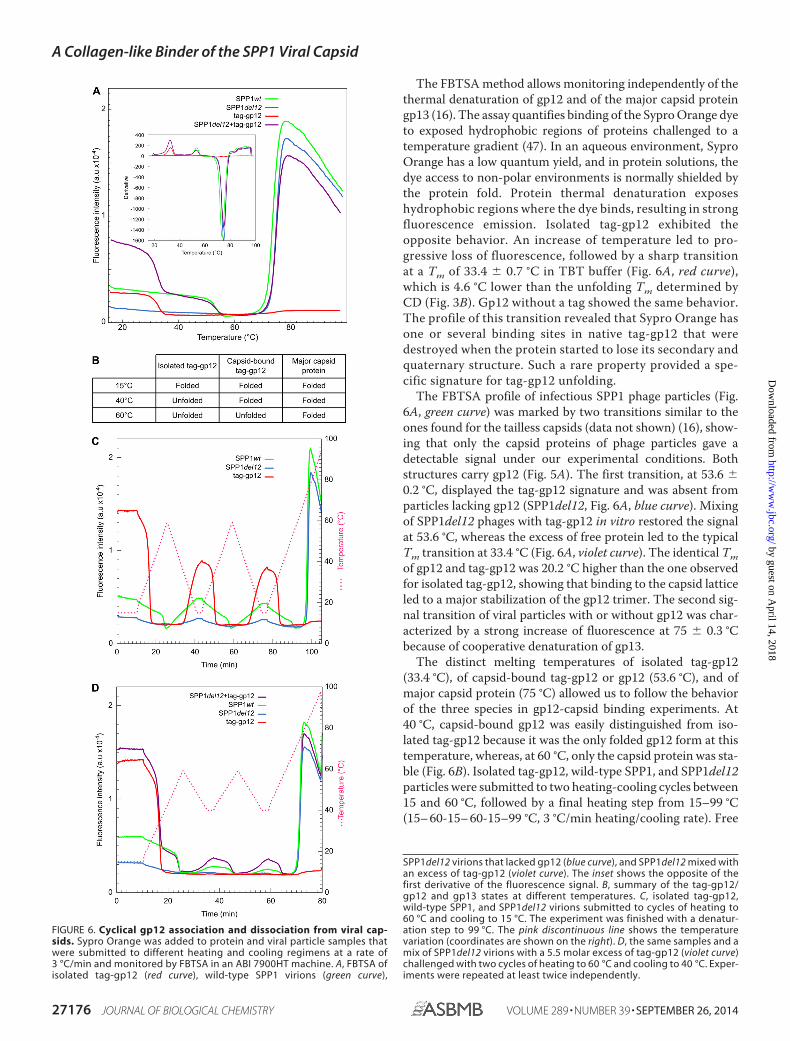
The FBTSA method allows monitoring independently of thethermal denaturation of gp12 and of the major capsid proteingp13 (16). The assay quantifies binding of the Sypro Orange dyeto exposed hydrophobic regions of proteins challenged to atemperature gradient (47). In an aqueous environment, SyproOrange has a low quantum yield, and in protein solutions, thedye access to non-polar environments is normally shielded bythe protein fold. Protein thermal denaturation exposeshydrophobic regions where the dye binds, resulting in strongfluorescence emission. Isolated tag-gp12 exhibited theopposite behavior. An increase of temperature led to pro-gressive loss of fluorescence, followed by a sharp transitionat a Tm of 33.4 � 0.7 °C in TBT buffer (Fig. 6A, red curve),which is 4.6 °C lower than the unfolding Tm determined byCD (Fig. 3B). Gp12 without a tag showed the same behavior.The profile of this transition revealed that Sypro Orange hasone or several binding sites in native tag-gp12 that weredestroyed when the protein started to lose its secondary andquaternary structure. Such a rare property provided a spe-cific signature for tag-gp12 unfolding.
The FBTSA profile of infectious SPP1 phage particles (Fig.6A, green curve) was marked by two transitions similar to theones found for the tailless capsids (data not shown) (16), show-ing that only the capsid proteins of phage particles gave adetectable signal under our experimental conditions. Bothstructures carry gp12 (Fig. 5A). The first transition, at 53.6 �0.2 °C, displayed the tag-gp12 signature and was absent fromparticles lacking gp12 (SPP1del12, Fig. 6A, blue curve). Mixingof SPP1del12 phages with tag-gp12 in vitro restored the signalat 53.6 °C, whereas the excess of free protein led to the typicalTm transition at 33.4 °C (Fig. 6A, violet curve). The identical Tmof gp12 and tag-gp12 was 20.2 °C higher than the one observedfor isolated tag-gp12, showing that binding to the capsid latticeled to a major stabilization of the gp12 trimer. The second sig-nal transition of viral particles with or without gp12 was char-acterized by a strong increase of fluorescence at 75 � 0.3 °Cbecause of cooperative denaturation of gp13.
The distinct melting temperatures of isolated tag-gp12(33.4 °C), of capsid-bound tag-gp12 or gp12 (53.6 °C), and ofmajor capsid protein (75 °C) allowed us to follow the behaviorof the three species in gp12-capsid binding experiments. At40 °C, capsid-bound gp12 was easily distinguished from iso-lated tag-gp12 because it was the only folded gp12 form at thistemperature, whereas, at 60 °C, only the capsid protein was sta-ble (Fig. 6B). Isolated tag-gp12, wild-type SPP1, and SPP1del12particles were submitted to two heating-cooling cycles between15 and 60 °C, followed by a final heating step from 15–99 °C(15– 60-15– 60-15–99 °C, 3 °C/min heating/cooling rate). Free
FIGURE 6. Cyclical gp12 association and dissociation from viral cap-sids. Sypro Orange was added to protein and viral particle samples thatwere submitted to different heating and cooling regimens at a rate of3 °C/min and monitored by FBTSA in an ABI 7900HT machine. A, FBTSA ofisolated tag-gp12 (red curve), wild-type SPP1 virions (green curve),
SPP1del12 virions that lacked gp12 (blue curve), and SPP1del12 mixed withan excess of tag-gp12 (violet curve). The inset shows the opposite of thefirst derivative of the fluorescence signal. B, summary of the tag-gp12/gp12 and gp13 states at different temperatures. C, isolated tag-gp12,wild-type SPP1, and SPP1del12 virions submitted to cycles of heating to60 °C and cooling to 15 °C. The experiment was finished with a denatur-ation step to 99 °C. The pink discontinuous line shows the temperaturevariation (coordinates are shown on the right). D, the same samples and amix of SPP1del12 virions with a 5.5 molar excess of tag-gp12 (violet curve)challenged with two cycles of heating to 60 °C and cooling to 40 °C. Exper-iments were repeated at least twice independently.
A Collagen-like Binder of the SPP1 Viral Capsid
27176 JOURNAL OF BIOLOGICAL CHEMISTRY VOLUME 289 • NUMBER 39 • SEPTEMBER 26, 2014
by guest on April 14, 2018
http://ww
w.jbc.org/
Dow
nloaded from

tag-gp12 exhibited a loss of signal upon heating, and its partialreacquisition when cooling to 15 °C showed that the fluoro-phore binding site(s) was/were not completely restored in thetag-gp12 population (Fig. 6C, red curve) in spite of the fact thatthe protein fully reacquired its quaternary structure CD sig-nature (Fig. 3). The temperatures of transition were remark-ably reproducible, revealing that tag-gp12 underwent disso-ciation/unfolding and folding/reassociation cycles. Gp12bound to SPP1 capsids exhibited a transition correspondingto a Tm of 53.6 °C (Fig. 6C, continuous green curve). Theprocess was reversible upon cooling and reheating, apartfrom a slight loss of fluorescence from one cycle to another.Therefore, gp12 dissociated/unfolded reversibly from wild-type capsids and maintained its binding activity to thecapsid.
To assess whether the capsid lattice influences gp12 refold-ing/reassociation, the cycling experiment was repeated withcooling steps to 40 °C (15– 60-40 – 60-40 –99 °C program, Fig.6D), a temperature at which free tag-gp12 remained unfoldedafter the first heating step (Fig. 3D, red curve). Gp12 bound tophage capsids kept its signature (Tm of 53.6 °C) in heating cyclesto 60 °C. Cooling to 40 °C led to recovery of some fluorescencesignals (Fig. 6D, green curve) but significantly less than when thetemperature was reduced to 15 °C (Fig. 6C). Therefore, at 40 °C,a subpopulation of gp12 rebound to phage capsids, yieldingfolded trimers that fixed Sypro Orange. Addition of a 5.5-foldmolar excess of exogenous tag-gp12 to wild-type capsids re-stored most of the gp12 signal associated with capsids aftereach 60 – 40 °C cycle (Fig. 6D, violet curve), showing thattag-gp12 had efficiently replaced gp12, which left its capsid
FIGURE 7. Capsid binding behavior of native and unfolded tag-gp12. Purified SPP1 tailless capsids lacking gp12 (capsid H�12) (blue characters and bluecurves on the left and blue rectangles above the gels on the right) and tag-gp12 (red) were preincubated separately, mixed, and treated with trypsin (except inA) according to the different combinations of incubation conditions used in the experiments in A–E (see “Results” for details), as outlined on the left of eachpanel. Samples treated with trypsin in B–E are identified by yellow rectangles above the gel lanes on the right. Capsids were then resolved by agarose gelelectrophoresis to assess their occupancy with tag-gp12. Wild-type SPP1 capsids with gp12 (H capsids) (green rectangles above the gels on the right) and H�12were used as controls. The schematics in the center of A show the electrophoretic mobility of capsid H (with gp12 represented in red) and H�12. The experimentwas repeated four times independently.
A Collagen-like Binder of the SPP1 Viral Capsid
SEPTEMBER 26, 2014 • VOLUME 289 • NUMBER 39 JOURNAL OF BIOLOGICAL CHEMISTRY 27177
by guest on April 14, 2018
http://ww
w.jbc.org/
Dow
nloaded from

sites upon denaturation. Restoration of the tag-gp12 signalat 40 °C occurred exclusively in presence of the capsid lat-tice, showing that this structure promoted tag-gp12 refold-ing and reassociation.
Native and Unfolded gp12 Binds to SPP1 Capsids in a DistinctWay—The finding that both native and unfolded tag-gp12bound to SPP1 phage capsids (Fig. 6, C and D) suggested twodistinct types of interaction, prompting their characterization.Tailless capsids without gp12 (H�12) and purified tag-gp12were preincubated separately at 16 or 45 °C, followed by mixingat different ratios for interaction at the two temperatures (Fig.7). Reactions were then incubated at 45 °C with trypsin, whichdegraded free gp12/tag-gp12 (Fig. 8A). The tag of capsid-boundtag-gp12 was prone to trypsin attack, but the gp12 moietyattached to the capsid remained intact (Fig. 8B), explaining thelower electrophoretic mobility of capsids loaded with tag-gp12not treated with trypsin when compared with those that weretrypsinated (Fig. 7, A and B). This step prevented subsequentinteractions of free tag-gp12 with capsids during downstreamsample manipulation at room temperature and separation bygel agarose electrophoresis. SPP1 capsids with gp12 (H capsids)or H�12 loaded with tag-gp12 had a slower electrophoreticmobility than capsids lacking gp12 (Fig. 7), most likely becausegp12/tag-gp12 reduces the capsid surface electronegativecharge. In contrast, gp12 does not have a major effect on capsiddiameter, which is almost identical in H and H�12 ( 610 Å(16)).
When H�12 capsids were mixed at 16 °C with increasingamounts of tag-gp12 native trimers, the capsid species shiftedfrom tag-gp12-free to capsids fully loaded with tag-gp12 (Fig. 7,A, B, and D). At a ratio (R 60 tag-gp12 trimers/capsid) of 0.5,most capsids lacked tag-gp12, but a minority was already satu-rated with tag-gp12, whereas, at R 1.5, almost all capsids weredecorated with tag-gp12. Species with intermediate electro-phoretic mobility were poorly detected, revealing that capsidspartially occupied with tag-gp12 were a minor population, evenat limiting amounts of tag-gp12 (e.g. R 0.5). We attribute thisbehavior to high cooperative binding of tag-gp12 trimers to its60 sites in the SPP1 capsid.
To characterize the interaction of unfolded tag-gp12 with thecapsid, the two species were preheated individually at 45 °C andmixed at the same temperature (Fig. 7C). A significant excess oftag-gp12 per capsid (R between 4 and 13) was needed to pro-mote a change of capsid electrophoretic mobility. Their dis-crete bands showed a migration pattern that progressed fromthe capsids lacking gp12 band behavior (R � 3) to the full tag-gp12-loaded capsid band (R 13) (Fig. 7C). Similar results wereobtained when the interaction reaction was prolonged over-night at 45 °C (not shown). Furthermore, preheating of capsidsat 16 °C or 45 °C showed that temperature did not affect theirbinding properties (Fig. 7). Stable interaction of unfolded tag-gp12 with H�12 capsids therefore required an excess of tag-gp12 that bound in an inefficient manner, leading to a popula-tion of capsids whose binding sites are only partially occupiedby tag-gp12 at molar ratios as high as R 11 (Fig. 7, C and E).The increase of occupancy with the rise of R correlated with anaugmentation of the tag-gp12 signal in FBTSA experiments,
consistent with the formation of tag-gp12 trimers in the capsidlattice (Fig. 9).
DISCUSSION
The 6.6-kDa gp12 polypeptide of bacterial virus SPP1 wasshown here to build an elongated trimer. Its properties indicate
FIGURE 8. Trypsin sensitivity of free and capsid-bound gp12. A, purifiedtag-gp12 and gp12 were incubated with trypsin either at 16 or at 45 °C. Bothproteins were completely digested by the protease at the tested tempera-tures, as assessed by Coomassie-stained SDS-PAGE (left panel) and Westernblot analysis (right panel) with polyclonal anti-SPP1 antibodies that recognizegp12 (Fig. 5B). B, trypsin digestion of the binding reaction between capsidsand tag-gp12 (labeled band 1 on the right of the figure) under the same con-ditions as in Fig. 7, B and C. Note that gp12 bound to H capsids is not sensitiveto trypsin, whereas the tag of tag-gp12 is partially (band 2) or fully (band 3)digested by trypsin. The Western blot analysis was developed with anti-SPP1antibodies that recognize gp12 but also, although giving a comparativelyweak signal, the major capsid protein gp13, whose band was used to controlthe normalized input of capsids in the binding reaction.
A Collagen-like Binder of the SPP1 Viral Capsid
27178 JOURNAL OF BIOLOGICAL CHEMISTRY VOLUME 289 • NUMBER 39 • SEPTEMBER 26, 2014
by guest on April 14, 2018
http://ww
w.jbc.org/
Dow
nloaded from

the presence of an intermolecular collagen-like triple helix thatcorrelates with presence of eight GXY repeats at the center ofthe gp12 sequence, revealing a modular organization in whichthe collagen-like elongated segment connects two short aminoand carboxyl terminus domains. Collagens are well studied pro-tein components of the extracellular matrix of animals. Theyare characterized by the presence of 4-hydroxyproline at posi-tion Y of the GXY triplet, which is considered a major determi-nant of collagen stability (42). However, stable triple helices arebuilt in synthetic model peptides by repeats longer than(GXY)9, showing that their association requires no amino acidposttranslational modifications (48). This property is consis-
tent with the presence of collagen-like segments in prokaryotesystems reported for streptococcal surface proteins (49, 50) andtail fibers of bacterial viruses (51), where several dozens of GXYtriplets assemble long, flexible filaments. The remarkable fea-ture of gp12 is that its short (GXY)8 stretch confers to the over-all polypeptide a collagen-like behavior with a characteristicloss of quaternary structure, corresponding to a sharp transi-tion at temperatures around 40 °C, like animal collagen (52, 53),that is rapidly and fully reversible upon cooling (Fig. 3). Theprocess is accompanied by physical separation of the polypep-tide chains that can reassociate into heterotrimers (Fig. 4). Theunusual binding of Sypro Orange dye to folded gp12 resulting influorescence emission reveals the presence of an accessiblehydrophobic binding environment in the trimer that is lost atthe beginning of denaturation, correlating with a fast drop offluorescence (Fig. 6). The gp12 native structure is, therefore,mainly stabilized by its intermolecular collagen-like triple helixrather than by a buried hydrophobic core that would becomeexposed for high-affinity binding of Sypro Orange upon unfold-ing, in contrast to the usual behavior of proteins (54).
The interaction of gp12 with SPP1 capsids does not changeits capacity to bind Sypro Orange (Fig. 6A). However, itincreases the protein gp12 thermal stability by 20.2 °C, to53.6 °C. Such stability is not limited anymore by the collagenfold intrinsic stability, being strongly enhanced by gp12 bindingto the capsid lattice. This stabilization mechanism ensures theperennial association of gp12 to viral particles that are liberatedto the environment when infected cells lyse. Native trimersbind cooperatively to their 60 sites in the capsid, as bestappraised when gp12 is provided in limiting concentrations tointeract with H�12 capsids. A mixed population of capsidswhose majority is either fully loaded with gp12 trimers ordevoided of this auxiliary protein is observed under such con-ditions (Figs. 7, A, B, and D, and 10A). We hypothesize thatinitial binding of one trimer to a capsid hexamer creates a tec-tonic effect that spreads across the overall icosahedral shell,promoting a conformational change of other hexamers thatstrongly favors interaction with gp12 trimers. Such a matura-tion event uncovers a novel dynamic role of the expanded cap-sid surface that has previously been viewed as a rather passivelattice of independent binding sites for auxiliary viral polypep-tides. The rearrangement resulting from the cross-talk betweenthe 60 gp12 attachment sites is subtle, leading to no detectable
FIGURE 9. FBTSA of H�12 incubated with increasing amounts of tag-gp12. Capsids and tag-gp12 were preincubated separately at 15 (green) or45 °C (red) and then mixed at the same temperature according to the exper-imental setup shown in the left panels of Fig. 7, A and C (not trypsinated),respectively. After coincubation, the samples were transferred to a QuantStu-dio 12Kflex machine for thermal denaturation at a heating rate of 3 °C/min inthe presence of Sypro Orange. The amplitude of the gp12 signal with itscharacteristic transition at 53.6 °C (cf. Fig. 6A) was plotted against the R ratio oftag-gp12 relative to the input of H�12 capsids. The experimental points areaverages of triplicates in two independent experiments.
FIGURE 10. Models of native gp12 trimers (A) and unfolded gp12 polypeptides (B) binding to capsids and their dissociation from the capsid lattice ina temperature-dependent fashion.
A Collagen-like Binder of the SPP1 Viral Capsid
SEPTEMBER 26, 2014 • VOLUME 289 • NUMBER 39 JOURNAL OF BIOLOGICAL CHEMISTRY 27179
by guest on April 14, 2018
http://ww
w.jbc.org/
Dow
nloaded from

difference when the structure of capsids before and after gp12binding is compared at nanometer resolution (16).
Denatured gp12 also binds to capsid lattices (Fig. 7, C and E),leading to assembly of folded trimers when present in molarexcess, as assessed in Sypro Orange binding experiments (Fig.9). Therefore, the SPP1 capsid provides a platform for attach-ment of unfolded gp12 polypeptide chains. When three chainsmeet at a gp13 hexamer interaction site, their physical proxim-ity likely provides a window of opportunity to twist together totrimerize at a temperature (45 °C) at which free gp12 chainsremain fully unstructured (Fig. 10B). If unfolded gp12 is pro-vided at less than a 13-fold excess relative to the number of itscapsid binding sites, the reaction yields a relatively homogene-ous population of capsids but those are only partially filled withgp12 (Fig. 7, C and E). Such behavior, resulting from the com-plexity of the interaction, contrasts with the very efficient coop-erative binding of folded trimers.
CONCLUSIONS
The precise architecture of viral particles achieved by tightlyregulated assembly of a few different polypeptides is an excel-lent system to understand how the polypeptides fold and howtheir physicochemical properties are exploited to build mega-dalton biomolecular assemblies of precise architecture withexquisite efficiency. The small capsid auxiliary protein gp12 ofbacterial virus SPP1 exhibits novel and noteworthy properties.It uses a collagen-like fold to assemble an elongated trimerwhose thermal stability properties render it a temperaturedependent binder to the capsid multivalent icosahedral plat-form. Cooperative binding ensures very efficient full occupancyof gp12 sites in the capsid (Figs. 7, A, B, and D, and 10A), pro-viding experimental evidence that an initial interaction of theviral auxiliary protein exerts long-range effects in the capsidlattice, favoring attachment to its other sites in the capsid.These properties of gp12, combined with its capacity toundergo fast reversible cycles of dissociation-unfolding andrefolding-reassociation to capsids, offer a versatile system toengineer the SPP1 viral particle.
Acknowledgments—We thank Anja Droge for generously communi-cating the initial identification of GXY repeats in the gp12 sequence.We also thank Manuela Argentini and David Cornu for mass spec-trometry analyses (mass spectrometry platform of IMAGIF); Chris-tophe Velours and Karine Madiona for CD and analytical ultracen-trifugation analyses (biophysics platform of IMAGIF); and IsabelleAuzat, Sandrine Brasiles, Charlene Cornilleau, Stephane Roche, andStephane Bressanelli for advice and experimental training (Unité deVirologie Moléculaire et Structurale).
REFERENCES1. Caspar, D. L., and Klug, A. (1962) Physical principles in the construction of
regular viruses. Cold Spring Harb. Symp. Quant. Biol. 27, 1–242. Baker, T. S., Olson, N. H., and Fuller, S. D. (1999) Adding the third dimen-
sion to virus life cycles: three-dimensional reconstruction of icosahedralviruses from cryo-electron micrographs. Microbiol. Mol. Biol. Rev. 63,862–922
3. Prasad, B. V., and Schmid, M. F. (2012) Principles of virus structural orga-nization. Adv. Exp. Med. Biol. 726, 17– 47
4. Casjens, S., and King, J. (1975) Virus assembly. Annu. Rev. Biochem. 44,
555– 6115. Prevelige, P. E., and Fane, B. A. (2012) Building the machines: scaffolding
protein functions during bacteriophage morphogenesis. Adv. Exp. Med.Biol. 726, 325–350
6. Ren, Z. J., Lewis, G. K., Wingfield, P. T., Locke, E. G., Steven, A. C., andBlack, L. W. (1996) Phage display of intact domains at high copy number:a system based on SOC, the small outer capsid protein of bacteriophageT4. Protein Sci. 5, 1833–1843
7. Yang, F., Forrer, P., Dauter, Z., Conway, J. F., Cheng, N., Cerritelli, M. E.,Steven, A. C., Pluckthun, A., and Wlodawer, A. (2000) Novel fold andcapsid-binding properties of the -phage display platform protein gpD.Nat. Struct. Biol. 7, 230 –237
8. Roos, W. H., Radtke, K., Kniesmeijer, E., Geertsema, H., Sodeik, B., andWuite, G. J. (2009) Scaffold expulsion and genome packaging trigger sta-bilization of herpes simplex virus capsids. Proc. Natl. Acad. Sci. U.S.A. 106,9673–9678
9. Parent, K. N., Khayat, R., Tu, L. H., Suhanovsky, M. M., Cortines, J. R.,Teschke, C. M., Johnson, J. E., and Baker T. S. (2010) P22 coat proteinstructures reveal a novel mechanism for capsid maturation: stability with-out auxiliary proteins or chemical crosslinks. Structure 18, 390 – 401
10. Tang, L., Gilcrease, E. B., Casjens, S. R., and Johnson, J. E. (2006) Highlydiscriminatory binding of capsid-cementing proteins in bacteriophage L.Structure 14, 837– 845
11. Effantin, G., Boulanger, P., Neumann, E., Letellier, L., and Conway, J. F.(2006) Bacteriophage T5 structure reveals similarities with HK97 and T4suggesting evolutionary relationships. J. Mol. Biol. 361, 993–1002
12. Lander, G. C., Evilevitch, A., Jeembaeva, M., Potter, C. S., Carragher, B.,and Johnson, J. E. (2008) Bacteriophage stabilization by auxiliary proteingpD: timing, location, and mechanism of attachment determined by cryo-EM. Structure 16, 1399 –1406
13. Yang, Q., Maluf, N. K., and Catalano, C. E. (2008) Packaging of a unit-length viral genome: the role of nucleotides and the gpD decoration pro-tein in stable nucleocapsid assembly in bacteriophage . J. Mol. Biol. 383,1037–1048
14. Li, Q., Shivachandra, S. B., Zhang, Z., and Rao, V. B. (2007) Assembly of thesmall outer capsid protein, Soc, on bacteriophage T4: a novel system forhigh density display of multiple large anthrax toxins and foreign proteinson phage capsid. J. Mol. Biol. 370, 1006 –1019
15. Qin L., Fokine, A., O’Donnell, E., Rao, V. B., and Rossmann M. G. (2010)Structure of the small outer capsid protein, Soc: a clamp for stabilizingcapsids of T4-like phages. J. Mol. Biol. 395, 728 –741
16. White, H. E., Sherman, M. B., Brasiles, S., Jacquet, E., Seavers, P., Tavares,P., Orlova, E. V. (2012) Capsid structure and its stability at the late stagesof bacteriophage SPP1 assembly. J. Virol. 86, 6768 – 6777
17. Lucon, J., Qazi, S., Uchida, M., Bedwell, G. J., LaFrance, B., Prevelige, P. E.Jr., and Douglas, T. (2012) Use of the interior cavity of the P22 capsid forsite-specific initiation of atom-transfer radical polymerization with high-density cargo loading. Nat. Chem. 4, 781–788
18. O’Neil, A., Prevelige, P. E., Basu, G., and Douglas T. (2012) Coconfinementof fluorescent proteins: spatially enforced communication of GFP andmCherry encapsulated within the P22 capsid. Biomacromolecules 13,3902–3907
19. Clark, J. R., and March, J. B. (2006) Bacteriophages and biotechnology:vaccines, gene therapy and antibacterials. Trends Biotechnol. 24, 212–218
20. Fischlechner, M., and Donath, E. (2007) Viruses as building blocks formaterials and devices. Angew. Chem. Int. Ed. Engl. 46, 3184 –3193
21. Tao, P., Mahalingam, M., Marasa, B. S., Zhang, Z., Chopra, A. K., and Rao,V. B. (2013) In vitro and in vivo delivery of genes and proteins using thebacteriophage T4 DNA packaging machine. Proc. Natl. Acad. Sci. U.S.A.110, 5846 –5851
22. Steinmetz, N. F., Lin, T., Lomonossoff, G. P., and Johnson, J. E. (2009)Structure-based engineering of an icosahedral virus for nanomedicine andnanotechnology. Curr. Top. Microbiol. Immunol. 327, 23–58
23. Strable, E., and Finn, M. G. (2009) Chemical modification of viruses andvirus-like particles. Curr. Top. Microbiol. Immunol. 327, 1–21
24. Droge, A., Santos, M. A., Stiege, A. C., Alonso, J. C., Lurz, R., Trautner,T. A., and Tavares, P. (2000) Shape and DNA packaging activity of bacte-riophage SPP1 procapsid: protein components and interactions during
A Collagen-like Binder of the SPP1 Viral Capsid
27180 JOURNAL OF BIOLOGICAL CHEMISTRY VOLUME 289 • NUMBER 39 • SEPTEMBER 26, 2014
by guest on April 14, 2018
http://ww
w.jbc.org/
Dow
nloaded from

assembly. J. Mol. Biol. 296, 117–13225. Oliveira, L., Tavares, P., and Alonso, J. C. (2013) Headful DNA packaging:
bacteriophage SPP1 as a model system. Virus Res. 173, 247–25926. Isidro, A., Henriques, A. O., and Tavares, P. (2004) The portal protein
plays essential roles at different steps of the SPP1 DNA packaging process.Virology 322, 253–263
27. Plisson, C., White, H. E., Auzat, I., Zafarani, A., Sao-Jose, C., Lhuillier, S.,Tavares, P., and Orlova, E. V. (2007) Structure of bacteriophage SPP1 tailreveals trigger for DNA ejection. EMBO J. 26, 3720 –3728
28. Auzat, I., Droge, A., Weise, F., Lurz, R., and Tavares, P. (2008) Origin andfunction of the two major tail proteins of bacteriophage SPP1. Mol. Mi-crobiol. 70, 557–569
29. Lurz, R., Orlova, E. V., Gunther, D., Dube, P., Droge, A., Weise, F., vanHeel, M., and Tavares, P. (2001) Structural organisation of the head-to-tailinterface of a bacterial virus. J. Mol. Biol. 310, 1027–1037
30. Alonso, J. C., Luder, G., Stiege, A. C., Chai, S., Weise, F., and Trautner,T. A. (1997) The complete nucleotide sequence and functional organiza-tion of Bacillus subtilis bacteriophage SPP1. Gene. 204, 201–212
31. Haima, P., Bron, S., and Venema, G. (1987) The effect of restriction onshotgun cloning and plasmid stability in Bacillus subtilis Marburg. Mol.Gen. Genet. 209, 335–342
32. Droge, A., and Tavares, P. (2000) In vitro packaging of DNA of the Bacillussubtilis bacteriophage SPP1. J. Mol. Biol. 296, 103–115
33. Becker, B., de la Fuente, N., Gassel, M., Gunther, D., Tavares, P., Lurz, R.,Trautner, T. A., and Alonso, J. C. (1997) Head morphogenesis genes of theBacillus subtilis bacteriophage SPP1. J. Mol. Biol. 268, 822– 839
34. Isidro, A., Santos, M. A., Henriques, A. O., and Tavares, P. (2004) Thehigh-resolution functional map of bacteriophage SPP1 portal protein.Mol. Microbiol. 51, 949 –962
35. Poh, S. L., el Khadali, F., Berrier, C., Lurz, R., Melki, R., and Tavares, P.(2008) Oligomerization of the SPP1 scaffolding protein. J. Mol. Biol. 378,551–564
36. Philo, J. S. (1997) An improved function for fitting sedimentation velocitydata for low-molecular-weight solutes. Biophys. J. 72, 435– 444
37. Jakutyte, L., Baptista, C., Sao-Jose, C., Daugelavicius, R., Carballido-Lopez,R., and Tavares, P. (2011) Bacteriophage infection in rod-shaped gram-positive bacteria: evidence for a preferential polar route for phage SPP1entry in Bacillus subtilis. J. Bacteriol. 193, 4893– 4903
38. Cole, C., Barber, J. D., and Barton, G. J. (2008) The Jpred3 secondarystructure prediction server. Nucleic Acids Res. 36, W197–W201
39. Soding, J., Biegert, A., and Lupas, A. N. (2005) The HHpred interactiveserver for protein homology detection and structure prediction. Nucleic
Acids Res. 33, W244 –W24840. Droge, A. (1998) Capsidmorphogenese des Bakteriophagen SPP1. Doctoral
thesis, Technische Universitat Berlin, Berlin, Germany41. Bella, J., Eaton, M., Brodsky, B., and Berman, H. M. (1994) Crystal and
molecular structure of a collagen-like peptide at 1.9 A resolution. Science266, 75– 81
42. Bella, J., Brodsky, B., and Berman, H. M. (1995) Hydration structure of acollagen peptide. Structure 3, 893–906
43. Cabre, F., Canela, E. I., and Canela, M. A. (1989) Accuracy and precision inthe determination of Stokes radii and molecular masses of proteins by gelfiltration chromatography. J. Chromatogr. 472, 347–356
44. Seifter, S., and Gallop, P. M. (1962) Collagenase from Clostridium histo-lyticum: collagen � H2O3 peptides gelatin � H2O3 peptides. MethodsEnzymol. 5, 659 – 665
45. Orlova, E. V., Gowen, B., Droge, A., Stiege, A., Weise, F., Lurz, R., van Heel,M., and Tavares, P. (2003) Structure of a viral DNA gatekeeper at 10 Åresolution by cryo-electron microscopy. EMBO J. 22, 1255–1262
46. Wallace, B. A., and Janes, R. W. (2001) Synchrotron radiation circulardichroism spectroscopy of proteins: secondary structure, fold recognitionand structural genomics. Curr. Opin. Chem. Biol. 5, 567–571
47. Ericsson, U. B., Hallberg, B. M., Detitta, G. T., Dekker, N., and Nordlund,P. (2006) Thermofluor-based high-throughput stability optimization ofproteins for structural studies. Anal. Biochem. 357, 289 –298
48. Persikov, A. V., Ramshaw, J. A., and Brodsky, B. (2005) Prediction of col-lagen stability from amino acid sequence. J. Biol. Chem. 280, 19343–19349
49. Xu, Y., Keene, D. R., Bujnicki, J. M., Hook, M., and Lukomski, S. (2002)Streptococcal Scl1 and Scl2 proteins form collagen-like triple helices.J. Biol. Chem. 277, 27312–27318
50. Mohs, A., Silva, T., Yoshida, T., Amin, R., Lukomski, S., Inouye, M., andBrodsky, B. (2007) Mechanism of stabilization of a bacterial collagen triplehelix in the absence of hydroxyproline. J. Biol. Chem. 282, 29757–29765
51. Ghosh, N., McKillop, T. J., Jowitt, T. A., Howard, M., Davies, H., Holmes,D. F., Roberts, I. S., and Bella J. (Jun 6, 2012) Collagen-like proteins inpathogenic E. coli strains. PLoS ONE 10.1371/journal.pone.0037872
52. Leikina, E., Mertts, M. V., Kuznetsova, N., and Leikin, S. (2002) Type Icollagen is thermally unstable at body temperature. Proc. Natl. Acad. Sci.U.S.A. 99, 1314 –1318
53. Cao, H., and Xu, S.-H. (2008) Purification and characterization of type IIcollagen from chick sternal cartilage. Food Chem. 108, 439 – 445
54. Kranz, J. K., and Schalk-Hihi, C. (2011) Protein thermal shifts to identifylow molecular weight fragments. Methods Enzymol. 493, 277–298
A Collagen-like Binder of the SPP1 Viral Capsid
SEPTEMBER 26, 2014 • VOLUME 289 • NUMBER 39 JOURNAL OF BIOLOGICAL CHEMISTRY 27181
by guest on April 14, 2018
http://ww
w.jbc.org/
Dow
nloaded from

Mohamed Zairi, Asita C. Stiege, Naima Nhiri, Eric Jacquet and Paulo TavaresSPP1 Viral Capsids
The Collagen-like Protein gp12 Is a Temperature-dependent Reversible Binder of
doi: 10.1074/jbc.M114.590877 originally published online July 29, 20142014, 289:27169-27181.J. Biol. Chem.
10.1074/jbc.M114.590877Access the most updated version of this article at doi:
Alerts:
When a correction for this article is posted•
When this article is cited•
to choose from all of JBC's e-mail alertsClick here
http://www.jbc.org/content/289/39/27169.full.html#ref-list-1
This article cites 53 references, 13 of which can be accessed free at
by guest on April 14, 2018
http://ww
w.jbc.org/
Dow
nloaded from