the circulatory and cardiovascular systems vocabulary important info headings
DESCRIPTION
The Circulatory and Cardiovascular Systems Vocabulary Important info Headings. The Heart. Surface Projection of the Heart. Superior right point at the superior border of the 3 rd right costal cartilage - PowerPoint PPT PresentationTRANSCRIPT

The Circulatory The Circulatory
and and
Cardiovascular SystemsCardiovascular Systems
VocabularyVocabulary
Important infoImportant info
HeadingsHeadings


Surface Projection of the Surface Projection of the HeartHeart
• Superior right point at the superior border of Superior right point at the superior border of the 3the 3rdrd right costal cartilage right costal cartilage
• Superior left point at the inferior border of the Superior left point at the inferior border of the 22ndnd left costal cartilage 3cm to the left of left costal cartilage 3cm to the left of midlinemidline
• Inferior left point at the 5Inferior left point at the 5thth intercostal space, 9 intercostal space, 9 cm from the midlinecm from the midline
• Inferior right point at superior border of the 6Inferior right point at superior border of the 6thth right costal cartilage, 3 cm from the midlineright costal cartilage, 3 cm from the midline

The Heart: Internal AnatomyThe Heart: Internal Anatomy• Four chambersFour chambers
– AtriaAtria• ReceivingReceiving chambers chambers
– Right atrium Right atrium – Left atriumLeft atrium
– VentriclesVentricles• DischargingDischarging chambers chambers
– Right ventricleRight ventricle– Left ventricleLeft ventricle
• Heart valvesHeart valves– Allow blood to flow in Allow blood to flow in one direction ONLYone direction ONLY
• Four valvesFour valves– Atrioventricular ValvesAtrioventricular Valves – between atria and ventricles – between atria and ventricles
• Bicuspid valveBicuspid valve ( (leftleft))• Tricuspid valveTricuspid valve ( (rightright) )
– Semilunar valvesSemilunar valves between ventricle and artery between ventricle and artery• Pulmonary semilunarPulmonary semilunar valve valve• Aortic semilunarAortic semilunar valve valve

Layers of Heart WallLayers of Heart Wall
• EpicardiumEpicardium– visceral layer of visceral layer of
serous serous pericardiumpericardium
• Myocardium Myocardium – cardiac musclecardiac muscle
layer is the bulk layer is the bulk of the heartof the heart
• EndocardiumEndocardium– chamber chamber lininglining & &
valvesvalves

Cardiac MyofibrilCardiac Myofibril

•Atria contract, blood fills ventricles through A-V valves
•Ventricles contract, blood pumped into aorta and pulmonary trunk through SL valves
Valve FunctionValve Function

Heart MurmurHeart Murmur• Heart murmurs are most Heart murmurs are most
often caused by often caused by defective defective heart valvesheart valves. .
• A valve may be unable to A valve may be unable to close completely. close completely.
• This leads to This leads to regurgitationregurgitation, , which is blood leaking which is blood leaking backward through the backward through the valve when it should be valve when it should be closedclosed
Normal heartbeatNormal heartbeat murmurmurmur
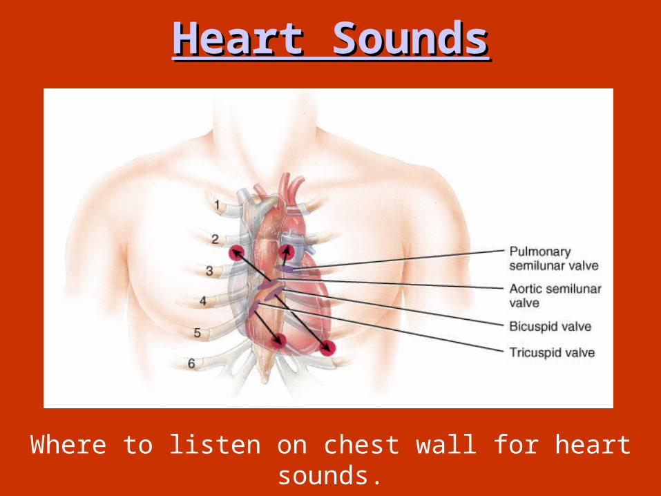
Heart SoundsHeart Sounds
Where to listen on chest wall for heart sounds.


What Causes the What Causes the Heartbeat?Heartbeat?

• Autorhythmic CellsAutorhythmic Cells– Cells fire spontaneously, act as Cells fire spontaneously, act as pacemakerpacemaker and form conduction and form conduction
system for the heartsystem for the heart• SA nodeSA node
– cluster of cells in wall of Rt. Atriacluster of cells in wall of Rt. Atria– begins heart activitybegins heart activity that spreads to both atria that spreads to both atria– excitation spreads to AV nodeexcitation spreads to AV node
• AV nodeAV node– in atrial septum, in atrial septum,
transmits signal transmits signal to to Bundle of HisBundle of His
• AV bundle of HisAV bundle of His – the connection the connection
between atria & between atria & ventriclesventricles
– divides into divides into bundle branches bundle branches & & purkinje fiberspurkinje fibers, , large diameter large diameter fibers that fibers that conduct signals conduct signals quicklyquickly
Conduction System of HeartConduction System of Heart

Rhythm of Conduction Rhythm of Conduction SystemSystem• SA node fires spontaneously SA node fires spontaneously
90-100 times per minute90-100 times per minute• AV node fires at AV node fires at 40-50 times per minute40-50 times per minute• If both nodes are If both nodes are
suppressed fibers suppressed fibers in ventricles by in ventricles by themselves fire themselves fire only only 20-40 times per 20-40 times per minuteminute
• Artificial pacemaker Artificial pacemaker needed if pace is needed if pace is too slowtoo slow
• Extra beats forming at Extra beats forming at other sites are called other sites are called Ectopic PacemakersEctopic Pacemakers– caffeine & nicotine caffeine & nicotine
increase activityincrease activity

Timing of Atrial & Ventricular Timing of Atrial & Ventricular ExcitationExcitation
• SA node setting pace since is the fastestSA node setting pace since is the fastest• In In 50 msec excitation50 msec excitation spreads through both spreads through both
atria and down to AV nodeatria and down to AV node• 100 msec delay at 100 msec delay at
AV nodeAV node due to due to smaller diameter smaller diameter fibers- allows atria fibers- allows atria to fully contract to fully contract filling ventricles filling ventricles before ventricles before ventricles contractcontract
• In 50 msec In 50 msec excitation spreads excitation spreads through both ventricles simultaneouslythrough both ventricles simultaneously

Depolarization & Depolarization & RepolarizationRepolarization
• DepolarizationDepolarization– Cardiac cell Cardiac cell restingresting membrane potential is membrane potential is -90mv-90mv– excitation spreads through gap junctionsexcitation spreads through gap junctions– fast Na+ channels open for rapid depolarizationfast Na+ channels open for rapid depolarization
• Plateau phasePlateau phase – 250 msec250 msec (only 1msec in (only 1msec in
neuron) neuron) – slow Caslow Ca+2+2 channels open, let Ca channels open, let Ca +2+2
enter from outside cell and from enter from outside cell and from storage in sarcoplasmic reticulum, storage in sarcoplasmic reticulum, while Kwhile K++ channels close channels close
– Ca Ca +2+2 binds to troponin to allow binds to troponin to allow for actin-myosin cross-bridge for actin-myosin cross-bridge formation & tension developmentformation & tension development
• Repolarization Repolarization – CaCa+2+2 channels close and K channels close and K++
channels open & channels open & -90mv is -90mv is restored as potassium leaves the cellrestored as potassium leaves the cell
• Refractory periodRefractory period – very long so heart can fillvery long so heart can fill

Physiology of Physiology of ContractionContraction
• Depolarization, plateau, repolarizationDepolarization, plateau, repolarization

Electrocardiogram---ECG or Electrocardiogram---ECG or EKGEKG
• EKGEKG– Action potentials of all active cells can be detected and Action potentials of all active cells can be detected and
recorded recorded
• P waveP wave– atrial depolarizationatrial depolarization
• P to Q intervalP to Q interval– conduction time from conduction time from
atrial to ventricular atrial to ventricular excitationexcitation
• QRS complexQRS complex – ventricular ventricular
depolarizationdepolarization
• T waveT wave– ventricular repolarizationventricular repolarization

One Cardiac CycleOne Cardiac Cycle• At 75 beats/min, one cycle requires 0.8 sec.At 75 beats/min, one cycle requires 0.8 sec.
– SystoleSystole ( (contractioncontraction) and ) and DiastoleDiastole ( (relaxationrelaxation) of both atria, plus the ) of both atria, plus the systole and diastole of both ventriclessystole and diastole of both ventricles
• End Diastolic Volume (EDV)End Diastolic Volume (EDV)– volumevolume in ventricle at end of diastole, about in ventricle at end of diastole, about 130 ml130 ml
• End Systolic End Systolic Volume (ESV)Volume (ESV)– volumevolume in in
ventricle ventricle at end of at end of systole, systole, about about 60 ml60 ml
• Stroke Volume Stroke Volume (SV)(SV)– the the volume volume
ejectedejected per per beat from beat from each ventricle, each ventricle, about about 70 ml70 ml
– SV = EDV - ESVSV = EDV - ESV
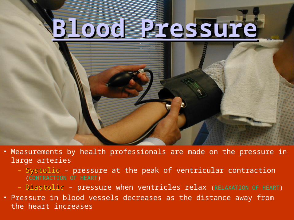
• Measurements by health professionals are made on the pressure in large arteries
– SystolicSystolic – pressure at the peak of ventricular contraction (CONTRACTION OF HEART)
– DiastolicDiastolic – pressure when ventricles relax (RELAXATION OF HEART)
• Pressure in blood vessels decreases as the distance away from the heart increases
Blood PressureBlood Pressure

PulsePulse
•PulsePulse – Pressure wave Pressure wave
of bloodof blood
•““Pressure Pressure Points”Points”– Area where Area where
pulse is easily pulse is easily palpatedpalpated
– Simple Simple monitoringmonitoring
Figure 11.16

Blood Pressure
Normal Systolic Normal Diastolic
140-120 mm Hg 80-75 mm Hg
“120/80”

Variations in Blood Variations in Blood PressurePressure
• Human normal range is variableHuman normal range is variable– Normal BPNormal BP
• 140–110 mm hg systolic140–110 mm hg systolic• 80–75 mm hg diastolic80–75 mm hg diastolic
– HypoHypotensiontension• Low systolic Low systolic
(below 110 mm hg)(below 110 mm hg)• Often associated Often associated
with illnesswith illness– HyperHypertensiontension
• High systolic High systolic (above 140 mm hg)(above 140 mm hg)
• Can be dangerous if it is Can be dangerous if it is chronicchronic

Measuring Arterial Blood Measuring Arterial Blood PressurePressure
Figure 11.18

Nervous System: Big Nervous System: Big BrotherBrother
• Nervous Nervous system system controls controls heartbeatheartbeat
• Sympathetic NSSympathetic NS = = fight or fight or flightflight
• ParasympathetiParasympatheti
c NSc NS = = relaxationrelaxation

What is your Resting What is your Resting Heart Rate?Heart Rate?
Normal = 60-75 Beats/Minute

THE THE BLOODBLOOD

The 3 Main Functions of The 3 Main Functions of BloodBlood::
1.1. TransportationTransportation
2.2. ProtectionProtection
3.3. RegulationRegulation• Blood is a Blood is a
connective tissueconnective tissue in in liquid formliquid form
• Greatest benefit Greatest benefit from homeostasis:from homeostasis:– Continuous flow of Continuous flow of
blood thru blood thru 60,000 60,000 miles of blood vesselsmiles of blood vessels

TRANSPORTATIONTRANSPORTATION::
• Blood moves thru Blood moves thru body where cells body where cells receive:receive:– Nutrients from Nutrients from
digestive organsdigestive organs– Oxygen from lungsOxygen from lungs– Hormones secreted Hormones secreted
from endocrine glandfrom endocrine gland
• Cells give blood waste Cells give blood waste •(CO(CO22, urea & uric , urea & uric
acid) & their acid) & their secretionssecretions

ProtectionProtection::• From harmful From harmful
microorganism & microorganism & their toxins their toxins – Through Through PhagocyticPhagocytic
white blood cells white blood cells – Specialized proteins Specialized proteins
called called AntibodiesAntibodies• Against fluid loss Against fluid loss
after an injury by after an injury by clottingclotting

RegulationRegulation:: • Regulates acid-base Regulates acid-base
balance of the body fluids balance of the body fluids – By way of By way of buffersbuffers
• Neutralize potential Neutralize potential harmful effects of: harmful effects of:
– too much co2too much co2– actic acidactic acid– other compoundsother compounds
• Body temp. by cooling or Body temp. by cooling or heating parts of bodyheating parts of body
• Controlled by Controlled by HypothalamusHypothalamus
• Controls volume of blood Controls volume of blood flow to diff. areas of bodyflow to diff. areas of body
Figure This figure highlights some of the major acute (short-term) effects on the body during
exercise.

Properties of BloodProperties of Blood::
•ColorColor•VolumeVolume•pHpH

COLORCOLOR • RED COLOR = RED COLOR =
– HEMOGLOBINHEMOGLOBIN (PIGMENT (PIGMENT PROTEIN)PROTEIN)
• Arterial blood the OArterial blood the O22 molecules are chemically molecules are chemically bound to hemoglobin bound to hemoglobin – Crimson-red colorCrimson-red color
• Venous blood OVenous blood O22 mol. are not mol. are not as prevalent & blood=as prevalent & blood=– Dark red color w/a slightly bluish Dark red color w/a slightly bluish
tinttint
• SEEN THROUGH SKIN VEINS SEEN THROUGH SKIN VEINS LOOK GREENISH- BLUE but it LOOK GREENISH- BLUE but it is is NOTNOT GREEN OR BLUE GREEN OR BLUE

VOLUMEVOLUME • 8% OF BODY WEIGHT8% OF BODY WEIGHT
– Most in vessels--rest Most in vessels--rest in heartin heart
• Does not vary much Does not vary much from day to day or from day to day or year to yearyear to year
• Avg. Avg. MaleMale = = – 5-6 liters5-6 liters of blood of blood
• Avg. Avg. FemaleFemale = = – 4-5 liters4-5 liters of blood of blood
• Difference due to avg Difference due to avg body weight not sexbody weight not sex
Apx. 8 pints

• Blood is Blood is thicker, thicker, denser, & more denser, & more adhesive than Hadhesive than H22OO
– Due to formed Due to formed elements (red blood elements (red blood cells)cells)
• Causes blood to flow Causes blood to flow 5x slower5x slower than H than H22OO
• Resistance to flow = Resistance to flow = viscosityviscosity
• Blood is a viscous Blood is a viscous substance b/c it resists substance b/c it resists flow more than waterflow more than water
Figure The shear rate dependence of
normal human blood viscoelasticity at 2 Hz and 22 °C.

• Slightly alkaline (aka: Slightly alkaline (aka: basicbasic) ) • pH = 7.35-7.45pH = 7.35-7.45• Range stays small despite change in:Range stays small despite change in:
– DietDiet– Cell secretionsCell secretions– Metabolic rateMetabolic rate
by buffering systems by buffering systems that remove h+ ionsthat remove h+ ions
• If buffers fail:If buffers fail:– BLOOD BLOOD TOO TOO ACIDICACIDIC (pH (pH below 6.0below 6.0))
•Body cells stop functioning Body cells stop functioning •No homeostasis = No homeostasis = AAcidosiscidosis
• Too little acid in blood = Too little acid in blood = AlkAlkalosis alosis (a lot (a lot less common)less common)

White Blood Cells-WBCWhite Blood Cells-WBC
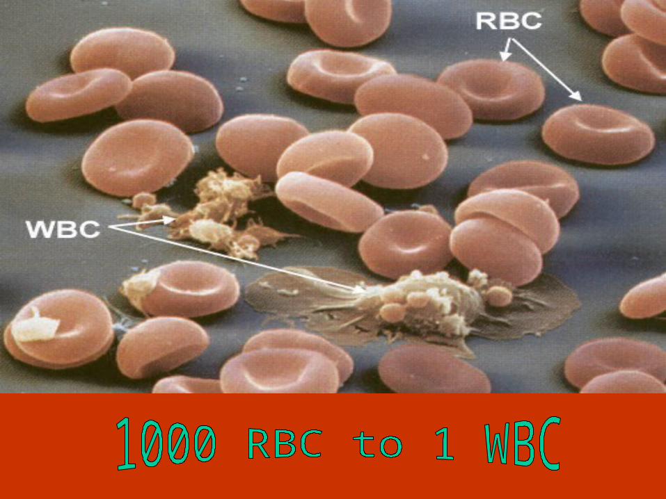

LeukocytesLeukocytes
• Less than 1% of Less than 1% of total blood total blood volumevolume
• 5000 TO 10,000 5000 TO 10,000 in cubic mmin cubic mm
• Any change in Any change in number…number…– High or low High or low
indicates a indicates a diseasedisease

TypesTypes::• All contain a nucleus All contain a nucleus
– (unlike the RBC’s) (unlike the RBC’s)
• Can wander outside the Can wander outside the Circ. SystemCirc. System
• Wbc cells differ in:Wbc cells differ in:– Nature of cytoplasmNature of cytoplasm– SizeSize– Shape of nucleusShape of nucleus
• Response to different Response to different staining techniquesstaining techniques
• Divided into 2 groups by Divided into 2 groups by cytoplasm differences:cytoplasm differences:– GranulocytesGranulocytes– AgranulocytesAgranulocytes

GranulocytesGranulocytes
• Cytoplasm Cytoplasm contains highly contains highly visible pebble-like visible pebble-like objects, known as objects, known as granulesgranules
• Twice the size of Twice the size of RBC’sRBC’s
• They contain a They contain a nucleus that is split nucleus that is split into sections called into sections called lobeslobes
• Produced in red Produced in red marrowmarrow

• Three types:Three types:– EosinophilsEosinophils– NeutrophilsNeutrophils– BasophilsBasophils
• Names come from the type of stain that brings out their Names come from the type of stain that brings out their distinguishing featuresdistinguishing features– NeutralNeutral– EosinEosin– BasicBasic

Neutrophil:Neutrophil:• Most abundantMost abundant = =
granulocytegranulocyte• Stain pink in a Stain pink in a
neutral stainneutral stain• Nucleus contains: Nucleus contains:
2 to 5 lobes2 to 5 lobes– Interconnected Interconnected
by thin bridgesby thin bridges• Make up about Make up about
60% of all wbc’s60% of all wbc’s in in a normal blood a normal blood samplesample

Eosinophils:Eosinophils:• 1 to 4%1 to 4% of of
WBC’s in a WBC’s in a normal blood normal blood samplesample
• Granules stain Granules stain red in an acid red in an acid stain that stain that contains a dye contains a dye known as known as eosineosin
• Nucleus = Nucleus = 2 2 lobeslobes

•Eosinophils are Eosinophils are notnot: : – Very mobileVery mobile – Or Or active active – But can phagocytize But can phagocytize
certain foreign certain foreign particles produced particles produced by allergic reactions by allergic reactions •Invading parasitesInvading parasites•Pollen grainsPollen grains•Mold sporesMold spores

Basophils:Basophils:
• RarestRarest0.5%0.5% or less of or less of wbc’s in bloodwbc’s in blood
• Large granules Large granules that stain that stain blue in basic blue in basic stainstain
• Nucleus is Nucleus is often bent into often bent into an an s-shape s-shape with with 2 lobes2 lobes

•Basophils & Mast Basophils & Mast
cells produce a cells produce a substance called substance called = = histaminehistamine– causes swelling causes swelling
or inflammationor inflammation•Swelling tells Swelling tells
other wbc’s other wbc’s where to find the where to find the site of infection site of infection http://link.brightcove.com
/services/link/bcpid236059233/bctid347806799
***Mast cells reside in tissues in the body,
and basophils are in the blood stream.

AgranulocytesAgranulocytes::• Contain very small Contain very small
amount of cytoplasmic amount of cytoplasmic granulesgranules
• 2 types of cells 2 types of cells – MonocytesMonocytes– LymphocytesLymphocytes
• Both produced in Both produced in red red bone marrowbone marrow
• Also produced by organs Also produced by organs of lymphatic system of lymphatic system – Lymph nodesLymph nodes– SpleenSpleen– ThalamusThalamus

Monocyte:Monocyte:• Largest cells in bloodLargest cells in blood• 3x larger than rbc’s3x larger than rbc’s• 2x larger than granulocytes2x larger than granulocytes• Nucleus can be round, oval, or lobedNucleus can be round, oval, or lobed• Often occupies most Often occupies most
of the cell volumeof the cell volume• 3 to 8% of wbc’s3 to 8% of wbc’s in in
a blood samplea blood sample

Lymphocyte:Lymphocyte:
• Same size as the rbcSame size as the rbc = the smallest wbc= the smallest wbc
• Nucleus is round and Nucleus is round and largelarge – Takes up almost all of Takes up almost all of
cell volumecell volume
• 25-33%25-33% of wbc’s in a of wbc’s in a blood sampleblood sample

Function:Function:• Protection from diseaseProtection from disease• Move out of vessels = Move out of vessels =
diapedesisdiapedesis • Once in the intestinal Once in the intestinal
fluid they act like fluid they act like ameba, extending ameba, extending streams of cytoplasmic streams of cytoplasmic arms called = arms called = pseudopodiapseudopodia
• To find infection they To find infection they sense chemicals sense chemicals released by invading released by invading microorganisms & microorganisms & damages cellsdamages cells http://video.search.yahoo.com/video/play?p=immune+r
esponse&n=21&ei=utf-8&js=1&fr=yfp-t-501-s&fr2=tab-web&tnr=20&vid=2317323

• Once found the wbc Once found the wbc
traps the microorganism traps the microorganism and engulfs it = and engulfs it = phagocytosisphagocytosis
• The The primaryprimary cells used cells used forfor phagocytosis phagocytosis ar the ar the neutrophils & neutrophils & monocytesmonocytes
• NeutrophilsNeutrophils are mobile are mobile & usually & usually arrive 1arrive 1stst at at site of infectionsite of infection
• Monocytes are very Monocytes are very active too, large size active too, large size allows for phagocytizing allows for phagocytizing whole cells & large # of whole cells & large # of bacteriabacteria
http://video.google.com/videoplay?docid=5946616451701404890

• When more wbc’s arrive When more wbc’s arrive at the site of infection at the site of infection they form a collection of they form a collection of living—dead—broken living—dead—broken cells and plasma = cells and plasma = puspus
• Not only phagocytosis to Not only phagocytosis to combat disease:combat disease:
• Highly Highly specific proteinsspecific proteins produced by the produced by the lymphocyteslymphocytes = =– Antibodies Antibodies
•These act against These act against foreign particles foreign particles and toxins that and toxins that enter bodyenter body
• Production of antibodies Production of antibodies = = immunityimmunity
http://video.yahoo.com/watch/697741/3134456

Platelets:Platelets:• Aka Aka ThrombocytesThrombocytes• Formed elements that Formed elements that
are are fragments of fragments of complex cellscomplex cells
• During development During development in in red bone marrowred bone marrow, , they are formed when they are formed when a large precursor cell a large precursor cell breaks apartbreaks apart
• In small fragments In small fragments platelets are released platelets are released into blood stream for into blood stream for circulationcirculation

• Larger fragments are broken down Larger fragments are broken down further to form more plateletsfurther to form more platelets
• Each platelet contains:Each platelet contains:– Cytoplasm surrounded by a plasma Cytoplasm surrounded by a plasma
membranemembrane– No nucleusNo nucleus but but
most organelles most organelles found in found in cytoplasmcytoplasm
• 1/10 the size of 1/10 the size of a RBCa RBC
• Shape = round or Shape = round or oval diskoval disk

• 150,000 to 360,000 150,000 to 360,000 platelets per cubic platelets per cubic mmmm in normal blood in normal blood sample = less sample = less numerous than rbcnumerous than rbc
• Prevention of fluid Prevention of fluid lossloss
• Initiate the formation Initiate the formation of of blood clotsblood clots
• This This plugs upplugs up the the breaks in the blood breaks in the blood vessel wall after an vessel wall after an injuryinjury

Blood GroupsBlood Groups
ABO & RhABO & Rh

• Blood grouping is Blood grouping is based on reaction based on reaction b/t surface proteins b/t surface proteins (on RBC plasma (on RBC plasma proteins) & special proteins) & special plasma proteinsplasma proteins
• AgglutinationAgglutination = = when cells when cells clumpclump together due to together due to being different being different blood typesblood types
• DeathDeath occurs due occurs due to destruction of to destruction of RBCRBC

• AntigenAntigen = = genetically genetically determined determined proteinsproteins that that are located are located on the on the surfacesurface of of the plasma the plasma membrane of membrane of a rbca rbc
• Also called = Also called = AgglutinogenAgglutinogen

AntibodyAntibody• AntibodyAntibody = = protein within the plasmaprotein within the plasma • Also called = Also called = AgglutininsAgglutinins• The rxn of an antigen & antibody determine if The rxn of an antigen & antibody determine if
blood will agglutinate or notblood will agglutinate or not

ABO SystemABO System• Only 2 antigensOnly 2 antigens
in the ABO in the ABO systemsystem– A and BA and B
• You can have You can have one, both or one, both or neither antigens neither antigens on your rbc on your rbc membranemembrane– A A (one)(one)– B B (one)(one)– AB AB (both)(both)– O O (neither)(neither)

TYPE A ANTIGEN A ANTIBODY B
ANTI-B
TYPE B ANTIGEN B ANTIBODY A
ANTI-A
TYPE AB ANTIGEN A
ANTIGEN B
NO ANTIBODY
TYPE O NO ANTIGEN ANTIBODY A
ANTIBODY B
ANTI-A / ANTI-B
RBC PLASMA

BLOOD TYPE CAN DONATE
BLOOD TOCAN RECEIVE BLOOD FROM
A A
AB
A
O
B B
AB
B
O
AB AB A B
AB O
O A B
AB O
O

Blood TransfusionsBlood Transfusions• If blood transfusion is If blood transfusion is
unsuccessful then rbc’s unsuccessful then rbc’s die & die & hemoglobin & hemoglobin & bilirubinbilirubin are are released released into the body which into the body which can cause can cause kidney kidney failurefailure & & deathdeath!!
• If you match the wrong If you match the wrong blood types blood types agglutination will occuragglutination will occur

Figure: Illustration Of The Forward And Reverse Grouping Reaction Patterns Of the ABO groups

Rh SystemRh System• Named after Named after rhesusrhesus
monkey where it was 1monkey where it was 1stst discovereddiscovered
• It was later found that It was later found that the Rh antigen is on the the Rh antigen is on the RBC membrane of RBC membrane of humanshumans
• If you have the Rh If you have the Rh antigen you are: antigen you are: – rh-positiverh-positive..
• If you don’t have the rh If you don’t have the rh antigen you are: antigen you are: – rh-negativerh-negative..

Rh-negative MOTHER PREGNANT FOR THE 1ST TIME WITH Rh-positive FETUS
Rh antigens MAY DIFFUSE THRU PLACENTA TO MOTHERS BLOODSTREAM
OVER TIME MOTHER WILL DEVELOP anti-Rh antibodies IN RESPONSE
THE 1ST CHILD WILL BE BORN BEFORE BEING AFFECTED BY antibodies
A 2ND Rh-positive FETUS MAY RECEIVE anti-Rh antibodies FROM THE MOTHER
IF THIS OCCURS THE FETUS’S RBC WILL BE DESTROYED IF NOT CAUGHT BY DOCTORS
SENSITIZATIONSENSITIZATION

• If mothers anti-rh antibodies If mothers anti-rh antibodies cross the placenta to the 2cross the placenta to the 2ndnd fetus then agglutination will fetus then agglutination will occur =occur = Erythroblastosis Erythroblastosis FetalisFetalis or or Hemolytic DiseaseHemolytic Disease
• The child will suffer from The child will suffer from anemia anemia & & hypoxiahypoxia (lack of (lack of o2) = o2) = brain damage or deathbrain damage or death – Unless a blood transfusion Unless a blood transfusion
is performed before birth is performed before birth which will provide more which will provide more rbc for o2 transport rbc for o2 transport
• If a 1If a 1stst time pregnant woman time pregnant woman knows she is rh-positive she knows she is rh-positive she can avoid sensitization by can avoid sensitization by receiving medical treatment receiving medical treatment with with rhogamrhogam

Blood Blood VesselsVessels

Arterial Supply of the BrainArterial Supply of the Brain
Figure 11.13

• CarotidCarotid & &
subclaviansubclavian arteries arteries supply supply headhead & & neckneck w/bloodw/blood
• Carotid is Carotid is the the major major suppliersupplier & & branches branches into into externalexternal & & internalinternal
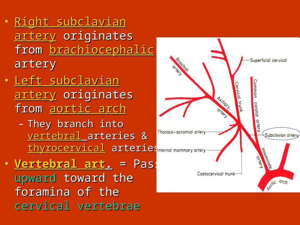
• Right subclavian arteryRight subclavian artery originates from originates from brachiocephalicbrachiocephalic arteryartery
• Left subclavian arteryLeft subclavian artery originates from originates from aortic aortic archarch– They branch into They branch into vertebralvertebral
arteries & arteries & thyrocervicalthyrocervical arteries arteries
• Vertebral artVertebral art.. = Pass = Pass upwardupward toward the toward the foramina of the foramina of the cervical cervical vertebraevertebrae

• Thyrocervical art.Thyrocervical art. Extend short Extend short distance to tissues in distance to tissues in neckneck which branch to which branch to supply: supply:
• Thyroid glands Thyroid glands • ParathyroidParathyroid• LarynxLarynx• Trachea Trachea • EsophagusEsophagus• Pharynx Pharynx • Muscles of head & Muscles of head &
neckneck
Figure Right subclavian arteriogram shows an aneurysm arising from the
thyrocervical trunk (arrow).

Systemic Systemic VeinsVeins• Large vessels that Large vessels that
are formed by are formed by convergence of convergence of smaller veins & smaller veins & venulesvenules
• Toward heartToward heart• Right atrium final Right atrium final
destinationdestination• Superior & inferior Superior & inferior
vena cavavena cava

Some veins don’t go to inferior vena cava but toward liverSome veins don’t go to inferior vena cava but toward liver
S U P E R IO R M E S E N TE R IC V E IN IN F E R IO R M E S E N TE R IC V E IN
H EPA TIC PO R TAL V E INO R IG IN A TE F R O M D IG E S TIV E TR A C THEPATIC PO RTAL SYSTEM SHUTS BLO O D
FRO M CAPILLARIES O F DIG . TRACT TO CAP. O F LIVER
LIVER
•Liver receives blood from 2 sources: Liver receives blood from 2 sources: •Hepatic Portal Vein Hepatic Portal Vein •Hepatic ArteryHepatic Artery
•Blood that is Blood that is high in O2high in O2 enters enters hepatic arteryhepatic artery•Blood Blood low in O2low in O2 enters enters hepatic hepatic portal veinportal vein

• Venous blood from dig Venous blood from dig organs is organs is low in O2low in O2 but but still still carries nutrientscarries nutrients absorbed by intestinesabsorbed by intestines
• Blood passes Blood passes slowlyslowly thru thru capillaries in livercapillaries in liver hepatic cells removehepatic cells remove materials used for materials used for metabolic functionsmetabolic functions phagocytic cells phagocytic cells eliminate bacteria etc eliminate bacteria etc that penetrate dig. liningthat penetrate dig. lining
• Blood passes thru liver Blood passes thru liver capcap collected by small collected by small veins that lead into veins that lead into hepatic veins hepatic veins emptied emptied into inferior vena cavainto inferior vena cava

Circulation to the Circulation to the FetusFetus

Blood Transport RoutineBlood Transport RoutineTaking blood (Aorta)to the tissues and back (Vena
Cavas)
– Arteries– Arterioles– Capillaries– Venules– Veins


Congestive Heart FailureCongestive Heart Failure • CausesCauses of CHF of CHF
– coronary artery disease, coronary artery disease, hypertension, MI, valve hypertension, MI, valve disorders, congenital defectsdisorders, congenital defects
• Left side heart failureLeft side heart failure– less effective pump so less effective pump so more more
blood remains in ventricleblood remains in ventricle– heart is heart is overstretchedoverstretched & &
even more blood remainseven more blood remains– blood backs up into lungs as blood backs up into lungs as
pulmonary edemapulmonary edema– suffocationsuffocation & lack of oxygen to & lack of oxygen to
the tissuesthe tissues
• Right side failure Right side failure – fluid builds up in tissues as fluid builds up in tissues as
peripheral edemaperipheral edema

Clinical ProblemsClinical Problems• MI = Myocardial InfarctionMI = Myocardial Infarction
– death death of area of heart of area of heart muscle from lack of Omuscle from lack of O22
– replaced with replaced with scar tissuescar tissue– results depend on size results depend on size
& location of damage& location of damage
• Blood ClotBlood Clot– use clot dissolving use clot dissolving
drugs streptokinase drugs streptokinase or t-PA & heparinor t-PA & heparin
– balloon angioplastyballoon angioplasty
• Angina PectorisAngina Pectoris– heart painheart pain from ischemia of from ischemia of
cardiac musclecardiac muscle


Myocardial Myocardial InfarctionInfarction• Myocardial infarction Myocardial infarction
means means heart attackheart attack, or , or coronary coronary thrombusthrombus..
• InfarctionInfarction = = death of death of musclemuscle, tissue or organ , tissue or organ as a result of a as a result of a blockage of the blood blockage of the blood supplysupply
• Blockage due to Blockage due to plaqueplaque buildup in arteries buildup in arteries because of because of high high cholesterolcholesterol and and saturated saturated fatsfats in diet in diet

Bypass SurgeryBypass Surgery

By-Pass GraftBy-Pass Graft Percutaneous Percutaneous Transluminal Transluminal
Coronary Coronary AngioplastyAngioplasty

Stent in an ArteryStent in an Artery
• Maintains patency of blood vesselMaintains patency of blood vessel

What's an Artificial Pacemaker?What's an Artificial Pacemaker?•“Artificial pacemaker" is a small, battery-operated device that helps the heart beat in a regular rhythm by sending electrical impulses to the heart to help it pump properly•An electrode is placed next to the heart wall and small electrical charges travel through the wire to the heart. •Most pacemakers are demand pacemakers. •They have a sensing device•It turns the signal off when the heartbeat is above a certain level•It turns the signal back on when the heartbeat is too slow.


• As the blood is pumped back to the heart, veins act as one-way valves to prevent the blood from flowing backwards.
• If the one-way valve becomes weak, some of the blood can leak back into the vein, collect there, and then become congested or clogged.
•This congestion will cause the vein to abnormally enlarge. These enlarged veins can be either vericose or spider veins.

•Lack of oxygen in the blood causes a bluish discoloration in the skin or mucous membranes called cyanosis.
•Most cyanosis is seen as a result of congenital heart disease, pulmonary disease, or as a terminal event as in cardiopulmonary arrest.

Desirable Levels of Blood Desirable Levels of Blood Cholesterol for AdultsCholesterol for Adults
• TC (total cholesterol) TC (total cholesterol) under 200 mg/dlunder 200 mg/dl
• LDL under 130LDL under 130 mg/dl mg/dl• HDL over 40HDL over 40 mg/dl mg/dl• Normally, Normally,
triglyceridestriglycerides are in are in the range of the range of 10-19010-190 mg/dl.mg/dl.
• Among the therapies Among the therapies used to reduce blood used to reduce blood cholesterol level are cholesterol level are exercise, diet, and exercise, diet, and drugs.drugs.

Exercise and the HeartExercise and the Heart• Sustained exercise Sustained exercise
increases oxygen increases oxygen demand in muscles.demand in muscles.
• Benefits of aerobic Benefits of aerobic exercise (any activity that exercise (any activity that works large body muscles works large body muscles for at least for at least 20 minutes, 20 minutes, preferably 3-5 times per preferably 3-5 times per weekweek) are;) are;– increased cardiac outputincreased cardiac output– increased HDL and increased HDL and
decreased triglyceridesdecreased triglycerides– improved lung functionimproved lung function– decreased blood pressuredecreased blood pressure– weight control.weight control.
