cardiovascular/circulatory system sports training and physiology kociuba
TRANSCRIPT

Cardiovascular/Circulatory System
Sports Training and Physiology
Kociuba
http://kidshealth.org/PageManager.jsp?lic=1&article_set=59298&cat_id=20607

Circulatory System - Objectives
• Describe the structures and functions of the Cardiovascular System
• Describe cardiac muscle
• Label the chambers of the heart
• Explain the process of blood flow throughout the heart
• Decipher what blood pressure is and how to take it

Circulatory System - Objectives
• Describe the structures and functions of arteries, veins, and capillaries
• List major arteries and veins in the body
• Explain how the systems responds to exercise and shock

Functions of the Heart
• Generating blood pressure– Contractions generate blood pressure
• Routing blood– Pulmonary circulation– Systemic circulation
• Regulating blood supply– Changes in rate and force of heart contraction
match blood flow to the demands of the body
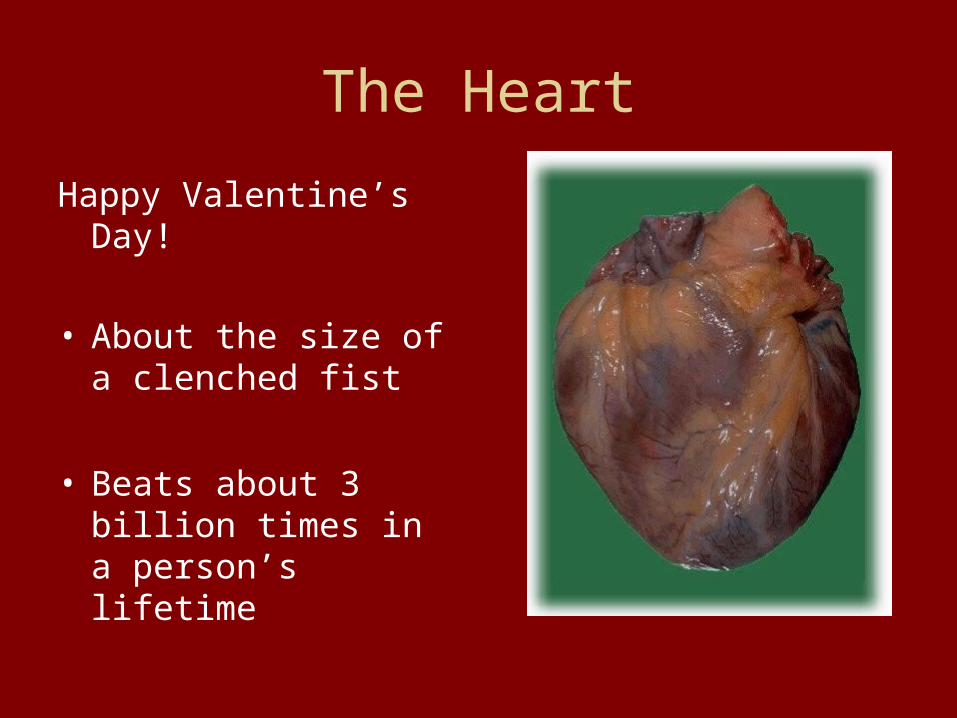
The Heart
Happy Valentine’s Day!
• About the size of a clenched fist
• Beats about 3 billion times in a person’s lifetime

The Heart Wall
Composed of 3 tissues• Epicardium
– Thin membrane forms the smooth outer surface of the heart
• Myocardium– Thick middle layer
composed of cardiac muscle
• Endocardium – Inner surface that
allows blood to move easily through the heart

Anatomy of the Heart and Major Blood Vessels
4 chambers in the heart• 2 atria
– Right – Left
• 2 ventricles– Right– Left
6 large veins• Superior vena cava• Inferior vena cava• 4 Pulmonary veins
2 large arteries• Pulmonary trunk• Aorta

Heart Valves
• Atrioventricular (AV) Valve– Tricuspid Valve: This AV valve located
between the right atrium and right ventricle– Bicuspid/Mitral Valve: This AV valve located
between the left atrium and left ventricle
• Semilunar Valves– Aortic semilunar valve: in the aorta– Pulmonary semilunar valve: in the pulmonary
trunk

Anatomy of the Heart

Conducting System of the Heart• The Sinoatrial Node or
SA node is capable of creating it’s own electrical impulse to get the heart started beating.
• This impulse then travels down to the Atrioventricular node or AV node and around the ventricles in the bundles and branches to create the contraction to push the blood out of the ventricles.

What exactly is our heart beat?

Heart Rate and Pulse
• Pulse is the beating of your heart that you can hear with a stethoscope and feel at pulse points
• Heart rate is the amount of times your heart beats in one minute.
Aging and the Heart
• By the age of 70 cardiac output is decreased by 1/3
• Aging cardiac muscle needs more time to contract and relax thus maximum heart rate is decreased
• Coronary Artery Disease (CAD) is age related
• Exercise prolongs these from happening

Blood Vessels and Circulation

Arteries
• Blood vessels that carry blood away from the heart. As they get further away, they the become smaller and smaller.
Classified As: • Elastic arteries• Muscular arteries• Small arteries
• Arterioles

Capillaries• The smallest of the blood vessels their
diameter is a little larger than a red blood cell
• The thin wall of the capillaries facilitate diffusion between the capillaries and the surrounding cells
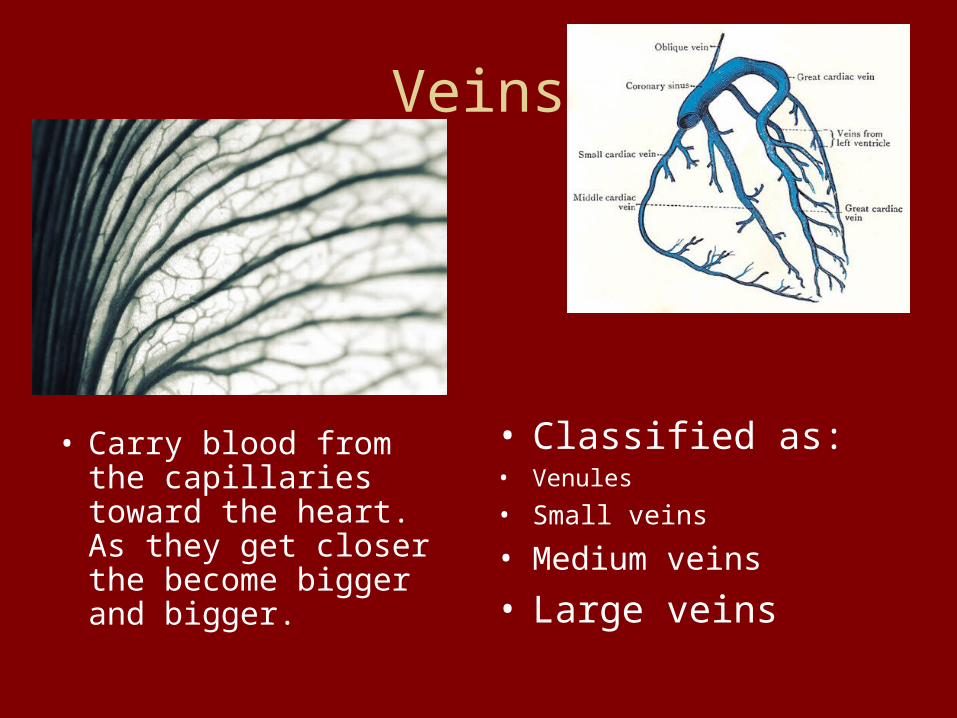
Veins
• Carry blood from the capillaries toward the heart. As they get closer the become bigger and bigger.
• Classified as: • Venules
• Small veins
• Medium veins
• Large veins
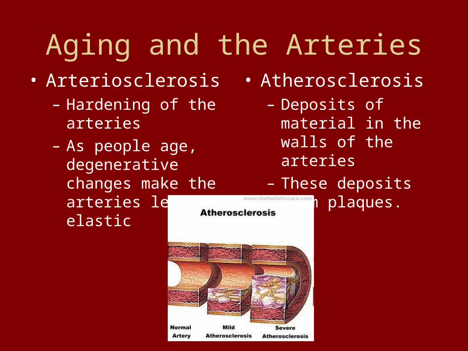
Aging and the Arteries• Arteriosclerosis
– Hardening of the arteries
– As people age, degenerative changes make the arteries less elastic
• Atherosclerosis– Deposits of material in
the walls of the arteries
– These deposits form plaques.
Blood Pressure
• Measure of the force that is exerted from blood against the blood vessel’s walls
• 2 measurements– Systolic: when the blood is forced out of the
ventricles and into the arteries– Diastolic: when the ventricles relax blood
moves at a minimum value
• We listen to blood pressure with a sphygmomanometer and stethoscope

Circulatory System Lab
Please read and we will go over how to take blood pressure…

How to take your blood pressure
1. Place the cuff of the sphygmomanometer on the participant’s arm right above the elbow. Giving them just enough room to bend the elbow.
2. Pump the cuff up enough that it is applying pressure to the upper part of the arm but not so tight that the participant is uncomfortable.

How to take your blood pressure
3. Place the stethoscope on the crease of the participant’s elbow.
4. Slowly release the pressure in the cuff while listening with the stethoscope.
5. When you hear the first sound of a heart beat, remember the number because this is the systolic number of their blood pressure.

How to take your blood pressure
6. Continue to release the pressure and note the number on the sphygmomanometer when the sounds disappear. This number is the participant’s diastolic pressure number.
7. Put the systolic number over the diastolic number and you have a resting blood pressure for your participant.