the c-cells: current concepts on normal histology … c cells.pdf · the c-cells: current concepts...
TRANSCRIPT

MORPHOL.–EMBRYOL., 1999–2004, XLV, p. 53–61
THE C-CELLS: CURRENT CONCEPTS ON NORMAL HISTOLOGY AND HYPERPLASIA
ANGELA BORDA*, NICOLE BERGER**, M. TURCU***, M. AL JARADI**, SORANA VEREŞ****
*Histology Department, University of Medicine and Pharmacy, Târgu Mureş; **Pathology Department, Lyon Sud Hospital; ***Pathology Department, University
of Medicine and Pharmacy, Târgu Mureş; ****”Petru Maior” University, Târgu Mureş
Summary. We describe the current concepts on the embryology, normal morphology and immunohistochemistry of a minor cell population of the thyroid, the C-cells. We also try to make delineation between the normal number of the C-cells and C-cell hyperplasia. The two types of C-cell hyperplasia, physiologic and neoplastic are defined and characterized from morphologic and genetic point of view. Their relation with thyroid pathology, especially with medullary thyroid carcinoma is discussed.
Key words: C-cell, C-cell hyperplasia, medullary thyroid carcinoma.
INTRODUCTION
Two distinct cell populations may be found in the thyroid parenchyma: the follicular cells, so far the most numerous, endodermally derived and synthesizing thyroglobulin, and the C-cells, a minor cell population, also known in the past as parafollicular cells, very different from the former in origin, morphologic appearance and function.
The C-cells were first described in animals (rats and dogs). In the human thyroid, Pearse, using silver stain methods, recognized them only in 1966. He suggested that these cells contain calcitonin and coined their name: the C-cells. His finding was confirmed by the development of the immunohistochemistry and the anti-calcitonin antibody, demonstrating the presence of this hormone in the C-cells cytoplasm (Bussolati et al., 1967). By that time, it was also demonstrated that medullary carcinoma, a distinct form of thyroid carcinoma described by Hazard (1959), consisted of C-cells (Williams et al., 1989).
EMBRYOLOGY
Knowing C-cells embryology is essential for better understanding their origin, distribution within the thyroid gland, and their pathology. The development

Angela Borda et al. 54
of C-cells is strongly related to that of the thyroid, which in humans develops from a median anlage and two lateral anlagen.
The median anlage is endodermally derived, from a depression at the base of the tongue. It gives rise to a structure, which becomes hollow and grows downward the anterior neck, known as the thyroglossal duct. At its end, it contains the developing thyroid gland, as a spherical and then bilobated structure. The thyroglossal duct undergoes atrophy, leaving as vestiges the pyramidal lobe. The follicular cells differentiate from the solid bilobated structure and are arranged in cords, nests and then in follicles. Early in the embryonic life the median anlage fuses with the lateral anlagen.
The paired lateral anlagen arise as ultimo-branchial bodies (UBBs) in close association to the primordium of the parathyroid IV, from the IVth–Vth branchial pouch complex. In their descent, the UBBs separate from the parathyroid IV, (which becomes the superior parathyroid, closely applied to the thyroid lobe) and fuse as solid masses of cells with the developing thyroid, in a posterior and lateral position, at the junction of the upper third with the medium third of its lateral lobes. Remnants of the UBBs are seen in postnatal life in the thyroid as so-called “solid cell nests” (SCN) (Harach, 1988; Martin et al., 2000) (Figure 1).
For many years, the origin of the C-cells was thought to be only the neural crest: during the embryogenesis, these cells migrate to the UBBs, which serve as a vector for their incorporation in the thyroid. The experimental works of Le Douarin and Le Lièvre, using quail chick transplant, proved this theory (Albores-Saavedra et al., 2001).
Other arguments for this origin, different from that of the follicular cells, are (Asa, 1997; LiVolsi, 1990; Papotti et al., 2000; Williams et al., 1989):
– the disposition of the C-cells in the thyroid: at the junction of the upper third with the medium third of the lobes, where the UBBs reach the thyroid.
– their close proximity to the remnants of the UBBs, the SCN. – the absence of C-cells in the lingual thyroid and in the thyroglossal duct
remnants. – identification of calcitonin secreting cells in the UBBs gland, in lower
animals, where the fusion with the median anlage does not take place. More recently, some authors raised the possibility of an additional source of
C-cells, based on the presence of the C-cells, even in a small number of cases, in the DiGeorge syndrome (Albores-Saavedra et al., 2001; Asa, 1997; Williams et al., 1989). This syndrome consists in complete or partial absence of derivates of the IIIrd and IVth–Vth pouch complexes, including UBBs. Nevertheless C-cells are found in approximately 25% of cases in this syndrome.
It is thought that the additional source of C-cells may be, as in the respiratory and digestive system, a stem endodermic cell, which could differentiate either in follicular or in C-cell. According to these authors, the stem endodermal cells could be at the origin of a minor part of C-cells and possibly give rise to the mixed

The C-cells: current concepts on normal histology and hyperplasia 55
medullary-follicular-derived carcinoma (Albores-Saavedra et al., 1990; Papotti et al., 2000). This theory does not have enough supporting evidence and necessitates further investigations.
MICROSCOPY
There are very few C-cells: only 0.1% of the glandular mass of the thyroid. They are localized at the junction of the middle and upper thirds of the lateral lobes. The highest concentrations are found at the sites of fusion between the UBBs and the medial thyroid anlage. For this reason, the C-cells are found in close association with the SCN, remnants of the UBBs (Harach, 1988). Their presence in the inferior third of the thyroid lobe or in the isthmus is very rare or questionable.
In the normal gland, the C-cells have an intrafollicular position. They are located individually or in small groups, between the basement membrane and the follicular cells, so that they are not in contact with the colloid.
C-cells have also been reported to occur in clusters in the interfollicular space, but now this situation is considered a misinterpretation of tangentional sections of the follicles in light microscopy (Bussolati et al., 1967; Carcangiu, 1997; LiVolsi, 1990).
Electron microscopy has improved the identification of the C-cell, demonstrating their intrafollicular position and the presence of two main types of secretory granules, that both contain calcitonin as demonstrated by immunohistochemistry.
Identification of C-cell in haematoxylin-eosin (HE) staining is very difficult and doubtful. They appear as polygonal or spindle-shape cells with pale staining, two or three times larger than the follicular cells. Their cytoplasm is finely granular and the nucleus round or ovoid, with a prominent nucleolus.
HISTOCHEMISTRY AND IMMUNOHISTOCHEMISTRY
Not reliably recognized by conventional stains, C-cells are detected by special techniques. In histochemistry, C-cells share the hallmarks of the diffuse endocrine cell system of Feyerter the most important being the argyrophilia, detected by the positivity of the Grimelius stain.
Widely used in the past, the Grimelius stain is now replaced by immunohistochemistry, which became the most precise and simple method for the identification of the C-cells.
In immunohistochemical techniques, C-cells are positive for neuroendocrine markers as chromogranin A, synaptophysin, and Neuron-Specific-Enolase (NSE). Moreover, the most specific marker is their main hormone product: the calcitonin (Carcangiu, 1997; LiVolsi, 1990).

Angela Borda et al. 56
HISTOPHYSIOLOGY
Calcitonin, the main product of C-cells is a peptide hormone of 32 amino acids that lowers the concentration of calcium in the blood by suppressing bone resorption achieved by osteoclasts. Massive degranulation occurs through experimentally induced hypercalcemia.
Nevertheless after total thyroidectomy the levels of serum calcium are within normal limits, demonstrating that in humans, calcitonin is of less importance than in other species.
The calcium level in the blood, which in turn depends upon the rate of demineralisation of the bone by osteoclasts, controls the calcitonin secretion. The release of the calcitonin results in diminishing the mineral resorption and returning calcium level to normal.
Calcitonin receptors are also found in the kidney where they regulate the calcium excretion in the urine. Calcitonin also enhances the production of vitamin D1 by the kidney. Calcitonin secretion is also stimulated by gastrin level and increase rapidly in response to pentagastrin stimulation.
NORMAL NUMBER VERSUS C-CELL HYPERPLASIA
The exact number of C-cells is unknown and for this reason it is difficult to define a true hyperplasia (Albores-Saavedra et al., 2001). The difficulties in establishing the exact number of the C-cells are due to their great variations according to age, sex, sampling method and associated thyroid pathology (Figure 2).
For their estimation, a correct sampling method must be used: horizontal sections in the two lateral lobes and vertical sections in the isthmus, at 3 mm intervals (Guyetant et al., 2003).
At least two sections from the junction of the middle with the upper thirds of the thyroid lobes must be included and examined. This region can be recognized at gross examination (especially when the gland is deformed by another pathology) by the presence of more prominent vessels.
Each section is studied in conventional HE stain and in calcitonin-stained slides. Many authors tried to make delineation between what we must consider normal or C-cells hyperplasia (CCH).
This number is very variable from one author to another and lacks specific quantification (Guyetant et al., 2003; LiVolsi, 1997).
The most accepted definition of CCH (Albores-Saavedra et al., 2001; Kaserer et al., 2001) is the presence, in at least one area with the highest estimated C-cell density, of more than 50 immunostained C-cells, per one low-power field (×100 magnification) in both thyroid lobes.

The C-cells: current concepts on normal histology and hyperplasia 57
In a recent paper, Guyetant et al. (2003) have more strict criteria: they defined CCH as more than 50 calcitonin positive cells found in three low-power fields (×100 magnification).
Morphologically, a distinction can be made among CCHs according to: – the growth pattern of the C-cells into the follicle, and – the morphology of the C-cells. Three types of CCH are distinguished by their pattern of growth (DeLellis,
1992; Perry et al., 1966): – focal CCH, corresponding to a segmental proliferation pattern within the
thyroid; – diffuse CCH defined by a C-cell proliferation forming a circumferential
intrafollicular collar, which pushes the follicular cells into the lumen; – nodular CCH diagnosed when C-cells clusters obliterate completely the
follicular lumen. The morphology of the C-cells defines a particular form of CCH, the
“neoplastic” CCH. The neoplastic C-cells have mild to moderate cytological atypia, nuclear pleomorphism, similar to those of invasive MTC, and most frequently have a nodular pattern of growth. Therefore, they are morphologically distinct from the follicular cells and recognizable in routine stains (HE) (Guyetant et al., 2003; Kaserer et al., 2001).
CCH AND THYROID PATHOLOGY
CCH, first recognized by Wolfe and Ljundberg in asymptomatic patients with MEN II A syndrome, was considered the precursor of genetically determined medullary thyroid carcinoma (MTC). Subsequently CCH unrelated to familial MTC was described, isolated or associated with diverse physiological or pathological thyroid conditions.
During the past 10 years, in many institutions, the preoperative serum calcitonin measurement, in order to detect MTC at an early stage, became systematic in all patients presenting with thyroid pathology. Consequently, the pathologists are confronted with a higher rate of diagnoses implying the pathology of the C-cells.
New entities were defined as the so-called “neoplastic CCH only”, which similarly to micro-MTC, can be responsible for mildly elevated calcitonin levels, leading to thyroidectomy in search of a MTC.
Furthermore, a highly sensitive and specific test, which identifies RET proto-oncogene germline mutations, is now available in order to detect patients with genetically determined MTC.
Nowadays, two types of CCH have been distinguished, especially on the molecular properties of the C-cells (the RET proto-oncogene analysis) and less on

Angela Borda et al. 58
morphologic ground (Guyetant et al., 2003; Kaserer et al., 2001; LiVolsi, 1997; Perry et al., 1966):
– the physiologic or reactive CCH, and – the neoplastic CCH.
PHYSIOLOGIC OR REACTIVE CCH
The main characteristics of physiologic CCH are (LiVolsi, 1997; Albores-Saavedra et al., 2001; Guyetant et al., 2003):
– focal or diffuse, rarely nodular, pattern of growth (Figures 3 and 4); – the hyperplastic C-cells are indistinguishable from adjacent follicular cells
in routine stains (HE); – no germ line mutation in RET proto-oncogene and no familial history of
MTC and MEN 2A syndrome; – possible mild to moderate elevation in serum calcitonin level with or
without pentagastrin stimulation. Using these criteria, a number of situations associated with reactive or
physiologic CCH were reported in the last 20 years (Albores-Saavedra et al., 2001; Barbot et al., Biddinger et al., 1991; Guyetant et al., 2003; Libbey et al., 1989):
– age-related CCH: in neonates and elderly subjects; – sex-related CCH: found twice as common in men as in women in an
autopsy study on normal thyroid; – CCH related to other conditions than thyroid pathology: prolonged
hypercalcemia, hyperparathyroidism and hypergastrinemia; – CCH related to thyroid pathology: previous hemithyroidectomy, Hashimoto
thyroiditis, thyroid non-Hodgkin lymphoma, tissue adjacent to neoplasms of follicular cell phenotype and nodular or diffuse hyperplasic goitre with or without hyperthyroidism (Figures 5 and 6).
More recently, CCH associated to sporadic, non-genetically determined MTCs was described in several studies (Guyetant et al., 2003; Kaserer et al., 2001). However, the significance of such association is doubtful; it gives raise to two hypotheses: CCH may represent either a reactive C-cell growth caused by the adjacent malignant tumor and/or a precursor of non-genetically determined MTC.
NEOPLASTIC CCH OR IN SITU MTC
Besides the morphology, which is very different from the physiologic CCH, neoplastic CCH has genetic and kinetic characteristics consistent with a malignant C-cell lesion (Diaz-Cano et al., 2001; Kaserer et al., 2001).
Its main characteristics are: – it is visible on microscopic examination in routine stains; neoplastic C-cells
are larger than normal and mildly to moderately atypical, similar to those of invasive MTC (Figure 7);

The C-cells: current concepts on normal histology and hyperplasia 59
– nodular and diffuse pattern of growth, often bilateral (Figure 8); – constant association with germ-line mutation of the RET proto-oncogene.
The most common mutations are detected in exons 10, 11, 13, 14, 15 and 16, rarely in 18 (Diaz-Cano et al., 2001; Krueger et al., 2000);
– the presence of a distinct basement membrane surrounding the hyperplasic nodules, which can be highlighted by periodic acid-Schiff stain or with anti-collagen IV antibodies immunostains. These stains can differentiate neoplastic CCH from early invasive micro MTC characterized by extension of C-cells through defects and/or reduplications of the basement membrane (Mears et al., 2003).
In the recent literature, it is pointed that the term “neoplastic CCH” is, for many authors, contradictory by itself and therefore a misnomer that should not be used. More appropriate would be the term “in situ MTC” or “thyroid intraepithelial neoplasia of C-cell” because this type of hyperplasia is a truly neoplastic process (Albores-Saavedra et al., 2001; Krueger et al., 2000; Mears et al., 2003).
RELATIONSHIP BETWEEN CCH AND MTC
CCH AND GENETICALLY DETERMINED MTC
Genetically determined MTC (25% of MTC, MEN IIA, MEN IIB and familial MTC) is preceded and accompanied by neoplastic CCH, which is considered the precursor of the disease, in male and female patients (Figure 9).
It can be diagnosed preoperatively in asymptomatic children and adolescents by detecting the germ line mutation of the RET proto-oncogene, even in the absence of serum calcitonin elevation.
In these young patients, the neoplastic CCH may be the only pathological finding, representing a preneoplasic stage of MTC. When tumours are already present, they are most often multifocal and bilateral and are always accompanied by neoplastic CCH (Krueger et al., 2000; LiVolsi, 1997).
Although these morphologic aspects are characteristic, they are unreliable in deciding if a familial risk exists or not. This will be decided only by the genetic test.
CCH AND SPORADIC MTC
In previous studies, sporadic MTC was considered not accompanied by CCH. These observations had been made in large tumours. In the past 10 years, preoperative serum calcitonin measurement, in order to detect sporadic MTCs in an earlier stage (micro MTC), led to the discovery of concomitant CCH in a rather high number of cases (Guyetant et al., 2003; Kaserer et al., 2001).
In his studies, Kaserer (Kaserer et al., 2001) found an inverse correlation between tumor size and the number of C-cells per low power field, which may

Angela Borda et al. 60
indicate that CCH regresses as the tumor grows in size and, maybe, higher amounts of calcitonin are produced.
A process of primary physiologic reactive growth stimulus to C cell may therefore exist initiating CCH, which in turn may initiate a sporadic MTC. Once the MTC evolved from CCH, the growth stimulus could disappear, either by the tumor size or by higher amounts of calcitonin produced. This relationship might be, according to Kaserer, one explanation for the high frequency of CCHs accompanying sporadic micro MTCs compared with previous studies where sporadic MTCs were larger.
When comparing concomitant CCH in sporadic and hereditary cases it has been found that CCH described in sporadic cases is more frequent in men than in women and has more often a “reactive” pattern of growth (exhibiting focal, diffuse or nodular types), and rarely “neoplastic” (Guyetant et al., 2003; Kaserer et al., 2001).
The rare association of neoplastic CCH and sporadic MTCs was found always in males and never in females. This fact should indicate that, in a certain number of cases, especially in men, sporadic MTC may evolve from neoplastic CCH and that in women, where alternative tumorigenesis pathways might also exist, it occurs only de novo. The consequence of these findings is that, in clinical practice, the finding of mild serum CT elevation in women is more significant of micro MCT than in men, where neoplastic CCH only is often found.
CONCLUSIONS
When discussing about CCH, a clear-cut distinction must be done between physiologic and neoplastic CCH, as they have a distinct morphologic pattern, genetic characteristics and clinical significance. Physiologic CCH is a reactive C cell growth and has no preneoplastic signification.
In contrast, neoplastic CCH (in situ MTC) is the precursor of genetically determined MTC in men and women and, it seems, that also of sporadic MTC in men. It can be detected preoperatively in asymptomatic patients by germ-line mutation of the RET proto-oncogene. This genetic examination is mandatory in all patients with neoplastic CCH, to rule out a hereditary disease.
REFERENCES
ALBORES-SAAVEDRA J.A., GORRAEZ DE LA MORA T., DE LA TORRE-RENDON F. et al., Mixed medullary carcinoma of the thyroid. A previously unrecognised variant of thyroid carcinoma, Hum Pathol, 1990, 21:1151–1155.
ALBORES-SAAVEDRA J.A., KRUEGER J.E., C-cell hyperplasia and medullary thyroid microcarcinoma, Endocr Pathol, 2001, 12:365–377.
ASA S.L., C-cell lesions of the thyroid, Pathol Case Rev, 1997, 2:210–217.

The C-cells: current concepts on normal histology and hyperplasia 61
BARBOT N., GUYETAN S., BELDENT V. et al., Thyroidite chronique auto-immune et hyperplasie des cellules C, Ann Endocrinol, 1991, 52:109–112.
BIDDINGER P.W., BRENNAN M.F., ROSEN P.P., Symptomatic C-cell hyperplasia associated with chronic lymphocytic thyroiditis, Am J Surg Pathol, 1991, 15:599–604.
BUSSOLATI G., PEARSE A.G.E., Immunofluorescent localisation of calcitonin in the C-cells of the pig and dog thyroid, J Clin Endocrinol, 1967, 37:205–209.
CARCANGIU M.L., Thyroid. In: STERNBERG, S.S. (ed), Histology for pathologists, 2nd edition, Lippincott-Raven Publishers, Philadelphia, 1997, 1075–1093.
DELELLIS R.A., C cell hyperplasia. In: ROSAI, J., CARCANGIU, M.L., DeLELLIS, R.A. (eds), Tumors of the thyroid gland, AFIP, Washington, 1992.
DIAZ-CANO S.J., DE MIGUEL M., BLANES A. et al., Germline RET 634 mutation positive MEN 2A-related C-cell hyperplasias have genetic features consistent with intraepithelial neoplasia, J Clin Endocrinol Metab, 2001, 86:3948–3957.
GUYETANT S., JOSSELIN N., SAVAGNER F. et al., C-cell hyperplasia and medullary thyroid carcinoma: clinicopathological and genetic correlations in 66 consecutive patients, Mod Pathol, 2003, 16:756–763.
HARACH H.R., Solid cell nests in the thyroid, J Pathol, 1988, 155:191–200. KASERER K., SCHEUBA C., NEUHOLD N. et al., Sporadic versus familial medullary thyroid
microcarcinoma. A histopathologic study of 50 consecutive patients, Am J Surg Pathol, 2001, 25:1245–1251.
KRUEGER J.E., MAITRA A., ALBORES-SAAVEDRA J.A., Inherited medullary microcarcinoma of the thyroid. A study of 11 cases, Am J Surg Pathol, 2000, 24: 853–858.
LIBBEY N.P., NOWAKOWSKI K.J., TUCCI J.R., C-cell hyperplasia in the thyroid in a patient with goitrous hypothyroidism and Hashimoto’s thyroiditis, Am J Surg Pathol, 1989, 13:71–77.
LiVOLSI V.A., Surgical pathology of the thyroid, WB Saunders Company, Philadelphia, 1990. LiVOLSI V.A., C-cell hyperplasia / neoplasia, J Clin Endocrinol Metab, 1997, 82:39–41. MARTIN V., MARTIN L., VIENNET G. et al., “Solid cell nests” et pathologies thyroïdiennes.
Etude rétrospective de 1390 thyroïdes, Ann Pathol, 3:196–201. MEARS L., DIAZ-CANO S.J., Difference between familial and sporadic medullary thyroid
carcinomas, Am J Surg Pathol, 2003, 27:266–267. PAPOTTI M., VOLANTE M., KOMMINOTH P. et al., Thyroid carcinomas with mixed follicular
and C-cell differentiation patterns, Semin Diagn Pathol, 2000, 2:109–119. PERRY A., MOLDBERG K., ALBORES-SAAVEDRA J.A., Physiologic versus neoplastic C-cell
hyperplasia of the thyroid. Separation of distinct histologic and biologic entities, Cancer, 1966, 77:750–756.
WILLIAMS E.D., TOYN C.E., HARACH H.R., The ultimobranchial gland and congenital thyroid abnormalities in man, J Pathol, 1989, 159:135–141.
Received: 3 May, 2004
Accepted: 10 September, 2004

Figure 2 – Normal number of the C cells, with intrafollicular position(calcitonin immunostaining, ×100)
Figure 1 – Solid cell nests (HPS, ×40)

Figure 3 – Reactive CCH in a nodular goiter (calcitonin immunostaining, ×40)
Figure 4 – Higher magnification to demonstrate focal (right) and diffuse (left) patternof growth of the same reactive CCH (calcitonin immunostaining, ×400)

Figure 5 – Reactive CCH in a Hashimoto's thyroiditis (calcitonin immunostaining, ×40)
Figure 6 – Higher magnification of a diffuse pattern of growth of the same reactive: C-cellsforming a circumferential intrafollicular (calcitonin immunostaining, ×200)
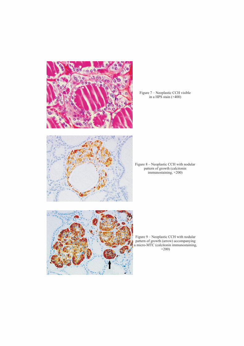
Figure 7 – Neoplastic CCH visiblein a HPS stain (×400)
Figure 8 – Neoplastic CCH with nodularpattern of growth (calcitonin
immunostaining, ×200)
Figure 9 – Neoplastic CCH with nodularpattern of growth (arrow) accompanying
a micro-MTC (calcitonin immunostaining,×200)