the bridge between science & society
TRANSCRIPT

The Bridge Between Science & Society
newsletterP. D. HINDUJA HOSPITAL
The best way to find yourself is to lose yourself
in the service of others.
- MAHATMA GANDHI
VOL 1 | ISSUE 01 | OCTOBER 2015 www.hindujahospital.com

On Cover - The famous photo of Mahatma Gandhi seeing Mycobacterium leprae through the microscope for the first time.

Bone and Joint Tuberculosis
DR. VIKAS AGASHE
DR. ZENA PATEL
DR. SUGANTHI IYER
06
CASE REPORT-Emergency Surgery for
Refractory Status Epilepticus
WEGENER’S GRANULOMATOSIS PRESENTING AS A TONSILLAR MASS
HINDUJA GRAND ROUNDS
EXPERIENCE-THE BEST TEACHER
CRITICAL CARE PEARLS
IN A NUTSHELL-ILDThrombocytopenia in Lupus
Prosthetic Joint Infection
LEGAL EYE
VIEW POINT -What Doctor’s Day mean to me?
AWARDS
QUIZ
07
11
13
17
20
22
27
29
35
32
04 OCTOBER 2015
DR. JAI MULLERPATTAN
DR. VRAJESH UDANI, DR. MILIND SANKHE,
DR. NEELU DESAI, DR. ABHIJEET BOTRE &
DR. SPOORTHI JAGADISH
MY JOURNEY
DR. KUSHRAO BHAJAN, DR. C. BALAKRISHNAN
DR. ANITA BHADURI
PULMONOLOGY DEPARTMENT
Oxygen-How much is too much
DR. FAHAD KAPADIA
DR. VIVEK SHETTY
DR. BALKRISHNA PADATE
DR. FARAM D. DASTUR
DR. FARAM D. DASTUR
DR. ASHIT HEGDE
CONTENTS
EDITORIAL TEAM
• Dr. C. Balakrishnan • Dr. Deepraj Bhandarkar
• Dr. Avinash N Katara • Dr. Lancelot Mark Pinto
• Dr. Balkrishna. Padate • Dr. Zena Patel
RADIOLOGIC - LUMPS & BUMPS26
DR. JAI MULLERPATTANRESPIRATORY REVIEW30
NEW CONSULTANT34
PRINTED & PUBLISHED byMarketing Department
P. D. Hinduja Hospital and Medical Research Centre Veer
Savarkar Marg, Mahim, Mumbai-400 016 India.
Tel: +91 - 22 - 24449199 / 24452222 / 24451515.
Fax: +91 - 22 - 2444 9199
Web: www.hindujahospital.com
Email: [email protected]

OCTOBER 2015 05
Valuable Inputs and Effort
From the Editor
Dear Friends,
Dr. (Mrs) Dalal after serving as chief editor of Hinduja Newsletter has recently relinquished this post and I have been entrusted the responsibility of editing the newsletter. I have formed a new team. The committee deliberated and opined that a change of format was in order. Accordingly the contents have been changed.
Apart from reviews and articles, we have introduced new sections like: My Journey - Hinduja Grand Rounds, Experience -The Best Teacher, Snapshot, Radio-logic, Legal eye, Viewpoint, Academic update etc.
I am honored to have been made the editor and will try my best to do justice. I thank all the editors thus far who have done a wonderful job. I also thank all my editorial team members for their valuable inputs and effort.
I trust the readers will enjoy this new edition.
We also will be happy to have your feedback.
Consultant Rheumatologist

My parents had migrated from Bombay some years earlier, my father was a general practitioner in a singly run practice, - it was a hard life, with little off time, I remember the bombing of London and later in 1942-
43 the ‘Doodle-bugs’ or pilotless planes programmed to ‘cut out’ over their target and cause maximum damage,- a forerunner of today’s drones.
Post 1945 I remember my school years as a happy time. I made some good friends, played cricket for the school and became a monitor. Academically I did not show any special aptitude, but chose medicine as it was a respected profession.
The next event in my journey came some two or three years later. I went with a friend to the last day of the yearly Promenade Concerts (Proms) held in July - August at the Albert Hall. The last night is typically attended mainly by students, the mood is lighthearted, somewhat irreverent, and ends with the singing of patriotic songs such as Land of Hope and Glory and Rule Brittania. Suddenly I felt uncomfortable and a sham to be joining in the singing when India had just gained its independence from the yolk of British, colonial rule. Of course I was Anglicized, and an admirer of certain British qualities, but was I one of them? No.
I made two trips to India before coming to stay permanently. The first was on a troop ship post war to ‘meet family’. The second was after passing MBBS when I came overland (except for the English Channel) in a journey lasting seven weeks with a cosmopolitan group, realizing this was a chance I would not get again once I started post-graduation. It was fascinating to see the gradual change in architecture, dress, customs, manners, eating habits, etc as we made our way from West to East. Three sites left an indelible impression on the mind. First, the Dome on the Rock mosque in Jerusalem for its exquisite carvings, Second, The Lost City of Petra in Jordan, now one of the seven wonders of the world. Third of course was the Taj Mahal at Agra. I found it is true when they tell you “travel broadens the mind”, but what they forgot to tell you is that it also loosens the bowels!!
We all got a bout of traveler’s diarrhoea at some stage or other.
After arriving in India in 1967 I landed a job in Pune at Jehangir Nursing Home as resident physician. It was a gentle way to get adjusted to Indian medical practice. I marveled at the ease with which consultants shifted from one language to another to interact with patients, - quite the opposite to my ‘kicheri’, - but people were very understanding..
My Journey
06 OCTOBER 2015
Another feature was on a matter of etiquette. When the consultant arrived to see the patient relatives would crowd into the room to witness everything. There was no privacy for doctor patient interview unless the consultant insisted on it. And lastly the obnoxious practice we have of carrying out CPR on all patients when the heart stops. It is not appropriate for incurable and frail patients waiting to die, - but our law makers don’t seem to see it that way.
I wanted to be in a teaching hospital and late 1969 I joined the KEM Hospital in Mumbai as a lecturer in Medicine. I spent a total of 17 years at the institution slowly climbing the ladder to become Professor and head of department of Medicine in 1986. In addition to my medicine duties I looked after the Tetanus Unit which was the subject of a number of investigations and publications regarding the disease. I also saw firsthand the power of immunization as the number of tetanus cases fell from 60 to 10 per month over 15 years. Over the years I taught a number of brilliant students who later distinguished themselves in India and abroad. I witnessed regrettably the “ splintering effect” of super specialization where loyalty to institution was slowly transferred to loyalty to one’s speciality only. I witnessed the major exodus of newly qualified doctors to the USA and UK each year. I was caught up in the strikes by junior doctors as they fought for better pay and working conditions, and recently for more security and protection from irate relatives. The Government only saw fit to react to crises, and unable to financially support medical institutions sufficiently to retain staff particularly as a growing private healthcare sector was offering better remuneration and working conditions. I was caught up in this web myself and in 1987 at the age of 51 years earning only Rs. 5500/month and feeling financially insecure, I resigned from KEM Hospital to join the new Hinduja Hospital.
My education has continued during my years at HNH. We realized early the importance of a postgraduate degree to motivate and retain essential JMS and are presently recognized in 24 specialities by the National Board of Examinations in Delhi for the DNB degree. The consultants have also distinguished themselves and today we are certainly one of the top academic institutions in the private sector. I have also learnt how complex the running of a hospital is especially with our ‘not for profit’ commitment. NABH has guided us along the road to accreditation, -the basic certificate of proficiency for an institution. In all of this the Hinduja family and hospital administration has been kind enough to let me participate and contribute at a time when I thought I was fit only for the rocking chair. And so I end with a thank you to all who helped me along ‘My Journey’ and with the following quotation:
Age is an opportunity no less
than youth itself though in another dress,And as the evening twilight fades away
The sky is filled with stars invisible by day
hen I look back to my early years I recognize they were different to those of myW
colleagues. I was born in London in 1935 and remember starting junior school with a satchel and a gas mask on my back for it was the beginning of the 2nd World War and there was a serious fear of nerve gas attacks.
DR. FARAM D. DASTUR
FOR MORE INFORMATION:
Email: [email protected]
Tel: 24447184
Ext: 8184
Director, Medical Education & Hospital Quality

Hinduja Hospital and Medical Research Centre, Mumbai Status epilepticus is a serious medical
practice with a potential for significant morbidity and mortality.
emergency in paediatric PD Almost 30-40 % of status epilepticus is refractory to first and second line treatment and needs coma producing therapy for controlling
seizures. A subset of these patients does not respond and are considered to be super-refractory. Treatment of this super-refractory status
epilepticus is extremely challenging with very poor literature on effective therapies. In selected cases and at experienced centers,
epilepsy surgery could be considered as a therapeutic option once medical management has failed.
Here we describe two children with medically refractory status epilepticus due to lesional pathology who responded well
to emergency epilepsy surgery.
PP was first referred to the Pediatric Neurology and Epilepsy
centre at the P. D. Hinduja National Hospital at 4.5 months of age
for neonatal onset drug-resistant epilepsy. The child had a
normal birth history. Seizures started from 15 days of life in the
form of flexor spasms. Initially these events were infrequent and
gradually increased to daily episodes. The jerks responded to
ACTH and topiramate. At this time his developmental milestones
and examination was normal. An MRI brain showed a definite
right, virtually hemispheric cortical dysplasia. (Fig 1) An EEG
showed predominantly right hemispheric epileptiform
discharges. The patient was subsequently lost to follow up.
CASE 1
Emergency surgery for refractory status epilepticus
At one year of age he returned with progressively increasing
spasms since 7 months of age. He was also noted to have
significant left hemiparesis. An option of hemispheric surgery was
declined by the parents. He responded to the ketogenic diet and
was also continued on clobazam, valproic acid and topiramate.
However, relentless seizures and worsening drwoiness with inabilty
to feed and subsequent status epilepticus brought him back to
P. D. Hinduja hospital for further management. He was in super-
refractory SE despite high level com producing therapy. The EEG
showed predominantly right sided ictal as well as interictal epileptic
discharges and right sided burst suppression pattern (Fig 1).
(b)(a)
OCTOBER 2015 07
DR. ABHIJEET BOTRE & DR. SPOORTHI JAGADISH
DR. VRAJESH UDANI DR. MILIND SANKHE DR. NEELU DESAIConsultant Pediatric Neurologist Consultant Neurosurgeon Consultant Pediatric
Neurologist & Epileptologist
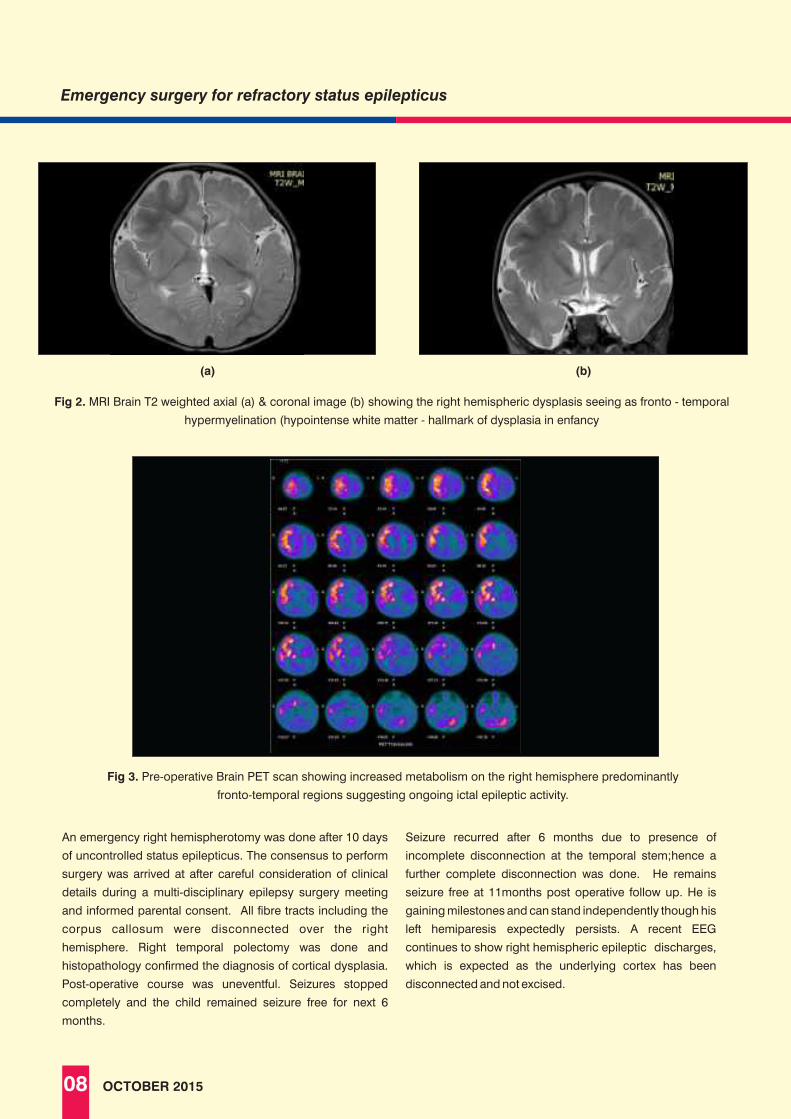
image image
(a) (b)
08 OCTOBER 2015
An emergency right hemispherotomy was done after 10 days
of uncontrolled status epilepticus. The consensus to perform
surgery was arrived at after careful consideration of clinical
details during a multi-disciplinary epilepsy surgery meeting
and informed parental consent. All fibre tracts including the
corpus callosum were disconnected over the right
hemisphere. Right temporal polectomy was done and
histopathology confirmed the diagnosis of cortical dysplasia.
Post-operative course was uneventful. Seizures stopped
completely and the child remained seizure free for next 6
months.
Seizure recurred after 6 months due to presence of
incomplete disconnection at the temporal stem;hence a
further complete disconnection was done. He remains
seizure free at 11months post operative follow up. He is
gaining milestones and can stand independently though his
left hemiparesis expectedly persists. A recent EEG
continues to show right hemispheric epileptic discharges,
which is expected as the underlying cortex has been
disconnected and not excised.
Emergency surgery for refractory status epilepticus

AB, a 7 year old boy presented to the Pediatric Neurology clinic at
PDHNH with a history of right sided brief, focal motor seizures
from 4 years of age following an possible encephalitis like illness
in his village 6 months earlier. Recent increase in seizure activity
was associated with a Todd’s paralysis. His MRI brain revealed ill-
defined patchy T2 hyperintensities in left frontal and peri- insular
cortex. He had failed trials of multiple antiepileptic drugs
(phenobarbitone, sodium valproate, levetiracetam and clobazam,
oxcarbazepine) with no benefit.
In March 2015 he presented with worsening seizures which later
evolved to a status epilepticus. His VEEG showed clinical as well
as subclinical seizures arising from the left temporal region.
Repeat MRI brain showed progressive left cerebral atrophy with
thinning of the left putamen and ill-defined patchy T2
hyperintensities in the left frontal and peri- insular cortex. This was
suggestive of Rasmussen’s encephalitis. He was tried on multiple
AEDs with no response. In view of refractory status epilepticus with
a left hemispheric onset and possible Rasmussens’ encephalitis,
an emergency hemispherotomy was performed after 7 day.
Histopathology of resected specimen showed inflammatory
changes confirming the diagnosis of Rasmussen’s encephalitis.
Seizures stopped completely after surgery and the child remains
seizure free at 3 months follow up. He has started speaking and is
ambulant.
Fig. 4 Interictal EEG showing left temporal spikes
CASE 2
Fig. 5 Ictal EEG shows a clear left temporo-occipital
seizure onset.
DISCUSSIONRefractory status epilepticus is a medical emergency with a substantial mortality rate. Despite the fact that it remains an important clinical
problem in all neurology centres worldwide, there is a remarkable lack of published data concerning effectiveness of treatment. Concerns
with treatment safety and outcome remain.
09OCTOBER 2015
Emergency surgery for refractory status epilepticus
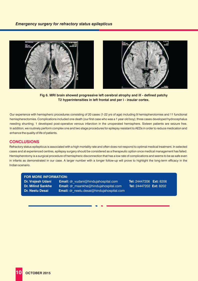
image image
Fig 6. MRI brain showed progressive left cerebral atrophy and ill - defined patchy
T2 hyperintensities in left frontal and per i - insular cortex.
CONCLUSIONSRefractory status epilepticus is associated with a high mortality rate and often does not respond to optimal medical treatment. In selected
cases and at experienced centres, epilepsy surgery should be considered as a therapeutic option once medical management has failed.
Hemispherotomy is a surgical procedure of hemispheric disconnection that has a low rate of complications and seems to be as safe even
in infants as demonstrated in our case. A larger number with a longer follow-up will prove to highlight the long-term efficacy in the
Indian scenario.
Our experience with hemispheric procedures consisting of 20 cases (1-22 yrs of age) including 9 hemispherotomies and 11 functional
hemispherectomies. Complications included one death (our first case who was a 1 year old boy); three cases developed hydrocephalus
needing shunting; 1 developed post-operative venous infarction in the unoperated hemisphere. Sixteen patients are seizure free.
In addition, we routinely perform complex one and two stage procedures for epilepsy resistant to AEDs in order to reduce medication and
enhance the quality of life of patients.
Emergency surgery for refractory status epilepticus
FOR MORE INFORMATION:
Dr. Vrajesh Udani Email: [email protected] Ext: 8206
Dr. Milind Sankhe Email: [email protected] Tel: 24447202 Ext: 8202
Dr. Neelu Desai Email: [email protected]
Tel: 24447206
10 OCTOBER 2015

Wegener’s granulomatosis is a necrotizing granulomatous vasculitis, which presents with upper or lower airway involvement, in a majority of patients. We describe a patient with this disease who presented with a necrotic tonsillar mass.
Investigatory reports were : Hb 7.6 gm/dl, wbc: 8290 / cumm, platelets : 525, ESR : 106 mm/hr, CRP: 192 mg/l, throat swab: negative, urine routine: normal, creatinine: 0.6 mg/dl, LDH: 323 mg/dl, ANA: negative, ANCA: c- ANCA lin20+++, x ray paranasal sinuses: normal, chest radiograph: normal and high resolution CT chest: trachea normal, few subcentimeter nodules superior segment of left lower lobe.
HPE of the necrotic mass was consistent with Wegener’s granulomatosis (figure 1).
A 13 yr old girl presented with cough, shortness of breath, throat pain and intermittent high-grade fever of 2 months duration. Since
20 days the throat pain had worsened and she has also noted flitting joint pains and transient rash. At admission she was unable to open
the mouth, speak or swallow. On examination vital signs were normal. There was severe trismus. A necrotic mass was evident in the right
tonsillar area with extensive ulceration of the right soft palate and anterior pillar. Vasculitic lesions were present in the hands and feet.
Systemic examination was unremarkable.
Investigatory reports were : Hb 7.6 gm/dl, wbc: 8290 / cumm, platelets : 525, ESR : 106 mm/hr, CRP: 192 mg/l, throat swab: negative, urine routine: normal, creatinine: 0.6 mg/dl, LDH: 323 mg/dl, ANA: negative, ANCA: c- ANCA lin20+++, x ray paranasal sinuses: normal, chest radiograph: normal and high resolution CT chest: trachea normal, few subcentimeter nodules superior segment of left lower lobe.
HPE of the necrotic mass was consistent with Wegener’s granulomatosis (figure 1).
DISCUSSIONWegener’s granulomatosis is a small/medium vessel vasculitis with protean manifestations (1). Head and neck manifestations, particularly in the sino-nasal tract are common and can affect as many as 90% of patients at presentation (2). In contrast oral lesions are less common and include oral ulceration, perforation of the palate, swelling and destruction of the lips (3-6). Although ischemic and necrotic tissue injury is common, tumorous lesions have been described.
REFERENCES
1) Fauci AS, Wolff SM. Wegener’s granulomatosus. Medicine 1973;52: 535-61.2) Handlers JP, Waterman J, Abrams AM, et al. Oral features of Wegener’s granulomatosus. Arch Otolaryngol Head Neck Surg
1985;111:267-70.3) Rahilly G, Rahilly M. A case of palatal Wegener’s granulomatosus. Oral Diseases. 2000;6(4):259-61.4) Knetcht K, Mishriki YY. More than a mouth ulcer. Oral ulcer due to Wegener’s granulomatosus. Postgraduate Medicine.
199;105(5):200-3.5) Eveson JW. Granulomatous disorders of the oral mucosa. Semin Diagnostic Pathol. 1996;13(2):118-27.6) Goulart RA, Mark EJ, Rosen S. Tumefactions as an extravascular manifestation of wegener’s granulomatosus. Am J Surg
Pathol 1995;19 (2):145-53.
12 OCTOBER 2015
Goulart et al reported 6 patients wherein the presenting manifestations were tissue swellings in the retroperitoneum, mediastinum
(two), breast, retro orbital tissue (two) and gingiva (7). The patient described presented with not only necrosis of the soft palate but,
also had a necrotic mass in the right faucial area. Although there was possible lung involvement, there was no kidney involvement.
The consistent histopathology and the strongly positive ANCA pointed towards the diagnosis.
FOR MORE INFORMATION:
Dr. Khushrao Bhajan Email: [email protected] Ext: 8262
Dr. C. Balakrishna Email: [email protected] Tel: 24447271 Ext: 8271
Dr. Anita Bhaduri Email: [email protected] Tel: 24447798 Ext: 7158
Tel: 24447262

HINDUJA GRAND ROUNDS
PULMONOLOGY DEPARTMENT
CASE 1
A 33 years old male, non-smoker, known case of bronchial asthma
and recurrent sinusitis presented to us with complains of chronic
cough with mucoid expectoration and dyspnea on exertion.
His serial chest radiographs showed fleeting opacities and blood
tests revealed elevated levels of Total IgE (5200U/L: Normal : 0-300
U/L) and Aspergillus specific IgE (5.2 mU/L Normal : 0-0.35 mU/L ).
A HRCT Chest was done which is displayed below.
WHAT IS THIS RADIOGRAPHIC SIGN CALLED?
WHAT IS THE FINAL DIAGNOSIS?
This radiographic sign is known as Finger in glove sign. It is the
characteristic finger-like appearance of mucous plugs within
dilated bronchiectatic central bronchi. It can be observed on CXR
and CT.
Final diagnosis is Allergic Bronchopulmonary Aspergillosis
(ABPA). It is an idiopathic inflammatory lung disease, occurring
commonly in patients suffering from asthma or cyctic fibrosis,
characterized by an allergic inflammatory response to the
colonization of aspergillus species in the lung. Repeated episodes
of inflammation, mucoid impaction and bronchial obstruction can
lead to bronchiectasis, fibrosis, and respiratory compromise. The
clinical picture of ABPA is dominated by asthma complicated by
recurrent episodes of fever, expectoration of brownish mucus
plugs, and hemoptysis. Investigations usually reveal peripheral
eosinophilia, raised serum Total IgE and Aspergillus specific IgE
and IgG, skin test reactivity to Aspergillus antigens. Treatment is
with inhaled and oral corticosteroids combined with a course of
antifungals such as itraconazole. We treated the patients with
steroids and itraconazole with good clinical and radiological
improvement.
image
CASE 2
A 40 years old housewife presented to us with 8 month history of
dyspnea on exertion and occasional dry cough. There was no
history of fever, weight loss or loss of appetite. She was treated with
empiric Anti-tuberculous medication for 3 months without any
improvement. On examination she had a room air saturation of 90%
and bilateral basal crackles were present. A HRCT was done which
is shown as below.
WHAT IS THIS CT PATTERN KNOWN AS?
WHAT IS THE LIKELY DIAGNOSIS?
This CT pattern is known as Crazy-paving pattern which refers to
the appearance of ground-glass opacity with superimposed
interlobular septal thickening and intralobular reticular thickening.
The likely diagnosis is Pulmonary alveolar proteinosis (PAP). It is a
diffuse lung disease characterized by the accumulation of
surfactant phospholipids and proteins in the distal air spaces. It is
not associated with inflammation and lung architecture is
preserved. Major symptoms of PAP are progressive dyspnea on
exertion, cough, weight loss, and low-grade fever. Bronchoalveolar
lavage in PAP has an opaque or milky appearance due to abundant
lipoproteinaceous material and cytological examination reveals
alveolar macrophages engorged with PAS-positive material.
Treatment options include whole lung lavage or lung
transplantation and other experimental options like exogenous
GM-CSF (Granulocyte Monocyte-Colony Stimulating Factor) and
plasmapheresis. We did a whole lung lavage for this patient and
there was clinical improvement and clearance of radiographic
shadows.
OCTOBER 2015 13
DR. ASHOK MAHASHUR DR. ZARIR F. UDWADIA DR. LANCELOT MARK PINTO | |Consultant Chest Physician Consultant Chest Physician Consultant Respirologist

CASE 3
A 26 year old female presented to us with six-month history of
cough with salty expectoration and chest pain. There was no
history of fever, weight loss or loss of appetite. She was started on
empiric anti tuberculous therapy but there was no improvement at
the end of three months. She had two pet dogs at home. A CT Scan
was done which is displayed below.
WHAT DOES THE CT SCAN SHOW ?
WHAT IS THE LIKELY DIAGNOSIS?
CT Scan of thorax shows a left side hydatid cyst of lung. The water-
lily sign is seen in hydatid disease when there is detachment of
the endocyst membrane which results in floating membranes
within the pericyst that mimic the appearance of a water lily.
Human cystic echinococcosis (CE) / hydatidosis is a dog-borne
zoonoses caused by infection with the larval stage of the dwarf
tapeworm of the genus Echinococcus. It occurs when humans act
as an accidental intermediate host and ingest viable eggs, which
have been shed in the faeces of the definitive host. The most
common symptoms of pulmonary cystic echinococcosis (CE)
include cough, chest pain, dyspnea and hemoptysis.
Approximately 60% of pulmonary hydatid disease affects the right
lung, multiple cysts are common. Approximately 20% of patients
with lung cysts also have liver cysts. Complications include cyst
rupture, secondary bacterial infections of the cyst.
CASE 4
A 34 year old female presented with complains of shortness of
breath on exertion and cough with occasional hemoptysis since
2 years. She gave history of Generalised tonic clonic seizures
three years ago. On examination she had bilateral basal crackles
and a soft fluctuating mass in right lumbar region.
WHAT IS SEEN IN THIS CT CHEST ?
WHAT IS THE LIKELY DIAGNOSIS?
Multiple thin wall cysts scattered throught the lung parenchyma.
The likely diagnosis is Lymphangioleiomyomatosis (LAM) LAM is
a rare lung disease characterized by Cystic destruction and
multifocal, nodular proliferation of immature smooth muscle and
perivascular epithelioid cells. There is a strong association with
the neurocutaneous syndrome known as tuberous sclerosis
complex (TSC). Marked female gender predominance is seen.
Nearly all patients are symptomatic at presentation, and
approximately 70 percent report dyspnea. Potential
complications of LAM include pneumothorax, hemoptysis,
chylothorax, chyloperitoneum, chylopericardium, and
development of lymphangioleiomyomas. Treatment is
symptomatic. New drugs like Sirolimus are being tried. Prevention
and management of complication is utmost important. We
counselled the patient regarding complications, avoidance of
pregnancy and also started her on Sirolimus.
The current treatment of hydatid disease of the lung is complete
excision of the cyst, including the germinative membrane, with the
maximum preservation of lung tissue. Adjunctive Albendazole
chemotherapy 10 mg/kg/day is prescribed for 4-6 weeks before
surgery to sterilise the cyst and for 2 months post-operatively to
reduce the recurrence rate. The patient underwent excision of the
cyst and also recieved Albendazole therapy.
14 OCTOBER 2015
Hinduja Grand Rounds

CASE 5
A 42-year-old diabetic man presented with recurrent episodes of
hemoptysis since the past five years. Sputum examination on
multiple occasions was negative for tuberculosis He had a past
history of pulmonary tuberculosis eight years back for which he
was treated with anti tuberculous drugs Chest CT was done which
is shown below.
WHAT IS THIS RADIOGRAPHIC SIGN CALLED?
WHAT IS THE FINAL DIAGNOSIS?
The radiographic sign is known as Crescent Sign reflecting the
presence of a fungus ball in a parenchymal cavity.
Aspergillomas are mass-like fungus balls that are typically
composed of Aspergillus fumigatus, and are a non-invasive form
of pulmonary aspergillosis. Aspergillomas occur in patients with
normal immunity but structurally abnormal lungs, with pre-existing
cavities, most commonly due to tuberculosis. Most patients of
pulmonary aspergilloma are asymptomatic. Symptomatic patient
commonly presents with hemoptysis (in 50 to 80% cases, which
may be life-threatening). Aspergillomas typically appear as
rounded or ovoid soft tissue attenuating masses located in a
surrounding cavity and outlined by a crescent of air. Altering the
position of the patient usually demonstrates that the mass is
mobile, thus confirming the diagnosis. An asymptomatic
aspergilloma does not necessarily require treatment, and the cavity
is essentially isolated from any systemic administration of anti-
fungals. In the setting of brisk haemoptysis, angiography may be
performed on an emergency basis and selective bronchial artery
embolisation can be life saving. Failing this, or in cases of repeated
haemoptysis surgical excision with a lobectomy remains the gold
standard. We subjected the patient to left upper lobectomy in view
of recurrent hemoptysis. The patient tolerated the procedure well
and is free of his symptoms.
CASE 6
A 40 year old male presented with symptoms of recurrent episodes
of fever and cough with purulent expectoration (half a cup everday)
since the past ten years. He had no past history of tuberculosis or
pneumonia. His personal and family history was non contributory.
He had undergone a bronchoscopy which revealed multiple
tracheal diverticuli and excessive collapsibility of lumen of trachea,
main bronchi and segmental bronchi during expiration and
coughing. Tracheal diverticuli biopsy showed loss of elastin fibres
with Verhoff’s stain. His CT scan is shown below.
WHAT IS THE FINAL DIAGNOSIS?
Aspergillomas are mass-like fungus balls that are typically
composed of Aspergillus fumigatus, and are a non-invasive form
of pulmonary aspergillosis. Aspergillomas occur in patients with
normal immunity but structurally abnormal lungs, with pre-existing
cavities, most commonly due to tuberculosis.
WHAT IS THIS RADIOGRAPHIC SIGN CALLED?
The radiographic sign is known as Crescent Sign reflecting the
presence of a fungus ball in a parenchymal cavity.
OCTOBER 2015 15
Hinduja Grand Rounds

Most patients of pulmonary aspergilloma are asymptomatic.
Symptomatic patient commonly presents with hemoptysis (in
50 to 80% cases, which may be life-threatening).
Aspergillomas typically appear as rounded or ovoid soft tissue
attenuating masses located in a surrounding cavity and
outlined by a crescent of air. Altering the position of the patient
usually demonstrates that the mass is mobile, thus confirming
the diagnosis. An asymptomatic aspergilloma does not
necessarily require treatment, and the cavity is essentially
isolated from any systemic administration of anti-fungals. In
the setting of brisk haemoptysis, angiography may be
performed on an emergency basis and selective bronchial
artery embolisation can be life saving. Failing this, or in cases
of repeated haemoptysis surgical excision with a lobectomy
remains the gold standard. We subjected the patient to left
upper lobectomy in view of recurrent hemoptysis. The patient
tolerated the procedure well and is free of his symptoms.
CASE 7
A 72 year old male presented to us with four month history of
gradually progressive breathlessness and dry cough. He was
a life time smoker. There was no history suggestive of
connective tissue disorder. There was no history of
occupational or environmental exposure. On examination he
was clubbed and had bilateral Velcro inspiratory crackles. His
Pulmonary Function Test revealed a restrictive pattern with a
reduced transfer factor. A HRCT was done which is shown
below.
CT Scan shows basal predominant, peripheral predominant
reticular abnormality with multiple layers of honeycombing.
WHAT DOES THE CT SCAN SHOW?
WHAT IS THE DIAGNOSIS?
The diagnosis is Idiopathic Pulmonary Fibrosis (IPF). IPF is a
chronic and ultimately fatal disease characterized by a
progressive decline in lung function. IPF belongs to a large group
of more than 200 lung diseases known as interstitial lung
diseases (ILD), characterized by the involvement of
lung interstitium. Lung tissue from people with IPF shows a
characteristic histopathologic pattern known as usual interstitial
pneumonia (UIP). The diagnosis of IPF requires exclusion of
other known causes of ILD and the presence of a typical
radiological pattern identified through high resolution computed
tomography(HRCT). It is generally seen above the age of 60 years
and presents with dyspnea on exertion and dry cough. Prognosis is dismal with the 5-year survival for IPF ranges between 20-40%,a
mortality rate higher than that of a number of malignancies,
including colon cancer, multiple myeloma and bladder cancer.
Treatment includes supportive management such as oxygen
therapy. Newer drugs such as pirfenidone and nintedanib have
been recently approved for use in IPF. The patient was started on
long term oxygen therapy and pirfenidone.
16 OCTOBER 2015
Hinduja Grand Rounds
FOR MORE INFORMATION:
Pulmonology Department
Email: [email protected]
Tel: 24447353
Ext: 8353

experience the best teacher
Vikas Agashe, consultant orthopaedic surgeon at P. D. Hinduja hospital in
Mumbai has been doing dedicated work on “Bone and Joint TB” for well
Has the behavior of bone and joint TB
changed?
The behavior of B&J seems to have changed over the past decade. The most important fact is the emergence of MDR and X D R T B (see below for the definition). The increased prevalence of HIV has also lead to an increase in the number of patients with resistant B&J TB. A large study of HIV patients published in 2004 by Maniar et al noted that 43.5% of HIV patients who developed TB did so in the extra-pulmonary sites. In this study 40% of patients who had cultures done had resistance to at least one drug. There is a fear that over the last decade, children have been exposed to drug resistant strains of mycobacterium tuberculosis at a very early age. This may result in development of primary complex with a multi drug resistant strain. If that is true, drug resistant TB will continue to be a major problem in the future.
What are the clinical features of Bone and Joint TB?
The most common area involved is the spine. The other areas
involved are hip, knee and shoulder. Soft tissue lesions are not
uncommon. B&J TB can present with local signs, constitutional
symptoms or both.
Patients with spinal tb generally present with gradually
increasing backache with stiffness.
Collapse of vertebrae may result in gibbus or kyphus deformity.
Occasionally some patients present with para paresis.
Local pain & swelling of the involved joint is the most common
presentation of extra spinal TB. The joint movements get
restricted gradually over weeks and months. Generally only one
joint is involved. However in non respondent cases the lesion is
often multifocal. 34/89 patients in our study had multifocal
disease.
The incidence in our study is high as only those who are not
responding to anti-tubercular therapy were included.
What are the radiological features of Bone and Joint TB?
X-rays although non-specific do help us in the diagnosis of B&J TB.
In the spine 5 types of lesions are identified.
This is the most common type. Disc space is
diminished & irregular. Most often, it is associated with para-spinal
soft tissue shadow, signifying soft tissue abscess. Multiple spinal
involvements are common. Collapse of vertebrae is well known
The body of vertebra is involved. The patient may not
have much pain for a long time as there is no involvement of the
joint. Occasionally the abscess can burst leading to collapsed
vertebra. Neurological involvement is common after the collapse
The disease spreads under anterior longitudinal
ligament and can have extensive spread. In the dorsal or lumbar
spine, can lead to smooth scalloping of the anterior aspect of the
vertebrae due to pulsations of the aorta
Involvement of only the posterior elements
occurs occasionally.
Seen as either dislocation or
increased pre vertebral soft tissue
Tuberculosis of other sites is characterized on plain radiographs by soft
tissue swelling, diminished and irregular joint space and erosions.
MRI changes by themselves are also non-specific but along with
the clinical scenario are very helpful. Though MRI is extensively
used to monitor the disease in clinical practice, the findings often
lag behind and may not represent the true status of the disease.
MRI is very useful to detect early lesions, define the extent
of bony involvement, delineate soft tissue abscesses and
detect skip lesions. It also helps in planning the tissue
diagnosis especially of deep-seated lesions
PLAIN RADIOLOGY
MRI
1. Para-discal:
2. Central:
3. Anterior:
4. Posterior elements:
5. True Arthritis in atlanto-axial joint:
OCTOBER 2015 17
DR. VIKAS AGASHE
DR.over 2 decades. Since 2009 under the auspices of the Bombay Orthopaedic Society,
he has embarked on a long term study of “Non-responders in Bone and Joint TB”
which, is funded by them (unpublished data). Till date 89 patients have been
enrolled.
BONE & JOINT TUBERCULOSIS
Consultant Orthopedic Surgeon
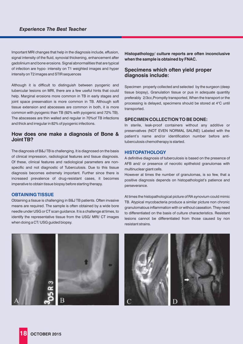
Important MRI changes that help in the diagnosis include, effusion,
signal intensity of the fluid, synovial thickening, enhancement after
gadolinium and bone erosions. Signal abnormalities that are typical
of infection are hypo- intensity on T1 weighted images and hyper
intensity on T2 images and STIR sequences
Although it is difficult to distinguish between pyogenic and
tubercular lesions on MRI, there are a few useful hints that could
help. Marginal erosions more common in TB in early stages and
joint space preservation is more common in TB. Although soft
tissue extension and abscesses are common in both, it is more
common with pyogenic than TB (92% with pyogenic and 72% TB).
The abscesses are thin walled and regular in 70%of TB infections
and thick and irregular in 82% of pyogenic infections.
How does one make a diagnosis of Bone & Joint TB?
The diagnosis of B&J TB is challenging. It is diagnosed on the basis
of clinical impression, radiological features and tissue diagnosis.
Of these, clinical features and radiological parameters are non-
specific and not diagnostic of Tuberculosis. Due to this tissue
diagnosis becomes extremely important. Further since there is
increased prevalence of drug-resistant cases, it becomes
imperative to obtain tissue biopsy before starting therapy.
Obtaining a tissue is challenging in B&J TB patients. Often invasive
means are required. The sample is often obtained by a wide bore
needle under USG or CT scan guidance. It is a challenge at times, to
identify the representative tissue from the USG/ MRI/ CT images
when doing a CT/ USG guided biopsy.
OBTAINING TISSUE
Histopathology/ culture reports are often inconclusive
when the sample is obtained by FNAC.
Specimens which often yield proper diagnosis include:
Specimen properly collected and selected by the surgeon (deep
tissue biopsy), Granulation tissue or pus in adequate quantity
preferably 2/3cc,Promptly transported, When the transport or the
processing is delayed, specimens should be stored at 4°C until
transported.
SPECIMEN COLLECTION TO BE DONE: In sterile, leak-proof containers without any additive or
preservatives (NOT EVEN NORMAL SALINE) Labeled with the
patient’s name and/or identification number before anti-
tuberculosis chemotherapy is started.
A definitive diagnosis of tuberculosis is based on the presence of
AFB and/ or presence of necrotic epitheloid granulomas with
multinuclear giant cells.
However at times the number of granulomas, is so few, that a
positive diagnosis depends on histopathologist’s patience and
perseverance.
At times the histopathological picture of RA synovium could mimic
TB. Atypical mycobacteria produce a similar picture non chronic
granulomatous inflammation with or without caseation. They need
to differentiated on the basis of culture characteristics. Resistant
lesions cannot be differentiated from those caused by non
resistant strains.
HISTOPATHOLOGY
18 OCTOBER 2015
Experience The Best Teacher

MICROBIOLOGY
MEDICAL TREATMENT
SURGICAL TREATMENT
Bone and joint TB is a pauci-bacillary disease. The culture positivity
is reported to be between 10-60%. The classic culture medium is
Lowenstein Jensen or solid medium culture and has a low
sensitivity and takes 6 to 8 weeks for growth. The recent cultures
methods like Bactec and MGIT (Mycobacterium Growth Indicator
Tube) are more sensitive. While Bactec is a semi-automatic method
and exposes the personnel to radiation, MGIT, is a very sensitive
test and yields a report in 2 to 3 weeks. However it is advisable to do
both do LJ as well as MGIT cultures and accept a negative report
only when both are negative.
What is the current management of Bone and Joint TB?
The medical treatment of B&J TB is similar to pulmonary TB.
However there are some issues which make medical management
of OA TB difficult. Most often in our country as the tissue diagnosis
is not done in every case, the diagnosis and treatment is empirical.
The change from intensive phase to maintenance phase is again
empirical. The end point is not clearly defined (unlike pulmonary TB
where 4 months after sputum turns negative is considered as the
end point). Various studies have recommended giving AKT for a
total of 9 months to 24 months. However in drug-sensitive cases,
the results of appropriately instituted and completed therapy are
good.
The protocol most commonly followed is
Isoniazide - 5mg/Kg All four drugs for 2-3 months as
Rifampicin - 10 m /kg intensive phase
Ethambutal - 15 to 20mg/kg
Pyrazinamide - 25mg/kg
This is followed by For 4 to 6 months as Maintenance
Isoniazide - 5mg/Kg phase
Rifampicin - 10 mg/kg
The treatment of MDR is generally initiated and managed by
infection disease specialists or chest physicians and are on similar
basis to resistant pulmonary TB
Surgical treatment is needed only in selected cases. The common
indications for surgical intervention in Pott’s spine are spinal
instability, late onset paraplegia, progressive paraplegia and very
large abscesses.
In other sites, surgical intervention is done for tuberculous
osteomyelitis with abscesses with large sequestrate and sclerotic
bones. Best to do surgery before putting the patient on ATT as the
chances of obtaining culture in sensitive cases reduces
significantly with therapy. Discharging sinuses not responding to
medical line of management also need surgical excision.
Totally destroyed joints, causing significant pain or disability, need
surgery. Excision, fusion and at times joint replacement are
needed. Generally replacement is done a few years after the
disease is quiescent however some surgeons prefer to do so even
earlier.
Bone and joint Tuberculosis, although not as prevalent and
infectious as its pulmonary counterpart, is increasing in incidence
and severity. It poses various diagnostic and therapeutic
challenges to a rheumatologist.
The symptoms and signs are often non-specific. In the early stages
it may difficult to get a tissue diagnosis. Imaging though very helpful
does not differentiate pyogenic from tubercular lesions. Since it is a
pauci-articular disease cultures from B&J TB patients do not have a
high yield. Along with the traditional culture medium, the newer
microbiological methods have improved diagnostic capabilities.
There still is no definite consensus as to how long to treat.
1.Pawar UM, Kundnani V, Agashe V, Nene A, Nene A. Multidrug-
resistant tuberculosis of the spine--is it the beginning of the end? A
study of twenty-five culture proven multidrug-resistant tuberculosis
spine patients. Spine 2009; 34(22): E 806-10.
2. Agashe V, Shenai S, Mohrir G, Deshmukh M, Bhaduri A, Deshpande
R, Mehta A, Rodrigues C. Osteoarticular tuberculosis--diagnostic
solutions in a disease endemic region. J Infect Dev Ctries. 2009 Aug 30;
3(7):511-6.
3.Patankar H, Kakatkar V, Shah C, Agashe V. Coracoacromial pins to
augment external fixation of the proximal humerus. J Bone Joint Surg
Br. 1995; July 77(4): 660-1.
SUMMARY
REFERENCES
}
}
OCTOBER 2015 19
FOR MORE INFORMATION:
Email: [email protected]
Experience The Best Teacher

In contemporary ICUs, pulse oximetry is ubiquitous and there is
really no reason to routinely use oxygen in all patients.
Conventionally we target a SO2 of > 85-90% in stable awake
patients. It is reasonable to give hypoxemic patients some
supplemental oxygen to keep the SO2 above the desired
target. It is much more important is to find & correct the cause of
the hypoxemia. Though it is widely assumed that we actually
help patients by reversing hypoxemia, the impact on clinical
outcomes is not well documented.
A few recent trials allow us to get some perspective as the
whether giving oxygen to most of our patients is really
necessary or even safe. In fact, two recent Randomized Control
Trials (RCT) & one very large observational trial suggest that
liberal use of oxygen may be causing significant harm.
(1)One RCT in acute exacerbation of COPD found that the risk of
death in all patients was significantly reduced by 58-78% with
titrated oxygen treatment compared to liberal oxygen. Another
RCT compared Air versus Oxygen non-hypoxemic patients with (2)ST elevation myocardial infarction . The use of oxygen in these
patients was associated with higher rates of arrhythmias (40.4
vs 31.4%) and recurrent myocardial infarctions (5.5 vs 0.9%). It
was also associated with larger initial infarct size as judged by
CK-MB & Troponin peak, and also larger infarct size at 6 months
as evaluated by cardiac MR.
A large observational study looked at clinical outcomes of (3)exposure to high oxygen concentrations following CPR . They
defined Hyperoxia as being > 300mmHg/, and hypoxemia as
being < 60 mmHg/. They noted that arterial hyperoxia was
independently associated with increased in-hospital mortality
compared with either hypoxia or normoxia. In other words, not
only did the patients who had a PaO > 300mmHg do worse 2
than those with a normal oxygen level; they also did worse than
patients who were hypoxemic.
What messages can we take home from these three large
trials. Mainly that oxygen must be used judiciously as it can
cause more harm that is superficially apparent.
Critical care pearls
Oxygen: How Much is Too Much?
20 OCTOBER 2015
Second, we need to ask ourselves why a patient needs
oxygen if the SO2 is normal. Unfortunately stopping using
oxygen routinely will require reversing years of conditioning
that oxygen is the breath of life and all patients should get it.
REFERENCES
1. Austin MA, Wills E, Blizzard L, et al. High flow oxygen
increases mortality in chronic obstructive pulmonary
disease patients in a pre-hospital setting: a randomized
trial. BMJ 2010; 341: c5462
2. AVOID investigators. Air Versus Oxygen in ST-Segment
Elevation Myocardial Infarction. Circulation. 2015 Jun
16;131(24):2143-50
3. Association Between Arterial Hyperoxia Following
Resuscitation From Cardiac Arrest and In-Hospital
Mortality. JAMA. 2010;303(21):2165-2171
Oxygen is often believed to be “the breath of life” and is routinely and
unquestioningly used in almost all sick patients, as well as in those undergoing
DR. FARHAD KAPADIA
surgical or invasive procedures.This may be because oxygen is relatively cheap and
widely available. In the pre pulse oximetry era, mild hypoxemia could not be detected
clinically and it made sense to use it liberally in any potentially hypoxemic patient.
FOR MORE INFORMATION:
Email: [email protected]
Tel: 24447112
Ext: 8112
Consultant Physician

Veer Savarkar Marg, Mahim, Mumbai - 400 016 (INDIA) Tel: 2445 1515 / 2445 2222 / 2444 9199 Fax: 2444 9151 • [email protected] www.hindujahospital.com

22 OCTOBER 2015
UIP: Usual interstitial pneumonia | NSIP: Non-specific interstitial pneumonia
BOOP: Bronchiolitis obliterans with obstructive pneumonia | AIP: Acute interstitial pneumonia LIP:
Lymphoid interstitial pneumonia | DIP: Desquamative interstitial pneumonia
In a nutshell - ILD Patterns
DR. JAI MULLERPATTANAssociate Consultant
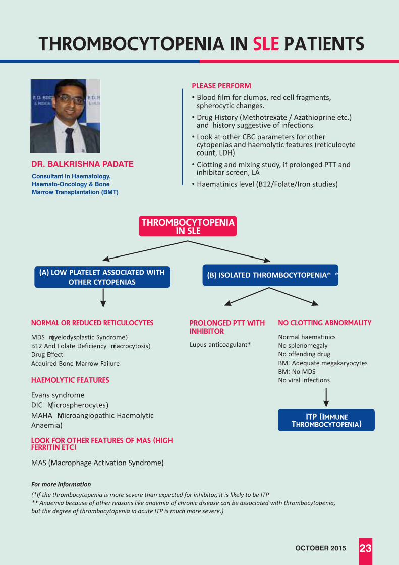
Thrombocytopenia in patientsSLE
DR. BALKRISHNA PADATE
PLEASE PERFORM
• Blood film for clumps, red cell fragments, spherocytic changes.
• Drug History (Methotrexate / Azathioprine etc.) and history suggestive of infections
• Look at other CBC parameters for other cytopenias and haemolytic features (reticulocyte count, LDH)
• Clotting and mixing study, if prolonged PTT and inhibitor screen, LA
• Haematinics level (B12/Folate/Iron studies)
(B) ISOLATED THROMBOCYTOPENIA**
Normal Or Reduced Reticulocytes
Haemolytic features
Look for other features of MAS (High ferritin etc)
MDS (myelodysplastic Syndrome)B12 And Folate Deficiency (macrocytosis)Drug Effect Acquired Bone Marrow Failure
Evans syndrome
DIC (Microspherocytes)MAHA (Microangiopathic Haemolytic Anaemia)
MAS (Macrophage Activation Syndrome)
Thrombocytopenia in SLE
Prolonged PTT with inhibitor Lupus anticoagulant*
No clotting abnormality
Normal haematinicsNo splenomegalyNo offending drugBM: Adequate megakaryocytes BM: No MDSNo viral infections
ITP (Immune Thrombocytopenia)
(*If the thrombocytopenia is more severe than expected for inhibitor, it is likely to be ITP** Anaemia because of other reasons like anaemia of chronic disease can be associated with thrombocytopenia, but the degree of thrombocytopenia in acute ITP is much more severe.)
(A) LOW PLATELET ASSOCIATED WITH OTHER CYTOPENIAS
For more information
Consultant in Haematology,
Haemato-Oncology & Bone
Marrow Transplantation (BMT)
OCTOBER 2015 23
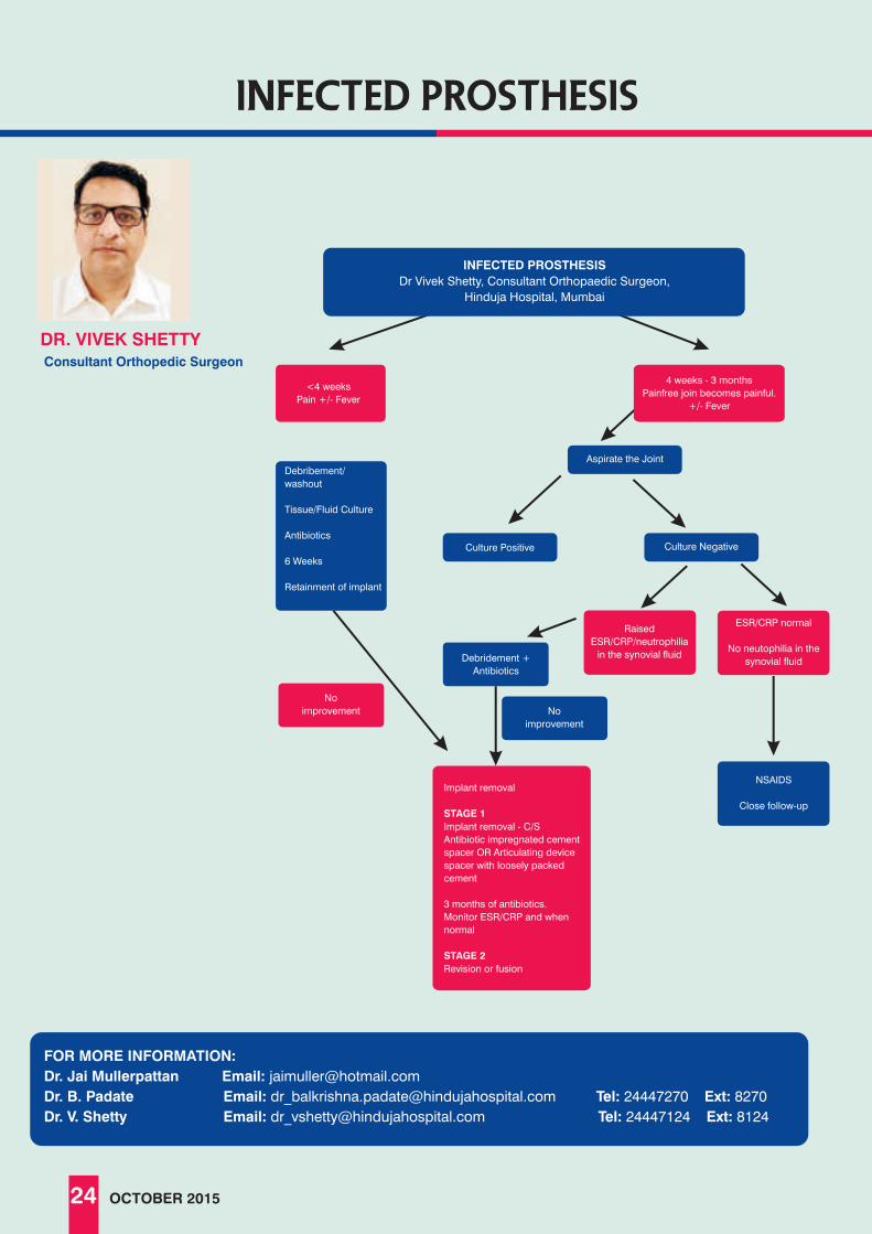
infected prosthesis
DR. VIVEK SHETTY
Aspirate the Joint
Culture NegativeCulture Positive
No
improvement
NSAIDS
Close follow-up
Debridement +
Antibiotics
Debribement/
washout
Tissue/Fluid Culture
Antibiotics
6 Weeks
Retainment of implant
No
improvement
Implant removal
STAGE 1
Implant removal - C/S
Antibiotic impregnated cement
spacer OR Articulating device
spacer with loosely packed
cement
3 months of antibiotics.
Monitor ESR/CRP and when
normal
STAGE 2
Revision or fusion
Raised
ESR/CRP/neutrophilia
in the synovial fluid
ESR/CRP normal
No neutophilia in the
synovial fluid
<4 weeks
Pain +/- Fever
4 weeks - 3 months
Painfree join becomes painful.
+/- Fever
INFECTED PROSTHESIS
Dr Vivek Shetty, Consultant Orthopaedic Surgeon,
Hinduja Hospital, Mumbai
FOR MORE INFORMATION:
Dr. Jai Mullerpattan Email: [email protected]
Dr. B. Padate Email: [email protected] Tel: 24447270 Ext: 8270
Dr. V. Shetty Email: [email protected] Tel: 24447124 Ext: 8124
24 OCTOBER 2015
Consultant Orthopedic Surgeon

100% Placement Assistance

tenderness over the deltoid muscle. Shoulder USG revealed a well
circumscribed oval lesion within the deltoid muscle. It had a
heterogeneous echotexture with lobular echogenic areas within and
was tender on applying probe pressure. These findings favour a solid
soft tissue intramuscular tumor. Further evaluation with MRI revealed an
enhancing well-defined posterior intradeltoid tumor ? nerve sheath
tumor.
Targeted pre-operative ultrasound localisation of the lesion was
performed as the lesion was small and changed position with
movement. Specific position of the arm was maintained that was
conveyed to the surgeon. This also helped in reducing the size of the
incison. Patient unerwent an excision biopsy and histopathology report
suggested vascular leiomyoma (SMA was strongly positive and S 100
was negative).
This case highlights the multipurpose role of ultrasound in soft tissue
lesions such determining the presence of an abnormality and then in
localising it, assessing internal morphology (solid, cystic, fat, etc.),
making a provisonal diagnosis, and when needed, localising it either for
biopsy or surgical removal. It is a quick and simple way to assess the
abnormality preliminary to a dedicated MRI, especially in small / non-
paplaple / mobile / deep lesions as in this case.
ACUTE ABDOMEN - EPIPLOIC APPENDAGITIS
A thirty-eight year old male came with an acute non-radiating right
flank pain. Severe point tenderness was noted on palpation. There
was no obvious evidence of acute appendicitis on ultrasound.
CT with oral and intravenous contrast revealed a focal oval fat density
area (about 3.5 cms in size) with surrounding inflammation in the right
flank between the ascending colon and redundant sigmoid colon.
Central linear area was noted possibly representing a thrombosed
vein. Constant relation was maintained between the lesion and
sigmoid colon. Findings represent inflamed fat, likely epiploic
appendagitis.
Torsion of the appendage (which is attached to the serosal surface of
the colon by a vascular stalk) leads to vascular occlusion, ischemia
and resultant acute pain. It is more common in males in the 4th and 5th
decade. Usual presentation is pain in the left iliac fossa with no fever
and leukocytosis.
In our case, there was right sided pain due to a redundant sigmoid
colon. Epilploic appendagitis is a self-limiting condition and resolves
with anti-inflammatory drugs. In recent times, it is diagnosed more
often because of the increasing use of CT in acute abdomen.
Knowledge of this entity avoids unnecessary hospitalization and
surgery. Differentials include an omental infarct.
radio logic
26 OCTOBER 2015
DR. ZENA PATEL
thirteen year old female complained of localised pain in the
right shoulder since 4 years. Clinically there was focal nodularA
FOR MORE INFORMATION:
Email: [email protected]
Lumps & Bumps-Ultrasound
Consultant Radiologist

Legal Eye - RES IPSA LOQUITUR
DR. SUGANTHI IYER
In all civil cases (including consumer), the onus of proof is on
the complainant to prove his complaint. This means that theI
Some examples of res ipsa loquitur applicable are:
• Development of meningitis after spinal anaesthesia
• Rupture of ear drum after ear syringing
• Permanent brain damage after appendidectomy
• Amputation of wrong limb or wrong digit or operation
• Burning of skin after strong antiseptic solution
• Injury,damage or death at a place where the attendants of patients
have no access (Labor room,ICU,OT)
• Leaving swabs or surgical instruments after an operation
OCTOBER 2015 27
The principle of application of res ipsa loquitur is clearly
illustrated in the following cases:
Mazumdar was admitted on the third floor in the "B" Hospital. He
was running high fever and was in a delirious state. After four
days, in the night around 2.20 am, the sister of the patient who
was staying with him in the room noticed the absence of the
patient from the room. She promptly informed the hospital staff
and a search was conducted to trace out the patient. The
security found him lying on the ground floor in the oncology
gallery of the hospital below the window of the room which the
patient was occupying. The patient had sustained multiple
fractures of lumbar vertebrae with complete dislocation of the
spinal cord and became a paraplegic.
The judge held that duty of a hospital is not limited to diagnosis
and treatment but extends to looking after the safety and
security of the patients. At the time of the incident, the patient
was an inpatient of the hospital. It was the duty of the hospital
authorities to take care of the patient who was suffering from
high fever and was in a delirious state. Due to absence of due
and reasonable care of the hospital authorities the incident
occurred, disabling the patient for the rest of his life and affecting
his employment and career prospects. Applying the principle of
res ipsa loquitur, the judge concluded that:
•Patient had sustained multiple injuries while he was an
inpatient in the hospital.
•The hospital has a duty towards the safety and security of its
patients.
•Had the hospital exercised due care, such accident would not
have occurred.
A.II(2014)CPJ 5(SC)SUPREME COURT OF INDIA {Mazumdar vs "B" Hospital}
JUDGMENT:
Deputy Director - Legal & Medical

In this case, Dr. G, had applied a plaster cast and Dr. S had
inserted a pin. But they had no records to prove that they
advised antibiotics or had taken any other steps to prevent gas
gangrene. Both these doctors committed egregious mistake
by not cleaning the wound properly. They were aware of the
fact that gas gangrene could be a complication under such
circumstances. Thus applying the principle of res ipsa
loquitur, the judge concluded that, that there was negligence,
inaction and passivity on their part which led to amputation of
the leg. Prompt action on part of Drs. G and S would have
prevented the gas gangrene and saved the leg of the patient.
Two and half lacs with 10% interest was awarded payable to
the complainant (30% by Dr. G and 70% by Dr. S)
• Hence the hospital is held liable and negligent for not
maintaining the necessary vigil in the hospital premises
to ensure safety of its patients.
Yogesh, who sustained multiple injuries and fracture in his
leg was taken initially to Dr. G who plastered his right leg.
After two days,patient approached Dr. S who operated him
and inserted a pin. Later on, Dr. S advised the patient to go
to a government hospital. Meanwhile,the foot emanated
foul smell since pus was present. The patient went to
Safdarjung Hospital who diagnosed that gas gangrene
had set in due to ill-treatment . AK amputation was done
immediately to save the life of the patient.
The court relied upon literature which clearly mentioned
that the organism Clostridia Botulinum of the gas gangrene
group multiply in anaerobic condition. Therefore as per
standard line of treatment while managing such a wound in
the leg, gas gangrene is better prevented by debridement
of all necrotic tissue, evacuation of pus, wound irrigation,
prophylactic antibiotics, gentle but effective application of
plaster to avoid compression of blood vessel. Above all,
the wound should not be tightly closed.
DAMAGES OF 7 LACS WITH 12% INTEREST WAS AWARDED .
JUDGEMENT:
B.II(2014)CPJ 499 (NC): Yogesh Vs DR.S & Ors.
28 OCTOBER 2015
FOR MORE INFORMATION:
Email: [email protected]
Tel: 24447723
Ext: 3223
Legal Eye - RES IPSA LOQUITUR
VIEW POINT
What Doctor’s Day Means To Me?
ach year when a new calendar arrives I check out the
national, holidays, - do they fall on Sat / Sun or not,Ethen the dates of important religious holidays, - how do the
dates vary from the previous year, and finally the dates of our
select hospital holidays, and then that’s it.
Today, doctors have fallen from grace and are seen by society as a profession steeped in corrupt practices, commercialism and greed. Alas this view is partly true and lies behind the several violent attacks on doctors reported from different parts of Mumbai by an enraged public seeking quick justice for fatalities they perceived as acts of medical negligence.
As leaders of the medical community what can we at HNH do to dispel this ‘radicalized’ view of our profession? We must be the flag bearers to restore again a strong doctor patient relationship which is at the heart of a noble profession.
But all is not gloom by any means. Great strides have been made in each and every specialty of medicine year on year. Technology is the driver behind many of these; - what was new yesterday risks being obsolete by next year. Only human nature remains largely unchanged, seeking succor in times of sickness and suffering. To those that combine compassion with science - Happy Doctors Day!
For other celebrity days such as Mother’s day, Father’s day, Teachers day and Doctors day I hardly give them a thought as I regard them as gimmicks put in place by a commercialized society. But now I find I am wrong.
Doctors Day is only commemorated by a handful of countries
wor ldwide of which Ind ia is one. We observe st1 July every year as it happens to be the birth anniversary of Dr.
B. C. Roy, an outstanding medical personality a freedom fighter,
a women’s rights advocate and a former chief minister of West
Bengal.
The practice was started in 1991 and has been followed yearly
thereafter. Seen from this sober perspective I feel we at HNH
should use the occasion for some healthy introspection.
OCTOBER 2015 29
DR. FARAM D. DASTUR
FOR MORE INFORMATION:
Email: [email protected]
Tel: 24447184
Ext: 8184
Director, Medical Education & Hospital Quality

30 OCTOBER 2015
Respiratory Review: July 19, 2015
Course Directors: DR. ASHOK MAHASHUR & DR. ZARIR F. UDWADIA
Organising Secretary: DR. JAI MULLERPATTAN
1. Dr. Camilla Rodrigues: TB diagnosis - standard of care in 2015
Dr. Rodrigues explained the various molecular genotypic tests available for TB diagnostics and also stressed
the need to identify the type of mutation which can predict high level or level resistance for the various drugs.
2. Dr. Zarir F Udwadia: Management of MDR TB - a case based approach
Dr. Udwadia presented three difficult cases of drug resistance TB each requiring varying approaches ranging
from new drugs such as bedaquiline, merpoenem and clavulanate and surgery or a combination of these.
3. Dr. Bhavin Jankharia: HRCT in the diagnosis of ILD’s
Dr. Jankharia elaborated on the various patterns of ILD as seen on an HRCT and clues to differentiate between
them.
4. Dr. Randeep Guleria: Newer drugs in COPD
Dr. Guleria gave an overview on the newer drugs for COPD both in the market and in the pipeline for various
stages in the pathogenesis right from smoking cessation, anti-inflammatories, bronchodilators and drugs to
prevent exacerbation.
5. Dr. Lancelot Pinto: Role of Pulmonary Rehabilitation in COPD
Dr. Pinto gave an excellent lecture on the need for pulmonary rehabilitation and the various components for the
same and the benefit they had in the quality of life and mortality of patients with COPD.
6. Dr. R Vijai Kumar: Asthma - COPD overlap syndrome
Dr. Vijai Kumar provided insights on this newly recognised entity along with clues to diagnosis and treatment
and differentiation from pure asthma or COPD.
7. Dr. Sujeet Rajan: Difficult asthma
Dr. Rajan expounded on difficult asthma on the basis of difficult disease, difficult patient and difficult doctors.
The need to educate patients and check the proper use of devices was emphasized as well as the fact that truly
difficult disease was a rare entity. Dr. Raja Dhar: Asthma phenotypes
8. Dr. Dhar provided an insight into various asthma phenotypes based on a case based approach along with a
need to individualise treatment in each of the cases such as obesity related asthma, neutrophilic asthma,
eosinophilic asthma etc.
9. Dr. Jai Mullerpattan: Management of severe pneumonia.
Dr. Mullerpattan educated the audience on the various severity scoring systems for community acquired
pneumonia as well as other devices to do the same such as biomarkers etc. An overview of the management
including antibiotic use and adjunctive therapy was also provided.

10. Dr. C Balakrishnan: Lung in Connective Tissue Diseases
Dr. Balakrishnan lectured on the differing pulmonary presentations in patients with connective tissue
disease and diagnosis/management of the same. Lung involvement in Rheumatoid arthrirtis, Systemic
sclerosis, Sjogrens syndrome, SLE and inflammatory myopathies were covered.
11. Dr. J C Suri: Sleep in respiratory diseases
Dr. Suri covered the change in patterns of breathing in sleep and the effect of the same in respiratory
diseases along with need for sleep study followed by nocturnal NIV or oxygen.
12. Dr. B K Smruti: Advances in the management of NSCLC
Dr. Smruti provided new insights in the advances of management of NSCLC while stressing the need to
obtain a histopathology diagnosis including type of NSCLC and presence of EGFR/ALK/K-Ras mutation so
that treatment can be tailored according to the same.
13. Dr. PP Prabhudesai: Management of the undiagnosed/difficult pleural effusion
Dr. Prabhudesai dwelled on length on the various causes of difficult pleural effusion along with clues to
diagnose them including biochemical tests and thoracoscopy.
OCTOBER 2015 31

32
QUIZ
1)
2)
3)
4)
5)
6)
7)
8)
9)
She qualified as a doctor from Grant Medical College during her reign as Miss World. Who is she
Name any Doctor who has also played test cricket
What is the abnormality seen on this X Ray
Why is July first celebrated as “Doctor’s Day”
Which Hollywood Actor who passed away recently, first achieved international fame for his role as a
Russian doctor.
This patient was depressed because of her disease and consumed a poison one night. The next morning
she was found smiling (instead of being found dead). Which disease? Which poison?
Siddhartha Mukherjee won the Pulitzer prize for his book “The Emperor of all Maladies”. What according to
him is the “Emperor of all maladies”
Who is the first Indian Doctor to scale Mount Everest
Which is India’s (and probably Asia’s) biggest Hospital (by bed strength)
OCTOBER 2015
DR. ASHIT HEGDE
Consultant Physician

OCTOBER 2015 33
10)
11)
12)
13)
14)
15)
In the movie “Anand” what was the disease that Anand was suffering from?
In the film “Trimurti”, the role of Kokha Singh was played by a doctor. What is his speciality
Which was the first hormone to be discovered
The Second nobel prize in medicine was awarded to a scientist for his work done
in India. Who was this scientist?
Both of these patients are suffering from Herpes zoster, which of them would you be more worried about
and why?
Who performed the first successful heart transplant and where
(1)2)3)4)
(5)(6)
(7)(8) (9) (10) (11)
(12)(13) (14)
(15)
Reita Faria Dr. W G Grace, Dr Ali Bacher Dextrocardia It is the birthday of Dr. Bidhan Chandra Roy (B.C Roy) a well-known
Physician and second Chief Minister of West Bengal Omar Shariff (Dr. Zhivago) Myasthenia Gravis , Organo phosphorous compound
Cancer Murad Lala of Hinduja Hospital Ahmedabad Civil HospitalLymphosarcoma of the Intestine Psychiatrist ( Mohan
Agashe) Secretin by Bayliss & Starling (not Insulin) Ronald Ross for his work on Malaria The second patient because there is a
lesion on the tip of his nose which means that he has a high risk of developing corneal involvement (The nasociliary nerve supplies both the tip of the
nose and the cornea) Dr. Christiaan Barnard in CapeTown
(((
Answers to quiz
FOR MORE INFORMATION:
Email: [email protected]
Tel: 24447263
Ext: 8263

Dr. Rucha KaushikMBBS, M.S.
Consultant Breast Cancer Surgeon
Dr. Shivkumar V. DalviM.S.(General Surgery, K.E.M.Hospital, Bombay)
(Surgery)Part Time Consultant in Health Check-up
Dr. Amresh Sudarshan BaliarsingMCh, DNB
Consultant Plastic Surgeon
Dr. kedar deogaonkarMS (Orth), DNB, MRCS, MSc,FRCS (Tr & Orth), CCT (UK)
Consultant Spinal Surgeon
Dr. nishant kumarMRCOphth (London), FRCOphth (London)
Consultant Ophthalmologist & Vitreo - Retinal Surgeon
Dr. satyakam krishna sawaimoonConsultant MD - Pathology & Surgical Pathology
new consultants
34 OCTOBER 2015
Dr. Hemanth Kumar PandharpurkarM.S. (General Surgery), M.R.C.S. (Edin.), Fellowship –
Vascular Surgery (Sree Chitra)
Consultant Vascular Surgeon
Dr. bhoomika thakoreMBBS, DNB Anaesthesiology
Post Doctoral Fellowship in Neuroanaesthesiolgy
Consultant Anaesthesia
Dr. pranjali advantMBBS, MD (Radiology), DNB, MNAMS, PDCC,
European Diploma in Radiology, EULAR Certified
Consultant Radiology

Best Multi Speciality Hospital-
Mumbai at Helthcare
Achievers Awards 2014
"Best Multi Specialty Hospital”
at e-HEALTH Maharashtra by
Department of Public Health,
Government of Maharashtra in 2014
Doc N Doc Awards
Best Multi-speciality
Hospital (Metro) 2014
ICICI Lombard and
CNBC TV18 Best Multi
Speciality Hospital in India -
Megapolis Award 2013 & 2011
Healthcare Leadership
Award by Stars of the
Industry 2014.
'Excellence in Community
Engagement' by Association
of Healthcare Providers
of India (AHPI) 2014.
Imc Ramkrishna Bajaj National
Quality Performance Excellence
Award 2014
In Healthcare Category
ICICI Lombard and
CNBC TV18 Best Multi
Speciality Hospital in India-
Megapolis Award 2014
AWARDS
"TB Champions" award by
Global Health Strategies, 2013
International Diamond Prize
for Customer Satisfaction,
by ESQR, 2012
IMC Ramkrishna Bajaj National
Quality Award, 2007
Excellence Award for CSR
project at The Asia
Healthcare Management
Awards, 2012
Excellence in Hospital
at the Medscape India
National Awards 2013
Best ICT enabled hospital
in Maharashtra at e-
Maharashtra Awards, 2013
Operational Excellence Award at
FICCI HEAL, 2013
Qimpro - Best Prax Benchmark,
Recogntion 2013 & 2010,
for Excellence in
Managerial Practices

Veer Savarkar Marg, Mahim, Mumbai - 400 016 (INDIA) Tel: 2445 1515 / 2445 2222 / 2444 9199 Fax: 2444 9151 • [email protected] www.hindujahospital.com