the “at-home lllt” in temporo-mandibular …...the “at-home lllt” in temporo-mandibular...
TRANSCRIPT

The “at-home LLLT” in temporo-mandibular disorders pain
control: a pilot study.
Fornaini C (1), Pelosi A (1), Queirolo V (1), Vescovi P (1) and Merigo E (1)
1) Department of Biomedical, Biotechnological and Translational Sciences (S.Bi.Bi.T.),
University of Parma, Italy
ABSTRACT
Objectives
The Temporo-Mandibular Disorders (TMD) are a set of dysfunctional
patterns concerning the temporo-mandibular joints (TMJ) and the
masticatory muscles; its main symptom is pain, probably caused by
inflammatory changes in the synovial membrane, alterations in the bone
marrow of the mandibular condyle and impingement and compression.
The aim of this preliminary study was to investigate the effectiveness in
the TMD pain reduction of a new laser device recently proposed by the
market that, due to its reduced dimensions and to be a class I laser
according the ANSI classification, may be used at home by the patient
himself.
Material and Methods
Twenty-four patients with TMD were randomly selected: the inclusion
criteria for the sample was the diagnosis of mono- or bi-lateral TMD, with
acute pain restricted to the joint area, associated with the absence of any
muscle tenderness during palpation.
The patients were randomly assigned to two groups:
Group 1 (12 patients): patients receiving real LLLT (experimental group).
Group 2 (12 patients): patients receiving inactive laser (placebo group).
The treatment was performed once a day for two weeks with a 808 nm
diode laser by the patient himself by irradiation of the cutaneous zone
corresponding to the TMJ for 15 minutes each side.
Each patient was instructed to express its pain in a visual analogue scale
(VAS) making a perpendicular line between the two extremes representing
the felt pain level.
Statistical analysis was realized with GraphPad Instat Software, where
P<0.05 was considered significant and P<0.01 very significant.
Results
The patient’s pain evaluation was expressed in the two study groups before
the treatment, 1 week and two weeks after the treatment.
The differences between the two groups result extremely significant with
p<0.0001 for the comparison of VAS value after 1 and 2 weeks.
Conclusion
This study, even if it may be considered such a pilot study, investigated a
new way to control the pain in the temporo-mandibular diseases by a at
home self administered laser device.
Results are encouraging but they will have to be confirmed by more
enlarged studies.
INTRODUCTION
Temporo-mandibular joint is one of the most fascinating and complex
synovial systems in the body. It is the area in which the mandible
articulates with the cranium [1]. The masticatory system is extremely
complex, which comprises primarily of bones, muscles, ligaments, and
teeth, all of which are responsible for activities like mastication, speech,
and deglutition. All these movements are regulated by an intricate
neurological controlling mechanism, which is important for the system to
function normally and efficiently. Lack of such harmony may lead to
disruptive muscle behaviour or structural damage to any of the
components [2]. The function of the TMJ is unique, in that the condyle
both rotates within the fossa and anteriorly translates along the articular
eminence. Because of the ability of the condyle to translate, the mandible
can have a much higher maximal incisal opening than would be possible
with rotation alone.
The Temporo-Mandibular Disorders (TMD) are a set of dysfunctional
patterns concerning the temporo-mandibular joints (TMJ) and the
masticatory muscles with frequent involvement of other structures of
various body districts with the result of making complicated the
classification and the diagnostic processes [3-6]; the average age of the
patients is 35.6 and the majority of patients are in the group of years
ranging from 26 to 40; the gender proportion is generally about 8 to 2 in
favour of the female [7]. Diseases related to the TMJ concern about a 1/3
of the general population [8].
The etiology of pain in TMD patients has not been clearly understood.
Occlusal disharmony and psychological distress are the two hypotheses,
which have dominated the literature. The psychological hypothesis

proposes that the disorder evolves as a consequence of psychological
distress that is usually due to the individual's stressful environment. The
psychological distress in turn leads to parafunctional habits (tooth
clenching and grinding) that result in muscle pain [9].
There are several possible sources of TMJ pain, such as inflammatory
changes in the synovial membrane including fluid resulting in joint
effusion [10-14], alterations in the bone marrow of the mandibular condyle
[15] and impingement and compression [16].
The roles of electro-physical modalities and surgery in the management of
TMD have not been fully elucidated [17]. Initial conservative therapy is
based on 3 general approaches: patient education, pharmacologic therapy,
and physical therapy. However, patients with chronic TMD usually need a
multidisciplinary approach involving a team of therapists, including a
dentist, psychologist, physical therapist, and even a chronic pain physician
[18].
Among the therapeutic procedures, low-level laser therapy (LLLT) has
recently been proposed to reduce pain intensity and improve maximal
mouth opening (MMO) in both acute and chronic TMD patients who had
received no previous TMD treatment (e.g., surgical treatment, occlusal
splint, or LLLT) [19]. A systematic review reported that LLLT is probably
more effective for the treatment of TMJ disorders, and less effective for
the treatment of masticatory muscle disorders [20].
The analgesic effect of LLLT acts at different levels and by different
mechanisms. Some explanations of this effect are: it increases beta-
endorphin level in spinal liquor, increases urinary excretion of gluco-
corticoids, which is a beta-endorphin synthesis inhibitor, increases the pain
threshold under pressure through a complex electrolytic nerve fibre
blocking mechanism, decreases histamine and acetylcholine release,
reduces bradykinin synthesis, increases ATP production, improves local
microcirculation, increases lymphatic flow thus reducing oedema [21, 22].
The aim of this preliminary study is to investigate the effectiveness in the
TMD pain reduction of a new laser device recently proposed by the market
that, due to its reduced dimensions and to be a class I laser according the
ANSI classification, may be used at home by the patient himself.
In fact, one of the problems related to the LLLT is represented by the
necessity, for the patients, to go to the therapist twice/three times for
treatments of some minutes. The appearing in the market of new LLLT
appliances, cheaper, smaller and able to be used at home by the patients
themselves might represent a solution to this problem, giving the
possibility to the patients to receive LLLT treatment also daily avoiding
the risk of overpower, due to this device has only a power setting.
MATERIAL AND METHODS
Patients
Twenty-four patients with TMD were randomly selected, informed about
their participation to the study and instructed about the modalities of the
test; all of them gave their consensus to participate to the protocol.
The inclusion criteria for the sample was the diagnosis of mono- or bi-
lateral TMD, with acute pain restricted to the joint area, associated with
the absence of any muscle tenderness during palpation.
The age of the patients ranged from 17 to 64 and among the 24 patients, 5
were of male and 19 of female gender.
The patients were randomly assigned to two groups:
Group 1 (12 patients): patients receiving real LLLT (experimental group).
Group 2 (12 patients): patients receiving inactive laser (placebo group).
The treatment was performed once a day for two weeks with a 808 nm Ga-
Al-As (Gallium-Aluminium-Arsenide) diode laser and it consisted in a “at-
home treatment” performed by the patient himself by irradiation of the
cutaneous zone corresponding to the TMJ for 15 minutes each side. Before
starting the treatment, all the patients were instructed by the same operator
about the procedures: to clean the area before the irradiation with
absorbent cotton socket with physiologic solution and to gently apply the
device in contact with the skin; in fact the beam source, in this way, is
maintained at a constant distance of 20 mm so irradiating an area of 4.5
cm2. It was not necessary, for the patients, to wear protective glasses, due
to fact that the appliance used is classified as class I device by the
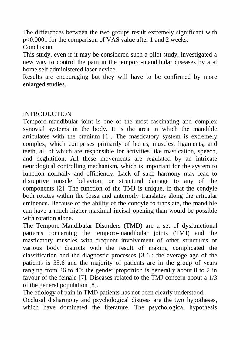
American National Standard Institute (Fig.1).
Figure 1: Laser application on the TMD.
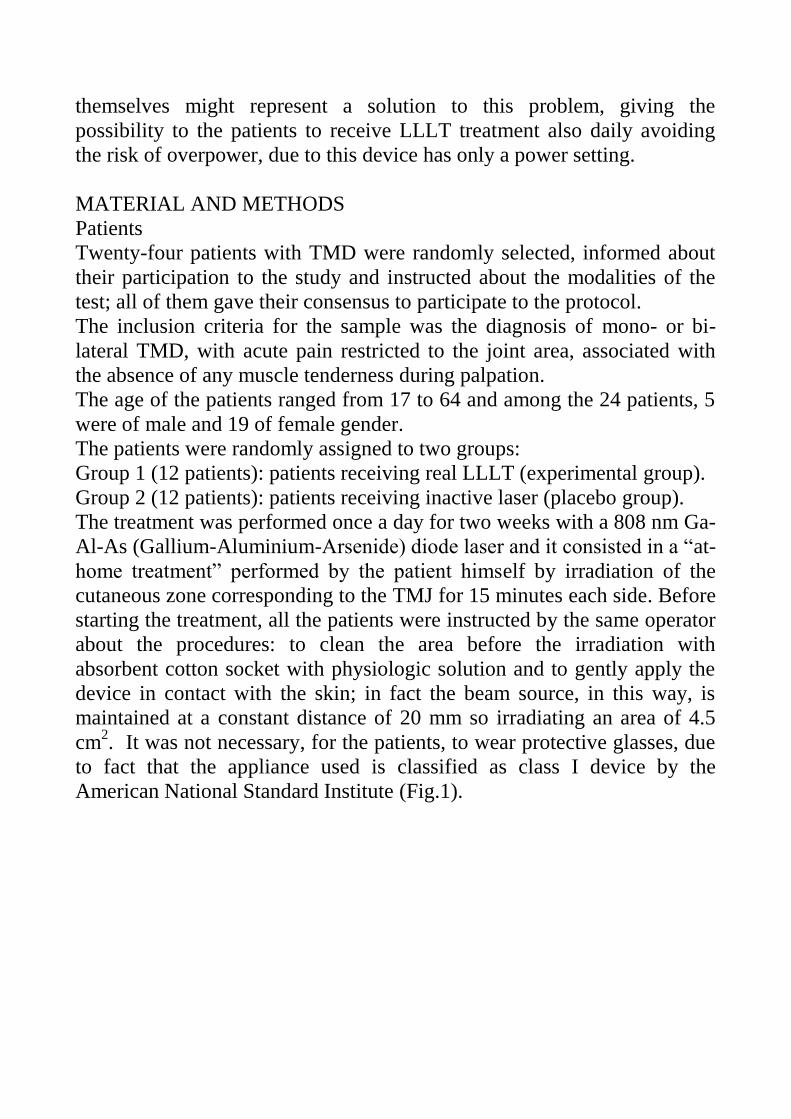
The laser apparatus was developed by the manufacturer in two identical
devices: one for the active laser and one for the inactive placebo laser, the
second one marked with a “P” letter. The lasers presented a green visible
guide light and only for the group 1 emitted also in the IR; the patients did
not know to which group they were assigned (Fig.2).
Figure 2: Devices used blindly by the patients: treatment device and
placebo device.
A visual analogue scale (VAS) allows the quantification of pain intensity.
This scale consists of a straight line measuring 10 cm in length, with
‘absence of pain’ written on the left end side and ‘worst pain ever felt’
written on the right end side. Each patient was instructed to make a
perpendicular line between the two extremes representing the felt pain
level [23].
Device
In this study it was used the B-Cure Laser (Israel). It is a Class I safety
little device (173 g of weight) emitting in the IR spectrum (808nm). A
green aiming beam is provided to show the irradiation area corresponding
to 4.5 cm2.
The Peak Power is 250 mW, given in micro-pulses with a Pulse frequency
of 15 kHz, for a Peak Energy per minute of 14.4 Joules.
Statistical analysis
To analyse the behaviour of the two groups, GraphPad Instat Software was
used, where P<0.05 was considered significant and P<0.01 very significant.
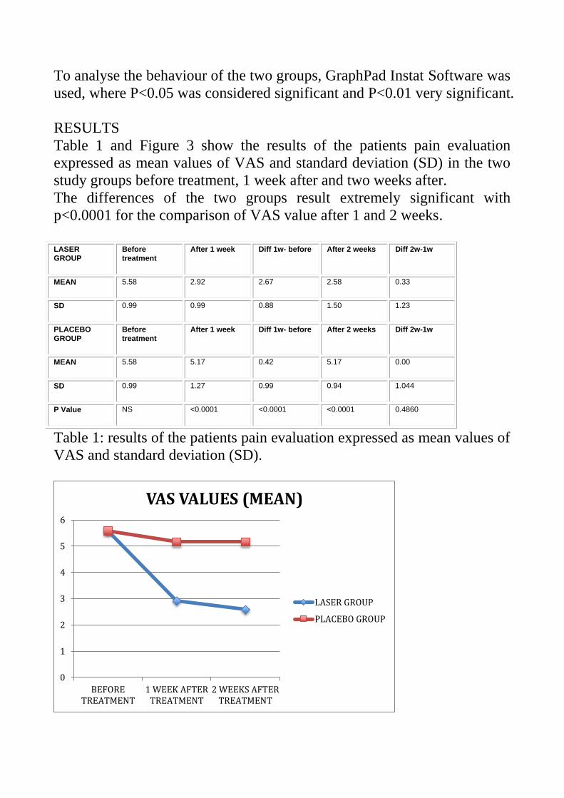
RESULTS
Table 1 and Figure 3 show the results of the patients pain evaluation
expressed as mean values of VAS and standard deviation (SD) in the two
study groups before treatment, 1 week after and two weeks after.
The differences of the two groups result extremely significant with
p<0.0001 for the comparison of VAS value after 1 and 2 weeks.
LASER
GROUP Before
treatment After 1 week Diff 1w- before After 2 weeks Diff 2w-1w
MEAN 5.58 2.92 2.67 2.58 0.33
SD 0.99 0.99 0.88 1.50 1.23
PLACEBO
GROUP Before
treatment After 1 week Diff 1w- before After 2 weeks Diff 2w-1w
MEAN 5.58 5.17 0.42 5.17 0.00
SD 0.99 1.27 0.99 0.94 1.044
P Value NS <0.0001 <0.0001 <0.0001 0.4860
Table 1: results of the patients pain evaluation expressed as mean values of
VAS and standard deviation (SD).
0
1
2
3
4
5
6
BEFORETREATMENT
1 WEEK AFTERTREATMENT
2 WEEKS AFTERTREATMENT
VAS VALUES (MEAN)
LASER GROUP
PLACEBO GROUP

Figure 3: results of the patients pain evaluation (means of VAS values) in
the two study groups before, 1 week after and two weeks after treatment.
DISCUSSION
In 2004, the World Association of Laser Therapy approved an agreement
on the format of clinical studies with LLLT for muscle and joint pain.
Lasers with an infrared wavelength are more suitable because they are
absorbed by the deeper layers of the tissue and not by the upper layer. The
most commonly used are located in the electromagnetic spectrum from
780 to 904 nm [24].
In a recent study, Kobayashi et al [25] hypothesized that one of the pain
relief mechanisms when using LLLT in the treatment of TMJ disorders is
the improved microcirculation in the temporal and masseter muscles. This
improved circulation helps to remove noxious deposits associated with
hypertension of the tissues. Pain relief is also felt by normalizing the
intramuscular pressure on sensory nerve endings. Other studies have
demonstrated that LLLT was shown to be effective for those with chronic
pain and in those who did not respond to other previous conservative
treatments [26].
The main important aspect regarding the LLLT treatment of the TMJ pain
concerns the correct parameters to use: in fact the intensity of the laser
must not harm the tissue, but can cause biochemical effects on cells, so the
laser is also known as the cold laser or soft laser [27].
The output power is normally less than 500 mW and the therapeutic doses
are less than 35 J/cm2 [28].
Due the applicable laser dosage follows the Arndt-Schultz rule, which
means that photo-bio-modulation only occurs when the dosage reaches the
threshold level [29], an effect would be suppressed if the dosage exceeds
the threshold.
CONCLUSION
This study, even if it may be considered such a pilot study due to its limits
(number of patients, number of data recorded, subjective evaluations)
investigated a new way to control the pain in the temporo-mandibular
diseases by a at home self administered laser device.
Results are encouraging but they will have to be confirmed by more
enlarged studies.

REFERENCES
1) Okeson JP. Management of Temporomandibular Disorders and
Occlusion. 5th edition. St. Louis, Mo, USA: Mosby; 2003.
2) McNeill C. Management of temporomandibular disorders: concepts and
controversies. J Prosthet Dent. 1997;77(5):510–522.
3) McNeill C, Mohl ND, Rugh JD, Tanaka TT. Temporomandibular
disorders: diagnosis, management, education, and research. J Am Dent
Assoc. 1990;120:253,255,257.
4) Mohl ND, Attanasio R. The Third Educational Conference to Develop
the Curriculum in Temporomandibular Disorders and Orofacial Pain:
introduction. J Orofac Pain. 2002;16:173–175.
5) List T, Walhund K, Wenneberg B, Dworkin SF. TMD in children and
adolescents: prevalence of pain, gender differences, and perceived
treatment need. Journal Of Orofacial Pain Winter. 1999
6) Al Ani MZ, Davies SJ, Sloan P, Glenny AM. Stabilization splint
therapy for TM pain dysfunction syndrome. The Cochrana Database of
Systematic Rewiews. 2004
7) Di Paolo C, Costanzo GD, Panti F, Rampello A, Falisi G, Pilloni A,
Cascone P, Iannetti G. Epidemiological analysis on 2375 patients with
TMJ disorders: basic statistical aspects. Ann Stomatol (Roma). 2013 Mar
20;4(1):161-9
8) Poirier F, Blanchereau C, Francfort E, Kolev T. Prise en charge
chirurgicale de l’articulation temporomandibulaire : à propos de 94 cas.
Rev Stomatol Chir Maxillofac. 2006;107:436–440.
9) Scarfe WC, Farman AG. Diagnostic imaging of the temporomandibular
joint. In: White SC, Pharoah MJ, editors. Oral Radiology, Principles and
Interpretation. 6th edition. Noida, India: Elsevier; 2010. pp. 473–478.
10) Haley DP, Schiffman EL, Lindgren BR, Anderson Q, Andreasen K.
The relationship between clinical and MRI findings in patients with

unilateral temporomandibular joint pain. J Am Dent Assoc 2001;132:476–
481.
11) Rudisch A, Innerhofer K, Bertram S, Emshoff R. Magnetic resonance
imaging findings of internal derangement and effusion in patients with
unilateral temporomandibular joint pain. Oral Surg Oral Med Oral Pathol
Oral Radiol Endod 2001;92:566–571.
12) Emshoff R, Brandlmaier I, Bertram S, Rudisch A. Risk factors for
temporomandibular joint pain in patients with disc displacement without
reduction - a magnetic resonance imaging study. J Oral Rehabil
2003;30:537–543.
13) Yano K, Sano T, Okano T. A longitudinal study of magnetic resonance
(MR) evidence of temporomandibular joint (TMJ) fluid in patients with
TMJ disorders. Cranio 2004;22:64–71.
14) Larheim TA. Role of magnetic resonance imaging in the clinical
diagnosis of the temporomandibular joint. Cells Tissues Organs
2005;180:6–21.
15) Emshoff R, Rudisch A. Temporomandibular joint internal
derangement and osteoarthrosis: are effusion and bone marrow edema
prognostic indicators for arthrocentesis and hydraulic distention?. J Oral
Maxillofac Surg 2007;65:66–73.
16) Güler N, Yatmaz PI, Ataoglu H, Emlik D, Uckan S.
Temporomandibular internal derangement: correlation of MRI findings
with clinical symptoms of pain and joint sounds in patients with bruxing
behaviour. Dentomaxillofac Radiol 2003;32:304–310.
17) Emshoff R, Bösch R, Pümpel E, Schöning H, Strobl H (2008) Low-
level laser therapy for treatment of temporomandibular joint pain: a
double-blind and placebo-controlled trial. Oral Surg Oral Med Oral Pathol
Oral Radiol Endod 105: 452–6
18) Okeson JP (2012) Temporomandibular Disorders in Chapter 5 -
Diseases of the Head and Neck. In: Bope ET, Kellerman RD, editors.

Conn's Current Therapy 2013, 1st ed., Pennsylvania: Saunders. pp. 320–
323.
19) Salmos-Brito JA, De Menezes RF, Teixeira CE, Gonzaga RK,
Rodrigues BH, et al. . (2013) Evaluation of low-level laser therapy in
patients with acute and chronic temporomandibular disorders. Lasers Med
Sci 28: 57–64
20) Melis M, Di Giosia M, Zawawi KH (2012) Low level laser therapy for
the treatment of temporomandibular disorders: a systematic review of the
literature. Cranio 30: 304–12
21) Simunovic Z. Low-level laser therapy with trigger points technique: a
clinical study on 243 patients. J Clin Laser Med Surg 1996;14:163–167.
22) Simunovic Z. Pain and practical aspects of its management. In: Laser
Medico, ed. Lasers in medicine and dentistry. Basic science and up-to-date
clinical application of low energy level laser therapy – LLLT. Rijeka:
European Medical Laser Association, 2000; cap. 14: 269–301.
23) Ferreira-Valente MA, Pais-Ribeiro JL, Jensen MP: Validity of four
pain intensity-rating scales. Pain 2011, 152:2399-2404.
24) Venancio RA, Camparis CM, Lizarelli RF. Low intensity laser therapy
in the treatment of temporomandibular disorders: a double-blind study. J
Oral Rehabil. 2005;32:800–807.
25) Kobayashi M, Kubota J. Treatment of temporomandibular joint (TMJ)
pain with diode laser therapy. J Laser Therapy 1999; 11.
26) Shiroto C, Ohshiro T. A new standard of Efficacy for Low Level Laser
Therapy (LLLT) in Pain Attenuation in Japan (a secondary publication).
Laser Ther. 2014 Sep 30;23(3):183-90.
27) Gonçalves RV, Novaes RD, Matta SL, et al. : Comparative study of
the effects of gallium-aluminum-arsenide laser photobiomodulation and
healing oil on skin wounds in wistar rats: a histomorphometric study.
Photomed Laser Surg, 2010, 28: 597–602

28) Aggarwal A, Keluskar V. Physiotherapy as an adjuvant therapy for
treatment of TMJ disorders. Gen Dent, 2012, 60: 119–122
29) Lubart R, Lavi R, Friedmann H, et al. Photochemistry and
photobiology of light absorption by living cells. Photomed Laser Surg,
2006, 24: 179–185.