the 3rd international forum on oxidative stress and aging · giorgio lenaz*, alessandra baracca,...
TRANSCRIPT

1
The 3rd International Forum on Oxidative Stress and Aging
September 11- 12, 2006
Nagoya University Lecture Hall (1F) at Kankyo Sogo-kan
Sponsored by: Daiko foundation
Center for Co-operative Research in Advanced Science and Technology. EcoTopia Science Institute, Nagoya University
Graduate School of Medicine, Nagoya University

2
Welcome to Nagoya It is my great pleasure to welcome everybody who is participating in this forum, and in particular speakers from abroad, and to have a chance to organize this triangular international forum consisting of Poland, Italy and Japan. The first international forum on Oxidative Stress and Aging was held in 2004 in Gdańsk、Poland organized by Professor Michał Woźniak, Medical University of Gdańsk, Gdańsk, and second time in 2005 in Pioraco, Italy, organized by Professor Giancarlo Falcioni, University of Camerino, Camerino. We would like to briefly explain the purpose of this forum on oxidative stress and aging. First of all, it is an attempt to bring together scientists standing on different research background, who working on oxidative stress related to apoptosis and aging. We are trying to provide a common platform for scientists working on various aspects of oxidative stress and apoptosis in cells. The aim is “union” rather than specialization. Now the time is ripe for synthesizing current biochemical, morphological and biophysical knowledge about the oxidative stress. Since I moved from University of Tokyo to Nagoya University according to the invitation by Professor Takashi Wakabayashi, I heard from him so many stories about Professor Jerzy Popinigis. I attended several times at Poland-Japan Forum on Biology and Sport Medicine organized by Professor Jerzy Popinigis, and became fascinated by his enormous friendship and faithfulness and ambition to science. Professor Jerzy Popinigis’s will has been succeeded and developed by his distinguished colleague, Professor Michał Woźniak. I do hope that all of participants will enjoy not only scientific discussions but also early autumn in Nagoya. September 11, 2006 Jiro Usukura
The Organizer of Nagoya Forum

3
Scientific Program September 11, 2006 9:00 – 9:05 Opening Remarks Jiro Usukura CCRAST, Nagoya University 9:05 – 9:10 Greeting Tsuneo Matsui Director of EcoTopia Science Institute, Nagoya University 9:10– 9:15 Greeting Michinari Hamaguchi Dean of Graduate School of Medicine Nagoya University Seminar 1 9:20 – 9:50 Chair: Giorgio Lenaz 9:20 – 9:50 Masashi Tanaka Tokyo Metropolitan Institute of Gerontology Polymorphism and mutations of mitochondrial genome contribute to human aging and degenerative diseases Seminar 2 9:50 – 10:40 Chair: Masashi Tanaka 9:50 – 10:20 Giorgio Lenaz University of Bologna Mitochondrial respiratory complex I in health and disease: From supramolecular organization to molecular mechanism of superoxide generation 10:20 – 10:40 Gian Paolo Littar ru Polytechnic University of the Marche New findings on coenzyme Q10 and cardiovascular disease 10:40 – 11:00 Coffee Break Seminar 3 11:00 – 11:45 Chair: Enrico Bertoli 11:00 – 11:15 Luca Tiano Polytechnic University of the Marche Extracellular SOD activity as a marker of endothelial functionality in cardiovascular disease. Effect of coenzyme Q10 treatment 11:15 – 11:30 Eva Bober Max-Plank-Institute, Bad Nauheim, Germany Inflammatory cardiomyopathy and premature aging in Sirt7 deficient mice 11:30 – 11:45 Edyta Niemczyk Medical University of Gdańsk Role of oxidative stress in the switch mechanism of the cell death mode from apoptosis to necrosis – involvement of plasma membrane NADPH oxidase 11:45 – 13:00 Lunch

4
Seminar 4 13:05 – 14:25 Chair: Masatoshi Hagiwara 13:00 – 13:05 Motoshi Suzuki Nagoya University Key note speak 13:05 – 13:25 Hideaki Tagami Nagoya City University Chromatin assembly and histone metabolism 13:25 – 13:45 Takeshi Urano Nagoya University How Aurora-A kinase activity is regulated during mitosis? 13:45 – 14:05 Akihiko Kikuchi Nagoya University Yeast NBP21S required for mitochondrial inheritance 14:05 – 14:25 Marinela Perpelescu Nagoya University Functional analysis of rsf complex in Hela cells 14:25 – 14:45 Coffee Break Seminar 5 15:05 – 16:05 Chair: Motoshi Suzuki 14:45 – 15:05 Masatoshi Hagiwara Tokyo Medical and Dental University; Alternative splicing reporter reveals cellular codes in vivo 15:05 – 15:25 Dean Rosenthal Georgetown University Cutaneous Responses to Carcinogens : Molecular Mechanisms of Skin Carcinogenesis Mediated by Id Proteins 15:25 – 15:45 Shunji Izuta Kumamoto University Regulation of eukaryotic DNA replication by PA2G4 15:45 – 16:05 Yoshiki Murakumo Nagoya University Analyses of ultraviolet-induced focus formation of hREV1, which is involved in DNA damage tolerance Distinguished Lecture (Open to public) 16:30 – 17:40 Chair: Kozo Kaibuchi John Heuser Professor of Biophysics and Cell Biology, Washington University School of Medicine, Fellow, National Academy of Science USA Imaging activities inside living cells by quick-freezing and electron microscopy.
5
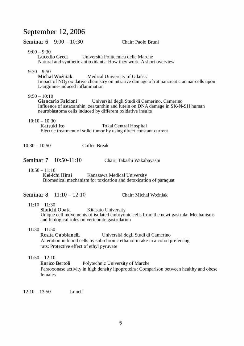
September 12, 2006 Seminar 6 9:00 – 10:30 Chair: Paolo Bruni 9:00 – 9:30 Lucedio Greci Università Politecnica delle Marche Natural and synthetic antioxidants: How they work. A short overview 9:30 – 9:50 Michał Woźniak Medical University of Gdańsk Impact of NO2 oxidative chemistry on nitrative damage of rat pancreatic acinar cells upon L-arginine-induced inflammation 9:50 – 10:10 Giancar lo Falcioni Università degli Studi di Camerino, Camerino Influence of astaxanthin, zeaxanthin and lutein on DNA damage in SK-N-SH human neuroblastoma cells induced by different oxidative insults 10:10 – 10:30 Katsuki Ito Tokai Central Hospital Electric treatment of solid tumor by using direct constant current 10:30 – 10:50 Coffee Break Seminar 7 10:50-11:10 Chair: Takashi Wakabayashi 10:50 – 11:10 Kei-ichi Hirai Kanazawa Medical University
Biomedical mechanism for toxication and detoxication of paraquat
Seminar 8 11:10 – 12:10 Chair: Michał Woźniak 11:10 – 11:30 Shuichi Obata Kitasato University Unique cell movements of isolated embryonic cells from the newt gastrula: Mechanisms and biological roles on vertebrate gastrulation 11:30 – 11:50 Rosita Gabbianelli Università degli Studi di Camerino Alteration in blood cells by sub-chronic ethanol intake in alcohol preferring rats: Protective effect of ethyl pyruvate 11:50 – 12:10 Enrico Bertoli Polytechnic University of Marche Paraoxonase activity in high density lipoproteins: Comparison between healthy and obese females 12:10 – 13:50 Lunch

6
Seminar 9 13:50 – 14:55 Chair: Giancarlo Falcioni 13:50 – 14:10 Paolo Bruni Università Politecnica delle Marche Ternary complexes liposome-DNA-Me2+ as synthetic vectors in gene transfer processes: The effect of DNA on the peroxidation of liposomes 14:10 – 14:25 Katarzyna Lechward Medical University of Vienna The function of ABCA class transporters in mouse tissues-Generation of tools 14:25 –14:40 Jedrzej Antosiewicz Jadrzej Sniadecki School of Physical Education and Sport c-Jun NH2-terminal kinase signaling pathway regulates ferritin degradation, labile iron pool, ROS and G2-M phase cell cycle arrest in human prostate cancer 14:40 – 14:55 Wiesław Ziółkowski Jedrzej Sniadecki University of Physical Education and Sport The effect of methyl-β-cyclodextrin on cholesterol efflux and rat liver mitochondria Bioenergetics 14:55-15:10 Zbigniew Śledziński Medical University of Gdańsk Oxidative stress markers during renal transplantation. 15:10 – 15:15 Closing Remar ks Shonen Yoshida Nagoya Kyoritu Hospital

7
Social Events
1. 10th in September (Sunday) 20:00~21:30 Get Together at Hotel MIELPARQUE 2. 11th in September (Monday) 18:00~20:00 Welcome Banquet at Hananoki Restaurant in Nagoya University (2 minutes walk from the lecture hall across the street) 3. 12th in September (Tuesday) 18:00~20:00 Farewell Party in Hotel MIELPARQUE (at Hagoromo room ) with KOTO Concert (Japanese Traditional harp)

8
Abstracts

9
POLYMORPHISMS AND MUTATIONS OF MITOCHONDRIAL GENOME CONTRIBUTE TO HUMAN AGING AND DEGENERATIVE DISEASES
Masashi Tanaka1, Yutaka Nishigaki1, Noriyuki Fuku1, and Yoshiji Yamada2 1 Department of Genomics for Longevity and Health, Tokyo Metropolitan Institute of Gerontology, Tokyo,
173-0015 Japan 2 Department of Human Functional Genomics, Life Science Research Center, Mie University, Tsu, Japan In spite of extensive efforts aiming at discovery of polymorphisms in the nuclear genome, only a few genetic factors predisposing to common multi-factorial, lifestyle-related, or age-dependent diseases, have been elucidated. The reason behind this failure may reside in the indifference of the ordinary researchers to the involvement of mitochondrial dysfunction in the pathogenesis of these disorders. We previously reported that haplogroup D is associated with longevity (Lancet 351: 185, 1998). The roles of mitochondria in a variety disease processes have been recently recognized. For example, mitochondrial dysfunction underlies impaired insulin secretion from the pancreatic beta cells as well as insulin resistance of skeletal muscles. To identify mtSNPs associated with age-related conditions such as longevity, Parkinson's disease, and Alzheimer's disease, as well as those related to energy metabolism such as obesity, thinness, and type-2 diabetes, or to atherosclerosis, we sequenced the entire mitochondrial genome of 672 individuals belonging to 7 different groups, with 96 individuals in each group, namely, centenarians, patients with Parkinson's disease, patients with Alzheimer's disease, young obese males, young non-obese males, and type 2 diabetes patients with or without severe vascular involvement (Genome Res 14: 1832, 2004). We constructed the human mitochondrial genome polymorphism database (http://www.giib.or.jp/mtsnp/index_e.shtml). On the basis of these mtSNP data we have developed a comprehensive mtSNP analysis system by use of fluorescent beads (Luminex). Our preliminary analysis has identified several haplogroups that are risk or protective factors for metabolic syndrome and/or type 2 diabetes. These studies are expected to pave the way to concerted investigations into treatments and therapies of both mitochondrial diseases and complex lifestyle-related disorders.

10
MITOCHONDRIAL RESPIRATORY COMPLEX I IN HEALTH AND DISEASE: FROM SUPRAMOLECULAR ORGANIZATION TO MOLECULAR
MECHANISM OF SUPEROXIDE GENERATION Giorgio Lenaz*, Alessandra Baracca, Christian Bergamini, Cristina Bianchi, Annalisa Biondi, Romana Fato, Maria Luisa Genova, Gianluca Sgarbi, Giancarlo Solaini Dipartimento di Biochimica, University of Bologna, Via Irnerio 48, 40126 Bologna, Italy *Corresponding author: [email protected] Mitochondrial Complex I (NADH Coenzyme Q oxidoreductase) is a very large enzyme catalyzing the first step of the mitochondrial electron transport chain. The total number of subunits in the bovine enzyme is 46 for a molecular mass of about 1000 KDa. The molecular mechanism of catalysis of this enzyme is not completely understood and the main reason is lack of detailed structural information. Recent structural and kinetic evidence suggests that Complex I is associated with a Complex III dimer in the form of a super-complex that may provide obligatory channelling of electrons via bound Coenzyme Q (CoQ) molecules. By application of metabolic flux analysis to NADH cytochrome c reductase activity in a reconstituted mitochondrial fraction from bovine heart, enriched with Complexes I and III, we have found that at high protein to phospholipids ratio these Complexes are associated in the form of a super-complex, whereas at low ratio they behave as independent units functionally linked by the CoQ pool. Peroxidation of the phospholipids used for reconstitution induces dissociation of the super-complex, with important implications for aging and age-related diseases where oxidative stress is an important pathogenic feature. One important feature of Complex I is its capacity to reduce oxygen to superoxide. By rational application of Complex I inhibitors and electron acceptors we have investigated the site of one-electron leak to oxygen in submitochondrial particles in which Complex I was functionally isolated by presence of mucidin that inhibits center o of the bc1 Complex but does not support superoxide generation from the Complex itself. We found that in presence of NADH and mucidin two classes of hydrophobic inhibitors can be distinguished on the basis of their ability to support superoxide formation: one class, including rotenone, piericidin A and rolliniastatins, enhances superoxide formation, whereas the other class, including stigmatellin, capsaicin and reduced short chain quinones, inhibits superoxide formation even in presence of an inhibitor of the first class. Hydrophilic quinone homologs, such as CoQ1, but not more hydrophobic analogs, such as decyl ubiquinone, enhance the prooxidant effect of rotenone-like inhibitors, indicating that a hydrophilic site is responsible of the electron leak. DCIP is reduced at three different sites, however its reduction at a distal site is inhibited by rotenone but not by stigmatellin. The bulk of these results suggest a bifurcated pathway of electrons flowing in Complex I where the acceptor ubiquinone is reduced by a two-step mechanism involving two different electron donors. The path involved in ubiquinone reduction to semiquinone is inhibited by rotenone, whereas the path involved in full reduction to ubiquinol is inhibited by stigmatellin. All findings may be explained by assuming that the ubisemiquinone is the electron donor to DCIP, whereas the stigmatellin-sensitive site is the donor to molecular oxygen. Block of electron transfer in Complex I, due to pathological alterations, such as in mitochondrial cytopathies, neurodegenerative diseases, aging and cancer, may differently affect the capability to generate superoxide, depending on the site affected. We present some data on Complex I alterations and superoxide generation in Leber’s Hereditary Optic Neuropathy, in aging and in a thyroid oncocytoma characterized by abnormal proliferation of mitochondria.

11
NEW FINDINGS ON COENZYME Q10 AND CARDIOVASCULAR DISEASE Gian Paolo Littarru
Institute of Biochemistry, Polytechnic University of the Marche, Ancona, Italy Abstract For a number of years Coenzyme Q (CoQ10 in humans) was known for its key role in mitochondrial bioenergetics; later studies demonstrated its presence in other subcellular fractions and in plasma, and deeply investigated its antioxidant role. These two roles constitute the basis on which research supporting the clinical use of CoQ10 is founded. Also at the inner mitochondrial membrane level, Coenzyme Q is recognized as an obligatory co-factor for the function of uncoupling proteins and a modulator of the transition pore. Recent data reveal that CoQ10 affects expression of genes involved in human cell signaling, metabolism and transport and some of the effects of exogenously administered CoQ10 might be due to this property. The clinical effects of CoQ10 in heart failure are commonly ascribed to its capacity of ameliorating the energy available for the various myocardial needs, such as contractility. In fact administration of an appropriate dosage of CoQ10 allowed us to measure an improvement of maximal oxygen uptake with the cardiopulmonary test. Furthermore we were able to demonstrate a significant improvement of contractility, by using the “low dobutamine dose” test.
A new concept, which has been addressed in the past few years, is the effect of CoQ10 on endothelial function. Impairment of endothelial function is a common feature in ischemic heart disease and this alteration often precedes the formation of the atherosclerotic plaque. It was shown that CoQ10 administration indeed ameliorates endothelial function in diabetic patients, as measured by the so-called flow mediated dilation (FMD). We extensively used this test with cardiovascular patients and were able to show that CoQ10 administration significantly improves FMD response. Among the biochemical reasons responsible for this effect, there could be an interaction of Coenzyme Q with superoxide anion, which could slow down the inactivation of NO· towards peroxynitrite. These observations were originally made on patients affected by ischemic heart disease, but the positive response to CoQ10 administration, in terms of amelioration of FMD was verified also in patients with heart failure. Keywords: CoQ10, atherosclerosis, endothelial function, myocardial contractility, FMD

12
EXTRACELLULAR SOD ACTIVITY AS A MARKER OF ENDOTHELIAL FUNCTIONALITY IN CARDIOVASCULAR
DISEASE. EFFECT OF COENZYME Q10 TREATMENT.
Dr. Luca Tiano, PhD
Institute of Biochemistry, Polytechnic University of the Marche, Ancona, Italy
Abstract Endothelial dysfunction reflects the impaired physiology of several endothelium-derived vasoactive factors, in particular nitric-oxide (NO), which plays a critical role in the endothelial homeostasis. It is involved in the regulation of arterial blood pressure, and also shows antiatherogenic properties. Oxidative stress has known repercussions on endothelial functionality through the inactivation and decreased synthesis of NO by reactive oxygen species such as superoxide. Extracellular superoxide dismutase (ecSOD) represent the major scavenger of superoxide anions in the arterial wall. The primary location of ecSOD in tissues is in the extracellular matrix and on cell surfaces where its concentration is 20 times higher than in plasma. The physiopathological role of ecSOD has been inquired in vascular related diseases, indicating a substantial reduction in vascular ecSOD activity in patients with coronary artery disease. Moreover, a strong correlation was found between endothelium bound ecSOD and flow dependent endothelial-mediated dilation, a functional parameter commonly used as a biomarker of vascular function. Finally, a relatively common missense mutation in codon 213 in exon 3 of the ecSOD gene (ecSOD-R213G). is associated with increased ischemic heart disease (IHD) risk. In fact, Heterozygous carriers of ecSODR213G have an ~10-fold plasma concentration of functional ecSOD due to accelerated release of ecSOD from the interstitial matrix. Consequently, despite high plasma concentration, the arterial wall may experience an oxidative stress condition. Coenzyme Q10 has a well acknowledged antioxidant role, besides its deeply inquired function in mitochondrial bioenergetics. Several studies have demonstrated that CoQ10 positively affects heart performance in congestive heart failure (CHF) and ischemic heart disease. Recent studies have also demonstrated an improvement in endothelial function after treatment with CoQ10 in diabetic patients. In the present study we aimed to determine whether oral CoQ10 supplementation (100 mg tid) was able to improve endothelium-dependent vasodilation and ecSOD activity in patients with coronary artery disease (CAD).

13
INFLAMMATORY CARDIOMYOPATHY AND PREMATURE AGING IN SIRT7 DEFICIENT MICE
Eva Bober
Max-Plank-Institute for Heart and Lung Research, Department of Cardiac Development and Remodeling, Parkstrasse
1, D-61231 Bad Nauheim, Germany [email protected] Single gene mutations that affect aging in a variety of organisms from yeast to mammals suggest
that evolutionary conserved universal mechanisms exist, which regulate aging and lifespan. Genes affecting the lifespan are organized in s. c. “longevity modules” and function in pathways that normally guarantee a better survival under harsh conditions such as food deprivation, cold or other kinds of environmental stress. Sir2 is a well-studied example of such a longevity gene that extends the lifespan in yeast, C. elegans and Drosophila when overexpressed. Sir2 encodes a histone/protein NAD+-dependent deacetylase and is involved in regulation of critical processes influencing stress resistance and metabolic pathways. In mammals seven Sir2-homologous genes, Sirt1 – Sirt7, also called sirtuins, exist and have been implicated in regulation of such different processes as transcription, replication, DNA-repair, stress resistance and metabolism. However, the molecular functions of mammalian sirtuins are only beginning to be uncovered.
In this paper, we show, that mice depleted of Sirt7 are susceptible to a variety of stresses, age prematurely, and have a decreased lifespan. Sirt7 seems to improve stress resistance through metabolic adaptation such as activation of fatty acid synthesis and through decreasing Ras-activated MAPK- and Akt-signaling pathways.
Sirt7 depleted mice are born alive and do not show any gross morphological abnormalities during the first months of life. However, beginning with approximately five months of age, several signs of premature aging become apparent: Sirt7 deficient mice are smaller than their littermates, they early develop kyphosis, have a reduced amount of visceral and subcutaneous fat, an increased level of inflammation, and acquire a reduced ratio of CD4/CD8 T-lymphocytes early in age. The majority of Sirt7 mutant animals die around the end of the first year of life (10 – 13 months) without any apparent cause of death.
Sirt7 mutants show a generally increased hyperactivity and exaggerated behavioral stress responses, which sometimes manifest in spontaneous epileptic seizures. Upon exposure to cold (4oC) stress after over night fasting, Sirt7 deficient mice are not able to keep a constant body temperature and die during the first three hours of cold exposure. Such deaths never occurred to wild-type animals. Sirt7 knockout mouse embryonic fibroblasts (MEFs) show a clearly decreased stress resistance as revealed by lower survival rates and higher apoptosis under adriamycin treatment. The increased genotoxic stress response correlates with p53 hyperacetylation. In fact, p53 acetylated peptides provide substrates for an efficient Sirt7-dependent deacetylation in vitro.
The majority of Sirt7 -/- mice suffer of mild to severe inflammatory cardiomyopathy. Apoptosis was increased in Sirt7 mutant myocard at about 50% as documented by TUNEL staining and electron microscopy. Furthermore, lipofuscin positive inclusions were also identified in Sirt7 deficient cardiomyocytes, while in the wild type cells lipofuscin staining was never observed at that age. Finally, degenerative changes were unambiguously demonstrated by an ongoing fibrosis, collagen VI accumulation and smooth muscle a-actin expression. In Sirt7 -/- hearts a significantly increased level of phosphorylated Akt indicating a hyperactivation of Akt-signaling, was observed. Since a long-term decrease of Akt/PKB pathway correlates with longevity in such different animals as C. elegans and mouse, the activation of Akt in the hearts of Sirt7 mutants confirms their premature aging phenotype.

14
ROLE OF OXIDATIVE STRESS IN THE SWITCH MECHANISM OF THE CELL DEATH MODE FROM APOPTOSIS TO NECROSIS –
INVOLVEMENT OF PLASMA MEMBRANE NADPH OXIDASE Edyta Niemczyk1*, Chieko Kurono2 Jakub Kedzior1, Rafal Januszewski3 and Takashi Wakabayashi1 1 Depatment of Cell Biology and Molecular Pathology, Medical University of Gdansk, Debinki1, Gdansk, Poland 2 Department of Anatomy, Nagoya City University Graduate School of Medicine, Nagoya, Japan 3 Department of Histology, Medical University of Gdansk, Debinki1, Gdansk, Poland *Corresponding author: [email protected] Cancer cells are very resistant to various drug treatment. However, it is possible to destroy these cells providing high reactive oxygen species (ROS) conditions. Intracellular ROS are mainly generated by mitochondria but recently it has been proven that also plasma membrane NADPH oxidase can participate in superoxide production. Cells can undergo two possible types of cell death: apoptosis and necrosis. Apoptosis is advantageous to organism helping to keep homeostasis whereas necrosis is very harmful causing local inflammations. The aim of this project is to find the molecular pathway which will force cancerous cells to undergo apoptosis during drug treatment, and also to find plasma membrane NADPH oxidase involvement in switch mechanism of cell death from apoptosis to necrosis in menadione treated human osteosarcoma 143B cell line. 143B cells were treated with an anticancer drug–menadion (MEN) (an artificial analogue of vitamin K3) for 6h, 9h and 24h. Menadion is known to induce superoxide formation which plays a role as signaling molecules. With flow cytometry method (using superoxide detecting probe-dihydroethidium) it was possible to visualize maximum of intracellular superoxide production at 6h of MEN treatment. At 9h the level of superoxide started to decrease while after 24h the superoxideproduction was almost completely abolished. These results correlated beautifully with electron microscopy data where it was possible to visualize apoptotic features of 143B cells after 6h of MEN treatment, intermediate cells after 9h and necrotic cells after 24h of MEN application. Up to now the existence of NADPH oxidase was confirmed in various types of cells like: epithelial cells, vascular smooth muscle cells, thyroid cells, fibroblasts, lymphocytes, spermatozoa and osteoclasts but so far no data on NADPH oxidase in 143B cells are available. The best recognized NADPH oxidase comes from phagocytes. This enzyme is composed of 6 subunits: plasma membrane bound cytochrome b558- a heterodimer of gp91phox and p22phox and the other 4 subunits existing in cytosol described as: p67phox, p47phox, p40phox and Rac1- a GTP-binding protein. To become active cytosolic subunits of NADPH oxidase are required to be phosphorylated. Only in that form they translocate to plasma membrane cytochrome b558 and assemble creating active form of the enzyme. To examine if superoxide generated from NADPH oxidase contributes to the transition of the cell death mode, specific inhibitors of NADPH oxidase were applied: diphenyleniodonium chloride, apocynin and N-vanillylnonanamide. All of them were effective in decreasing superoxide levels. To detect plasma membrane NADPH oxidase subunits in human osteosarcoma 143B cell line Western blot method was applied. With confocal microscopy it was possible to visualize the intracellular localization of NADPH oxidase subunits before and after MEN application. We succeeded to prove that in 143B cells NADPH oxidase does exist and its superoxide prodution participates in switch mechanism of death cell mode from apoptosis to necrosis. However, the exact pathway of these mechanisms still remains unknown.

15
Chromatin assembly and histone metabolism Hideaki Tagami (Grad. School Natural Sciences, Nagoya City University)
The chromatin-encoded information that includes histone modifications and histone variants
organizes specification of cell types. Despite of numerous studies about histone chaperones which
escort histones to DNA, the molecular events leading to the maintenance and transmission of
epigenetic information have been obscure. Recent insights into the histone complexes have
provided new aspects to the issue. The histone deposition machineries specialized for the variant
histones participate in the distinct nucleosome assembly pathways. The existence of histone H3-H4
dimer in the pre-deposited complexes can be envisaged potential implications of the epigenetic
inheritance (Cell (2004) 116, 51-61).
I will discuss our recent findings on the histone complexes in yeast. The histone complexes are
highlighted as a strategy to understand molecular mechanisms and the regulations of chromatin
dynamics.

16
How Aurora-A kinase activity is regulated during mitosis? Takeshi URANO
Department of Biochemistry II, Nagoya University Graduate School of Medicine,
65 Tsurumai-machi, Showa-ku, Nagoya 466-8550, Japan
Mammalian Aurora-A is related to a serine/threonine protein kinase that was
originally identified by its close homology with Saccharomyces cerevisiae Ipl1p and
Drosophila melanogaster aurora that are key regulators in the orchestration of mitotic
events. Aurora proteins are frequently overexpressed in human cancers, and Aurora-A
has also been described as an oncogene. This kinase family thus has considerable potential
as a molecular therapeutic target.
The protein level of Aurora-A, its peak kinase activity during mitosis and its
activation have been attributed to phosphorylation. We previously reported that 1)
Thr288 within the activation loop of Aurora-A is a critical residue for activating
phosphorylation events and that it is spatiotemporally restricted to a brief window at
mitosis on duplicated centrosomes and on spindle microtubules proximal to the poles
using phospho-specific Aurora-A monoclonal antibodies; 2) activated Aurora-A forms
complexes with the negative regulator protein serine/threonine phosphatase type 1
that was negatively phosphorylated on Thr320 by Cdk1/cyclin B and 3) Aurora-A is
regulated by ubiquitin-mediated proteolysis (the E3 ligase is the Cdh1-related form of
the anaphase-promoting complex/cyclosome) in late mitosis. We recently identified
Ca2+/calmodulin pathway plays a role in regulating Aurora-A function. We would like to discuss
how Aurora-A kinase activity is regulated.

17
YEAST NBP2 IS REQUIRED FOR MITOCHONDRIAL INHERITANCE
K. OHKUNI AND A. KIKUCHI
Nbp2p is a Nap1-binding protein in Saccharomyces cerevisiae identified by its interaction with Nap1 by a two-hybrid system. NBP2 encodes a novel protein consisting of 236 amino acids with an Src homology3 (SH3) domain. We showed that NBP2 functions to promote mitotic cell growth at high temperatures and cell wall integrity. Loss of Nbp2 results in cell death at high temperatures and in sensitivity to calcofluor white. Cell death at high temperature is thought not to be due to a weakened cell wall. Additionally, we have isolated several type-2C serine threonine protein phosphatases (PTCs) as multicopy suppressors and MAP kinase-kinase (MAPKK), related to the yeast PKC MAPK pathway, as deletion suppressors of the nbp2 mutant. Screening for deletion suppressors is a new genetic approach to identify and characterize additional proteins in the Nbp2-dependent pathway. Genetic analyses suggested that Ptc1, which interacts with Nbp2 by the two-hybrid system, acts downstream of Nbp2 and that cells lacking the function of Nbp2 fail to distribute mitochondria to daughter cells at high temperature, and to localize Nap1 and Ptc1 in the cytoplasm.

18
FUNCTIONAL ANALYSIS OF RSF COMPLEX IN HELA CELLS
Marinela Perpelescu1, Hua Yang1, Naohito Nozaki2, Chikashi Obuse3, Kinya Yoda1
1Nagoya Univ., Biosci. Biotech. Center, 2Kanagawa Dental College,
Dept Gene Mech., 3Kyoto Univ., Grad. Sch. Biostud.
Known as the two subunits of RSF complex, human ISWI-type of chromatin
remodeling complex, Rsf1 (ICEN2) and hSNF2H (ICEN8) were found as components of
ICEN (interphase centromere) complex in the CENP-A affinity precipitates isolated by NChIP
(native chromatin immuno-precipitation) (Obuse et al, 2004, Genes to Cells). Initially, RSF
complex was identified as a factor that favors in vitro DNA transcription initiation along with
FACT complex (LeRoy et al, 1998, Science) comprising of FACTp140 (ICEN6) and FACTp120
(ICEN12). In this work we addressed questions about the function of RSF at centromere
considering the possibility of its implication in remodeling of centromeric chromatin.
Indirect immuno-staining of HeLa cells using monoclonal antisera showed that Rsf1 and
SNF2H transiently co-localized with centromeres at late S-G2 stages of cell cycle. NChIP with
both anti-Rsf1 and anti-SNF2H antibodies co-precipitated CENP-A chromatin, indicating that
Rsf1-SNF2H complex physically associates with CENP-A chromatin in interphase.
siRNA-depletion of Rsf1/RSF caused conspicuous accumulation of prometaphase cells, and
increased misaligned chromosomes suggesting that RSF complex is necessary for normal
kinetochore function. Rsf1 or RSF depletion inhibits in vivo exogenous GFP-CENP-A loading
to centromeric chromatin, suggesting that Rsf1 is necessary for CENP-A uploading to
centromeres. Purified RSF complex can in vitro reconstitute and space CENP-A nucleosomes as
well as H3 nucleosomes. Given above data, we propose that RSF complex takes a role for
CENP-A chromatin remodeling and spacing.

19
ALTERNATIVE SPLICING REPORTER REVEALS CELLULAR CODES IN VIVO
Hidehito Kuroyanagi1, 2, Tetsuo Kobayashi3, 4, Shohei Mitani3, 4 & Masatoshi Hagiwara1, 2
1School of Biomedical Science and 2Medical Research Institute, Tokyo Medical and Dental University, Yushima, Bunkyo-ku, Tokyo 113-8510, Japan, 3Department of Physiology, Tokyo Women’s Medical University School of Medicine, Kawada-cho, Shinjuku-ku, Tokyo 162-8666, Japan and 4CREST, JST, Hon-cho, Kawaguchi, Saitama 332-0012, Japan.
Alternative splicing of pre-mRNAs enables multicellular organisms to create a huge diversity of
proteome from a finite number of genes. Many alternative splicing events have been shown to be
regulated in cell-type-dependent and/or developmentally regulated manners. However, extensive
studies in vitro or in cultured cells have not fully elucidated “cellular codes” that determine the
specific splicing patterns in living organisms. Here we show that a splicing reporter worm
visualized expression profiles of mutually exclusive exons at a single cell level in vivo. By utilizing
the worm, we screened for splicing mutants defective in the tissue-specific expression pattern with a
fluorescence-assisted worm sorter and identified a novel trans-acting factor and a cis-element
evolutionarily conserved in metazoan. The mutant was defective in alternative splicing of the
endogenous gene and showed corresponding specific phenotypes. This approach may open the way
to systematically decipher cellular codes regulating alternative splicing in vivo.
20
Cutaneous Responses to Carcinogens : Molecular Mechanisms of Skin Carcinogenesis Mediated by Id Proteins
Dean S. Rosenthal and Cynthia M. Simbulan-Rosenthal
Georgetown University School of Medicine
We are studying the response of skin to DNA damaging agents such as solar ultraviolet irradiation (UVB) and sulfur mustard. Chronic UVB stress deregulates the balance between survival and programmed cell death, promoting clonal expansion of initiated (mutated) cells and development of skin cancer. We found that immortalized human keratinocytes (KC) were sensitized to UVB-induced apoptosis, possibly representing a transient regression-prone interme-diate stage equivalent to the precancerous state known as “actinic keratosis”. We showed that the increased UVB sensitivity is related to upregulation of inhibitor of differentiation (Id) genes. The four members of the Id family (Id1-4) possess an HLH domain and act as dominant negative inhibitors of bHLH transcription factors including Rb, E2F, cyclins, ETS family proteins, and myoD, preventing them from binding DNA and exerting their transcriptional effects. Id proteins, thus, regulate various cellular processes such as cell cycle progression, apoptosis, and differentiation. Id1 and Id3 can delay or eliminate senescence by suppressing the p16/Ink4a promoter in human KC. Id2, a dominant negative inhibitor of Rb, binds Rb and prevents interaction between Rb and E2F-1, and allows hyperproliferation of certain cells. We found that Id3 is induced by UVB only in immortalized cells and kills them via Bax upregulation and mitochondrial destabilization, while Id2 is induced by UVB only in primary cells. Ectopic expression of Id2 in primary human KC suppressed differentiation, similar to the effects of UVB, and Id2 siRNA blocked this UVB response. These results suggest that UVB suppresses differentiation of primary KC at least in part via upregulation of Id2. Thus, upregulation of Id2 by UVB may predispose keratinocytes to carcinogenesis by preventing their normal differentiation program. Interestingly, ectopic expression of Id2 in Id2-knockout (KO) fibroblasts increased cell proliferation, induced properties of transformation, and formed solid skin tumors 10-14 days after grafting or subcutaneously injection into nude mice. Id3, on the other hand, induces a mitochondrial/ Bax/ caspase-9 – dependent apoptosis and mediates UVB-sensitization of immortalized human keratinocytes. Since UVB has been shown to generate reactive oxygen species (ROS) in UVB-exposed human KC and skin equivalent models, and Id3 has been identified as a redox-sensitive gene in other cell types, we are currently investigating whether UVB upregulates Id3 via ROS generation. DNA damage from sulfur mustard leads to skin blistering, and results from selective apoptosis of the basal layer of the epidermis leading to its separation from the dermis. Exposure of human KC as well as human skin grafts on the back of athymic mice showed that death and blistering is due to the activation of Fas, and caspases -3, -8, and -9. We are now testing inhibitors of these pathways to block UVB induced skin carcinogenesis and sulfur mustard blistering by exploiting our knowledge of the pathways we have delineated.

21
Regulation of eukaryotic DNA replication by PA2G4
Hiroyuki Kawasaki and Shunji Izuta
Graduate School of Science and Technology, Kumamoto University, 2-39-1 Kurokami, Kumamoto
860-8555, Japan
DNA replication is an essential event for proliferation in both prokaryotic and eukaryotic cells.
Although several protein factors involved in eukaryotic DNA replication are identified and
characterized, the exact regulation mechanism of DNA replication during S-phase has not been
clarified. The egg extract of African claw frog, Xenopus laevis, is often used as a cell-free DNA
replication system. Using this system, many replication factors are identified. To search a novel
regulatory factor of replication, the Xenopus egg extract was fractionated into both precipitate and
supernatant fractions with 50% ammonium sulfate. The supernatant fraction was further purified
and the protein factor that affected DNA replication in the extract with the single-stranded DNA
template was obtained. This factor was composed of a single polypeptide of 50 KDa on
SDS-polyacrylamide gel electrophoresis, and designate as Xep50. Analysis of Xep50 revealed that
this protein might be a Xenopus homolog of mouse p38-2G4 or human PA2G4. PA2G4 is known as
a proliferation-associated protein or a cell cycle-regulated protein that appears in the nuclei from
late G1 to early S phase and diminished at late G2 during cell cycle. Therefore, PA2G4 is thought to
have an important role on DNA replication or cell cycle progression. PA2G4 is also reported to bind
to Rb protein, DNA repair enzymes or histone deacetylase 2. Furthermore, it is reported that the
gene of this protein is highly mutated in HNPCC patients. The biological role of PA2G4 on the
regulation of DNA replication will be discussed.

22
Analyses of ultr aviolet-induced focus formation of hREV1, which is involved in DNA damage tolerance
Yoshiki Murakumo
Department of Pathology, Nagoya University Graduate School of Medicine
Translesion DNA synthesis (TLS) is one of the DNA damage tolerance mechanisms that allow cells
with DNA damage to continue DNA replication. Each of the mammalian Y-family DNA
polymerases (Polh, Poli, Polk, and REV1) has been shown to carry out TLS by itself or in a
combination with another enzyme in vitro. Recently, the C-terminal region of mammalian REV1
(the total 1251 residues in human) was found to interact with Polh, Poli, and Polk, as well as with
the REV7 subunit of another TLS enzyme Polz. Thus, it is proposed that REV1 plays a pivotal role
in TLS in vivo. To assess the DNA damage response of REV1 protein in cells, we examined the
localization of human REV1 protein (hREV1) in non-damaged and ultraviolet (UV)-irradiated cells.
Ectopically expressed hREV1 in mammalian cells was localized to the nucleus and exhibited
dozens of tiny foci in approximately 3% of non-damaged cells. The percentage of focus-forming
cells markedly increased after UV irradiation in a time- and dose-dependent manner. The focus
formation was associated with UV-induced DNA damage. Interestingly, although the hREV1 foci in
S-phase cells co-localized with PCNA foci, suggesting the association of hREV1 with the
replication machinery, hREV1 focus formation was observed not only in S-phase but also in
G1-phase. These findings demonstrate a possibility that hREV1 may be recruited at DNA damaged
sites before the replication starts and play some roles for DNA repair in G1 phase.

23
Imaging activities inside living cells by quick-freezing and electr on microscopy John Heuser, M.D.,
Biophysics and Cell Biology, Washington University School of Medicine, St. Louis, MO 63110
In recent years, enormous improvements have been made in imaging the inner workings of molecules by x-ray diffraction, and in imaging whole macromolecular machines by cryo-EM-tomography. But these gains have only been applicable to crystallized fragments of molecules (for x-ray) or isolated and purified macromolecules (for EM). This has precipitated an acute need to image macromolecular activities in their natural cellular context, in vivo, inside living cells. Here, improvements have lagged, with x-ray microscopy being a total failure and EM-tomography being applicable only to the most minute of free-living organisms. The roadblocks to advancement here have been the classical ones: the need to thin-section larger cells and tissues before imaging, and the inability to adequately freeze whole cells before sectioning. Consequently, the field has remained 'mired' in antiquated techniques of chemical prefixation that have not improved significantly since the days when the Egyptians of antiquity embalmed their kings and princes for perpetual rest in the pyramids, or the early electron microscopists struggled to cut sections thin enough to clearly discriminate cellular membranes from the crowded background of macro-molecules that exist free in the cytoplasm of cells. Against this discouraging impasse, one technique has stood out as a simple, robust means to totally circumvent the above technical problems: namely, "slam-freezing" of cells onto a copper block cooled to 4šK with LHe, followed by freeze-fracture and "deep-etching" to see macromolecules in their natural cellular context. For over a quarter of a century, this alternative approach has yielded a wealth of information about the most fleeting of macromolecular activities and the most delicate of macromolecular interactions, all within the interior of cells frozen directly from life but never embalmed or thin sectioned at all. In this talk, examples of the successes of this approach that have been achieved over the years will be illustrated, and these will be contrasted with fledgling and largely futile attempts to accomplish the same goals by alternative EM techniques, especially by cryo-EM-tomography. Additionally, the argument will be made that the "deep-etch" EM technique is not just a "poor man's alternative" to imaging molecular activities in living cells (although it is vastly simpler and cheaper to do than EM tomography), but is in fact the only proper approach to this goal, which deserves intense application by current electron microscopists and represents a rich substrate for further development and improvement by the next generation of biophysicists, engineers, and microscopists.

24
NATURAL AND SYNTHETIC ANTIOXIDANTS: HOW THEY WORK. A SHORT OVERVIEW
Paola Astolfi, Patricia Carloni, Elisabetta Damiani, Lucedio Greci * Dipartimento di Scienze dei Materiali e della Terra, Università Politecnica delle Marche, Via Brecce Bianche, I-60131 Ancona, Italy. * Corresponding author: [email protected] Peroxidative processes caused by free radicals are directly or indirectly responsible for some pathologies such as inflammation, carcinogenesis, ischemia-reperfusion damage and ageing.1 Oxygen, which is a vital species, may become toxic through its metabolites such as superoxide anion, hydroperoxyl and hydrogen peroxide,2 the so called reactive oxygen species (ROS) all involved in "oxidative stress". It is well known that when a piece of meat is exposed to air it becomes rancid (lipid peroxidation) due to the reactions of the lipids present in the tissue with oxygen; but when it is part of a live animal, this kind of degradation does not occur due to the antioxidant load possessed, produced or consumed by the live organism. All biological systems undergo peroxidative degradation in different ways1 likewise in polymer systems, either in plastics or elastomers.3 In order to maintain their physical and chemical properties, these organic systems have to be protected by antioxidants. Not all antioxidants exert their inhibitory action through the same mechanism. The antioxidants reported in Figure 1 have been chosen to explain the different mechanisms (hydrogen abstraction, electron transfer and radical coupling) involved in their antioxidant action.
OH
CH3
CH2CH2OH
OH
OH
CH2CH2OH
OHO
CH3
OH
CH3
CH3
CH3CH3
CH2(CH2CH2CHCH2)3HOH
OH
CH CHCOOH
OH
OMe
CH3 CH3
CH3
CH3
CH3 CH3CH3CH3
CH3 CH3 O
OH OH
CHOHCH2OH
O
N CH3
O
O
Ph
. N CH3
NPh
O
Ph
.N
O.
BHT Nitroxide 1 Nitroxide 2
a-Hydroxytyrosole Tyrosole Caffeic Acid a-Tocopherol
BHA
Ascorbic Acidb-Carotene
A)
B)
TEMPO
Tyrosole
Figure 1: A) Selected natural antioxidants; B) Selected synthetic antioxidants. 1. Halliwell B and Gutteridge JM., Free Radicals in Biology and Medicine, Oxford
University Press, Oxford 2001. 2. McCord JM: The evolution of free radicals and oxidative stress, Amer J Med 2000; 108:
652-659. 3. Pospisil J, Klemchuk PP: Oxidation inhibition of organic material. CRS Press Inc., Boca Raton,
Florida 1990.
25
IMPACT OF NO2 OXIDATIVE CHEMISTRY ON NITRATIVE DAMAGE TO RAT PANCREATIC ACINAR CELLS UPON L-ARGININE INDUCED
INFLAMMATION Michał Woźniak Department of Medical Chemistry, Medical University of Gdańsk, Gdańsk, Poland Nitric oxide (nitrogen monoxide, NO) produced by the constitutive nitric oxide synthase plays a critical physiological role in regulating vasomotor tone. On the contrary, the larger amounts produced by inducible nitric oxide synthase are cytotoxic. Though NO is fairly stable and relatively unreactive toward biological targets, reactive nitrogen species may promote significant oxidative chemistry inside the cell. Nitric oxide has been found by us, to be an important precursor for highly reactive nitric dioxide radical (nitrogen dioxide, NO2) production in L-arginine induced acute pancreatitis leading to profound cell injury and finally necrotic death (BBRC 326, 2005, 331-320). NO2 as a powerful oxidant can stimulate lipid peroxidation and damage proteins but it is not yet clear which particular targets of chemical attack by NO2 are the most critical in vivo. In the current studies we show that nitrogen dioxide generated in vivo through biotransformation of L-arginine promotes significantly oxidation of thiol groups buried in the hydrophobic clefts of endoplasmic reticulum proteins. Quite opposite, NO2 promotes only negligible lipid peroxidation within endoplasmic reticulum membranes. This could mean that under L-arginine supplementation insufficient lipid peroxides were formed in the membrane and that membrane protein thiol groups could act by preferential scavenging of NO2 and so protecting critical lipid moieties. These observations give rise to a novel free radical pathway involved in NO transformations into NO2. Selectivity of biological targets oxidation in L-arginine induced pancreatitis follow the rate constant of nitrogen dioxide reactions with important cellular constituents.

26
INFLUENCE OF ASTAXANTHIN, ZEAXANTHIN AND LUTEIN ON DNA DAMAGE IN SK-N-SH HUMAN NEUROBLASTOMA CELLS INDUCED BY
DIFFERENT OXIDATIVE INSULTS
Marcello Santoconoa, Monica Zurriaa, Manuel Carlonib, Donatella Fedelib,
Giancarlo Falcionib
aResearch, Development & Innovation Department, SIFI SpA, Via E. Patti 36, Lavinaio (CT), Italy bDipartimento di Biologia MCA, Università degli Studi di Camerino, Via Camerini, 2 I-62032 Camerino (MC), Italy.
E-mail:[email protected]
In order to gain more knowledge about the antioxidant role of the predominant carotenoids (lutein and zeaxanthin) of the human retina, we investigated their antioxidant activity and capacity. Astaxanthin was also included in this study because its structure is very close to that of lutein and zeaxanthin. The antioxidant activity of these molecules was evaluated using chemiluminescence techniques, with lucigenin and luminol as chemiluminogenic probes for the superoxide anion and hydrogen peroxide respectively. The scavenging activity versus reactive nitrogen species was followed by spectrophotometric analysis in the visible region. It was found that all three carotenoids have scavenging activity versus both, reactive oxygen (ROS) and nitrogen (RNOS) species. Possible antioxidant capacity of these three compounds was sought using a biological system consisting of SK.N.SH human neuroblastoma cells subjected to oxidative stress from exposure to UVA radiation or to different RNOS donors. In particular, we determined wheter these compounds were capable of minimizing DNA damage. DNA damage was assessed using the “comet assay” a rapid and sensitive single-cell gel electrophoresis technique able to detect primary DNA damage in individual cells. The alkaline comet assay revealed that both the exposition to UVA or to RNOS induced DNA damage in neuroblastoma cells. The presence of carotenoids during UVA exposition increased the damage. On the contrary, in the case of RNOS, the ability of zeaxanthin, lutein and astaxanthin to reduce the DNA damage depends on the type of RNOS donor and the carotenoid concentration used. The data from this study provide additional information on the antioxidant and prooxidant activities of the predominant macular pigment carotenoids of the human retina.

27
ELECTRIC TREATMENT OF SOLID TUMOR BY USING DIRECT ELECTRIC CURRENT Katsuki Ito1,2
1Tokai Central Hospital, Gifu, 2Nagoya University,Nagoya, Japan Introduction:
The direct electric current through the platinum electrodes as a cancer therapy was successfully applied to the lung tumors by Nordenstroem, Sweden, in 1978. Thereafter many researchers focused on the basic principle of this therapeutic method. In the present study animal experiments were done to study the mechanisms and the effectiveness of the method to solid tumors and to explore the conditions to obtain the good results. Materials and Methods:
One million Yoshida sarcoma cells were implanted intradermally to the abdominal wall of male Donryu rats aged 8 weeks, weighing 300- 350 gm. On the 5th post-implanted day, when the tumor size became about 12 mm in diameter, the rats were separated into 4 groups and were given the direct electric treatment through the pair of platinum electrodes for 1 hour daily for 4 days continuously with 3 mA 0.05 volt, 1 mA 0.02 volt, 0.05 mA 0.01volt and 0 mA 0 volt, to group A, B, C and the control group, respectively. To know the mechanisms of the effectiveness of the method, the specimens of the tumors reduced in their sizes after the treatment were obtained to extract DNA, and electrophoresis was done on the agarose gel by the method of Wyllie. Results:
Decreases of the size of the tumors began on the 8th day in 13 among 16 cases of group B (1 mA 0.02 volt) and completely disappeared on the 23rd day without any sign of recurrence for more than 3 months. The method employed was not effective in the rest of 3 animals in group B and they died in a month. The decrease of the size of tumors occurred to 4/10 of group A, 5/14 of group C and 0/10 in control group. The average survival periods were 14.5 days for group A, 17 days for group C, and 12.5 days for the control group. All animals in-group B survived for more 130 days. The gel-electrophoresis of DNA of the tumor after the treatment, the ladder patterns were observed. Conclusion:
Based on the results of this study the electric treatment of the solid tumors using a small constant direct current seems to be useful. As for the mechanisms of the disappearing of the tumor by this method, the histological study including DNA analysis indicates that tumor cells die not due to the simple tissue burn or necrosis but to apoptosis.

28
BIOMEDICAL MECHANISMS FOR TOXICATION AND DETOXICATION OF PARAQUAT
Kei-Ichi Hirai, Hiroki Shimada and Eriko Simamura Department of Molecular and Cell Structural Science, Kanazawa Medical University, 1-1 Daigaku, Uchinada, Ishikawa 920-0293, Japan. *Corresponding author: [email protected] Since 1961 when the potent herbicide paraquat dichloride (1,1’-dimethyl-4,4’-bipyridinium dichloride) was invented in England, it brought great development in agriculture. However, at the same time, paraquat caused many deaths because of severe multiorgan disorders and chronic lung fibrosis in patients who were exposed to it. Furthermore, it is recently appeared that paraquat may be a risk factor of Parkinson’s disease. The acute cytotoxic mechanisms of paraquat are mediated by ROS, but it was long time speculated that the ROS might be formed by microsomal drug-metabolizing enzyme systems. In 1985, however, we found that the dose of paraquat did not alter endoplasmic reticulum (corresponding to microsomes) ultrastructures, but mitochondria were selectively destroyed in rat lungs (Hirai et al: Exp Mol Path 1985, 43: 242-52). In this Forum we describe our research on the recent mechanisms of paraquat toxication and detoxication. Detoxication Mechanisms The survival rate of mice receiving 50 mg/kg paraquat was 40% at 7 days and significantly rose with pretreatment of phenobarbital, an inducer of microsomal drug-metabolizing enzyme systems (Hirai et al: Toxicology 1992, 72: 1-16). In 2002, we also demonstrated that pretreatments of mice with phenytoin, phenobarbital or rifampicin induced NADPH-cytochrome P450 reductase, CYP3A and CYP2B, and then paraquat was first metabolized to paraquat-monopyridone in the postmicrosomal cytosol and subsequently hydroxylated by the microsomal enzymes, resulting in the reduction of the mortality (Shimada et al: Arch Biochem Biophys 2002, 402: 149-57). In these animal livers, the smooth endoplasmic reticulum was vigorously hyperplastic. For further detoxication mechanisms, treatments with ROS scavengers may be useful for survival from paraquat, since alpha-tocopherol and antioxidative propofol significantly saved paraquat-exposed animals (Ariyama et al: Intens Care Med 2000, 26: 981-6). Toxication Mechanisms The most important findings were due to the evidence that mitochondria were always selectively destroyed at the earlier stage of paraquat acute cytotoxicity even in animal bodies (Hirai et al: Exp Mol Path 1985, 43: 242-52), cultured cells (Wang et al: J Elect Micros 1992, 41: 181-4) and isolated mitochondria (Hirai et al: 1992). As for the mechanisms, we found that mitochondria produced ROS in the presence of NADH and paraquat in combination (Hirai et al: J Elect Micros 1999, 48: 289-96), and that a novel NADH-quinone oxidoreductase activity was identified as the mitochondrial outer membrane function on paraquat (Shimada et al: Arch Biochem Biophys 1998, 351: 75-81). Finally, we propose the attractive mechanisms in which voltage dependent anion channel (VDAC) for controlling mitochondrial membrane permeability may play a role in paraquat toxicity, because in the VDAC-over expressed cells transfected with vdac1 cDNA, paraquat intensely generated ROS and damaged mitochondria, but in the VDAC-knocked down cells transfected with VDAC siRNA, paraquat failed to form mitochondrial ROS, resulting in the cells being unaffected (unpublised results).

29
UNIQUE CELL MOVEMENTS OF ISOLATED EMBRYONIC CELLS FROM THE NEWT GASTRULA; MECHANISMS AND BIOLOGICAL ROLES ON
VERTWBRATE GASTRULATION
Shuichi Obata1*, Kazuhiro Takano2, Shinji Komazaki2, Tsutomu Oinuma3, Makoto Asashima4,5 1College of Liberal Arts and Sciences, Kitasato University, 1-15-1 Kitazato, Sagamihara, Kanagawa, Japan 2Department of Anatomy, Saitama Medical School, Iruma, Saitama, Japan 3Department of Anatomy, School of Medicine, Miyazaki University, Kiyotake, Miyazaki, Japan 4Graduate School of Arts and Sciences, The University of Tokyo, Komaba, Meguro, Tokyo, Japan 5ICORP/JST, Japan *Corresponding auther: [email protected] INTRODUCTION: Gastrulation is an important step for morphogenesis in most species of multicellular animal embryos. The gastrulation is a complex phenomenon combined with cell movements, cell shape changes, cell-cell adhesion, cell-matrix adhesion, and so on. The cell movements and cell shape changes are important to form an archenteron (primitive gut) and mesoderm layer. In 1943, Holtfreter reported two types of unique cell movements, named creeping movement of vermiform cells and circus movement, in isolated embryonic cells from amphibian gastrulae. He suggested that these cell movements were important in gastrulation of amphibian embryos. For instance, the creeping movement was suggested to play roles in drawing blastopore into the embryo. However, little is known about mechanisms of these movements and their roles playing in gastrulation yet. We investigated mechanisms of the creeping movement to understand the physiological roles of this movement in amphibian gastrulation. MATERIALS AND METHODS: Japanese newt, Cynops pyrrhogaster, gastrulae (stage 12) were used. Presumptive mesoderm and endoderm were cut in Ca2+, Mg2+-free Steinberg’s solution with fine tungsten needles, and cultured on agarose bed in the same solution. The cell movement of the dissociated cells was observed under upright microscope through water immersion objective lens (10 X or 20 X) as reflective images (not transmission images), and recorded them through cooled CCD camera. In some cases, embryos were chemically fixed and observed under binocular, upright microscope, or electron microscope. RESULTS: In early gastrula embryo, presumptive mesodermal cells and a part of presumptive endodermal cells invaginating into the embryo transformed into sausage-like shape. They elongated toward the direction of surface of the archenteron. The elongated cells were also observed both in the dissociated mesodermal and endodermal cells, and they are called vermiform cells. They showed the creeping movement. In the vermiform cell, a hyaline pseudopodium was formed at protruded end, and a small knob at the opposite end. We analyzed the creeping movement in the vermiform cells by time-lapse microscopy. The cells moved with the hyaline pseudopodium at the head, which was like a worm. Several contracted sites were observed at the surface of the cell, simultaneously. The contraction waves at the cell surface moved backward, from the pseudopodium side to the knob side. Yolk granules and pigment granules also changed their positions inside the cells. By treatment of cytochalasin D (inhibitor for actin polymerization), the sausage-like shape of the vermiform cell quickly changed into round one. The elongation of the cells, contraction waves, and streaming of yolk granules and pigment granules were also completely inhibited by this treatment. Nocodazole (inhibitor for microtubule polymerization), however, did not affect them. We report the roles of cytoskeleton, intracellular Ca2+ concentration changes, and membrane traffic playing in the creeping movement of the vermiform cells.

30
ALTERATION IN BLOOD CELLS BY SUB-CHRONIC ETHANOL INTAKE IN ALCOHOL PREFERRING RATS: PROTECTIVE EFFECT OF ETHYL PYRUVATE Rosita Gabbianelli1, Donatella Fedeli1, Robet A. Olek2, Carlo Cifani3, Maurizio Massi4, Carlo Polidori4, Giancarlo Falcioni1
1 Dipartimento di Biologia MCA, Università degli Studi di Camerino, Via Camerini, 2 I-62032 Camerino (MC), Italy E-mail: [email protected] 2 Department of Bioenergetics, Jedrzej Sniadecki University School of Physical Education, Wiejska 1, 80-336 Gdansk, Poland 3 Dottorato di Ricerca in “Invecchiamento e Nutrizione”, Università degli Studi di Camerino, Via Camerini, 2 I-62032 Camerino (MC), Italy 4 Dipartimento di Medicina Sperimentale e Sanità Pubblica, Università degli Studi di Camerino di Camerino, Via Scalzino,3 2 I-62032 Camerino (MC), Italy Background Adult human consumption of wine or super-alcoholic drinks is a world-wide behaviour and alcohol over-consumption has been associated with different pathologies such as cirrhosis, acute and chronic pancreatitis, cardiovascular disease, psychological and neurological dysfunction and immune system damage. Numerous experimental data indicate that “free radicals” contribute to ethanol induced cell injury. This study has been conducted to examine the feasibility of preventing oxy radical-dependent oxidative stress to rats following ethanol consumption, using ethyl pyruvate as an antioxidant. Methods Genetically selected alcohol-preferring rats were given for ten weeks water or 10% ethanol in absence and in presence of different ethyl-pyruvate amounts. Animal body weight as well as food and fluid intake were recorded weekly. Food was available ad libitum for the entire period. At the end of the sub-chronic treatment their blood was collected and the different cell types separated. Erythrocytes were tested for their antioxidant enzyme activities and plasma fluidity. The “comet assay” was employed to determine the DNA status on white blood cells. Measurement of monocyte respiratory burst was monitored by chemiluminescence technique after PMA activation. Results We found evidence of changes on membrane physico-chemical properties and antioxidant enzyme activities of erythrocytes following ethanol intake. A protective role for ethyl pyruvate in mitigating the ethanol intake effect was measured. Also the DNA damage produced with sub-chronic alcohol treatment was reduced by the presence of ethyl pyruvate. Measurement of monocyte respiratory burst indicates a higher superoxide anion production in ethanol group compared to the control group. The presence of ethyl pyruvate reduces considerably the superoxide anion production. Conclusions Ten weeks of sub-chronic ethanol treatment produces significant damage to the different blood cells even to a strain of rats genetically selected for high ethanol intake. Cytotoxicities due to alcohol intake could be alleviated by redox reactants such as ethyl pyruvate.

31
PARAOXONASE ACTIVITY IN HIGH DENSITY LIPOPROTEINS: COMPARISON BETWEEN HEALTHY AND OBESE FEMALES
Enrico Bertoli*,Tiziana Bacchetti, Simona Masciangelo, Gianna Ferretti Institute of Biochemistry, Faculty of Medicine, Polytechnic University of Marche, 60100, Ancona, Italy *Corresponding author: [email protected] Several studies have shown modifications of lipid and lipoprotein metabolism in obese subjects. Hypercholesterolemia, high levels of triglycerides and of low density lipoproteins (LDL)and low levels of high density lipoprotein (HDL) are frequently observed in human obesity both in adult and paediatric patients. Moreover, alterations of lipoprotein heterogeneity have been demonstrated in these subjects. The modifications of lipoprotein levels and composition are likely related to the higher risk of cardiovascular disease associated with obesity. Several studies have demonstrated an increase of oxidative stress in obese subjects, with a higher susceptibility to lipid peroxidation of LDL isolated from obese subjects compared to healthy subjects. It has been suggested that the increase in oxidative damage could be due to a decrease in antioxidant properties. To further investigate the relationship between oxidative stress and lipoprotein alterations in human obesity, we studied the activity of paraoxonase and the levels of lipid hydroperoxides in HDL isolated from plasma of obese subjects (O-HDL) (n=12) with respect to HDL from age-sex-matched controls (C-HDL) (n=31). Paraoxonase is a calcium-dependent esterase associated with HDL (HDL-PON) that plays an important role in the protective effect exerted by HDL against oxidative damage of plasma lipoproteins and cell membranes. The enzymatic activity of HDL-PON varies widely among healthy humans and it has been suggested that subjects with low PON activity may have a greater risk of developing diseases in which oxidative damage and lipid peroxidation are involved in comparison with subjects with high PON activity Our results demonstrated, for the first time, that the activity of HDL-PON in obese subjects is significantly lower with respect to controls (p<0.001) and showed modifications in protein and lipid composition of O-HDL. Moreover, higher levels of lipid hydroperoxides in plasma lipoproteins (HDL and LDL) isolated from obese subjects (p<0.001) were observed. The negative correlations established between HDL-PON activity and the levels of lipid hydroperoxides in lipoproteins (HDL and LDL) confirm the relationship between paraoxonase and lipid peroxidation both in healthy and obese subjects (p<0.001). In addition, plasma levels of leptin correlated positively with the levels of lipid hydroperoxides (p<0.001) and negatively with PON activity (p<0.001) in HDL of obese subjects, suggesting a relationship between leptin and oxidative stress. In conclusion, this study confirms the alterations of lipoprotein composition and the increase of oxidative stress in obese subjects. Oxidative stress and oxidation of lipoproteins are implicated in the development of coronary heart disease and atherosclerosis. Therefore the lower paraoxonase activity and the compositional changes of lipoproteins could contribute to the higher risk of cardiovascular disease associated with human obesity.

32
TERNARY COMPLEXES LIPOSOME-DNA-ME2+ AS SYNTHETIC VECTORS IN GENE TRANSFER PROCESSES: THE EFFECT OF DNA ON
THE PEROXIDATION OF LIPOSOMES. Paolo Bruni1, Michela Pisani1, Oriano Francescangeli2 1Depatment of “Scienze dei Materiali e della Terra”, Università Politecnica delle Marche, Ancona. Italy 2Department of Fisica ed Ingegneria dei Materiali e del Territorio, Università Politecnica delle Marche, Ancona. Italy *Corresponding author: [email protected]
Gene therapy, because of its aim to eliminate causes rather than symptoms of diseases, is believed by many researchers to be the therapy of the 21st century. Recent completion of the working draft of the human genome has convinced scientists about the reliable possibility of using gene medicines to combat genetic diseases. Gene delivery vectors commonly used in gene therapy are both viral and non-viral ones. It is now well-established that the development of gene therapy is strictly depending on safe and efficient gene delivery reagents. In the next future perfect gene delivery vectors will be synthetic systems incorporating the advantages acquired by the viruses over millions of years of evolution, but avoiding the pathogenicity and the immune reactions produced by the viruses themselves. The entire field of non-viral gene therapy has known a great renaissance since the pioneeristic studies by Felgner. In these studies, cationic liposomes (CLs), self-closed structures composed of several curved concentric membranes, have revealed themselves as efficient alternatives to viruses. Our contribution to synthetic vectors for DNA involves the use of ternary “neutral liposome-DNA-Metal ions” (L-DNA-Me2+) complexes that show the advantage of a very week, if any, cytotoxicity. Some encouraging results have been recently obtained by us in an in vitro transfection of plasmid DNA experiment. Besides the necessity of determining the structure and the factors involved in the trasfection processes (both in vitro and in vivo), a large know how of the chemistry of such complexes is necessary. We started by studying their answer to oxidation processes; two different approaches have been so far undertaken in our laboratories. As a first contribution, the peroxidation promoted by ferrous ions (Fenton reaction) has been studied at the physiological pH value: a catalytic effect of DNA on the peroxidation of the liposome moiety of various complexes is operating. The lag time reduces or is completely annihilated and an increase, both of the yields and of the rates of the reactions, has been observed. This effect has been attributed to the ability of the three components, liposomes, DNA and Fe2+, to form a stable ternary complex, which produces a reduction of the undulatory fluctuations of the hydrocarbon tails of liposomes and strengthens the packing between the acyl chains in the lipid bilayers, with the consequence of enhancing the liposome crystallinity. As a second contribution, the autoxidation of phosphatidylcholine liposomes promoted by Cytochrome C and/or tert-butylhydroperoxide has been studied and the effect of DNA on the reaction checked. Different reactions have been performed at pH values ranging from 4 to 8, either on multilamellar (MLV) and unilamellar (ULV) liposomes of phosphatidylcholine. Simple MLV’s show a decrease both of the extent and the rate of oxidation on going from pH=4 to pH=8, while in the same time the extent and rate of oxidation of the complex MLV/DNA increases; starting from a higher degree of oxidation of simple MLV with respect to the complex MLV/DNA at pH=4, one reaches a higher degree of oxidation of the complex MLV/DNA, with respect to simple MLV, at pH=8. The inversion is observed at pH=6.6-7. The conclusion is that DNA is able to act as a pro-oxidant at higher pH and as an antioxidant at lower pH values. This result asks for different pH driven mechanisms involved in the processes. The extent of oxidation depends also on Cytochrome C concentration and the addition of tert-butylhydroperoxide: while the addition of the peroxide results in an increase of the oxidation extent due to Cytochrome C at acidic pH, at basic pH the peroxide is necessary to induce oxidation. Analogous results are obtained with ULV.
33
THE FUNCTION OF ABCA CLASS TRANSPORTERS IN MOUSE TISSUES-GENERATION OF TOOLS.
Katarzyna Lechward, Walter Glaser, Sussane Falkner and Karl Kuchler Medical University of Vienna, Department of Medical Biochemistry, Division of Molecular Genetics, Vienna, Austria.
ABC transporters are the ATP driven pumps, that contain an ATP binding cassette and regulate efflux/influx of variety of substrates, such as aminoacids, sugars and whole range of synthetic and naturally occurring products like drugs and toxins. Their topology models show a core structure, composed of two sets of transmembrane helices connected with a ATP binding loop, that forms so called nucleotide binding domain. Mammalian ABCs fall into six major families designated A – F. Based on sequence similarities further clusters can be distinguished within each of the families. After years of studying the roles of ABC proteins, there is still limited information available on their mechanisms of substrate recognition/transport and selectivity. We focused our research on mouse ABCA family and decided to develop tools that will prompt us to more detailed studies on expression, cellular localization and function(s) of its members. Our first goal is to obtain specific polyclonal antibodies that recognize their external loops. We predicted the most antigenic and ordered regions and expressed them as GST-fusion proteins in E.coli. On the average, we obtained 2mg of each antigen from a liter of bacterial culture. Presently, we are in the process of producing antibodies. Our next aim is to employ our antisera to detect localization of distinct ABCA transporters in a panel of mouse tissues.
34
c-JUN NH2-TERMINAL KINASE SIGNALING PATHWAY REGULATES FERRITIN DEGRADATION, LABILE IRON POOL, ROS AND G2-M PHASE
CELL CYCLE ARREST IN HUMAN PROSTATE CANCER Jedrzej Antosiewicz
Department of Bioenergetics Jedrzej Sniadecki School of Physical Education and Sport c-Jun NH2-terminal kinase (JNK) is a stress activate protein kinase present in every cell of human body. It has been generally accepted that reactive oxygen species (ROS) in addition to many other factors, are important activators of JNK. Although, recently it has been shown that activation of JNK can lead to ROS generation however the mechanism of this increase is not well understand. We hypothesize that JNK regulates ROS formation by regulating ferritin an iron storage protein, degradation. Ferritin degradation will lead to increase in labile iron pool (LIP) and ROS formation. We used DU145 and PC-3 human prostate cancer cells treated with diallyl trisulfide (DATS), which is a highly promising anticancer constituent of processed garlic as a model. Previously we demonstrated that DATS induced JNK activation, generation of reactive oxygen species (ROS) and G2-M phase cell cycle arrest. The present study was undertaken to gain insights into the mechanism of DATS-mediated ROS production using. The DATS-induced ROS generation and G2-M phase cell cycle arrest in DU145 and PC-3 cells was significantly attenuated in the presence of desferrioxamine (DFO), an iron chelator, but this protection was not observed with iron-saturated DFO. DATS treatment caused a marked increase in the level of labile iron that was accompanied by degradation of light chain of iron storage protein ferritin. Interestingly, DATS-mediated ferritin ubiquitination and degradation, increase in labile iron pool, ROS generation and cell cycle arrest were significantly attenuated by ectopic expression of a catalytically inactive mutant of JNKK2 as well as RNA interference of stress-activated protein kinase/extracellular signal-regulated kinase 1 (SEK1), which are upstream kinases in JNK signal transduction pathway. In addition proteosomal inhibitor blocks ferritin degradation and an increase in LIP. In conclusion, the present study provides experimental evidence to indicate existence of a novel signaling pathway involving JNK signaling pathway in regulation of ferritin ubiquitination and proteosomal degradation. Moreover the role of mitochondria in ROS formation in prostate cancer cells will also be discussed.

35
THE EFFECT OF METHYL- -CYCLODEXTRIN ON CHOLESTEROL EFFLUX AND RAT LIVER MITOCHONDRIA BIOENERGETICS.
Wiesław Ziółkowski*, 1 Miachał Szkatuła, 2 Artur Nurczyk, 1, Takashi Wakabayashi, 3 Wojciech Bogusławski2, Emilia Syta, 4 Jan Jacek Kaczor,1 Michał Woźniak2 1Department of Biochemistry, Jedrzej Sniadecki University of Physical Education and Sport, Wiejska 1, 80-336 Gdansk, Poland. 2Department of Medical Chemistry, Medical University of Gdansk, Debinki 1, 80-211 Gdansk,Poland.3Depatment of Cell Biology and Molecular Pathology, Medical University of Gdansk, Debinki 1, 80-211 Gdansk, Poland. 4Department of Biophysics, Medical University of Gdansk, Debinki 1, 80-211 Gdansk,Poland. *Corresponding author: [email protected]
The mitochondrial cholesterol is mainly located in mitochondrial outer membrane (MOM), and comparing with inner membrane (MIM) its level is about four times higher [Echegoyen et al. 1993]. Appropriate cholesterol content in mitochondria is important for normal function of that organelle [Echegoyen et al. 1993]. However its over-accumulation especially in MOM may be deleterious for the cell [Campbell et al. 2002]. On the other hand, Keatisuwan et al. [1991] documented lowered cholesterol content in cardiac mitochondria after exercise, while in experimental conditions cholesterol efflux especially from the plasma membrane may be induced by methyl -cyclodextrin (M- -CD) [Ohtani et al. 1989]. Recently we used M- -CD to study the effect of controlled cholesterol efflux on rat liver mitochondria function [Ziolkowski et al. 2004]. We observed that 2,5 min incubation of isolated RLM with 2% but not 1% of M- -CD caused decrease in function and coupling of mitochondria. Garofalo et al. [2005] also showed that the disruption of lipid microdomains in isolated mitochondria by M- -CD prevented mitochondria depolarization and cytochrome c release induced by GD3 or t-Bid. Therefore it seems that changes in mitochondrial cholesterol level may be involved in bioenergetics, life and death of mitochondria. The aim of the study was to check the effect of 2% and 4% of M- -CD on: 1) function (RCI) and coupling (ADP/O) of RLM in two temperatures 25 and 37 C, 2) cholesterol release form mitochondria 3) activities of cytochrome oxidase -COX (located in MIM) and rotenon insensitive NADH
cytochrome c reductase (located in MOM) Seven rats were sacrificed by decapitation, and the livers were used for the experiments. Mitochondria were isolated according to Adachi et al. [1991] method. The protein content in mitochondria was estimated with the Lowry et al. method [1951]. Oxygen uptake of mitochondria was measured at 25 and 37 C with a Clark oxygen electrode in Gilson Polarograph. Mitochondria were incubated for 2,5 min with respiration medium (control) or 2% and 4% of M CD. Respiratory control index (RCI) and ADP/O were calculated from oxygen electrode traces according to Estabrook [1967]. After incubation of 30 mg of mitochondrial protein with contol buffer or 2% and 4% of M CD, solutions were centrifugated 50000xg 30 min, and radioactive cholesterol content was measured in supernatants. COX and rotenon insensitive NADH cytochrome c reductase activities were assesed spectrophotometrically. Decreased function of RLM incubated with 2 and 4% of M CD were more evident in in 37 C then 25 C. Similary lowered ADP/O ratio was seen after RLM incubation with M CD in 37 C then in 25 C. 2 and 4% of M CD also caused release of cholesterol from mitochondria into the supernatant. The level of cholesterol in supernatants after 2% and 4% of M CD was respectively 13-fold and 23-fold higher compered with control. There was no effect of cholesterol removal agent on COX activities, but M CD decreased activity of rotenon insensitive NADH cytochrome c reductase.

36
OXIDATIVE STRESS MARKERS DURING RENAL TRANSPLANTATION Maciej Biernacki1, Justyna Bigda1, Kamil Jankowski1, Michał Woźniak2, Zbigniew Śledziński1
Dept of General, Endocrine and Transplantation Surgery1, Medical University of Gdansk Dept of Medical Chemistry2 Medical University of Gdansk
Introduction: Free radicals reactions are involved in many conditions. Among them, ischemia-reperfusion phenomena seems to be one of the most interesting. It occurs during kidney transplantation as a constant element of the operation. Aims: We investigated serum level of markers of free radical reactions during kidney transplantation. Material: 50 Patients who underwent the kidney transplantation in Dept of General, Endocrine and Transplantation Surgery of Medical University of Gdansk (1999-2002) were included in the study. Control group (n=10) consisted of patient without metabolic and neoplasmatic diseases, who underwent elective laparoscopic cholecystectomy. Methods: Blood samples were collected in five time points: during anesthetic induction and in 1-2, 10, 15, 60 min after vessels clamps removing. The serum levels of protein carbonyl groups, malonyl dialdehyde (MDA) and protein sulphydryl groups were measured. In control group, blood samples were taken just before surgery, and after 60 minutes of operation. Result: Increased levels of carbonyl groups, malonyl dialdehyde were found. The strongest increase of these parameters (especially protein carbonyl groups). Sulphydryl groups amount remained at almost the same level during observation. In control group oxidative stress markers levels did not change after surgery. Conclusions:
37
List of Pre-registered Participants (Alphabetical order in participant countries and names) AUSTRIA Katarzyna Lechward Medical University of Vienna, Department of Medical Biochemistry, Division of Molecular Genetics, Vienna, Austria
[email protected] GERMANY Eva Bober Max-Plank-Institute for Heart and Lung Research, Department of Cardiac Development and Remodeling, Parkstrasse 1, D-61231 Bad Nauheim, Germany [email protected] ITALY Enrico Bertoli Institute of Biochemistry, Faculty of Medicine, Polytechnic University of Marche, 60100, Ancona, Italy [email protected] Paolo Bruni Depatment of “Scienze dei Materiali e della Terra”, Università Politecnica delle Marche, Ancona. Italy [email protected] Giancarlo Falcioni Dipartimento di Biologia MCA, Università degli Studi di Camerino, Via Camerini, 2 I-62032 Camerino (MC), Italy. [email protected] Rosita Gabbianelli Dipartimento di Biologia MCA, Università degli Studi di Camerino, Via Camerini, 2 I-62032 Camerino (MC), Italy [email protected] Lucedio Greci Dipartimento di Scienze dei Materiali e della Terra, Università Politecnica delle Marche, Via Brecce Bianche, I-60131 Ancona, Italy. [email protected] Giorgio Lenaz Dipartimento di Biochimica, University of Bologna, Via Irnerio 48, 40126 Bologna, Italy [email protected] Gian Paolo Littarru Institute of Biochemistry, Polytechnic University of the Marche, Ancona, Italy [email protected]
Luca Tiano Institute of Biochemistry, Polytechnic University of the Marche, Ancona, Italy [email protected]

38
JAPAN Masatoshi Hagiwara School of Biomedical Science and Medical Research Institute, Tokyo Medical and Dental University, Yushima, Bunkyo-ku, Tokyo 113-8510, Japan [email protected]
Michinari Hamaguchi Department of Cancer Biology, Nagoya University Graduate School of Medicine, 65 Tsurumai-cho, Nagoya 466-8550, Japan [email protected] Kei-Ichi Hirai Department of Molecular and Cell Structural Science, Kanazawa Medical University, 1-1 Daigaku, Uchinada, Ishikawa 920-0293, Japan. [email protected] Katsuki Ito Tokai Central Hospital, 4-6-2 Sohara Higashijima-cho, Kagamihara, Gifu 504-8601 [email protected] Shunji Izuta Graduate School of Science and Technology, Kumamoto University, 2-39-1 Kurokami, Kumamoto 860-8555, Japan [email protected] Akihiko Kikuchi Division of Molecular Micology and Medicine Nagoya University Graduate School of Medicine Tsurumai-cho 65, Showa-ku, Nagoya 466-8550, Japan [email protected] Nobuhiro Morone Department of Ultrastructural Research, National Institute of Neuroscience, National Center of Neurology and Psychiatry 4-1-1 Ogawahigashi-cho, Kodaira, Tokyo 187-8502 Japan [email protected] Yoshiki Murakumo Department of Pathology, Nagoya University Graduate School of Medicine, Tsurumai-cho 65, Showa-ku, Nagoya 466-8550, Japan [email protected] Shuichi Obata College of Liberal Arts and Sciences, Kitasato University, 1-15-1 Kitazato, Sagamihara, Kanagawa, Japan [email protected] Marinela Perpelescu Nagoya Univ., Biosci. Biotech. Center, Chikusa-ku, Furo-cho, Nagoya [email protected]

39
Motoshi Suzuki Division of Molecular Carcinogenesis Nagoya University Graduate School of Medicine Tsurumai-cho 65, Showa-ku, Nagoya 466-8550, Japan [email protected] Hideaki Tagami Graduate School of Natural Sciences Nagoya City University, 1 Yamanohata, Mizuho, Nagoya, Aichi 467-8501, JAPAN [email protected] Kenji Tanaka National Institute of Technology and Evaluation 2-5-8 Kazusakamatari, Kisarazu, Chiba, 292-0818 Japan [email protected] Masashi Tanaka Department of Genomics for Longevity and Health, Tokyo, Metropolitan Institute of Gerontology, Tokyo, 173-0015 Japan [email protected] Takeshi Urano Department of Biochemistry II, Nagoya University Graduate School of Medicine, Tsurumai-cho 65, Showa-ku, Nagoya 466-8550, Japan [email protected] Jiro Usukura Center for Cooperative Research in Advanced Science and Technology, Nagoya University, 1-1 Furo-cho Nagoya 464-8603 Japan [email protected] Shonen Yoshida Nagoya Kyoritu Hospital, 1-172 Hokke, Nagoya 454-0933 Japan [email protected] POLAND Jedrzej Antosiewicz Department of Bioenergetics Jedrzej Sniadecki School of Physical Education and Sport Gdańsk Wiejska 1, 80-336 Gdansk, Poland [email protected] Edyta Niemczyk Department of Cell Biology and Molecular Pathology, Medical University of Gdańsk,Debinki1, Gdańsk Poland [email protected] Zbigniew Śledziński Dept of General, Endocrine and Transplantation Surgery, Medical University of Gdansk, Debinki1, Gdańsk Poland [email protected] Takashi Wakabayashi Department of Cell Biology and Molecular Pathology, Medical University of Gdańsk, Debinki1, Gdańsk Poland [email protected]

40
Michał Woźniak Department of Medical Chemistry, Medical University of Gdańsk, Debinki1,Gdańsk, Poland [email protected] Wiesław Ziółkowski Department of Biochemistry, Jedrzej Sniadecki University of Physical Education and Sport, Wiejska 1, 80-336 Gdansk, Poland. [email protected] USA Dean Rosenthal Georgetown University School of Medicine Cynthia Rosenthal 3900 Reservoir Rd. NW, Washington DC 20057, USA [email protected]