tethered function assays: an adaptable approach to study rna
TRANSCRIPT
C H A P T E R F O U R T E E N
M
IS
*{
ethods
SN 0
CenteDepa
Tethered Function Assays: An
Adaptable Approach to Study RNA
Regulatory Proteins
Jeff Coller* and Marv Wickens†
Contents
1. In
in
076
r fortme
troduction and Rationale
Enzymology, Volume 429 # 2007
-6879, DOI: 10.1016/S0076-6879(07)29014-7 All rig
r RNA Molecular Biology, Case Western Reserve University, Cleveland, Ohiont of Biochemistry, University of Wisconsin, Madison, Wisconsin
Else
hts
300
2. T
he Basic Design of the Tethered Function Assay 3022
.1. P osition of the tethering site 3023. T
he Tether 3033
.1. T he MS2 bacteriophage coat protein as a tether 3033
.2. N -peptide as a tether 3043
.3. U 1A protein and IRP as tethers 3043
.4. N -terminal or C-terminal fusions 3053
.5. T rans-effects 3054. T
he Reporter mRNA 3054
.1. T he number and location of tethered binding sites 3065. A
Priori Considerations About the Logic of the Assay 3075
.1. M ultiprotein complexes 3075
.2. T he role of RNA binding in function 3075
.3. A nalyzing function without knowing the target 3085
.4. A nalyzing the function of essential genes 3086. Im
portant Controls 3087. E
xamples of the Tethered Function Assay in the Literature 3127
.1. A nalyzing essential genes 3127
.2. S eparation of multiple functions that reside within thesame protein
3127
.3. D issecting complexes 3137
.4. M utagenesis of tethered proteins can also be useful inidentifying unique gain-of-function alleles
3147
.5. T ethering of proteins to different areas of the reporter canhave different effects
315vier Inc.
reserved.
299
300 Jeff Coller and Marv Wickens
7
.6. Id entifying mRNA localization functions and visualizing taggedmRNAs in vivo
3157
.7. T ethered function can be used to detect both stimulatory andinhibitory events
3177
.8. A nalyzing mRNA modifying enzymes 3178. P
rospects 318Ack
nowledgments 318Refe
rences 318Abstract
Proteins and protein complexes that regulate mRNA metabolism must possess
two activities. They bind the mRNA, and then elicit some function, that is,
regulate mRNA splicing, transport, localization, translation, or stability. These
two activities can often reside in different proteins in a complex, or in different
regions of a single polypeptide. Much can be learned about the function of the
protein or complex once it is stripped of the constraints imposed by RNA
binding. With this in mind, we developed a ‘‘tethered function’’ assay, in
which the mRNA regulatory protein is brought to the 30 UTR of an mRNA reporter
through a heterologous RNA–protein interaction. In this manner, the functional
activity of the protein can be studied independent of its intrinsic ability to
recognize and bind to RNA. This simple assay has proven useful in dissecting
numerous proteins involved in posttranscriptional regulation. We discuss the
basic assay, consider technical issues, and present case studies that exemplify
the strengths and limitations of the approach.
1. Introduction and Rationale
In studying proteins that regulate mRNAmetabolism, it often is usefulto experimentally separate function from mRNA binding. In manyinstances, the natural mRNA target for a given protein is unknown; anyassay of function must therefore be performed independent of the naturalRNA–protein interaction. In addition, because posttranscriptional regu-latory steps often are coupled, genetic analysis of functions in vivo can becomplicated by indirect effects. Lastly, mutations in many critical RNA-binding proteins have pleiotropic effects on the cell and make it impossibleto deduce which functions are direct. To circumvent these problems, wehave developed a useful technique that allows the function of a protein to beanalyzed, unconstrained by that protein’s natural ability to interact with itsmRNA target. We commonly refer to the technique as a ‘‘tethered functionassay.’’ The approach is adaptable and overcomes multiple complications inthe study of mRNA-binding proteins.
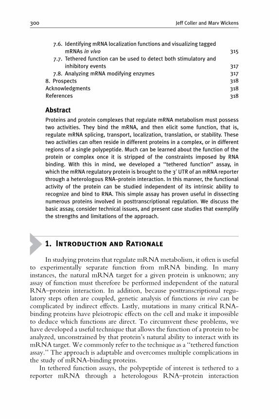
In tethered function assays, the polypeptide of interest is tethered to areporter mRNA through a heterologous RNA–protein interaction
Tethered Function Assays 301
(Fig. 14.1). Usually, the tethering site lies in the 30 untranslated region(UTR) of the mRNA; this region is relatively unconstrained evolutionarily,and the natural site of action of many mRNA regulators. Tethered functionassays have been used to show the role of proteins in control of mRNAtransport, translation, localization, and stability (Coller and Wickens, 2002).Different reporters need to be used to assay each of these processes.
The tethered function assay takes advantage of the observation thatmany nucleic acid-binding proteins are modular. For example, manyDNA transcription factors are bipartite, with separate DNA-binding andtranscriptional activation domains (Hope and Struhl, 1986; Keegan et al.,1986). Often the activities of these two domains are autonomous andseparable; in other instances, they reside in distinct members of a multi-polypeptide complex. RNA-binding proteins display similar modularity.The rationale of the tethered function approach is to examine solely the‘‘functional’’ activity of an RNA-binding protein tethered artificially to anmRNA, circumventing the constraints imposed by natural RNA binding.
Poly(A)
Assay mRNA translation,stability, etc.
Reporter
Poly(A)
Tetherbinding site
X
Tether
Tether
X
Reporter
Figure 14.1 Tethered function assays using the 30 UTR. A protein (X) is brought to areporter mRNAthrough an artificial RNA^protein interaction (tether). In this exam-ple, the tethered binding site has been shown in the 30 UTR of the reporter, but otherlocations have been used. The function of the tethered protein in any aspect of themRNA’smetabolismor function can then be assayed byconventional methodology.

302 Jeff Coller and Marv Wickens
In some cases, RNA binding and function may not be readily separable.For example, in nucleases and helicases, the nucleic acid-binding site is alsothe active site of the protein. Moreover, the interaction of a protein with itsnatural RNA-binding site can regulate the protein’s activity; in theseinstances, it may be impossible to assay the function of the tethered proteinin the absence of its cognate site.
2. The Basic Design of the Tethered
Function Assay
The design of the tethered function assay is relatively straightforward.To determine the effects of a protein X on mRNA metabolism, a chimericprotein is expressed in vivo in which protein X is continuous with a tetheringpolypeptide (see Fig. 14.1). The tethering protein is an RNA-bindingprotein that recognizes an RNA tag sequence with high specificity andaffinity. The effect of the fusion protein on mRNA metabolism is deter-mined by coexpressing the chimera with anmRNA reporter (such as lacZ orluciferase) into which a tag RNA sequence has been embedded. The fusionprotein’s effects on mRNA metabolism are assayed by conventional means[i.e., Western blot, Northern blot, reverse transcriptase polymerase chainreaction (RT-PCR), etc.]. While the assay is relatively straightforward,several issues discussed in the following sections should be considered atthe outset in designing a tethering experiment.
The assay, though powerful, is artificial. Only positive results are mean-ingful: lack of effects cannot be interpreted. Some RNA-binding proteinsmay require other proteins or their cognate RNA-binding sites to function,or be inactive as chimeras, or require appropriate positioning on themRNA.
2.1. Position of the tethering site
A first consideration when designing a tethered function assay is the positionin the mRNA of the tag sequence (i.e., the tethering site). While differentlaboratories have used tethered function assays and placed tag sequenceswithin all regions of the mRNA, the most useful and common site is the 30UTR (Coller andWickens, 2002). The tethering of proteins to the 30 UTRhas particular biological and experimental advantages. Importantly, manysites that regulate diverse steps in an mRNA’s life, including its transport,cytoplasmic localization, stability, and translational activity, often reside inthe 30 UTR. Thus, tethering to that region places regulators where theymight well function. In addition, it is known that the exact location ofseveral 30 UTR regulators is not critical for their function, implying that
Tethered Function Assays 303
precise spatial positioning is not critical. Lastly, the 30 UTR has fewerconstraints than either the 50 UTR (which can affect translational initiationfrequency) or the open reading frame. The intercistronic region of bicis-tronic mRNAs also is relatively unconstrained and has been used fortethered function experiments using the same rationale (De Gregorioet al., 1999, 2001; Furuyama and Bruzik, 2002; Shen and Green, 2006;Spellman et al., 2005; Wang et al., 2006).
3. The Tether
In choosing which protein to use as the tether, it is necessary toconsider affinity and specificity for the RNA tag, subcellular localization,and impact of the tether on the activity of the test protein. The mostcommon tether is the bacteriophage MS2 coat protein (Beach et al., 1999;Bertrand et al., 1998; Coller et al., 1998; Collier et al., 2005; Dickson et al.,2001; Dugre-Brisson et al., 2005; Gray et al., 2000; Kim et al., 2005; Longet al., 2000; Lykke-Andersen et al., 2000, 2001; Minshall and Standart,2004; Minshall et al., 2001; Ruiz-Echevarria and Peltz, 2000). However,the iron response element binding protein (IRP), a derivative of bacterio-phage l N-protein (De Gregorio et al., 1999, 2001), and the spliceosomalU1A protein have been used successfully (Brodsky and Silver 2000; Finouxand Seraphin, 2006). In the following sections we will discuss each of thesespecific tethers and their merits and drawbacks.
3.1. The MS2 bacteriophage coat protein as a tether
The MS2 coat protein has been a popular choice for several reasons. First,this protein is relatively small (14 kDa), thus minimizing potential disrup-tions to the test protein. Second, the biochemistry of theMS2 coat’s bindingto its target sequence has been well established. Specifically, the MS2 coat isknown to bind with high specificity and selectivity to a 21-nucleotide RNAstem–loop (Kd ¼ 1 nM; Carey and Uhlenbeck, 1983). In addition, muta-tions in the binding site are available that increase or decrease affinity. Inparticular, the substitution of a single U within the stem–loop to a Cincreases affinity 50-fold over wild type (Lowary and Uhlenbeck, 1987).Moreover, use ofMS2 allows a high dosage of tethered proteins to be presenton the mRNA: the MS2 coat interacts with its target sequence as a dimer;thus for every stem–loop present in the mRNA reporter, two tetheredproteins are present. Lastly, MS2 binds cooperatively to two stem–loops,further increasing the occupancy of sites (Witherell et al., 1990). In someapplications, the more protein that is bound, the better; each of these factorscontribute to a strong signal in the functional assay.
304 Jeff Coller and Marv Wickens
On the other hand, theMS2 coat protein is not the simplest optionwhen itis necessary to carefully control the number of tethered protein moleculesbound. Since the MS2 coat protein binds as a dimer to a single site, andinteracts with adjacent sites cooperatively, a large (and not trivial to determine)number of protein molecules may be bound to the targeted mRNA.
3.2. N-peptide as a tether
The bacteriophage l N protein is often used in the tethered function assay(Baron-Benhamou et al., 2004). N-protein regulates bacterial transcrip-tional antitermination by binding to a 19-nucleotide RNA hairpin withinearly phage operons called boxB (Scharpf et al., 2000). Importantly, theN-peptide/boxB interaction occurs with high affinity (Kd ¼ 1.3 nM). Theparticular advantage of the N-peptide in tethering assays is the result of itsextremely small size; only 22 amino acids are required for the high affinityinteraction with boxB RNA. Because of this, many laboratories have optedto use the N-peptide rather than MS2 coat protein, reasoning that itminimizes potential interference with the fusion protein’s function(Baron-Benhamou et al., 2004). Another desirable feature of N-peptide isthat unlike the MS2 coat, the protein binds 1:1 to its RNA target.
3.3. U1A protein and IRP as tethers
Both the U1A protein and IRP have been used successfully as tethers (DeGregorio et al., 1999; Finoux and Seraphin, 2006). U1A is a U1 smallnuclear ribonucleoprotein (snRNP)-specific protein that binds with highspecificity and affinity to a 30-nt RNA hairpin (Kd ¼ 5 nM; van Gelderet al., 1993). IRP also binds to a 30-nt RNA hairpin that normally resideswithin the UTRs of target mRNAs with high affinities (Kd ¼ 90 pM;Barton et al., 1990). Like N-peptide, the concentration of both U1A andIRP on the reporter mRNA is theoretically 1:1 (protein:RNA tag). UnlikeN-peptide, however, both of these proteins are relatively large: 38 kDa forU1A and 97 kDa for IRP. As a result, they have not commonly been usedin tethered function assays.
In general, the MS2 coat provides the highest concentration of tetheredproteins to be bound to the reporter per binding site. This may allowphenotypes to be observed without greatly increasing the overall length ofthe mRNA reporter, an undesired situation in some applications. N-peptide,on the other hand, allows the delivery of a single tethered protein per bindingsite. The cost of this control of stoichiometry can be a need to introducemultiple tandem binding sites (more than four) in order to observe a robustphenotype (see below); the trade-off is an increase in reporter length. None-theless, the relative merits of MS2 coat protein, N-peptide, U1A, or IRP aresituation specific. All have been successfully used tomeasure effects onmRNA
Tethered Function Assays 305
translation, turnover, and transport. Direct comparisons between differenttethers have not been made.
3.4. N-terminal or C-terminal fusions
The relative positions of the tethering protein and the protein of interest canbe important. For example, in our own experience, tethering the MS2 coatprotein to the N-terminus of the poly(A)-binding protein (PAB) resulted inmuch more activity than if the tether was located at the C-terminus (datanot shown). This will have to be determined on a case-by-case basis; bothorientations should be tested.
3.5. Trans-effects
A third important issue to consider is that the fusion protein may have trans-acting effects. Often, the tethered function assay is performed in a wild-typebackground with the endogenous copy of the test protein present. Thepresence of the tethering moiety may create a dominant negative allele thatblocks the function of the normal protein in vivo, seriously complicatinganalysis. As a result, controls to ensure that any observed effects occur onlyin cis with respect to the mRNA reporter are important (see below).
4. The Reporter mRNA
The tethered function assay can be adapted to measure the effect of atethered protein on many steps in mRNA metabolism and function. Theadaptability comes mainly from the choice of reporter mRNA and the finalassay performed. We will discuss only some of the reporters and assays thathave been put into practice.
The choice of reporter mRNA obviously is dictated by the effect to beassayed. For example, translational activity can be measured in yeast usingthe LacZ, HIS3, and CUP1 mRNAs, while in metazoans, luciferase, CAT,and epitope tags are most common (De Gregorio et al., 1999, 2001; Grayet al., 2000; Pillai et al., 2004). In determining the effects of a tetheredprotein on mRNA stability, MFA2, PGK1, and YAP1 have been used asreporter mRNAs in yeast, and b-globin and luciferase have been used inmammalian systems (Amrani et al., 2006; Chou et al., 2006; Coller et al.,1998; Finoux and Seraphin, 2006; Kim et al., 2005; Lykke-Andersen et al.,2001, 2001; Ruiz-Echevarria and Peltz, 2000).
The intrinsic behavior of the reporter mRNA is an important consider-ation. To determine whether a tethered protein stabilizes an mRNA, themRNA must be unstable in the absence of the protein; conversely, todetermine whether a tethered protein destabilizes the mRNA, the

00 1 2 3 5
25
50
75
100
Per
cent
tra
nsla
tion
Number of binding elements
Poly(A)
NAgo2
( )1–5 boxBelements
Ago2/N fusionAgo2 alone
A
B
Reporter
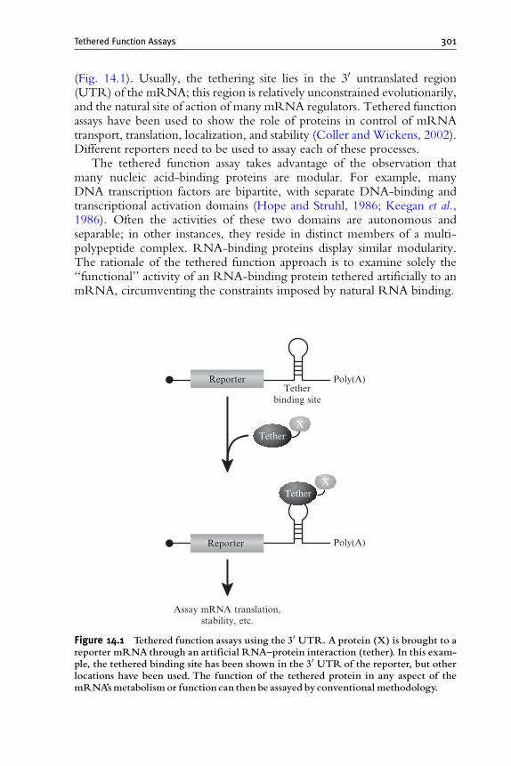
Figure 14.2 The number of tethered binding sites can influence phenotypic read-out.(A) Shown is the effect of increasing the number of tethered binding sites on transla-tional repression mediated by tethered Ago2 (Pillai et al. 2004). Specifically, 0,1, 2, 3, or 5boxB elements were introduced into the 30 UTRof a reporter gene expressing Renillaluciferase (RL). (B) The reporters were transfected into HeLa cells expressing eitherAgo2 (black bars) or anN-peptideAgo2 fusion (gray bars) and translation measured byenzymatic assay. As shown, increasing the numberof tethered binding sites dramaticallyinfluences the repression observed.
306 Jeff Coller and Marv Wickens
mRNA reporter must be stable without the protein. The same reasoningapplies to effects on other aspects of mRNA metabolism such as translationand subcellular localization.
4.1. The number and location of tethered binding sites
The number and location of tether binding sites are important variables.First, it should be decided where the tethered sites should be positioned,i.e., the 50 UTR, 30 UTR, or coding region. This depends on the suspectedrole of the protein in mRNA metabolism. For example, a protein thoughtto regulate polyadenylation might logically be placed in the 30 UTR. It isimportant that the placement of the tethered binding sites not interfere on
Tethered Function Assays 307
its own with the mRNA. For example, in testing the role of tethered PABon mRNA stability, sites were placed in a region of theMFA2 30 UTR thatwas known not to affect the mRNA’s half-life (Coller et al., 1998; Muhlradand Parker, 1992). Placement elsewhere would have dramatically alteredthe normal turnover rate of this message. It is helpful, therefore, to select as areporter an mRNA whose cis-acting sequences are well characterized.Obviously these issues make it important that the behavior of the reportermRNA with and without tethering sites be compared in the absence of thechimeric protein (see below, and Fig. 14.2).
A second issue in designing a reporter concerns the number of tetheredbinding sites. In many cases using the MS2 bacteriophage coat as the tether,two stem–loops have been sufficient to observe an effect (Coller et al., 1998;Gray et al., 2000; Minshall et al., 2001; Ruiz-Echevarria and Peltz, 2000).However, many more sites have been used, ranging from 6 to 24 (Bertrandet al., 1998; Fusco et al., 2003; Lykke-Andersen et al., 2000, 2001; Pillaiet al., 2004). The effect of the number of binding sites has been evaluatedsystematically in two reports (Lykke-Andersen et al., 2000; Pillai et al.,2004). Increasing the number of binding sites can increase the signal andenhance the assay’s sensitivity. In Fig. 14.2, the extent of translationalrepression achieved by a tethered protein is proportional to the number ofbinding sites (Pillai et al., 2004).
5. A Priori Considerations About the Logic of
the Assay
5.1. Multiprotein complexes
mRNA regulatory events often occur through multiprotein complexesformed via protein–protein and protein–RNA interactions. In such cases,RNA binding may occur via one critical protein, which tethers the activityof another protein to the mRNA. Thus, the ‘‘active’’ protein may notdirectly contact the RNA. One strength of the tethered approach is itsability to assay the ‘‘activity’’ independent of RNA binding.
5.2. The role of RNA binding in function
The interaction between RNA and protein in some cases is essential foractivity. RNA–protein interactions can change the conformation of theRNA, the protein, or both; not surprisingly, some complexes are biologi-cally active, while the free RNAs or proteins are not (Williamson, 2000).Certain RNA ligands likely can influence activation or repression activity,much as in DNA-induced allosteric effects on transcription factors (Lefstinand Yamamoto, 1998; Scully et al., 2000). In addition, the context of the

308 Jeff Coller and Marv Wickens
natural binding site may be important for the protein’s activity becauseessential factors are bound in the neighborhood.
These considerations have two implications. First, negative results in atethered function assay are meaningless, even if the RNA and protein dointeract on the reporter. Second, the outcome seen—for example, mRNAstabilization by a particular tethered protein—may differ when the protein isassociated with its natural RNA-binding site. The same issues apply toDNA-binding transcription factor complexes, which have been powerfullydissected via comparable tethering approaches.
5.3. Analyzing function without knowing the target
In many cases, putative RNA-binding proteins have been identified, buttheir respective RNA target is unknown. One asset of the tetheringapproach is that a protein’s activity can be determined without knowingthe natural RNA target.
5.4. Analyzing the function of essential genes
In some cases, the RNA-binding protein under investigation is essential forcell viability; as a result, traditional genetic techniques are complicated bypleiotropic effects. The tethered function assay allows the function of theprotein to be examined on just one mRNA species in an otherwisewild-type cell.
6. Important Controls
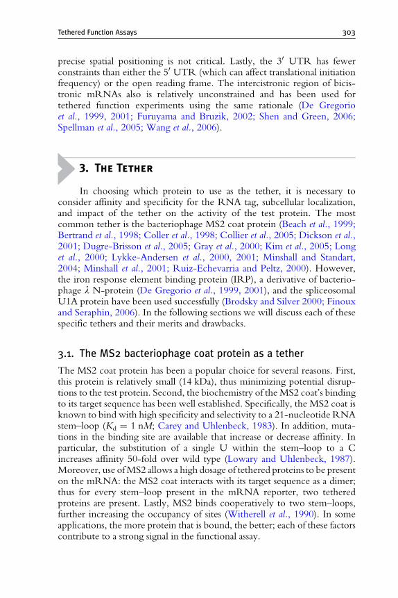
Several controls are critical in tethered function assays, and shouldalways be performed (Fig. 14.3). It is necessary to ensure that (1) the tetheredbinding site does not affect the mRNA on its own, (2) the tethering proteinalone (e.g., MS2 coat protein) does not have an impact, and (3) any observedeffects should occur only in cis (that is, when the protein is bound to themRNA). To control for possible trans-acting effects, the chimeric proteinshould be expressed alongside a reporter that lacks binding sites. This set ofcontrols can ensure that an observed effect is specific to the protein ofinterest, and occurs only when it is associated with the mRNA in cis (seeFig. 14.3).
This concludes the general discussion of the design of a basic tetheredfunction assay. In the following section we discuss a few specific exampleswith the aforementioned general principles considered. These case studiesare not meant to be comprehensive of the literature but rather provide asample of the uses of the tethered function assay to address certain biologicalissues. An overview is provided in Table 14.1.

ProteinTethering
siteHalf-life(min)
None
MS2-PAB
MS2
MS2-SXL
AntisenseMS2
MS2-PAB
MS2-PAB
MS2
MS2
MS2
MS2
None
4
23
4
5
3
3
Poly(A)
MS2PAB
Poly(A)
MS2PAB
Poly(A)
MS2PAB
Poly(A)
MS2SXL
Poly(A)
MS2
Poly(A)Reporter
Reporter
Reporter
Reporter
Reporter
Reporter
Figure 14.3 Importantcontrols toconsiderwhenperforming atethered function assay.Shown is a representation of experiments we performed to demonstrate the effects ofPAB on mRNA stability (Coller et al.,1998). First, the effect of the tether was evaluatedby determining half-lives of the reporter in cells expressing just the MS2 coat proteinalone orMS2 fused to Sxl-lethal, a distinct RNA-binding protein of similar size to PAB(MS2-SXL). Second, we determined that the observed increase in mRNA stability wasa consequence of tethering PAB in cis, bymeasuring reporter half-lifewhen the mRNAcannotbindMS2-PAB; either the tethering siteswere not present or the siteswere in theantisense orientation.This latter experiment also controlled for the contribution of thetethering sites to the stability of the reporter. From these controls it was possible to con-clude that the observed reporter stabilization was specific to PAB and occurred onlywhen it was tethered.
Tethered Function Assays 309
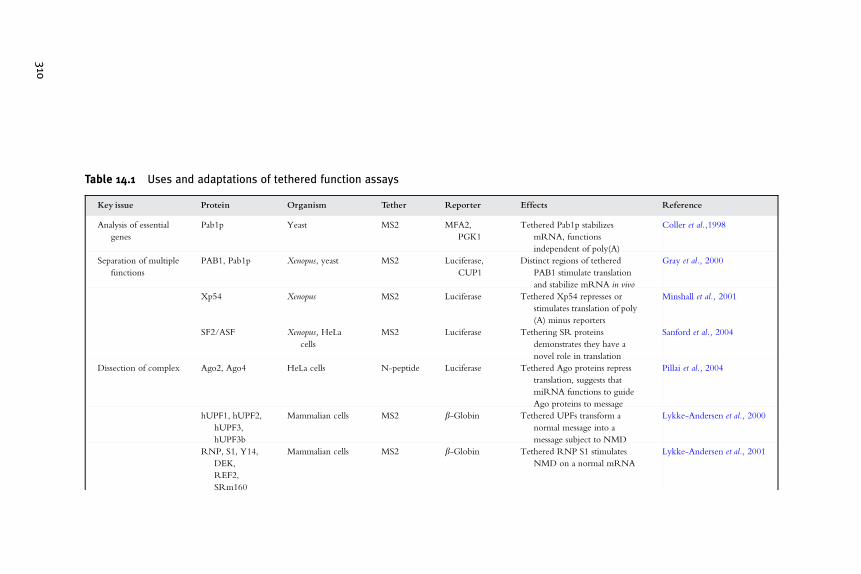
Table 14.1 Uses and adaptations of tethered function assays
Key issue Protein Organism Tether Reporter Effects Reference
Analysis of essential
genes
Pab1p Yeast MS2 MFA2,
PGK1
Tethered Pab1p stabilizes
mRNA, functions
independent of poly(A)
Coller et al.,1998
Separation of multiple
functions
PAB1, Pab1p Xenopus, yeast MS2 Luciferase,
CUP1
Distinct regions of tethered
PAB1 stimulate translation
and stabilize mRNA in vivo
Gray et al., 2000
Xp54 Xenopus MS2 Luciferase Tethered Xp54 represses or
stimulates translation of poly
(A) minus reporters
Minshall et al., 2001
SF2/ASF Xenopus, HeLa
cells
MS2 Luciferase Tethering SR proteins
demonstrates they have a
novel role in translation
Sanford et al., 2004
Dissection of complex Ago2, Ago4 HeLa cells N-peptide Luciferase Tethered Ago proteins repress
translation, suggests that
miRNA functions to guide
Ago proteins to message
Pillai et al., 2004
hUPF1, hUPF2,
hUPF3,
hUPF3b
Mammalian cells MS2 b-Globin Tethered UPFs transform a
normal message into a
message subject to NMD
Lykke-Andersen et al., 2000
RNP, S1, Y14,
DEK,
REF2,
SRm160
Mammalian cells MS2 b-Globin Tethered RNP S1 stimulates
NMD on a normal mRNA
Lykke-Andersen et al., 2001
310
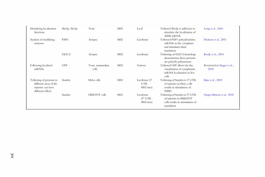
Identifying localization
functions
She2p, She3p Yeast MS2 LacZ Tethered She2p is sufficient to
stimulate the localization of
ASHI mRNA
Long et al., 2000
Analysis of modifying
enzymes
PAP1 Xenopus MS2 Luciferase Tethered PAP1 polyadenylates
mRNAs in the cytoplasm
and stimulates their
translation
Dickson et al., 2001
GLD-2 Xenopus MS2 Luciferase Tethering of GLD-2 homologs
demonstrates these proteins
are poly(A) polymerases
Kwak et al., 2004
Following localized
mRNAs
GFP Yeast, mammalian
cells
MS2 Various Tethered GFP allows for the
visualization of cytoplasmic
mRNA localization in live
cells
Reviewed in Singer et al.,
2005
Tethering of proteins to
different areas of the
reporter can have
different effects
Staufen HeLa cells MS2 Luciferase (30
UTR
MS2 sites)
Tethering of Staufen to 30 UTR
of reporter in HeLa cells
results in stimulation of
NMD
Kim et al., 2005
Staufen HEK293T cells MS2 Luciferase
(50 UTR
MS2 sites)
Tethering of Staufen to 50 UTR
of reporter in HEK293T
cells results in stimulation of
translation
Dugre-Brisson et al., 2005
311

312 Jeff Coller and Marv Wickens
7. Examples of the Tethered Function Assay in
the Literature
7.1. Analyzing essential genes
Tethered function assays allow the presence of essential RNA-bindingproteins to be modulated on a target mRNAwithout affecting cell viability.For example, in Saccharomyces cerevisiae, PAB is an essential gene involved inmany different aspects of mRNA metabolism. Studies of PAB1 functionusing conditional alleles or genetic suppressors have shown that this proteinis required for efficient mRNA translation, coupled deadenylation anddecay, and polyadenylation. Detailed analysis of these functions in vivo iscomplicated by the breadth of PAB’s roles and the fact that it is essential.Tethered function assays were used to circumvent these pleiotropic effects.Using this approach, PAB was shown to stabilize an mRNA to which it wastethered (Coller et al., 1998). The activities of mutant forms of PAB (astethered proteins) have been determined, and the active regions identified,even though yeast carrying the equivalent mutants would not be viable(Coller et al., 1998; Gray et al., 2000).
Tethered function assays have also facilitated analysis of essential transla-tion initiation factors. For example, eukaryotic initiation factor (eIF)4G, acritical member of the cap-binding complex, is thought to recruit the 40Sribosome to the mRNA by simultaneously binding both cap-bindingfactors (eIF4E) and a 40S ribosome-associated complex (eIF3). A wealthof biochemical data has illuminated the contribution of eIF4G to translationin vitro. De Gregorio et al. (1999) used a tethered function approach toreveal mechanisms of eIF4G action in vivo. They first determined thateIF4G tethered to the intergenic region of a bicistronic reporter mRNAwas sufficient to drive mRNA translation independent of the cap. Thisenabled identification of a conserved core domain of eIF4G that is requiredfor translational stimulation (De Gregorio et al., 1999). Similar studies withtranslational initiation factor eIF4E demonstrated that it stimulates transla-tion independent of its ability to bind the cap (De Gregorio et al., 2001).This latter study pioneered the use of N-peptide as a tethering device(Baron-Benhamou et al., 2004).
7.2. Separation of multiple functions that reside within thesame protein
Many posttranscriptional events are coupled. For example, splicing and 30polyadenylation influence one another and these events influence transport,degradation, and translation of the mRNA. In several cases, proteinsinvolved in an upstream event can also have a dramatic role in a downstream
Tethered Function Assays 313
event. This complicates the use of conventional mutational analysis inpinpointing the protein’s direct effects. In such cases, tethered functionassays can help determine which of many affected steps are due directly tothe activity of the protein.
In one example of this approach, SR proteins were shown to directlyaffect both splicing and translation (Sanford et al., 2004). SR proteins are alarge family of nuclear phosphoproteins required for constitutive and alter-native splicing. A subset of SR proteins is known to shuttle between thenucleus and cytoplasm, suggesting that these proteins play important cyto-plasmic roles in mRNA metabolism. Since many alterations in SR proteinsin vivo impact splicing, it was difficult to determine whether any observedeffects on translation were a direct effect of the SR defect or an indirectconsequence of the splicing defect. To overcome this limitation, Sanfordet al. (2004) used a tethered function assay in which they injected reportermRNA bearing the MS2-RNA binding element with an MS2-SF2/ASF(an SR protein) protein fusion into the cytoplasm of Xenopus oocytes. Thedata demonstrated that tethered SF2/ASF stimulated translation by approx-imately 6-fold over the appropriate controls. This was also shown to be ageneral property of SF2/ASF by demonstrating that similar phenotypeswere observed in HeLa cell-free translation extracts.
These findings resulted in the conclusion that SR proteins can promotemRNA translation after they are deposited on the mRNA via splicing.From the standpoint of this review, the important point is that the tetheredfunction assay allowed the elucidation of a role for SR proteins in mRNAtranslation by removing the complication of the upstream event, i.e.,splicing.
7.3. Dissecting complexes
Tethered function assays can be particularly useful when genetics is complexor unsuited to the problem. Many regulatory events are controlled bymultiprotein complexes. Discrete components of the complex provideRNA binding and recognition, which in turn recruit the functional activityto the site of regulation.
7.3.1. Protein complexes: NMDAnalysis of non-sense-mediated decay (NMD) is exemplary. MammalianmRNAs are targeted for rapid turnover when they contain a stop codonthat is greater than 50 nucleotides upstream of the last exon–exon boundary, aprocess termed NMD. A group of proteins binds to the exon–exon (E/E)junction of mammalian mRNA subject to NMD (Le Hir et al., 2000a,b;Singh and Lykke-Andersen, 2003). Although this complex is primarily foundon NMD substrates, it was unclear if their presence was a cause or effectof the transcript being targeted for NMD. Lykke-Andersen et al. (2001)
314 Jeff Coller and Marv Wickens
used a tethered function approach to test whether the placement of any ofthese proteins on a normal mRNA would elicit an NMD response. Whilethe E/E complex consists of at least five proteins, only tethered RNP S1elicited NMD. In this case, the tethered function approach revealed a roleof a specific protein in eliciting the function of a multiprotein complex (E/E complex), and showed it was a cause, rather than an effect, of the NMDprocess.
7.3.2. RNA–protein complexes: miRNAsThe tethered function assay has helped identify key components in theRNA protein complex associated with miRNA-mediated gene silencing.Ten years ago, a small, noncoding RNA of approximately 21 nucleotides,lin-4, was shown to bind the 30 UTR of lin-14 mRNA in the nematodeCaenorhabditis elegans, and to silence its translation (Pasquinelli et al., 2005).Since that initial discovery, miRNAs have emerged as ubiquitous regulatorsof mRNA translation and stability.
Numerous factors are required for miRNAmaturation and for the assem-bly of the miRNA into a ribonucleoprotein (RNP) complex that repressestranslation of the target mRNA. The RNA interference silencing complex(RISC) has been shown to be necessary for cessation of mRNA translation byan miRNA (Filipowicz, 2005; Sontheimer, 2005). Tethered function assaysmade it possible to dissect the repression function of RISC from the miRNA:specific components of RISC, namely Ago1–2, are sufficient to translationallyrepress reporter mRNAs to which they are artificially bound (Behm-Ansmantet al., 2006; Pillai et al., 2004; Rehwinkel et al., 2005).
7.4. Mutagenesis of tethered proteins can also be useful inidentifying unique gain-of-function alleles
Because the effects of a tethered protein are examined on a single reportermRNA, the effects of many manipulations of the protein sequence can beexamined readily and conclusively. This can reveal novel molecular propertiesin the protein.
This general approach has been applied to the Dhh1p/RCK1/p54family of RNA helicases (Minshall and Standart, 2004; Minshall et al.,2001). The Xenopus homolog, Xp54, is sufficient to repress the translationof an mRNA to whose 30 UTR it is tethered. Interestingly, mutants withinthe putative DEAD box motif of this protein transform this helicase from atranslational repressor into a translational stimulator. These results mayindicate that Xp54 may serve two roles in mRNA metabolism that aredependent on modulation of its conformation or helicase activity.
Tethered Function Assays 315
7.5. Tethering of proteins to different areas of the reportercan have different effects
It should be noted that the tethered function assay measures the effect of anmRNP complex in its nonnative context and thus may induce emergentproperties of the protein. Moreover, the protein of interest may havedistinct functions when positioned differently on the mRNA reporter.Indeed, it has been documented that similar proteins when tethered todifferent areas of an mRNA can have distinct outcomes.
For example, the conserved mRNA-binding protein Staufen is impor-tant during early embryonic development in Drosophila and has been iden-tified as an important regulator of mammalian mRNA processes. Tetheringof mammalian Staufen to the 50 UTR of reporter mRNAs stimulatestranslation without impacting mRNA stability in HEK293T cells and rabbitreticulocyte lysates (Dugre-Brisson et al., 2005). Interestingly, tetheringmammalian Staufen to the 30 UTR in HeLa cells does not stimulatetranslation, but instead destabilizes the mRNA (Kim et al., 2005). Thesetwo reports are from distinct cells types, and so require further analysis.However, it may be that Staufen possesses different activities, dependent onits location in the mRNA. This property would echo that of IRP; bound tothe 50 UTR of ferritin mRNA, it inhibits translation; bound to the 30 UTRof transferrin mRNA, it inhibits mRNA decay (Hentze et al., 2004). It mayturn out to be important to compare the effects of proteins tethered todifferent locales to reveal region-specific differences.
7.6. Identifying mRNA localization functions and visualizingtagged mRNAs in vivo
Proteins that cause an mRNA to move to a particular location within a cellcan be assayed using the tethered function approach. For example, yeastShe2p and She3p are present in a complex on theASH1 30 UTR. Tetheringeither She2p or She3p to the 30 UTR of a reporter gene was sufficientto stimulate that mRNA’s localization to the bud tip (Long et al., 2000).These findings directly demonstrate a localization function, and shouldenable its genetic dissection away from formation of the complex or bindingto RNA.
Several adaptations of the tethered function assay have been developedto tag an mRNA for further analysis, rather than study a particular protein’seffects. Although these are not strictly tethered function assays (as theprotein is merely a tag), we mention them here because they are so closelyrelated technically. They now are widely used, and have been reviewed intheir own right (Beach et al., 1999; Singer et al., 2005); we discuss only asingle, early pioneering example.
316 Jeff Coller and Marv Wickens
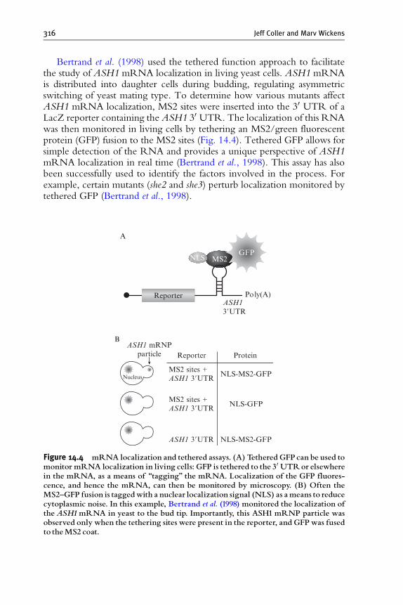
Bertrand et al. (1998) used the tethered function approach to facilitatethe study of ASH1 mRNA localization in living yeast cells. ASH1 mRNAis distributed into daughter cells during budding, regulating asymmetricswitching of yeast mating type. To determine how various mutants affectASH1 mRNA localization, MS2 sites were inserted into the 30 UTR of aLacZ reporter containing the ASH1 30 UTR. The localization of this RNAwas then monitored in living cells by tethering an MS2/green fluorescentprotein (GFP) fusion to the MS2 sites (Fig. 14.4). Tethered GFP allows forsimple detection of the RNA and provides a unique perspective of ASH1mRNA localization in real time (Bertrand et al., 1998). This assay has alsobeen successfully used to identify the factors involved in the process. Forexample, certain mutants (she2 and she3) perturb localization monitored bytethered GFP (Bertrand et al., 1998).
ASH13�UTR
MS2 sites +ASH1 3�UTR
Reporter Protein
NLS-MS2-GFP
MS2 sites +ASH1 3�UTR
NLS-GFP
ASH1 3�UTR NLS-MS2-GFP
Nucleus
ASH1 mRNPparticle
A
B
Poly(A)
MS2
Reporter
GFPNLS
Figure 14.4 mRNA localization and tethered assays. (A) Tethered GFP can be used tomonitor mRNA localization in living cells: GFP is tethered to the 30 UTRor elsewherein the mRNA, as a means of ‘‘tagging’’ the mRNA. Localization of the GFP fluores-cence, and hence the mRNA, can then be monitored by microscopy. (B) Often theMS2^GFP fusion is taggedwith a nuclear localization signal (NLS) as ameans to reducecytoplasmic noise. In this example, Bertrand et al. (1998) monitored the localization oftheASH1mRNA in yeast to the bud tip. Importantly, this ASH1 mRNP particle wasobserved only when the tethering sites were present in the reporter, and GFP was fusedto theMS2 coat.
Tethered Function Assays 317
7.7. Tethered function can be used to detect both stimulatoryand inhibitory events
As mentioned, the tethered function assay is highly adaptable. Tetheredfunction assays have been used to monitor stimulatory and inhibitory effectsof mRNA metabolism factors. For instance, in Xenopus it was demonstratedthat tethered DAZL stimulates translation (Collier et al., 2005), while usingthe same reporters others have shown that tethered Xp54 inhibits mRNAtranslation in Xenopus (Minshall and Standart, 2004; Minshall et al., 2001).Similar results have been seen for assaying effects on mRNA stability.Certain classes of AU-rich binding proteins will stabilize mRNA whentethered, while others destabilize the mRNA ( Barreau et al., 2006; Chouet al., 2006). Thus, tethered function assays provide flexibility in allowing arange of phenotypes to be observed.
7.8. Analyzing m R NA modifying enzymes
Tethered function assays have been used to identify enzymes involved inmRNA processing. Sequences near the 30 end of an mRNA recruit acomplex of proteins that promotes 30 end cleavage and polyadenylation.By tethering the relevant poly(A) polymerase directly to the 30 end of thereporter, that enzyme was shown to be sufficient for the elongation of poly(A) tails in oocytes and to stimulate translation as a result ( Dickson et al.,2001). Sites for interaction with other components of the complex aredispensable (Dickson et al., 2001). The same general approach has beenused to identify other divergent poly(A) adding enzymes, termed theGLD-2 family, from C. elegans, flies, frogs, mice, and humans (Kwaket al., 2004; J. E. Kwak et al ., unpublished observations; Wang et al., 2002).
A strength of the tethered approach is that many candidate open readingframes (ORFs) can be tested rapidly. A limitation is that false negatives arise.For example, two Saccharomyces cerevisiae proteins, Trf4p and Trf5p, that areknown to be poly(A) polymerases, differ dramatically as tethered proteins.Trf5p is active, and Trf4p is not ( J. E. Kwak et al., unpublished observations).This may reflect a difference in their substrate specificity, requirements forRNA or protein partners, or be an artifactual consequence of an inactiveconformation in one chimeric protein.
Tethering assays can reveal unanticipated biochemical activities. In thesame group of tethering experiments that identified the GLD-2 family,certain relatives of these PAPs turn out not to add poly(A) at all, but to addpoly(U) instead ( J. E. Kwak et al., unpublished observations). Investigationsinto the biological role of these newly discovered poly(U) polymerases arecurrently underway. The key point here is that tethered assays enabled facilebiochemical identification of the RNA modifications they catalyze.
318 Jeff Coller and Marv Wickens
8. Prospects
Tethered function assays provide a simple means to address the role ofspecific RNA-binding proteins on mRNAmetabolism and function. Theiruse is certainly not limited to the few examples mentioned here and inTable 14.1. The tethered function approach provides a unique platform forthe study of suspect regulators of mRNA metabolism that have unknowntarget specificity and/or functional activity. Of particular interest are simplephenotypic screens that allow the rapid identification of tethered proteinson the metabolism of a given reporter.
As the genome sequences of more species become available, methods toanalyze function beyond familial sequence resemblance are needed. Tetheredfunction assays may provide a rapid screen to sort proteins into functionalfamilies.
ACKNOWLEDGMENTS
We thank many individuals who have contributed their thoughts and ideas to this review, mostnotably, Drs. Jens Lykke-Anderson, Scott Ballantyne, Kristian Baker, Kris Dickson, Niki Gray,Stan Fields, Mattias Hentze, Allan Jacobson, Roy Parker, Stu Peltz, Daniel Seay, Rob Singer,Nancy Standart, and Joan Steiz. We also thank Drs. Wenqian Hu and Thomas J. Sweet forcritical reading of the manuscript. Work in theWickens laboratory is supported by grants fromthe National Institutes of Health (NIH). Dr. Coller is supported by a grant from the AmericanCancer Society and the NIH.
REFERENCES
Amrani, N., Dong, S., He, F., Ganesan, R., Ghosh, S., Kervestin, S., Li, C., Mangus, D. A.,Spatrick, P., and Jacobson, A. (2006). Aberrant termination triggers nonsense-mediatedmRNA decay. Biochem. Soc. Trans. 34, 39–42.
Baron-Benhamou, J., Gehring, N. H., Kulozik, A. E., and Hentze, M.W. (2004). Using thelambdaN peptide to tether proteins to RNAs. Methods Mol. Biol. 257, 135–154.
Barreau, C., Watrin, T., Beverley Osborne, H., and Paillard, L. (2006). Protein expression isincreased by a class III AU-rich element and tethered CUG-BP1. Biochem. Biophys. Res.Commun. 347, 723–730.
Barton, H. A., Eisenstein, R. S., Bomford, A., and Munro, H. N. (1990). Determinants ofthe interaction between the iron-responsive element-binding protein and its binding sitein rat L-ferritin mRNA. J. Biol. Chem. 265, 7000–7008.
Beach, D. L., Salmon, E. D., and Bloom, K. (1999). Localization and anchoring of mRNAin budding yeast. Curr. Biol. 9, 569–578.
Behm-Ansmant, I., Rehwinkel, J., Doerks, T., Stark, A., Bork, P., and Izaurralde, E. (2006).mRNA degradation by miRNAs and GW182 requires both CCR4:NOT deadenylaseand DCP1:DCP2 decapping complexes. Genes Dev. 20, 1885–1898.
Bertrand, E., Chartrand, P., Schaefer, M., Shenoy, S. M., Singer, R. H., and Long, R. M.(1998). Localization of ASH1 mRNA particles in living yeast. Mol. Cell 2, 437–445.

Tethered Function Assays 319
Brodsky, A. S., and Silver, P. A. (2000). Pre-mRNA processing factors are required fornuclear export. RNA 6, 1737–1749.
Carey, J., and Uhlenbeck, O. C. (1983). Kinetic and thermodynamic characterization of theR17 coat protein-ribonucleic acid interaction. Biochemistry 22, 2610–2615.
Chou, C. F., Mulky, A., Maitra, S., Lin, W. J., Gherzi, R., Kappes, J., and Chen, C. Y.(2006). Tethering KSRP, a decay-promoting AU-rich element-binding protein, tomRNAs elicits mRNA decay. Mol. Cell. Biol. 26, 3695–3706.
Coller, J., and Wickens, M. (2002). Tethered function assays using 30 untranslated regions.Methods 26, 142–150.
Coller, J. M., Gray, N. K., and Wickens, M. P. (1998). mRNA stabilization by poly(A)binding protein is independent of poly(A) and requires translation. Genes Dev. 12,3226–3235.
Collier, B., Gorgoni, B., Loveridge, C., Cooke, H. J., and Gray, N. K. (2005). The DAZLfamily proteins are PABP-binding proteins that regulate translation in germ cells. EMBOJ. 24, 2656–2666.
De Gregorio, E., Preiss, T., and Hentze, M.W. (1999). Translation driven by an eIF4G coredomain in vivo. EMBO J. 18, 4865–4874.
De Gregorio, E., Baron, J., Preiss, T., and Hentze, M.W. (2001). Tethered-function analysisreveals that elF4E can recruit ribosomes independent of its binding to the cap structure.RNA 7, 106–113.
Dickson, K. S., Thompson, S. R., Gray, N. K., and Wickens, M. (2001). Poly(A) polymer-ase and the regulation of cytoplasmic polyadenylation. J. Biol. Chem. 276, 41810–41816.
Dugre-Brisson, S., Elvira, G., Boulay, K., Chatel-Chaix, L., Mouland, A. J., andDesGroseillers, L. (2005). Interaction of Staufen1 with the 50 end of mRNA facilitatestranslation of these RNAs. Nucleic Acids Res. 33, 4797–4812.
Filipowicz, W. (2005). RNAi: The nuts and bolts of the RISC machine. Cell 122, 17–20.Finoux, A. L., and Seraphin, B. (2006). In vivo targeting of the yeast Pop2 deadenylase
subunit to reporter transcripts induces their rapid degradation and generates new decayintermediates. J. Biol. Chem. 281, 25940–25947.
Furuyama, S., and Bruzik, J. P. (2002). Multiple roles for SR proteins in trans splicing. Mol.Cell. Biol. 22, 5337–5346.
Fusco, D., Accornero, N., Lavoie, B., Shenoy, S. M., Blanchard, J. M., Singer, R. H., andBertrand, E. (2003). Single mRNA molecules demonstrate probabilistic movement inliving mammalian cells. Curr. Biol. 13, 161–167.
Gray, N. K., Coller, J. M., Dickson, K. S., and Wickens, M. (2000). Multiple portions ofpoly(A)-binding protein stimulate translation in vivo. EMBO J. 19, 4723–4733.
Hentze, M. W., Muckenthaler, M. U., and Andrews, N. C. (2004). Balancing acts:Molecular control of mammalian iron metabolism. Cell 117, 285–297.
Hope, I. A., and Struhl, K. (1986). Functional dissection of a eukaryotic transcriptionalactivator protein, GCN4 of yeast. Cell 46, 885–894.
Keegan, L., Gill, G., and Ptashne, M. (1986). Separation of DNA binding from thetranscription-activating function of a eukaryotic regulatory protein. Science 231,699–704.
Kim, Y. K., Furic, L., Desgroseillers, L., and Maquat, L. E. (2005). Mammalian Staufen1recruits Upf1 to specific mRNA 30UTRs so as to elicit mRNA decay. Cell 120,195–208.
Kwak, J. E., Wang, L., Ballantyne, S., Kimble, J., and Wickens, M. (2004). MammalianGLD-2 homologs are poly(A) polymerases. Proc. Natl. Acad. Sci. USA 101, 4407–4412.
Lefstin, J. A., and Yamamoto, K. R. (1998). Allosteric effects of DNA on transcriptionalregulators. Nature 392, 885–888.
Le Hir, H., Izaurralde, E., Maquat, L. E., and Moore, M. J. (2000a). The spliceosomedeposits multiple proteins 20–24 nucleotides upstream of mRNA exon-exon junctions.EMBO J. 19, 6860–6869.
320 Jeff Coller and Marv Wickens
Le Hir, H., Moore, M. J., and Maquat, L. E. (2000b). Pre-mRNA splicing alters mRNPcomposition: Evidence for stable association of proteins at exon-exon junctions. GenesDev. 14, 1098–1108.
Long, R. M., Gu,W., Lorimer, E., Singer, R. H., and Chartrand, P. (2000). She2p is a novelRNA-binding protein that recruits the Myo4p-She3p complex to ASH1 mRNA.EMBO J. 19, 6592–6601.
Lowary, P. T., and Uhlenbeck, O. C. (1987). An RNA mutation that increases the affinityof an RNA-protein interaction. Nucleic Acids Res. 15, 10483–10493.
Lykke-Andersen, J., Shu, M. D., and Steitz, J. A. (2000). Human Upf proteins target anmRNA for nonsense-mediated decay when bound downstream of a termination codon.Cell 103, 1121–1131.
Lykke-Andersen, J., Shu, M. D., and Steitz, J. A. (2001). Communication of the position ofexon-exon junctions to the mRNA surveillance machinery by the protein RNPS1.Science 293, 1836–1839.
Minshall, N., and Standart, N. (2004). The active form of Xp54 RNA helicase in transla-tional repression is an RNA-mediated oligomer. Nucleic Acids Res. 32, 1325–1334.
Minshall, N., Thom, G., and Standart, N. (2001). A conserved role of a DEAD box helicasein mRNA masking. RNA 7, 1728–1742.
Muhlrad, D., and Parker, R. (1992). Mutations affecting stability and deadenylation of theyeast MFA2 transcript. Genes Dev. 6, 2100–2111.
Pasquinelli, A. E., Hunter, S., and Bracht, J. (2005). MicroRNAs: A developing story. Curr.Opin. Genet. Dev. 15, 200–205.
Pillai, R. S., Artus, C. G., and Filipowicz, W. (2004). Tethering of human Ago proteins tomRNA mimics the miRNA-mediated repression of protein synthesis. RNA 10,1518–1525.
Rehwinkel, J., Behm-Ansmant, I., Gatfield, D., and Izaurralde, E. (2005). A crucial role forGW182 and the DCP1:DCP2 decapping complex in miRNA-mediated gene silencing.RNA 11, 1640–1647.
Ruiz-Echevarria, M. J., and Peltz, S. W. (2000). The RNA binding protein Pub1modulates the stability of transcripts containing upstream open reading frames. Cell101, 741–751.
Sanford, J. R., Gray, N. K., Beckmann, K., and Caceres, J. F. (2004). A novel role forshuttling SR proteins in mRNA translation. Genes Dev. 18, 755–768.
Scharpf, M., Sticht, H., Schweimer, K., Boehm, M., Hoffmann, S., and Rosch, P. (2000).Antitermination in bacteriophage lambda. The structure of the N36 peptide-boxB RNAcomplex. Eur. J. Biochem. 267, 2397–2408.
Scully, K. M., Jacobson, E. M., Jepsen, K., Lunyak, V., Viadiu, H., Carriere, C.,Rose, D. W., Hooshmand, F., Aggarwal, A. K., and Rosenfeld, M. G. (2000). Allostericeffects of Pit-1 DNA sites on long-term repression in cell type specification. Science 290,1127–1131.
Shen, H., and Green, M. R. (2006). RS domains contact splicing signals and promotesplicing by a common mechanism in yeast through humans. Genes Dev. 20, 1755–1765.
Singer, R. H., Lawrence, D. S., Ovryn, B., and Condeelis, J. (2005). Imaging of geneexpression in living cells and tissues. J. Biomed. Opt. 10, 051406.
Singh, G., and Lykke-Andersen, J. (2003). New insights into the formation of activenonsense-mediated decay complexes. Trends Biochem. Sci. 28, 464–466.
Sontheimer, E. J. (2005). Assembly and function of RNA silencing complexes. Nat. Rev.Mol. Cell. Biol. 6, 127–138.
Spellman, R., Rideau, A., Matlin, A., Gooding, C., Robinson, F., McGlincy, N.,Grellscheid, S. N., Southby, J., Wollerton, M., and Smith, C. W. (2005). Regulationof alternative splicing by PTB and associated factors. Biochem. Soc. Trans. 33, 457–460.

Tethered Function Assays 321
van Gelder, C. W., Gunderson, S. I., Jansen, E. J., Boelens, W. C., Polycarpou-Schwarz, M., Mattaj, I. W., and van Venrooij, W. J. (1993). A complex secondarystructure in U1A pre-mRNA that binds two molecules of U1A protein is required forregulation of polyadenylation. EMBO J. 12, 5191–5200.
Wang, L., Eckmann, C. R., Kadyk, L.C, Wickens, M., and Kimble, J. (2002). A regulatorycytoplasmic poly(A) polymerase in Caenorhabditis elegans. Nature 419, 312–316.
Wang, Z., Xiao, X., Van Nostrand, E., and Burge, C. B. (2006). General and specificfunctions of exonic splicing silencers in splicing control. Mol. Cell 23, 61–70.
Williamson, J. R. (2000). Induced fit in RNA-protein recognition. Nat. Struct. Biol. 7,834–837.
Witherell, G. W., Wu, H. N., and Uhlenbeck, O. C. (1990). Cooperative binding of R17coat protein to RNA. Biochemistry 29, 11051–11057.