tentative assessment of hematology and serum chemistry...
TRANSCRIPT

Open Veterinary Journal, (2011), Vol. 1: 13-20 ISSN: 2218-6050
Original Article
________________________________________________________________________________________________________
*Corresponding Author: Department of Physiology, Biochemistry and Pharmacology, Chittagong Veterinary and Animal
Sciences University, Khulsi, Chittagong-4202, Bangladesh. Email: [email protected]
13
______________________________________________________________________________ Submitted: 10/11/2010 Accepted: 08/12/2010 Published: 23/02/2011
Hematology and serum chemistry reference values of stray dogs in Bangladesh
S.A. Khan1,2,*
, J.H. Epstein2, K.J. Olival
2, M.M. Hassan
1, M.B. Hossain
1, K.B.M.A. Rahman
4, M.F. Elahi
1,
M.A. Mamun1, N. Haider
3 , G. Yasin
1 and J. Desmond
2
1 Chittagong Veterinary and Animal Sciences University, Chittagong, Bangladesh
2 EcoHealth Alliance (formerly Wildlife Trust), New York, USA
3 International Centre for Diarrhoeal Disease Research, Dhaka, Bangladesh 4 Food and Agricultural Organization- AI Technical unit, DLS, Bangladesh
______________________________________________________________________________________________
Abstract Hematology and serum chemistry values were obtained from 28 male and 22 female stray dogs in Chittagong
Metropolitan area, Bangladesh. The goal of the study was to establish reference value for hematology and serum
chemistry for these semi wild animals in relation to age, sex, reproductive stage and body condition. No significant
differences were found for mean values of hemoglobin, packed cell volume, mean corpuscular hemoglobin
concentration, white blood cell, differential leukocyte count, total protein, albumin, glucose, cholesterol, phosphorus
and potassium among or between sexes, ages, reproductive states or body conditions. Significant differences were
noted for erythrocyte sedimentation rate (p<0.02) between sexes. Among different age groups significant differences
were found for total red blood cell count (p<0.001). Different body conditions have significant differences in red
blood cell count, mean corpuscular volume and mean corpuscular hemoglobin (p<0.001). Pregnant and non-pregnant
females differed significantly in their red blood cell count, mean corpuscular volume and mean corpuscular
hemoglobin (p<0.001).
Keywords: Hematology, Serum chemistry, Reference value, Stray dog, Bangladesh.
______________________________________________________________________________________________
Introduction Hematology and serum clinical chemistry analyses are used for health assessment of domestic animals and a
wide range of captive wildlife (Smith, 2000; Fowler
and Miller, 2003; Kaneko et al., 2008).
However, for semi-wild animals, such as stray dogs,
no published information is available in Bangladesh or
other regions of south-east Asia. Dog population is
estimated to be 37.5 million in the south-east Asia
region with an increase of about 10% annually (Ali et
al., 1977). There are no available data for stray dog
populations in Bangladesh (WHO, 1998). Numerous
wildlife species are natural reservoirs of rabies virus
and on rare occasions act as a source of transmission
to humans.
Blood is an important medium in assessing the health
status of animals. Both the physiological and
pathological conditions of animals can be assessed by
the evaluation of hematological and biochemical
analyses of the blood. Though ample work has been done on establishing the baseline values of
biochemical and hematological parameters of dogs,
there has been little work done on stray dogs and no
studied groups in Bangladesh.
The aim of this first study was to establish reference
values for selected hematology and clinical chemistry
analyses that may contribute to the assessment of feral
animal health in Bangladesh in the future.
Materials and Methods
Study area: Dogs were captured in Chittagong metropolitan area in
Bangladesh. The study was conducted in an area
containing 200 small homes where humans live in
close contact with dogs. Dogs in this area forage
primarily on household wastes and have no
vaccination history.
Animals: Once identified, the most individuals were captured
using baiting methods, then restrained and muzzled.
Upon capture, blood samples were collected,
animals were classified by age and sex and
geographic location was recorded. Body condition
score was assessed for each individual.
Sample processing: Blood samples were collected from the jugular or
cephalic vein of each animal and immediately dispensed in two tubes, one with EDTA and the other
was plain to obtain serum. Hematological parameters
including: red blood cells (RBCs) and white blood
cells (WBCs) count and hemoglobin concentration
(Hb) were manually estimated according to Campbell
(1995). Packed cell volume (PCV) was determined

http://www.openveterinaryjournal.com
S.A. Khan et al. Open Veterinary Journal, (2011), Vol. 1: 13-20
14
according to Howlett et al. (2002). Mean corpuscular
volume (MCV), mean corpuscular hemoglobin (MCH)
and mean corpuscular hemoglobin concentration
(MCHC) were calculated as described by Campbell
(1995).
Blood smears were immediately prepared from EDTA
blood samples and were stained with Diff Quick stain
(EMD Chemicals, Inc., Gibbstown, New Jersey 08027,
USA) (Fig. 1) and 200 leukocytes were differentiated
in smears prepared from each animal.
Biochemical evaluation: Serum total protein, albumin, phosphorus, potassium,
cholesterol and glucose were analyzed in a biochemical analyzer (Model: PLD-951/951A/951B).
Statistical analysis: All statistical analyses were performed using PASW
Statistics 18.0 software (SPSS Inc., Chicago, IL,
USA). Mean and standard deviation for hematology
and serum chemistry values were generated initially by
using the combined dataset (n=50). Independent t-tests
for equality of means were used to test for a significant
effect of sex or pregnancy status on blood variables. T-
statistics, mean differences between groups, standard
errors of those differences, and p-values were reported.
One-way ANOVAs were used to test for significant
interactions of age and BCS against blood cell counts
and chemistry values. F-statistics, degrees of freedom
and p-values were reported. Significance was
determined when p<0.05. Results
All stray dogs seemed healthy. The mean hematology
and serum chemistry values were within the reference
range for dogs with the exception of the total protein
which was higher (92.6±21.6, 52-78 mg/dl) (Table 1).
Hematology and serum chemistry values of male and
female dogs showed no significant differences except
the ESR values, which were significantly higher in
male dogs (3.6±0.3) as compared to their female
counterparts (3.3±0.2) ( p<0.02) (Table 2).
Pregnant and non-pregnant dogs showed significant
differences in RBC (10.5±4.1 4.1±0.4 x10⁶ cells/µl),
MCV (38.4±20.7 112.4±34.6 fl), MCH (13.8±5.5
29.8±5.3 pg) (p<0.001), total protein (116.8±45.9
87.4±10.6 mg/dl) (p<0.004) and glucose (47.2±19.7
86.5±48.2 mg/dl) (p<0.02) values. Other hematology
and serum chemistry values did not show significant
differences (Table 3).
There were no significant differences for hematology and serum chemistry among different age groups
except for RBC and eosinophil counts. RBC count was
higher in adults (6.3±3.3 x10⁶ cells/µl) in comparison
to juveniles (5.8±1.9x10⁶ cells/µl) and puppies
(5.7±2.3x10⁶ cells/µl) (p<0.001), and eosinophil count
was higher in adults (5.9±2.8) and juveniles (4.5±2.4)
than in puppies (1.6±2.8) (p<0.01) (Table 4).
Dogs with poor body condition showed significantly
higher RBC count (14.8±0.0 x10⁶
cells/µl) compared
with dogs with fair (5.5±1.8 x10⁶
cells/µl) and dogs
with good body condition (5.5±1.7 x10⁶
cells/µl).
There was also a significant impact for body condition
on values of MCV (20.1±0.0 fl) and MCH (8.7±0.0
pg) (p<0.001) (Table 5). Other hematology and serum
chemistry parameters were more or less similar in
dogs with differing body conditions (Table 5).
Discussion Glucose and cholesterol in stray dogs were
significantly lower than those observed in the
reference range, which may indicate starvation or at least poor food intake. Starvation and mal-absorption
are considered to be the main causes of low blood
sugar in dogs, but other factors such as a hepatic
problem, insulin treatment, hypothyroidism, increased
excretion in the urine (renal glucosuria) and idiopathic
conditions (unknown cause) in some toy breeds of
dogs are also associated with glucose problems.
Hypoglycemia demonstrated in microfilariaemic dogs
suggested liver dysfunction secondary to circulatory
disturbance.
In addition, the hypoglycemia was attributed to
glucose consumption by the Dipetalonema viteae and
Brugia pahangi parasites (Court et al., 1986).
Increased total Protein on the other hand can indicate
contact with tick-transmitted diseases such as
ehrlichiosis and babesiosis.
Protein profile of serum samples showed an increase in total protein and globulin concentration with a
decrease in the albumin values in dogs infested with
microfilariae as compared to non-infested dogs.
Observed hyperproteinemia can be attributed to an
increase in the γ-globulin concentration in response to
the parasitic antigens or to release of hemoglobin from
destructed erythrocytes (Moustafa et al., 1991). The
obtained hypoalbuminemia likely corresponds to
degenerative changes in the haemoparasitized organs
(mainly liver). Similar results have been previously
reported (Safwat and El-Abdin, 1982; Kitagawa et al.,
1998). The large standard deviation in the hematology
and serum chemistry may be due to different groups of
animals within 50 dogs such as different stage of
estrous cycle (proestrous, estrous, diestrous and
anestrous), different age group (adult, juvenile, and
puppy) and different body conditions as well as
nutritional stage of individual stray dogs.
Age: In the study, RBC count was significantly higher in
puppies as was noted in previous literature.
Erythrocyte numbers were high at birth, but fell
rapidly as puppies began nursing.
Reduction of TEC values continued during the first
month of life.

http://www.openveterinaryjournal.com
S.A. Khan et al. Open Veterinary Journal, (2011), Vol. 1: 13-20
15
Fig. 1 Normal blood cells stained with Diff Quik, 100x. A = Lymphocyte, B = Mature neutrophil, C = Band neutrophil,
D = Basophil, E = Monocyte and F = Eosinophil.
Table 1: Mean values of hematology and serum chemistry of 50 stray dogs
Test Mean±SD Observation range Reference range
RBC (x106 cells/µl) 6.1±2.8 3.4-14.8 5.5-8.5
Hb (g/dl) 12.4±2.0 7.0-15.6 14.2-19.2
PCV (%) 40.1±10.2 24-62 29-55
ESR (% in first hour) 3.4±0.3 3-4 0-6
MCV(fl) 78.2±35.6 20.2-104.2 65-80
MCH (pg) 23.4±8.0 8.5-34.2 12.2-25.4
MCHC (%) 32.8±9.4 18.9-52.1 32-36
WBC (x103) 10.2±3.7 4.2-18.1 5.9-16.6
Band neutrophil (%) 13.0±7.4 1-30 0-4
Mature neutrophil (%) 39.0±15.8 7-69 51-84
Monocyte (%) 3.0±2.9 0-11 1-9
Lymphocyte (%) 47.0±13.9 16-72 8-38
Eosinophil (%) 5.1±2.8 0-10 0-9
Basophil (%) 0.9±0.8 0-2 0-1
Total protein (mg/dl) 92.6±21.6 56.8-182.3 52-78
Albumin (mg/dl) 26.0±3.9 19.5-38.0 23-31
Glucose (mg/dl) 55.9±27.8 17.9-139.4 80-120
Cholesterol (mmol/l) 76.1±13.6 53.7-112.0 110-330
Phosphorus (mg/dl) 3.3±2.2 1.9-12.9 2.5-7.7
Potassium (mmol/l) 4.1±0.9 2.5-7.0 4.4-6.1
http://www.openveterinaryjournal.com
S.A. Khan et al. Open Veterinary Journal, (2011), Vol. 1: 13-20
16
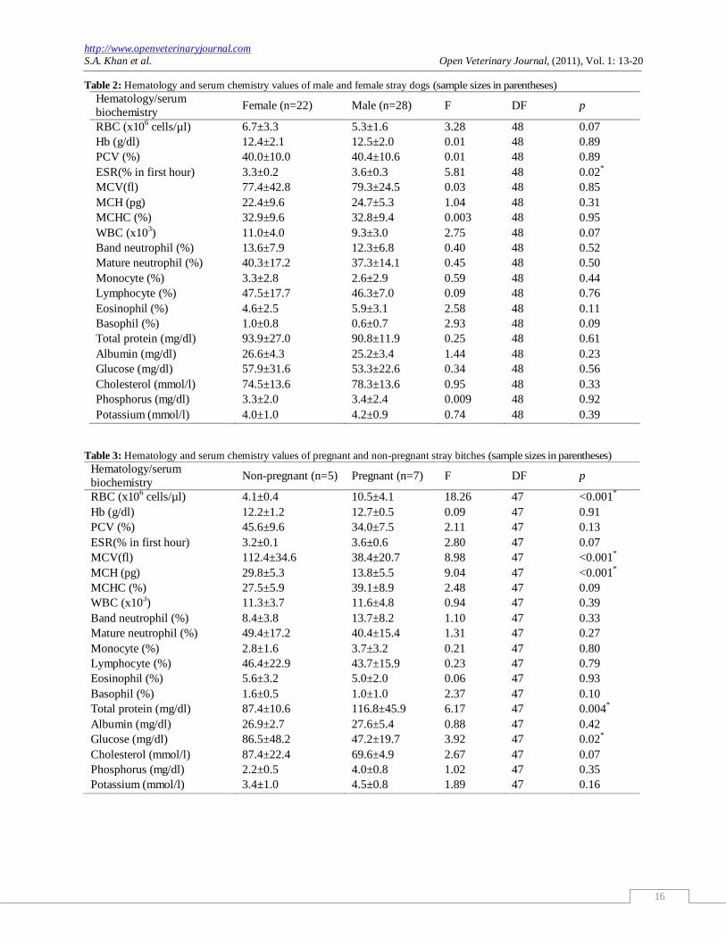
Table 2: Hematology and serum chemistry values of male and female stray dogs (sample sizes in parentheses)
Hematology/serum
biochemistry Female (n=22) Male (n=28) F DF p
RBC (x106 cells/µl) 6.7±3.3 5.3±1.6 3.28 48 0.07
Hb (g/dl) 12.4±2.1 12.5±2.0 0.01 48 0.89
PCV (%) 40.0±10.0 40.4±10.6 0.01 48 0.89
ESR(% in first hour) 3.3±0.2 3.6±0.3 5.81 48 0.02*
MCV(fl) 77.4±42.8 79.3±24.5 0.03 48 0.85
MCH (pg) 22.4±9.6 24.7±5.3 1.04 48 0.31
MCHC (%) 32.9±9.6 32.8±9.4 0.003 48 0.95
WBC (x103) 11.0±4.0 9.3±3.0 2.75 48 0.07
Band neutrophil (%) 13.6±7.9 12.3±6.8 0.40 48 0.52
Mature neutrophil (%) 40.3±17.2 37.3±14.1 0.45 48 0.50
Monocyte (%) 3.3±2.8 2.6±2.9 0.59 48 0.44
Lymphocyte (%) 47.5±17.7 46.3±7.0 0.09 48 0.76
Eosinophil (%) 4.6±2.5 5.9±3.1 2.58 48 0.11
Basophil (%) 1.0±0.8 0.6±0.7 2.93 48 0.09
Total protein (mg/dl) 93.9±27.0 90.8±11.9 0.25 48 0.61
Albumin (mg/dl) 26.6±4.3 25.2±3.4 1.44 48 0.23
Glucose (mg/dl) 57.9±31.6 53.3±22.6 0.34 48 0.56
Cholesterol (mmol/l) 74.5±13.6 78.3±13.6 0.95 48 0.33
Phosphorus (mg/dl) 3.3±2.0 3.4±2.4 0.009 48 0.92
Potassium (mmol/l) 4.0±1.0 4.2±0.9 0.74 48 0.39
Table 3: Hematology and serum chemistry values of pregnant and non-pregnant stray bitches (sample sizes in parentheses)
Hematology/serum
biochemistry Non-pregnant (n=5) Pregnant (n=7) F DF p
RBC (x106 cells/µl) 4.1±0.4 10.5±4.1 18.26 47 <0.001
*
Hb (g/dl) 12.2±1.2 12.7±0.5 0.09 47 0.91
PCV (%) 45.6±9.6 34.0±7.5 2.11 47 0.13
ESR(% in first hour) 3.2±0.1 3.6±0.6 2.80 47 0.07
MCV(fl) 112.4±34.6 38.4±20.7 8.98 47 <0.001*
MCH (pg) 29.8±5.3 13.8±5.5 9.04 47 <0.001*
MCHC (%) 27.5±5.9 39.1±8.9 2.48 47 0.09
WBC (x103) 11.3±3.7 11.6±4.8 0.94 47 0.39
Band neutrophil (%) 8.4±3.8 13.7±8.2 1.10 47 0.33
Mature neutrophil (%) 49.4±17.2 40.4±15.4 1.31 47 0.27
Monocyte (%) 2.8±1.6 3.7±3.2 0.21 47 0.80
Lymphocyte (%) 46.4±22.9 43.7±15.9 0.23 47 0.79
Eosinophil (%) 5.6±3.2 5.0±2.0 0.06 47 0.93
Basophil (%) 1.6±0.5 1.0±1.0 2.37 47 0.10
Total protein (mg/dl) 87.4±10.6 116.8±45.9 6.17 47 0.004*
Albumin (mg/dl) 26.9±2.7 27.6±5.4 0.88 47 0.42
Glucose (mg/dl) 86.5±48.2 47.2±19.7 3.92 47 0.02*
Cholesterol (mmol/l) 87.4±22.4 69.6±4.9 2.67 47 0.07
Phosphorus (mg/dl) 2.2±0.5 4.0±0.8 1.02 47 0.35
Potassium (mmol/l) 3.4±1.0 4.5±0.8 1.89 47 0.16

http://www.openveterinaryjournal.com
S.A. Khan et al. Open Veterinary Journal, (2011), Vol. 1: 13-20
17
Table 4: Hematology and serum chemistry values of stray dogs in different age groups
Hematology/serum
biochemistry Adult (n=29) Juvenile (n=18) Puppy (n=3) F DF p
RBC (x106 cells/µl) 6.3±3.3 5.8±1.9 5.7±2.3 0.18 47 0.001
*
Hb (g/dl) 12.4±1.3 13.1±2.4 8.6±1.3 7.52 47 0.27
PCV (%) 41.4±10.4 39.5±10.0 31.6±5.5 1.33 47 0.51 ESR (% in first hour) 3.4±0.3 3.5±0.3 3.4±0.3 0.68 47 0.66
MCV(fl) 82.1±43.4 73.3±20.7 70.4±23.0 0.40 47 0.35
MCH (pg) 23.4±8.3 24.4±7.5 17.0±7.8 1.07 47 0.35
MCHC (%) 31.9±8.6 35.1±10.8 28.0±8.0 1.06 47 0.83
WBC (x103) 10.6±3.6 10.4±3.6 6.1±3.1 2.10 47 0.13
Band neutrophil (%) 11.5±5.8 15.2±9.5 15.3±4.5 1.56 47 0.22
Mature neutrophil (%) 43.2±13.2 33.3±15.5 33.0±32.1 2.54 47 0.09
Monocyte (%) 3.1±3.4 2.6±1.6 4.6±3.0 0.67 47 0.51
Lymphocyte (%) 45.8±14.8 48.8±10.2 47.0±26.9 0.24 47 0.78
Eosinophil (%) 5.9±2.8 4.5±2.4 1.6±2.8 4.29 47 0.01*
Basophil (%) 0.9±0.9 0.8±0.6 0.6±1.1 0.26 47 0.76
Total protein (mg/dl) 98.1±24.7 83.6±14.0 92.7±5.8 2.68 47 0.07
Albumin (mg/dl) 25.6±3.8 26.5±4.4 26.6±2.4 0.34 47 0.71
Glucose (mg/dl) 55.5±27.5 57.2±31.2 51.5±5.4 0.05 47 0.94
Cholesterol (mmol/l) 78.9±14.9 71.5±10.2 77.2±15.5 1.68 47 0.19
Phosphorus (mg/dl) 3.1±1.1 3.8±3.4 2.8±0.7 0.65 47 0.52
Potassium (mmol/l) 4.3±1.1 3.7±0.6 3.8±0.3 2.85 47 0.06
Table 5: Hematology and serum chemistry values of stray dogs with different body conditions
Hematology/serum
biochemistry Good (n=21) Fair (n=26) Poor (n=3) F DF p
RBC (x106
cells/µl) 5.5±1.7 5.5±1.8 14.8±0.0 39.10 47 0.001*
Hb (g/dl) 12.9±1.6 12.0±2.4 13.0±0.0 1.17 47 0.31
PCV (%) 40.4±8.9 41.1±11.2 30.0±0.0 1.65 47 0.20
ESR (% in first hour) 3.5±0.2 3.4±0.3 3.9±0.0 3.21 47 0.04*
MCV(fl) 75.5±27.7 87.2±37.2 20.1±0.0 5.80 47 0.001*
MCH (pg) 24.9±6.9 23.9±7.8 8.7±0.0 6.56 47 0.001*
MCHC (%) 33.3±7.7 31.3±10.6 43.3±0.0 2.32 47 0.10
WBC (x103) 10.0±3.7 10.1±3.6 12.9±4.5 0.84 47 0.43
Band neutrophil (%) 10.3±4.3 14.7±8.3 17.6±11.5 2.88 47 0.06
Mature neutrophil (%) 40.6±14.1 36.8±17.7 47.0±6.0 0.72 47 0.49
Monocyte (%) 4.0±3.7 2.3±2.0 2.0±0.0 2.08 47 0.13
Lymphocyte (%) 42.4±13.7 50.0±14.2 52.0±0.0 2.00 47 0.14
Eosinophil (%) 6.0±2.8 4.3±2.8 7.0±0.0 2.81 47 0.07
Basophil (%) 0.8±0.8 0.8±0.7 2.0±0.0 3.19 47 0.05*
Total protein (mg/dl) 100.5±28.3 87.3±13.3 83.0±6.0 2.65 47 0.08
Albumin (mg/dl) 26.1±3.6 25.4±3.6 30.7±7.2 2.54 47 0.08
Glucose (mg/dl) 60.8±29.4 54.1±27.2 36.9±15.1 1.07 47 0.35
Cholesterol (mmol/l) 77.1±15.3 75.7±13.2 73.1±0.4 0.13 47 0.87
Phosphorus (mg/dl) 4.2±3.1 2.7±0.7 3.3±0.3 2.99 47 0.05*
Potassium (mmol/l) 4.3±1.0 3.9±0.9 3.8±1.0 0.80 47 0.45
These changes were related to increased
destruction of fetal erythrocytes as well as
the rapid growth of the puppy. Circulating
red cell mass was significantly reduced
(Lee et al., 1976).
In the second month of a puppy’s life, a
gradual increase in RBC takes place and
continues until adult levels are attained at
about one year of age (Anderson and Gee,
1958).

http://www.openveterinaryjournal.com
S.A. Khan et al. Open Veterinary Journal, (2011), Vol. 1: 13-20
18
A number of studies done on German Shepherds
revealed no significant difference in RBC, WBC, Hb,
MCV, MCH, MCHC and differential leucocytes
counts between adults and juveniles (Konrad et al.,
1980).
Young canids tend to have lower RBC, hemoglobin
and hematocrit values than mature adults. In beagles,
there were increases with age in the hematocrit,
hemoglobin and RBC values, and maximum values
were reached only between 13 and 24 months of age
(Bulgin et al., 1970).
Other studies in dogs have shown that the hemoglobin
and hematocrit increased until 18 months of age (Weiner and Bradley, 1972). Blood loss due to
infestations with external and internal parasites could
also lead to anemia.
Sex: Differences in ESR between the sexes were of little
practical value. Sedimentation of erythrocytes in blood
has been studied extensively in the past and it is
known that this property of the blood is influenced by
red cell and plasma characteristics.
This is reflected in species differences seen in the ESR
in health and changes in ESR during disease. Several
technical factors are also known to affect the ESR
(Ham and Curtis, 1938; Lloyd, 1958; Miale, 1967;
Whitby and Britton, 1969; Williams and Trainer,
1971).
It has been found that ESR increases with the length of
the tube and height of the blood column, and that it decreases when the bore size of the tube is less than
2.5 mm (Ham and Curtis, 1938). In our present study
the ESR values were higher in male dogs than in
females.
This finding was in contrast with results of previous
studies. The extent of ESR variation is affected by a
variety of factors. In a study involving 382 male and
382 female beagles between 8 and 16 months of age,
age-related changes in RBC, Hb and PCV were seen
but no sex influence was noted (Brunk and Becker-
Berger, 1980).
Some investigators have reported higher RBC,
hematocrit and hemoglobin values in male dogs, but
others have observed no differences between the sexes
(Anderson and Gee 1958, Michaelson et al., 1966;
Brunk and Becker-Berger 1980). No significant
differences between the sexes were found in the blood
values of conditioned wild coyotes or pen-raised coyotes (Jain, 1986).
Reproductive stage: RBC count was higher in pregnant bitches.
Erythrocyte production increases during pregnancy
while erythrocyte mass per unit of body weight
remains constant throughout the entire pregnancy and
hemoglobin and hematocrit progressively decrease
into the third trimester (Lund and Donovan, 1967;
Peck and Arias, 1979; Heilmann, 1987).
Physiological anemia is solely due to a dilution
decrease in hemoglobin concentration. There is
increased plasma volume of about 50% and red cell
mass of about 18-25%, which was very much
consistent with our findings. Moreover, PCV
decreases in pregnant bitches due to a shorter life span
of erythrocyte and hemo-dilatation (McFee, 1973;
Lurie, 1993; Cavill, 1995).
The MCV provides an indication of the status or size
of erythrocytes and epitomizes either normal or
abnormal cell division during erythropoiesis (Nussey et al., 1995). The MCV values were lower in pregnant
bitches due to higher RBC number during pregnancy
periods in canine populations. The different stages of
estrous cycle in stray dogs had frequently influenced
the progesterone and estrogen concentration, which
can determine the serum chemistry profile in these
semi-wild animals.
Body condition: In this study, dogs with poor body condition displayed
high RBC counts in comparison with fair and good
body conditioned dogs. This is seen in dehydrated
animals as their blood becomes more concentrated.
This is also noted in other conditions, such as some
cases of shock, response to high altitudes (the air is
'thinner,' containing less oxygen, so more RBC’s are
put into circulation), diseases of the lungs, etc.
Conditions decreasing the amount of oxygen reaching the tissues of the body will cause higher numbers of
RBCs to be found in the complete blood cell count
(CBC).
MCV and MCH values are inversely related to RBC
count or hematocrit value. Erythrocytosis, also called
polycythemia, is defined by an increase in total RBC
number, PCV, and Hb concentration above reference
intervals.
Erythrocytosis occurs frequently in dogs and can arise
due to a number of causes. Erythrocytosis may be
relative, due to a decrease in total plasma fluid
volume, or absolute, due to an increase in RBC
production (Sharma and Joshi, 2002).
Conclusion Stray dogs live closely with humans in Bangladesh. In
recent years, the sources of emerging and reemerging
diseases were mainly of animal origin, especially
originating from wildlife. Chances of disease emergence from stray dogs are of
great concern in Bangladesh. These findings provide
important baseline data with which to examine the
health status of stray dogs in south and south-east
Asia. The study examined the effect of sex, pregnancy
status, age, and body condition score on blood cell
counts and biochemistry values.

http://www.openveterinaryjournal.com
S.A. Khan et al. Open Veterinary Journal, (2011), Vol. 1: 13-20
19
This data may help to understand the emergence of
new diseases from this semi-wild animal including the
deadly rabies virus.
Acknowledgment Funding for this study was provided, in part, by the
Eppley Foundation for Research and the Rockefeller
Foundation
____________________________________________
References Ali, W., Khan, F.K., Doulah, S., Majumdar, J.U. 1977.
Surveillance of rabies in Dacca, Bangladesh. Med.
Res. Co. Bull. 3(2), 117-123.
Anderson, A.C. and Gee, W. 1958. Normal blood values in the Beagle. Vet. Med. 53, 135-138.
Brunk, R. and Becker-Berger, S. 1980. Statistical
examination of age and sex specific differences in
blood parameters in English beagle dogs. Berl
Munch Tierarztl Wochenschr. 93, 128-132.
Bulgin, M.S., Munn, S.L. and Gee, W. 1970.
Hematologic changes to 4 and one-half years of
age in clinically normal beagles. J. Am. Vet. Med.
Assoc. 157, 1064-1070.
Campbell, T.W. 1995. Avian haematology and
cytology, 2nd
Ed. Iowa state Uni. Press. Ames.
Cavill, I. 1995. Iron and erythropoisis in normal
subjects and in pregnancy. J. Perinat. Med. 23, 47-
50.
Court, J.P., Martin-Short, M. and Lees, G.M. 1986. A
comparison of the response of Dipetalonema viteae
and Brugia pahangi adult worms to antifilarial agents. Trop. Med. Parasitol. 37(4), 375-380.
Fowler, M.E. and Miller, R.E. (eds). 2003. Zoo and
Wild Animal Medicine. 5th
ed. St. Louis, MO:
Saunders, 485-486.
Ham, T.H. and Curtis, F.C. 1938. Plasma fibrinogen
response in man. Influence of the nutritional state,
induced hyperpyrexia, infectious diseases and liver
damage. Med. 17, 413-445.
Heilmann, L. 1987. Blood theology and pregnancy.
Bailliere Clin. Haem. 1(3), 777-799.
Howlett, J.C., Bailey, T.A., Samour, J.H., Naldo, J.L.
and D’aloia, M. 2002. Age-related hematologic
changes in captive-reared houbara, white-bellied,
and rufouscrested bustards. J. Wildlife Dis. 38,
804-816.
Jain, N.C. 1986. Normal values in blood of laboratory,
fur-bearing, and miscellaneous zoo, domestic, and
wild animals. In Schalm’s Veterinary Hematology. 4
th ed. Ed N. C. Jain. Philadelphia, Lea & Febiger,
337.
Kaneko, J.J., Harvey, J.W. and Bruss, M.L. 2008.
Clinical Biochemistry of Domestic Animals. 6th
ed.
San Diego, CA: Academic Press, 493, 889-895.
Kitagawa, H., Kitoh, K. and Ohba, Y. 1998.
Comparison of laboratory test results before and
after surgical removal of heartworms in dogs with
vena caval syndrome. J. Am. Vet. Med. Assoc. 213
(8), 1134-1136.
Konrad, J., Kupak, M. and Husak, S. 1980.
Hematology of the clinically healthy dog. Vet.
Med. (Praha). 25(7), 405-412.
Lee, P., Brown, M.E. and Hutzler, P.T. 1976. Blood
volume changes and production and destruction of
Erythrocytes in Newborn Dogs. Am. J. Vet. Res.
37(5), 561-575.
Lloyd, H.E. 1958. Estimation of the erythrocyte
sedimentation rate of capillary blood; description
of a new method. Ann. Rheum. Dis. 17, 234-239.
Lund, C.J. and Donovan, J.C. 1967. Blood volume during pregnancy: significance of plasma and red
cell volumes. Am. J. Obstet. Gynaecol. 98, 393-
403.
Lurie, S. 1993. Changes in age distribution of
erythrocytes during pregnancy, A longitudinal
study. Gynaecol. Obstet. Invest. 36, 141-144.
Mcfee, J.G. 1973. Anemia in pregnancy a reappraisal.
Obstet. Gynaecol. Surv. 28, 769-793.
Miale, B. 1967. Laboratory Medicine-Hematology, 3rd
ed. The C. V. Mosby Company, St. Louis.
Michaelson, S.M., Scherr, K. and Gilt, S. 1966. The
blood of the normal beagle. J. Am. Vet. Med.
Assoc. 148, 532-534.
Moustafa, A.M., Agag, B., Esmat, M. and Selim, A.M.
1991. Studies on filariasis in Egyptian buffaloes.
III. Clinical observations and electrophoretic
patterns in sera of naturally infested buffaloes with microfilaria before and after treatment with
stipophon. Zagazig Vet. J. 19, 583-595.
Nussey, G., Van Vuren, J.H.J. and Du Preez, H.H.
1995. Effect of copper on the haematology and
osmoregulation of the Mozambique tilapia,
Oreochromis mossambicus (Cichlidae). Comp.
Biochem. Phys. Part: 111A, 369-380.
Peck, T.M. and Arias, F. 1979. Hematologic changes
associated with pregnancy. Clin. Obstet. Gynaecol.
22, 485-498.
Safwat, M.S. and El-Abdin, Y.Z. 1982. Some
biochemical studies on the serum of infested and
non-infested camels with Dipetalonema evansi.
Egyptian J. Vet. Sci. 19, 141-145.
Sharma, M. and Joshi, C. 2002. Serum mineral and
haemato-biochemical profile of microfilariae
infested cattle in India: Its effects on production
and therapy. Asian-Australasian J. Anim. 15(3), 357-365.
Smith, S.A. 2000. Specific species appropriate
hematology. In: Feldman B.F., Zinkl J.G., Jain
N.C., eds. Schalm’s Veterinary Hematology. 5th
ed.
Baltimore, MD: Lippincott Williams & Wilkins,
1055-1224.
Weiner, D.J. and Bradley, R.E. 1972. The hemogram
and certain serum protein fractions in normal

http://www.openveterinaryjournal.com
S.A. Khan et al. Open Veterinary Journal, (2011), Vol. 1: 13-20
20
beagle dogs. Vet. Med. Sm. Anim. Clin. 67, 393-
398.
Whitby, L. and Britton, C.J.C. 1969. Disorders of the
Blood, 10th
ed., p.85, London, Churchill.
WHO. 1998. Report. Informal consultation. Regional
strategies for elimination of Rabies (WHO, South-
East Asia Regional Office). New Delhi, p-7.
Williams, J.L. and Trainer, D.O. 1971. A
hematological study of snow, blue and Canada
geese. J. Wildlife Dis. 7, 258-265.