tenascin is a serum marker of lobulak but not portal fibrogenesis in chronic liver diseases . dept....
TRANSCRIPT
HEPATOLOGY Vol. 22, No. 4, P t . 2, 1995 A A S L D A B S T R A C T S 369A
1049 FIBRONECTIN ISOFORM GENE EXPRESSION IN RAT HEPATIC STELLATE AND PARENCHYMAL CELLS OF NORMAL AND FIBROTIC LIVER. G Xu. I Virtanen(l~. V Ro~iers(2). T Niki. PJ De Bleser. E Wi~s¢. and A Geerts. Laboratory for Cell Biology and Histology, (1)Labora- tory for Toxicology, Free University Brussels (V.U.B.), and (2)Department of Anatomy, University of Helsinki.
Hepatic fibrosis is a hallmark of most chronic liver diseases and precedes cir- rhosis. Fibmnectin (FN) is a major component of liver biomatrix and seems to play a pacemaker role in liver fibrogenesis. We investigated FN isoform gene expression in hepatic stellate cells (HSC) and parenchymal cells (PC). HSCs are the most important connective tissue producing cells in injured liver. PCs are the main plasma FN secreting cells. FN isoform mRNAs were stud- ied by Northern hybridization in freshly isolated normal HSCs and PCs, CCI 4 treated HSCs and PCs, and HSCs at day 3, 6 and 12 in culture. Five specific cDNA probes that recognize different domains of FN mRNA were kindly donated by Dr. R. Hynes. FN isoform proteins in primary culture and subcul- tured HSCs were examined by immunoprecipitation using specific mono- clonal antibodies. Rat total FN transcript was detectable in freshly isolated CCI 4 treated HSCs and in cultured HSCs, but not in PCs. EIlIB fragment was only present in cultured HSCs. V95 fragment and SR270 fragment were found in all investigated cells except in freshly isolated normal HSCs. Immunoprecipitation confirmed the Northern hybridization analysis. We found that EIIIA- and EIIIB-contalning FNs were synthesized and secreted by primary cultured and subcultured HSCs. We conclude that none of theFN isoforms were present in quiescent HSCs. Untreated and CC14 treated PCs express EIIIA'-EIIIB--V95 + FN. In vivo activated HSCs express a variant form of FN (EIIIA+-EIIIB'-V95 +) during transition to the myofibroblast-like phe- notype. This variant was also produced and secreted bY cultured HSCs. Currently, we are examining whether EIIIA-containing FN can be used as a blood marker for detecting activation of HSCs in chronic liver disease.
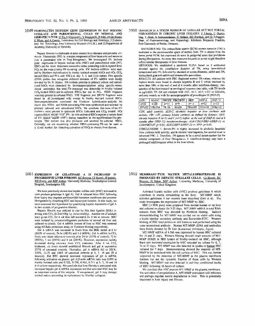
1050 TENASCIN IS A SERUM MARKER OF LOBULAR BUT NOT PORTAL FIBROGENESIS 1N CHRONIC LIVER DISEASES. P Libuda. C Oester- ling, T AkstL R Somasundaram. U St61zel. EO Riecken. and D Schuppan. Dept. of Gastroenterology and Hepatology, Klinikum Benjamin Franklin, Free University of Berlin, Germany.
BACKGROUND: The extracellular matrix (ECM) protein tenascin (TN) is restricted to the perisinusoidal space of normal liver. TN is absent from the dense portal ECM, but expressed de novo in periportal areas that proliferate during flbrogenesis. An assay that measures tenascin in serum might therefore reflect lobular flbrogenesis in liver diseases. METHODS: We established a sandwich ELISA based on 2 antibodies directed against the constitutive domains of TN, using immobilized monoclonal anti-TN, followed by standard or serum-dilutions, rabbit anti-TN, biotinylated goat anti-rabbit and streptavidin-peroxidase. RESULTS: All patients with PBC displayed normal TN-values, whereas the highest levels were found in chronic hepatitis B and C Which declined by more than 50% at the end of and at 6 months after interferon-therapy, irre- spective of the biochemical or serological response (see table, with TN-levels in }ig/I+SD). TN did not correlate with AST, ALT, ALP, ~/-GT or bilirubin, and only weakly so with the aminopropeptide of procollagen lII (pIIINP).
ON ALH-C PBCI/I1 PBCIII/IV nHE__P HEP-6 HEP-12 =22 n=20 n=ll I n=9 n=12 n=12 7+32 318+_104 ] 78+26 I 84+-/9 785+169 341+124 354+112
CON, adult controls; ALH-C, alcoholic hepatitis or liver cirrhosis with activity; PBC I-IV, primary biliary cirrhosis as defined by Scheuer; HEP, chronic hepatitis B (n=5) and C (n=7) before, at the end of (HEP-6) and at 6 months after (HEP-12) interferon-therapy. ALH-C/HEP/HEP-6/HEP-12 vs. CON/PBCI-IV: p<O.01; ALH-C/HEP-6/HEP-12 vs. HEP: p<O.05.
CONCLUSIONS: 1. Serum-TN is highly increased in alcoholic hepatitis/ liver cirrhosis with activity, and in chronic viral hepatitis, but normal even in advanced PBC. 2. Therefore, TN appears to be a novel serum-marker for the lobular component of liver fibrogenesis. 3. Interferon-therapy may have a prolonged antifibrogenic effect in the liver lobule.
1051 EXPRESSION OF GELATINASE A IS INCREASED IN PROGRESSIVE LIVER FIBROSIS. RC Benyon, CJ Hovell, JP Iredale, WF Ferris and MJP Arthur. University Medicine, Southampton General Hospital, Southampton UK.
We have previously shown that hepatic stellate ceils (HSC) activated in vitro produce gelatinase A (gel A). Gel A released from HSC following liver injury may degrade perisinusoidal basement membrane and promote flbrogenesis by disturbing HSC and hepatocyte function. In this study, we have examined this hypothesis by quantifying hepatic expression of gel A in two models of progressive fibrosis.
Hepatic fibrosis was induced in rats by bile duct ligation (BDL) or dosing with CCI 4 (0.2ml/100g i.p. twice weekly). Another set of animals were given CC14 for 4 wk then left untreated for 4 wk to recover. HSC were isolated by pronase/collagenase perfusion of normal rat liver and cultured on plastic. Gel A mRNA content of liver or HSC was assessed using RNAase protection assay or Northern blotting respectively.
Gel A mRNA was increased in livers from the BDL model at 6 hr (362% of control), 24 hr (383%) and 3 d (400%). Gel A mRNA in CCI 4 livers was raised relative to controls after 24 hr (157% of control), 72 hr (595%), 1 wk (295%) and 4 wk (289%). However, expression rapidly decreased during recovery from CCI 4 treatment. After 4 wk CCI 4 treatment, rat livers showed established fibrosis and gel A expression 537% of untreated controls. Thereafter, gel A mRNA fell to 201%, 210%, 111% and 148% of untreated controls at 3, 7, 14 and 28 d recovery. Rat HSC showed increased expression of gel A mRNA following activation on plastic: gel A/fl-actin mRNA ratio was 0.099 in freshly isolated cells and 0.322, 0.758, 0.903, 0.711 at 5, 7, 10 and 14 d of culture respectively. We conclude that liver fibrosis is associated with increased hepatic gel A mRNA expression and that activated HSC may be an important source of this enzyme. If unsupressed, gel A may damage normal matrix promoting its replacement by fibrillar collagens.
1052 MEMBRANE-TYPE MATRIX METALLOPROTEINASE IS PRODUCED BY HEPATIC STELLATE CELLS. CJ Hovell, RC Benyon, JE Baker, MJP Arthur. University Medicine, University of Southampton, United Kingdom.
Activated hepatic stellate cells (HSC) produce gelatinase A which contribute to matrix remodelling in the liver. MT-MMP which activates gelatinase A has recently been described (Sato et al). The study investigates the expression of MT-MMP by HSC.
HSC (> 90% pure) were prepared from normal human or rat liver and cultured on plastic for 3-21 days. MT-MMP mRNA in total RNA extracts from HSC was detected by Northern blotting. Indirect immunolabelling for MT-MMP was carried out on viable cells using a biotin labelled secondary antibody and Extravidin-FITC. Western blotting of HSC total protein or celt fractions was performed using the same monoclonal antibody. Human MT-MMP cDNA and antibodies were kindly donated by Dr Sato (Kanazawa University, Japan).
MT-MMP mRNA of 4.5kb was expressed by human HSC cultured for 14 and 21 days. Western blotting showed small amounts of MT- MMP (63kD) in SDS lysates of freshly-isolated rat HSC, although there was increased expression by HSC activated by culture for 4, 7, 14 or 21 days. MT-MMP was also detected in lysates of human HSC cultured for 7 days. Immunostaining showed the majority of MT- MMP to be associated with the cell surface of HSC. This was further supported by the detection of MT-MMP in the plasma membrane fraction but not the cytosolic fraction of these cells by Western blotting. MT-MMP was also detected in cell-free conditioned media of HSC following 16 hours of culture.
We conclude that HSC express MT-MMP at the plasma membrane. Via activation of progelatinase A, MT-MMP expression will influence and perhaps regulate matrix degradation in liver. This is potentially important in liver injury and fibrosis.