tenascin-c and mechanotransduction in the development and ... filecardiovascular research institute,...
TRANSCRIPT

REVIEW ARTICLEpublished: 29 July 2014
doi: 10.3389/fphys.2014.00283
Tenascin-C and mechanotransduction in the developmentand diseases of cardiovascular systemKyoko Imanaka-Yoshida1,2* and Hiroki Aoki3
1 Department of Pathology and Matrix Biology, Mie University Graduate School of Medicine, Tsu, Japan2 Mie University Research Center for Matrix Biology, Tsu, Japan3 Cardiovascular Research Institute, Kurume University, Kurume, Japan
Edited by:
Kersti K. Linask, University of SouthFlorida Morsani College ofMedicine, USAMichiko Watanabe, Case WesternReserve University School ofMedicine, USA
Reviewed by:
Richard L. Goodwin, University ofSouth Carolina School of Medicine,USARaymond Bruce Runyan, Universityof Arizona, USA
*Correspondence:
Kyoko Imanaka-Yoshida, Departmentof Pathology and Matrix Biology,Mie University Graduate School ofMedicine, 2-174 Edobashi,Tsu 514-8507, Japane-mail: [email protected]
Living tissue is composed of cells and extracellular matrix (ECM). In the heart and bloodvessels, which are constantly subjected to mechanical stress, ECM molecules formwell-developed fibrous frameworks to maintain tissue structure. ECM is also important forbiological signaling, which influences various cellular functions in embryonic development,and physiological/pathological responses to extrinsic stimuli. Among ECM molecules,increased attention has been focused on matricellular proteins. Matricellular proteinsare a growing group of non-structural ECM proteins highly up-regulated at active tissueremodeling, serving as biological mediators. Tenascin-C (TNC) is a typical matricellularprotein, which is highly expressed during embryonic development, wound healing,inflammation, and cancer invasion. The expression is tightly regulated, dependent on themicroenvironment, including various growth factors, cytokines, and mechanical stress.In the heart, TNC appears in a spatiotemporal-restricted manner during early stages ofdevelopment, sparsely detected in normal adults, but transiently re-expressed at restrictedsites associated with tissue injury and inflammation. Similarly, in the vascular system, TNCis strongly up-regulated during embryonic development and under pathological conditionswith an increase in hemodynamic stress. Despite its intriguing expression pattern,cardiovascular system develops normally in TNC knockout mice. However, deletion ofTNC causes acute aortic dissection (AAD) under strong mechanical and humoral stress.Accumulating reports suggest that TNC may modulate the inflammatory response andcontribute to elasticity of the tissue, so that it may protect cardiovascular tissue fromdestructive stress responses. TNC may be a key molecule to control cellular activity duringdevelopment, adaptation, or pathological tissue remodeling.
Keywords: extracellular matrix, tenascin-C, matricellular protein, mechanotrasduction, coronary artery, heart,
aortic dissection
INTRODUCTIONLiving tissue is composed of cells and extracellular matrix (ECM).In the heart and blood vessels, which are constantly subjected tomechanical stress, ECM molecules form well-developed fibrousframeworks to maintain the tissue structure by supporting theshape and position of cells, integrating and transmitting mechan-ical forces generated inside the cells to whole tissue. ECM isalso important for biological signaling, which influences variouscellular functions in embryonic development, and physiologi-cal/pathological responses to extrinsic stimuli. Tenascin-C (TNC)is a non-structural ECM protein highly expressed in morphogen-esis and tissue remodeling, and has a wide range of effects on cellresponses. Emerging evidence suggests that TNC may be involvedin mechanotransduction in response to mechanical stress. In thisreview, we will focus on the adaptive role of TNC in the mechan-ical stress response in the development and pathological state ofthe cardiovascular system.
OVERVIEW OF EXTRACELLULAR MATRIX INCARDIOVASCULAR SYSTEMFIBROUS EXTRACELLULAR MATRIXOf all the organs of the body, the large arteries, particularly theaorta, are subject to the greatest mechanical stress. They have awell-organized fibrous framework. In the tunica media, multi-layered elastin sheets (lamellae) connected by fine elastin fibersform a three-dimensional continuous network that links smoothmuscle cells. This elastin network of the arterial wall functions asan elastic reservoir protecting the tissue from destructive stress.The outermost layer, the tunica adventitia, consists of a collagen-rich ECM and helps prevent vascular rupture at extremely highpressures (Wagenseil and Mecham, 2009). In the heart, the majorstructural component of the ECM is collagen, which also formsa three-dimensional network interconnecting myocytes to eachother and to the vasculature (Caulfield and Borg, 1979; Borg andCaulfield, 1981). The fibrous skeleton composed of collagen is
www.frontiersin.org July 2014 | Volume 5 | Article 283 | 1

Imanaka-Yoshida and Aoki Tenascin-C and mechanical stress
continuous with the annulus fibrosus cordis, the support appara-tus of the tricuspid, mitral, and aortic valves to the cardiac musclein a manner analogous to the attachment of tendons to skeletalmuscle (Hinton and Yutzey, 2011). This stress-tolerant collage-nous network not only contributes to passive elastic propertiesof the heart but also to the transmission of mechanical forces toand from the cardiomyocytes (reviewed in Sussman et al., 2002;Bowers et al., 2010; Borg and Baudino, 2011).
NON-STRUCTURAL MATRIX, MATRICELLULAR PROTEINIn addition to the fibrous ECM, a unique functional cate-gory of non-structural ECM, matricellular proteins, are receiv-ing increasing attention (Bornstein, 2009). Matricellular proteinsconstitute a growing family (Table 1) that originally includedthrombosondin-1 (TSP1), SPARC (secreted protein, acid andrich in cysteine; osteonectin), and TNC (Sage and Bornstein,1991), and then TSP2, osteopontin, CCN1, CTGF (CCN2), andtenascin-X were added (Bornstein and Sage, 2002). They havecommon unique properties: (1) expressed at high levels dur-ing development and in response to injury; (2) do not subservestructural roles but function as modulators of cell-matrix inter-actions; (3) bind to many cell-surface receptors, other ECMmolecules, growth factors, cytokines, and proteases; (4) gen-erally induce de-adhesion, in contrast to the positive adhesiv-ity of most matrix proteins (Bornstein and Sage, 2002). Theterm has become used more widely and new members, such asgalectins and periostin, have joined the group (Bornstein, 2009).In cardiovascular development, significant roles of periostinhave been reported (Conway and Molkentin, 2008; Inai et al.,2008; Norris et al., 2008, 2009; Ghatak et al., 2014). It is alsonoteworthy that some members, such as SPARC, osteopontin,and periostin, have been found to be related to developing
Table 1 | Matricellular proteins.
Thrombospondins
TSP-1
TSP-2
Secreted protein acidic and rich in cysteine (SPARC/osteonectin)
Tenascin family
Tenascin-C
Tenascin- X
Osteopontin
CCN family
CCN1 Cysteine-rich angiogenic inducer (CYP-61)
CCN2 Connective tissue growth factor (CTGF)
CCN3 Nephroblastoma overexpressed (Nov)
CCN4 Wnt-induced secreted protein-1 (WISP-1)
CCN5 WISP-2 connective tissue growth factor-like protein (CTGF-L)
CCN6 WISP-3
Periostin
Galectins
Plasminogen activator inhibitor type 1 (PAI-1)
Fibulin-5
Small leucine-rich proteoglycans (Biglycan, Decorin, Lumican,
Fibromodulin)
bone and teeth, which are subjected to strong mechanicalstress.
TENASCIN-CTHE TENASCIN FAMILYTenascins are a family of multimeric ECM glycoprotein character-ized by an N-terminal globular domain and heptad repeats, whichfacilitate multimerization; one or more tenascin-type epidermalgrowth factor (EGF)-like repeats; a series of fibronectin (FN) typeIII domains, and a C-terminal fibrinogen-related domain. Thereare six names for the tenascin gene products: tenascin-C, X, R, Y,W, and N (Tucker et al., 2006; Tucker and Chiquet-Ehrismann,2009). TNC was the first tenascin found to be highly expressed intendons and embryonic ECM (Chiquet-Ehrismann et al., 1986).It was discovered independently in several laboratories as gliomamesenchymal ECM antigen, myotendinous antigen, cytotactin,and J1 glycoprotein (reviewed in Tucker et al., 2006; Chiquet-Ehrismann and Tucker, 2011). Tenascin-R is the second memberand is predominantly expressed in the central and peripheralnervous systems (Rathjen et al., 1991). Tenascin-X is a mam-malian tenascin primarily expressed in loose connective tissuesuch as the dermis, epimysium, and blood vessels (Matsumotoet al., 1992; Bristow et al., 1993) Mutations in tenascin-X canlead to a type of Ehlers–Danlos Syndrome (reviewed in Bristowet al., 2005) Tenascin-Y is an avian tenascin similar to mammaliantenascin-X (Hagios et al., 1996). Tenascin-W (Weber et al., 1998)is found primarily in pre-osteogenic areas, the kidney, smoothmuscle, and most prominently also in cancer stroma. Tenascin-Nis most recently discovered tenascin and is similar to tenascin-W(Neidhardt et al., 2003).
BIOLOGICAL ROLE OF TENASCIN-CTNC is the best characterized member of the family (Orend andChiquet-Ehrismann, 2006; Midwood and Orend, 2009; Chiquet-Ehrismann and Tucker, 2011; Midwood et al., 2011; Udalovaet al., 2011; Brellier and Chiquet-Ehrismann, 2012; Chiquet-Ehrismann et al., 2014) and is a typical matricellular protein. Itis a huge molecule of approximately 220–400 kDa as an intactmonomer and is assembled as a hexamer. TNC is found in manydeveloping organs of embryos, down-regulated after birth toa few tissues bearing high tensile stress and locations of highcell turnover, but highly up-regulated during injury, inflamma-tion, regeneration, and cancer (Chiquet-Ehrismann et al., 2014).A number of in vitro studies suggest that TNC has a widerange of effects on cell adhesion, motility, differentiation, growthcontrol, and ECM organization via multiple cell surface recep-tors including integrins α9β1, αvβ3, and αvβ6, Toll-like recep-tor 4 (TLR4) and syndecan-4 (Orend and Chiquet-Ehrismann,2006; Midwood and Orend, 2009). As in the case of targetdisruption of several other matricellular protein genes, TNCknockout mice develop normally (Saga et al., 1992; Forsberget al., 1996). Recent detailed investigations of various diseasemodels using TNC KO have suggested that TNC may pro-mote tissue healing but enhances inflammation and fibrosis(Midwood et al., 2011; Udalova et al., 2011; Brellier and Chiquet-Ehrismann, 2012; Imanaka-Yoshida, 2012; Chiquet-Ehrismannet al., 2014).
Frontiers in Physiology | Biophysics July 2014 | Volume 5 | Article 283 | 2

Imanaka-Yoshida and Aoki Tenascin-C and mechanical stress
During embryogenesis and tissue remodeling, TNC isexpressed transiently at specific sites, suggesting that the expres-sion of TNC is tightly regulated dependent on the cell typeand tissue microenvironment (Tucker and Chiquet-Ehrismann,2009). Many different growth factors, such as TGFβ, FGF, PDGF,and proinflammatory cytokines, are able to induce TNC expres-sion (for a review, see Orend and Chiquet-Ehrismann, 2006;Tucker and Chiquet-Ehrismann, 2009).
A variety of signaling pathways and transcription factors areknown to stimulate TNC transcription (reviewed in Chiquet-Ehrismann and Tucker, 2011). These include TGF/Smad 3/4(Jinnin et al., 2004), TLR4/NFkB (Goh et al., 2010), c-Jun/NFkB(Mettouchi et al., 1997), Notch (Sivasankaran et al., 2009), Sox4(Scharer et al., 2009), PDGF/Ets (Jinnin et al., 2006), and MEF2cwith scleraxis (della Gaspera et al., 2009). Conversely, TNC cantrigger a variety of signaling pathways via multiple cell surfacereceptors. Interestingly, it affects some of the same signaling path-ways that initially trigger the expression leading to negative orpositive feedback loops (Chiquet-Ehrismann and Tucker, 2011).For example, PDGF can induce TNC expression via the phospho-inositide 3-kinase/Akt pathway (Jinnin et al., 2006) and MAPKpathways (Chiquet et al., 2004) and, in turn, TNC enhancesPDGF signaling by cross-talk between PDGFR-β and integrinαvβ3 with activation of focal adhesion kinase and Src tyrosinekinase (Ishigaki et al., 2011). In contrast, a negative feedbackloops is created in the case of small GTPase RhoA as discussedin the next section.
INDUCTION OF TENASCIN-C BY MECHANO-STRESSMechanical stress is also a strong inducer of TNC. Just as oneof its original names, “myotendinous antigen,” suggests, TNC ishighly expressed at the myotendinous and osteotendinous junc-tions (Jarvinen et al., 1999, 2000, 2003) at sites subjected tomechanical stress. High expression of TNC is often observed atthe branching point of arteries (Mackie et al., 1992), although theexpression level of TNC is generally low in adult blood vessels.Based on this distribution of the molecule, the close associationof mechanical stress and TNC has been proposed. Supportingthis possibility, load-induced bone remodeling or muscle over-load up-regulates the expression of TNC (Webb et al., 1997; Flucket al., 2000; Mikic et al., 2000; Mackey et al., 2011), while immobi-lizing tendons down-regulates the expression. In culture, variousmechanical stresses including stretching (Chiquet et al., 2004),compression (Jagodzinski et al., 2008), and shear stress (Tan et al.,2013), up-regulate TNC synthesis by fibroblasts, chondrocytes,smooth muscle cells, and endothelial cells.
Several types of cell-surface proteins, including stretch-sensitive ion channels, are known to sense mechanical forces andtranslate them into biochemical signals (Kung, 2005). Mechanicalinputs can be also detected by mechanosensing apparatus ofthe focal adhesion complex and transduced to the cytoskele-ton (Wang et al., 2009). Chiquet and coworkers have shown amechanism by which a mechano-signal is transduced at the link-age between the ECM and cytoskeleton, which controls TNCtranscription mediated by megakaryoblastic leukemia 1 (MALor MKL1)/myocardin-related transcription factor A (MRTFA)(Chiquet et al., 2007, 2009; Asparuhova et al., 2009, 2011; Brosig
et al., 2010). The cycle stretch of fibroblasts up-regulates TNCtranscription, independent of de novo protein synthesis, paracrinefactors such as TGFβ, and mitogen-activated protein kinases(MAPKs), but depends on actomyosin contractility controlled bythe RhoA/ROCK pathway (Sarasa-Renedo et al., 2006) (Figure 1).Mechanical stimuli activate the signaling pathway involving inte-grin β1 (Chiquet et al., 2007) and integrin-linked kinase (ILK)(Maier et al., 2008), which induces actin assembly and stressfiber formation via mDia and ROCK (Ridley and Hall, 1992).MAL/MLK1/MRTFA is a coactivator of serum response factor(SRF) and is predominantly localized in the cytoplasm throughan interaction with G-actin (Miralles et al., 2003; Guettler et al.,2008). Therefore, depletion of the cytoplasmic G-actin pool fol-lowing Rho activation causes translocation of MAL into thenucleus, where it induces TNC transcription, partly dependenton SRF (Asparuhova et al., 2011).
RhoA-dependent mechanotransduction requires pericellularfibronectin (Lutz et al., 2010). TNC binds fibronectin at the bind-ing site to syndecan-4, a coreceptor for integrin α5β1, and hasa negative impact on focal adhesion formation and activation ofRhoA (Midwood et al., 2006; Lange et al., 2008; Van Obberghen-Schilling et al., 2011). Therefore, mechanically induced TNC maylead to negative feedback from the mechanotrasduction signal.Moreover, since TNC is an elastic molecule that can be stretchedto several times its resting length in vitro (Oberhauser et al.,1998; Marin et al., 2003), it may contribute to tissue elasticity andprotect against mechanical stress.
FIGURE 1 | Diagram of the molecular pathway of the
mechano-induction of tenascin-C. Mechanical strain activates RhoA infibroblasts, depending on fibronectin, integrin β1 and integrin-linked kinase(ILK), which causes the reduction of monomeric G-actin by inducing actinassembly and stress fiber formation. Depletion of the G-actin pool freesMAL/myocardin-related transcription factor-A (MRTF-A)/megakaryoblasticleukemia-1 (MKL1) to enter the nucleus, which induces TNC expressionpartly depending on serum response factor (SRF). Meanwhile, TNC bindsfibronectin at the syndecan-4 binding sites and interferes withfibronectin-mediated RhoA activation, and finally suppresses TNCtranscription. Adapted from Asparuhova et al. (2009), Imanaka-Yoshida et al.(in press).
www.frontiersin.org July 2014 | Volume 5 | Article 283 | 3
Imanaka-Yoshida and Aoki Tenascin-C and mechanical stress
MECHANOTRANSDUCTION AND TENASCIN-C INCARDIOVASCULAR DEVELOPMENTDuring heart development, ECM not only provides structuralsupport for embedded cells but plays an important biological rolein the orchestration of cell behavior to form a complex structurewith 4 chambers and 4 valves. Accumulated studies have showndiverse functions of various ECM molecules, including hyaluro-nan, proteoglycans, the collagen family, fibronectin, and periostin(reviewed in Lockhart et al., 2011).
HEART DEVELOPMENT AND TENASCIN-CSpecific roles of TNC in heart morphogenesis have long beenanticipated based on its strictly regulated temporal expression atspecific sites closely associated with cell migration and epithelial-mesenchymal/mesenchymal-epithelial transition: (Wagenseil andMecham, 2009) differentiation of precardiomyocytes, (Caulfieldand Borg, 1979) cushion tissue formation, (Borg and Caulfield,1981) valve formation, and (Hinton and Yutzey, 2011) coronaryvessel formation (Imanaka-Yoshida et al., 2003).
During the development of mouse embryos, the initial expres-sion of TNC is detected in mesodermal cells in the first heart field(FHH), which undergo mesenchymal-epithelial transition anddifferentiate to cardiomyocytes and endocardial cells. Once thecells differentiate to cardiomyocytes, they rapidly stop express-ing TN-C, while endocardial cells continue to express TNC. TNCexpression is also detected at the recruitment of precardiac cellsfrom the second heart field (SHF) (Imanaka-Yoshida et al., 2003).Interestingly, cardiomyocytes from the SHF in the outflow tractmaintain the expression of TN-C during looping and shortening.
Endocardial cushion and tenascin-CThe primitive heart consists of the inner endocardium and outermyocardium and cardiac jelly, composed predominantly of theproteoglycan glycosaminoglycan hyaluronan between the two lay-ers. After cardiac looping, the cardiac jelly expands within the AV
canal and outflow tract regions and endocardial cells undergoepithelial–mesenchymal transformation (EMT) and invade it,forming an endocardial cushion (Eisenberg and Markwald, 1995;Person et al., 2005), which is the initial step in valvulogenesis.
A number of reports have demonstrated the expression ofTNC in cushion tissue closely associated with EMT of endocar-dial cells (Hurle et al., 1990; Crossin and Hoffman, 1991; Zhanget al., 1993; Hiltgen et al., 1996; Sugi and Markwald, 1996; Boyeret al., 1999). Indeed, TNC promotes EMT of cancer cells in vitro(Nagaharu et al., 2011; Katoh et al., 2013).
Furthermore, Garita et al. have recently reported interestingresults suggesting that TNC may provide a structural commu-nication or mechano-communication between the myocardiumand endocardium during looping. Using four-dimensional opti-cal coherence tomography (OCT), they found that the endo-cardium was consistently oriented between the midline of theventral floor of the foregut and the outer curvature of the myocar-dial wall throughout the cardiac cycle and that TN-C co-localizedwith FN at the attachment areas at the outer curvature of the heartwall to the ventral floor of the foregut (Garita et al., 2011).
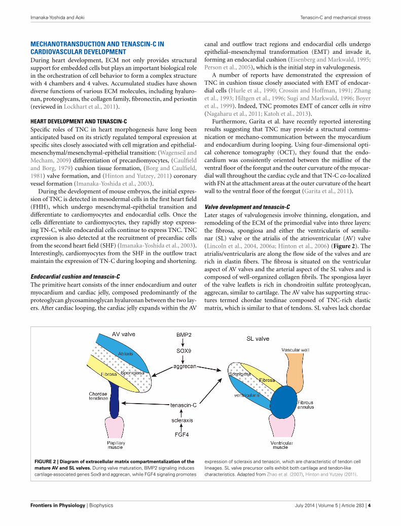
Valve development and tenascin-CLater stages of valvulogenesis involve thinning, elongation, andremodeling of the ECM of the primordial valve into three layers:the fibrosa, spongiosa and either the ventricularis of semilu-nar (SL) valve or the atrialis of the atrioventricular (AV) valve(Lincoln et al., 2004, 2006a; Hinton et al., 2006) (Figure 2). Theatrialis/ventricularis are along the flow side of the valves and arerich in elastin fibers. The fibrosa is situated on the ventricularaspect of AV valves and the arterial aspect of the SL valves and iscomposed of well-organized collagen fibrils. The spongiosa layerof the valve leaflets is rich in chondroitin sulfate proteoglycan,aggrecan, similar to cartilage. The AV valve has supporting struc-tures termed chordae tendinae composed of TNC-rich elasticmatrix, which is similar to that of tendons. SL valves lack chordae
FIGURE 2 | Diagram of extracellular matrix compartmentalization of the
mature AV and SL valves. During valve maturation, BMP2 signaling inducescartilage-associated genes Sox9 and aggrecan, while FGF4 signaling promotes
expression of scleraxis and tenascin, which are characteristic of tendon celllineages. SL valve precursor cells exhibit both cartilage and tendon-likecharacteristics. Adapted from Zhao et al. (2007), Hinton and Yutzey (2011).
Frontiers in Physiology | Biophysics July 2014 | Volume 5 | Article 283 | 4

Imanaka-Yoshida and Aoki Tenascin-C and mechanical stress
tendineae, but instead have comparable supporting tissue in theaortic and pulmonic roots and hinge regions (Zhao et al., 2007).
Remodeling of the heart valve primordia shares a regula-tory pathway with developing cartilage/tendons (Lincoln et al.,2006b; Hinton and Yutzey, 2011). In the development of limbbuds, diversification of cartilage and tendon cells from a com-mon precursor is antagonistically regulated by BMP and FGFsignaling pathways. BMP2 not only promotes chondrogenesisbut also inhibits tendon development, while FGF4 promotes ten-don differentiation (Edom-Vovard et al., 2002; Edom-Vovard andDuprez, 2004).
Similarly, BMP2 signaling activates valve progenitor cells toexpress Sox9 transcription factor and the aggrecan gene as wellas cartilage precursors in limb buds (Lincoln et al., 2006a; Zhaoet al., 2007). In contrast, FGF4 signaling activates scleraxis andTNC expression in the valve-supporting apparatus as well as indeveloping tendons (Lincoln et al., 2006a; Zhao et al., 2007).Hemodynamics is often proposed to be one of the driving forcesin valve development (Combs and Yutzey, 2009); however, thereis no evidence indicating that mechano-stress might be involvedin the induction of TNC during the development of the valves.
VASCULAR DEVELOPMENT AND TENASCIN-CAnother possibility is that TNC may play a role in blood ves-sel development. In coronary vessels, most vascular progenitorscome from the proepicardial organ (PEO) between the primi-tive heart and the liver bud. Mesenchymal cells from the PEOmigrate to the heart and form the epicardium. Epicardial cellsundergo EMT, differentiate into endothelial cells and vascularsmooth muscle cells (VSMCs), and form a primitive capillary net-work, which eventually connects to the aorta (see Nakajima andImanaka-Yoshida, 2013, for review). During this process, TNC istransiently expressed in PEO before cell migration and at epicar-dial EMT. It is worthy of note that TNC is highly up-regulated andassociated with thickening of the vascular wall after the prema-ture vessels are linked with the aorta (Ando et al., 2011), possiblypromoting the recruitment of vascular mural cells by facilitatingPDGF-BB/PDGFRβ signaling (Ishigaki et al., 2011).
Similar up-regulation of TNC in the vascular wall associatedwith hemodynamic change is observed during the developmentof the aorta (Imanaka-Yoshida et al., in press). In E12-13 mouseembryos, weak expression of TNC is detected in the ascending,arch and descending aorta. After ED14-15, when the systemiccirculatory system is established, TNC expression is evidently up-regulated and becomes even stronger after birth. In normal adults,the expression of TNC in the aortic wall is generally reduced,although the infra-renal aorta continues to express TNC.
Despite its intriguing expression pattern during cardiovasculardevelopment, targeting deletion of the TNC gene causes a grosslynormal phenotype (Saga et al., 1992; Forsberg et al., 1996). Ourrecent preliminary data suggested that over-expression of TNCin the heart may not cause a distinct phenotype, either (unpub-lished data). Compensatory mechanisms should be present intissue morphogenesis of the embryo although it is not identi-fied. However, increasing number of studies indicate that TNC is a“stress protein” whose importance becomes apparent when organhomeostasis is challenged by injury or destructive stress such as
mechanical overload (Chiquet-Ehrismann et al., 2014), while it ismasked during embryonic development.
MECHANOTRANSDUCTION AND TENASCIN-C INCARDIOVASCULAR DISEASEMECHANOTRANSDUCTION IN HEART DISEASEIn the heart, extracellular and intercellular mechanical loadsare linked to the myofibrils in cardiomyocytes via vari-ous mechanosensing complexes (McCain and Parker, 2011).Cadherins links with myofibrils of neighboring cells at interca-lated disks, while integrins attach Z-discs laterally to the con-nective tissue at costameres (Pardo et al., 1983a,b). Costameresare structures related to the focal adhesion complex and criticalcytoskeletal elements involved in environmental mechanochem-ical signal transduction into cardiomyocytes (Samarel, 2005;Russell et al., 2010). They are also the sites where contractileforces generated within cardiomyocytes are transmitted to thesurrounding interstitial collagen network (Danowski et al., 1992;Imanaka-Yoshida et al., 1996, 1999, 2004). Costameres may corre-spond to the myotendinous junction in the sense of transmittingcontraction forces of muscle to connective tissue.
Although TNC is not detected in the normal myocardium,it transiently appears upon tissue injury and inflammation invarious heart disease (Imanaka-Yoshida, 2012; Okamoto andImanaka-Yoshida, 2012).
In an acute myocardial infarction model animal, TNC is exclu-sively localized at the border zone between the intact and infarctedlesion, the most active site of tissue remodeling (Imanaka-Yoshidaet al., 2001; Nishioka et al., 2010). As a typical matricellu-lar protein, TNC could loosen the strong costameric adhesion(Imanaka-Yoshida et al., 2001). This “de-adhesion” function maybe useful to release surviving cardiomyocytes to reorganize theirshape and arrangement; on the other hand, it should reduce theefficiency of the transduction of contraction force of cardiomy-ocytes. Furthermore, the border zone should be sites subjectedto strong stress due to the difference in the physical property ofthe intact myocardium and necrotic tissue. By exploiting its elas-tic properties (Oberhauser et al., 1998; Marin et al., 2003), asdiscussed in the previous section TNC may protect surviving car-diomyocytes in the border zone as a shock absorber. However,there is no formal proof of this concept. In fact, deletion of TNCattenuates adverse ventricular remodeling and improves cardiacfunction after myocardial infarction in model mice (Nishiokaet al., 2010). Therefore, the adaptive role of TNC in heart tissueremodeling has remained elusive.
MECHANOTRANSDUCTION AND TENASCIN-C IN AORTIC DISEASERecently, we found that TNC plays an adaptive role in main-taining the tissue strength of the aorta upon hemodynamic andhumoral stress and protects aortic tissue from destructive events(Kimura et al., 2014). In this section we summarize our find-ings and propose the logic of a maintenance mechanism of tissuestrength involving TNC. The aorta must maintain tensile strengthto tolerate blood pressure, and must also maintain mechani-cal flexibility and elasticity to accommodate the stroke volumeduring the systolic phase and to keep the blood flowing dur-ing the diastolic phase. Because the blood pressure and stroke
www.frontiersin.org July 2014 | Volume 5 | Article 283 | 5

Imanaka-Yoshida and Aoki Tenascin-C and mechanical stress
volume fluctuate during the cardiac cycle, circadian rhythm, anddepending on physical and mental activities, aortic tissue musthave a mechanism that locally optimizes these mechanical prop-erties to meet the changes in hemodynamic demands. The failureof such a mechanism would lead to a mismatch between themechanical properties and hemodynamic demands, causing cen-tral arterial hypertension in the case of excessive aortic stiffness(Agabiti-Rosei et al., 2007) or destructive aortic tissue remodel-ing including aortic aneurysm and aortic dissection (Cronenwettand Johnston, 2010). Because the mechanical properties of aortictissue are determined mainly by the composition and architec-ture of ECM (Cronenwett and Johnston, 2010), the maintenancemechanism of aortic mechanical properties is expected to betightly coupled with the ECM metabolism. TNC is one of thecandidate molecules to maintain the strength of the tissue againstmechanical stress.
Acute aortic dissectionAcute aortic dissection (AAD) is a medical emergency and themost common aortic disease that is life-threatening (Cronenwettand Johnston, 2010). Patients usually experience the sudden onsetof chest or back pain that typically migrates along with theprogression of the tearing of the aortic wall. Because patientsexperience no preceding symptoms, the exact sequence of theevents during AAD onset is unknown. However, it is generallyaccepted that AAD starts with the tearing of the intimomediallayer of the aortic wall, followed by circumferential and longi-tudinal tearing of the aortic medial wall due to blood rushinginto the pseudolumen that is formed between the inner andouter layers of the torn medial layer of the aortic wall. Severalgenetic disorders are known to predispose the suffering individu-als to AAD, including Marfan syndrome, Loeys-Dietz syndrome,vascular Ehlers-Danlos syndrome, bicuspid aortic valve, Turnersyndrome, and familial thoracic aortic aneurysm and dissection.However, these genetic disorders account for up to 10% of AADcases (Cronenwett and Johnston, 2010) and little is known aboutthe etiology of other cases. In addition, the molecular pathogen-esis of AAD is largely unknown, partly because animal modelsthat recapitulate the pathological features of human AAD are notavailable, except for those that are models of genetic disorders.
Aortic stress model in miceDuring the investigation into the pathophysiological role of TNCin the aorta under mechanical and humoral stress, we discov-ered that deletion of TNC renders mice susceptible to AAD(Kimura et al., 2014). We created a mouse model of aorticstress by inducing aortic stiffness and hypertension (Figure 3),known risk factors for AAD (Jondeau et al., 1999). Aortic stiff-ness was induced by periaortic treatment of the infrarenal aortaby 0.5 M CaCl2, which causes disruption of the elastic lamellaeand strong periaortic fibrosis (Ca treatment). Hypertension wasinduced by continuous infusion of angiotensin II (1 μg/kg/min;AngII treatment), which is known to induce constriction and aproinflammatory response in the vasculature.
The increase in stress in this model was verified by the directmeasurement of aortic pressure waves with catheterization. Catreatment caused an increase in the maximal dP/dt of the dis-tal aorta, while AngII infusion increased that of the proximal
FIGURE 3 | Mouse model of aortic stress. Mouse model of aortic stresswas created by inducing hypertension with angiotensin II infusion andaortic stiffness with periaortic application of CaCl2 in the infrarenal aorta(brackets). Top panel: Hemodynamic stress on aortic wall was evaluated bymeasuring dP/dt (mmHg/s) with aortic catheterization. Thick lines indicateAngII-treated groups. Closed circles indicate Ca-treated groups. Bottompanel: Stress response of aortic wall was evaluated by X-gal staining of theaorta from Tnc reporter mouse, in which blue staining indicates Tnc geneactivity. Adapted from Kimura et al. (2014).
aorta. The combination of Ca and AngII treatments (Ca+AngII)increased the dP/dt throughout the aorta. The expression of TNC,as monitored in TNC reporter mice into which the lacZ gene wasintroduced into one of the Tnc loci, was observed exclusively inmedial smooth muscle cells and faithfully followed the increasein dP/dt.
Acute aortic dissection in miceTo understand the function of TNC in this aortic stress model,we applied Ca+AngII treatment to TNC knockout mice (TNC-KO). Remarkably, only TNC-KO mice developed AAD in thesuprarenal aorta (Figure 4), while WT mice showed only aorticwall thickening in the same region of the aorta. Treatment withCa alone or AngII alone did not induce AAD in either WT orTNC-KO mice. It should be noted that AAD developed in thesuprarenal aorta, which is distant from the Ca-treated infrarenalaorta, and in almost all of the cases of AAD we observed anormal-looking segment of the aorta in between. This observa-tion indicated that direct propagation of the inflammation fromthe Ca-treated infrarenal aorta cannot explain AAD developmentin the suprarenal aorta. The finding that Ca+AngII treatmentgreatly enhanced hemodynamic stress led us to conclude that the
Frontiers in Physiology | Biophysics July 2014 | Volume 5 | Article 283 | 6

Imanaka-Yoshida and Aoki Tenascin-C and mechanical stress
FIGURE 4 | AAD in TNC-KO mice. Morphology of mouse AAD in TNC-KOmice. Top panels: A macroscopic image, 3D-reconstitution of opticalsections obtained by optical coherence tomography with a cut-out view,and a schematic of the cut-out view. Bottom panels: Elastica van Giesonstaining of the suprarenal aorta. True (tr) and false (fl) lumens are indicatedin TNC-KO with Ca+AngII treatment. The inset is the magnified view of thedissection site (thick arrow) as indicated by the rectangle. Adapted fromKimura et al. (2014).
augmented hemodynamic stress was at least partly responsible forAAD development in TNC-KO mice.
AAD in TNC-KO mice recapitulated the main features of thehuman aorta, including disruption of the intimomedial layerswith otherwise preserved elastic lamellar architecture, intramuralhematoma, and formation of a pseudolumen with a double-barrelappearance. One important feature of human AAD was miss-ing; longitudinal dissection of the medial layer. This is probablybecause the medial layer of the human aortic wall consists ofabout a 100 layers of elastic lamellae, while that of the mouse aor-tic wall consists of only 4–7 layers. Therefore, disruption of onlya few elastic lamellae would result in complete disruption of theintimomedial layers, leaving only adventitia.
Transcriptome analysis before AAD development revealed theimpaired induction of ECM protein genes and exaggerated theinduction of proinflammatory genes in the suprarenal aorta ofTNC-KO compared to WT (Kimura et al., 2014). Measurementof the tensile strength of the suprarenal aorta in WT showeda transient reduction 1 week after Ca+AngII treatment, which
recovered 6 weeks after Ca+AngII, probably due to the inductionof ECM proteins. In contrast, the strength of the suprarenal aortaof TNC-KO mice showed more marked weakening 1 week afterCa+AngII treatment, likely reflecting the impaired induction ofECM proteins. Thus, deletion of the Tnc gene and the resultantimpairment of ECM gene induction showed a significant impacton the adaptive response in reinforcing tissue strength against theincrease in hemodynamic stress.
The exaggerated induction of proinflammatory genes in theTNC-KO aorta may also have a significant impact on the home-ostasis of aortic tissue. Indeed, imaging cytometric analysis ofthe TNC-KO aorta showed much more infiltration of CD45-positive inflammatory cells that showed stronger activation ofNFκB and STAT3 compared to the WT aorta before AAD devel-opment, probably reflecting the proinflammatory environment inthe TNC-KO aorta. Interestingly, activation of SMAD2, a down-stream molecule of TGFβ signaling, was reduced in VSMCs in theTNC-KO aorta, concomitant with the reduction in the expres-sion of smooth muscle α-actin, indicative of compromised VSMCdifferentiation. Impaired TGFβ signaling may explain the impair-ment of both the differentiation of VSMCs and induction ofECM genes, because TGFβ is a strong inducer of VSMC dif-ferentiation (Kumar and Owens, 2003) and a master regulatorof ECM genes (Bobik, 2006). Consistently, TGFβ is reported toprotect the aorta from rupture by angiotensin II infusion inApoE-deficient mice (Wang et al., 2010), possibly by stabilizingthe inflamed aortic tissue (Dai et al., 2005), in contrast to itspathogenic role in Marfan syndrome (Dietz, 2010). Modulationof the cytokine environment may explain the marked reductionin the tensile strength of the aorta and AAD development uponaortic stress by Ca+AngII treatment in TNC-KO mice, althoughexactly how TNC modulates the cytokine environment remainsto be elucidated.
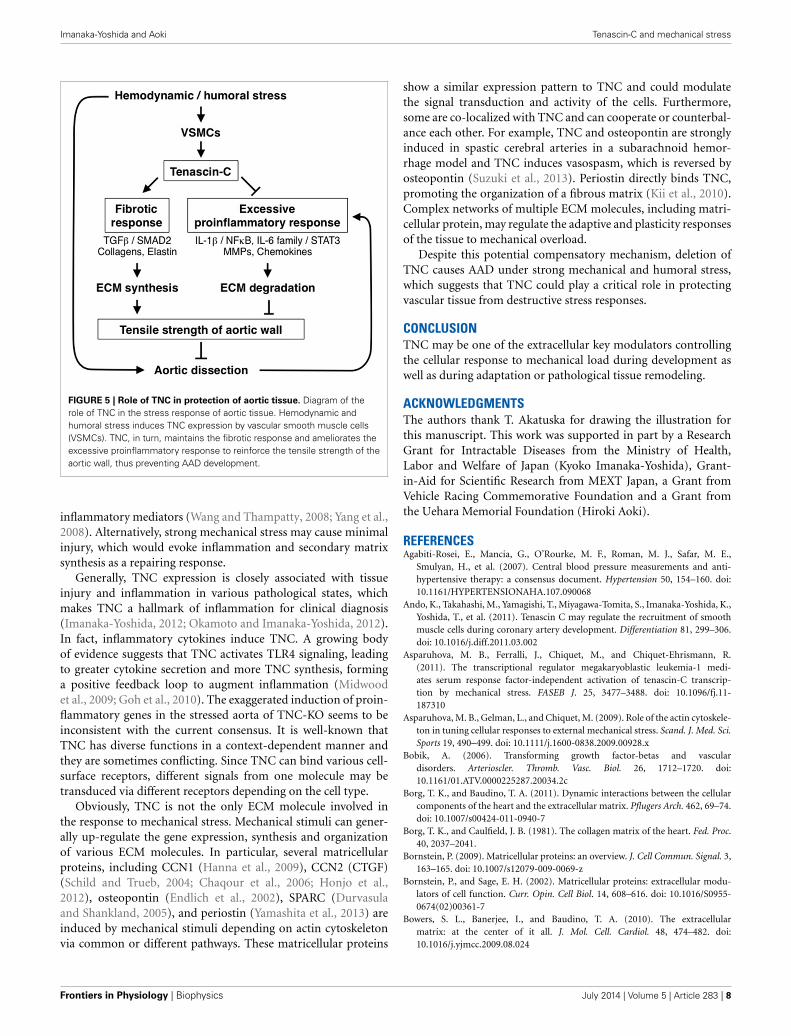
Role of tenascin-C in the protection of aortic tissueFrom the viewpoint of aortic homeostasis and AAD pathogene-sis, TNC can be regarded as a stress-activated molecular damper(Figure 5); it is inactive under normal conditions, but once thetissue experiences high mechanical stress it is activated and worksto reinforce tissue strength by inducing ECM proteins and atthe same time by ameliorating the excessive proinflammatoryresponse. These findings may be clinically relevant, because ele-vation of tissue and serum TNC levels has been reported in bothStanford type A and type B human AAD (Nozato et al., 2013;Trescher et al., 2013). It is also noteworthy that in TNC-KOmice, aortic wall stiffness was increased only in the infrarenalabdominal aorta where TNC was expressed at a low level (ourunpublished data). This suggests that TNC may also participatein the maintenance of the flexibility of aortic walls in certainsituations.
ADAPTIVE ROLE OF TENASCIN-C IN THE MECHANICALSTRESS RESPONSEAs observed in the aortic stress model discussed above, adap-tive or destructive tissue remodeling upon hemodynamic andhumoral stress could be associated with the inflammatoryresponse. Indeed, mechanical forces influence the production of
www.frontiersin.org July 2014 | Volume 5 | Article 283 | 7
Imanaka-Yoshida and Aoki Tenascin-C and mechanical stress
FIGURE 5 | Role of TNC in protection of aortic tissue. Diagram of therole of TNC in the stress response of aortic tissue. Hemodynamic andhumoral stress induces TNC expression by vascular smooth muscle cells(VSMCs). TNC, in turn, maintains the fibrotic response and ameliorates theexcessive proinflammatory response to reinforce the tensile strength of theaortic wall, thus preventing AAD development.
inflammatory mediators (Wang and Thampatty, 2008; Yang et al.,2008). Alternatively, strong mechanical stress may cause minimalinjury, which would evoke inflammation and secondary matrixsynthesis as a repairing response.
Generally, TNC expression is closely associated with tissueinjury and inflammation in various pathological states, whichmakes TNC a hallmark of inflammation for clinical diagnosis(Imanaka-Yoshida, 2012; Okamoto and Imanaka-Yoshida, 2012).In fact, inflammatory cytokines induce TNC. A growing bodyof evidence suggests that TNC activates TLR4 signaling, leadingto greater cytokine secretion and more TNC synthesis, forminga positive feedback loop to augment inflammation (Midwoodet al., 2009; Goh et al., 2010). The exaggerated induction of proin-flammatory genes in the stressed aorta of TNC-KO seems to beinconsistent with the current consensus. It is well-known thatTNC has diverse functions in a context-dependent manner andthey are sometimes conflicting. Since TNC can bind various cell-surface receptors, different signals from one molecule may betransduced via different receptors depending on the cell type.
Obviously, TNC is not the only ECM molecule involved inthe response to mechanical stress. Mechanical stimuli can gener-ally up-regulate the gene expression, synthesis and organizationof various ECM molecules. In particular, several matricellularproteins, including CCN1 (Hanna et al., 2009), CCN2 (CTGF)(Schild and Trueb, 2004; Chaqour et al., 2006; Honjo et al.,2012), osteopontin (Endlich et al., 2002), SPARC (Durvasulaand Shankland, 2005), and periostin (Yamashita et al., 2013) areinduced by mechanical stimuli depending on actin cytoskeletonvia common or different pathways. These matricellular proteins
show a similar expression pattern to TNC and could modulatethe signal transduction and activity of the cells. Furthermore,some are co-localized with TNC and can cooperate or counterbal-ance each other. For example, TNC and osteopontin are stronglyinduced in spastic cerebral arteries in a subarachnoid hemor-rhage model and TNC induces vasospasm, which is reversed byosteopontin (Suzuki et al., 2013). Periostin directly binds TNC,promoting the organization of a fibrous matrix (Kii et al., 2010).Complex networks of multiple ECM molecules, including matri-cellular protein, may regulate the adaptive and plasticity responsesof the tissue to mechanical overload.
Despite this potential compensatory mechanism, deletion ofTNC causes AAD under strong mechanical and humoral stress,which suggests that TNC could play a critical role in protectingvascular tissue from destructive stress responses.
CONCLUSIONTNC may be one of the extracellular key modulators controllingthe cellular response to mechanical load during development aswell as during adaptation or pathological tissue remodeling.
ACKNOWLEDGMENTSThe authors thank T. Akatuska for drawing the illustration forthis manuscript. This work was supported in part by a ResearchGrant for Intractable Diseases from the Ministry of Health,Labor and Welfare of Japan (Kyoko Imanaka-Yoshida), Grant-in-Aid for Scientific Research from MEXT Japan, a Grant fromVehicle Racing Commemorative Foundation and a Grant fromthe Uehara Memorial Foundation (Hiroki Aoki).
REFERENCESAgabiti-Rosei, E., Mancia, G., O’Rourke, M. F., Roman, M. J., Safar, M. E.,
Smulyan, H., et al. (2007). Central blood pressure measurements and anti-hypertensive therapy: a consensus document. Hypertension 50, 154–160. doi:10.1161/HYPERTENSIONAHA.107.090068
Ando, K., Takahashi, M., Yamagishi, T., Miyagawa-Tomita, S., Imanaka-Yoshida, K.,Yoshida, T., et al. (2011). Tenascin C may regulate the recruitment of smoothmuscle cells during coronary artery development. Differentiation 81, 299–306.doi: 10.1016/j.diff.2011.03.002
Asparuhova, M. B., Ferralli, J., Chiquet, M., and Chiquet-Ehrismann, R.(2011). The transcriptional regulator megakaryoblastic leukemia-1 medi-ates serum response factor-independent activation of tenascin-C transcrip-tion by mechanical stress. FASEB J. 25, 3477–3488. doi: 10.1096/fj.11-187310
Asparuhova, M. B., Gelman, L., and Chiquet, M. (2009). Role of the actin cytoskele-ton in tuning cellular responses to external mechanical stress. Scand. J. Med. Sci.Sports 19, 490–499. doi: 10.1111/j.1600-0838.2009.00928.x
Bobik, A. (2006). Transforming growth factor-betas and vasculardisorders. Arterioscler. Thromb. Vasc. Biol. 26, 1712–1720. doi:10.1161/01.ATV.0000225287.20034.2c
Borg, T. K., and Baudino, T. A. (2011). Dynamic interactions between the cellularcomponents of the heart and the extracellular matrix. Pflugers Arch. 462, 69–74.doi: 10.1007/s00424-011-0940-7
Borg, T. K., and Caulfield, J. B. (1981). The collagen matrix of the heart. Fed. Proc.40, 2037–2041.
Bornstein, P. (2009). Matricellular proteins: an overview. J. Cell Commun. Signal. 3,163–165. doi: 10.1007/s12079-009-0069-z
Bornstein, P., and Sage, E. H. (2002). Matricellular proteins: extracellular modu-lators of cell function. Curr. Opin. Cell Biol. 14, 608–616. doi: 10.1016/S0955-0674(02)00361-7
Bowers, S. L., Banerjee, I., and Baudino, T. A. (2010). The extracellularmatrix: at the center of it all. J. Mol. Cell. Cardiol. 48, 474–482. doi:10.1016/j.yjmcc.2009.08.024
Frontiers in Physiology | Biophysics July 2014 | Volume 5 | Article 283 | 8
Imanaka-Yoshida and Aoki Tenascin-C and mechanical stress
Boyer, A. S., Erickson, C. P., and Runyan, R. B. (1999). Epithelial-mesenchymaltransformation in the embryonic heart is mediated through distinct per-tussis toxin-sensitive and TGFbeta signal transduction mechanisms. Dev.Dyn. 214, 81–91. doi: 10.1002/(sici)1097-0177(199901)214:1<81::aid-dvdy8>3.0.co;2-3
Brellier, F., and Chiquet-Ehrismann, R. (2012). How do tenascins influencethe birth and life of a malignant cell? J. Cell. Mol. Med. 16, 32–40. doi:10.1111/j.1582-4934.2011.01360.x
Bristow, J., Carey, W., Egging, D., and Schalkwijk, J. (2005). Tenascin-X, collagen,elastin, and the Ehlers-Danlos syndrome. Am. J. Med. Genet. C Semin. Med.Genet. 139C, 24–30. doi: 10.1002/ajmg.c.30071
Bristow, J., Tee, M. K., Gitelman, S. E., Mellon, S. H., and Miller, W. L. (1993).Tenascin-X: a novel extracellular matrix protein encoded by the humanXB gene overlapping P450c21B. J. Cell Biol. 122, 265–278. doi: 10.1083/jcb.122.1.265
Brosig, M., Ferralli, J., Gelman, L., Chiquet, M., and Chiquet-Ehrismann, R. (2010).Interfering with the connection between the nucleus and the cytoskeleton affectsnuclear rotation, mechanotransduction and myogenesis. Int. J. Biochem. CellBiol. 42, 1717–1728. doi: 10.1016/j.biocel.2010.07.001
Caulfield, J. B., and Borg, T. K. (1979). The collagen network of the heart. Lab.Invest. 40, 364–372.
Chaqour, B., Yang, R., and Sha, Q. (2006). Mechanical stretch modulates thepromoter activity of the profibrotic factor CCN2 through increased actin poly-merization and NF-kappaB activation. J. Biol. Chem. 281, 20608–20622. doi:10.1074/jbc.M600214200
Chiquet, M., Gelman, L., Lutz, R., and Maier, S. (2009). From mechanotransduc-tion to extracellular matrix gene expression in fibroblasts. Biochim. Biophys. Acta1793, 911–920. doi: 10.1016/j.bbamcr.2009.01.012
Chiquet, M., Sarasa-Renedo, A., and Tunc-Civelek, V. (2004). Induction oftenascin-C by cyclic tensile strain versus growth factors: distinct contributionsby Rho/ROCK and MAPK signaling pathways. Biochim. Biophys. Acta 1693,193–204. doi: 10.1016/j.bbamcr.2004.08.001
Chiquet, M., Tunc-Civelek, V., and Sarasa-Renedo, A. (2007). Gene regulation bymechanotransduction in fibroblasts. Appl. Physiol. Nutr. Metab. 32, 967–973.doi: 10.1139/H07-053
Chiquet-Ehrismann, R., Mackie, E. J., Pearson, C. A., and Sakakura, T. (1986).Tenascin: an extracellular matrix protein involved in tissue interactions dur-ing fetal development and oncogenesis. Cell 47, 131–139. doi: 10.1016/0092-8674(86)90374-0
Chiquet-Ehrismann, R., Orend, G., Chiquet, M., Tucker, R. P., andMidwood, K. S. (2014). Tenascins in stem cell niches. Matrix Biol. doi:10.1016/j.matbio.2014.01.007. [Epub ahead of print].
Chiquet-Ehrismann, R., and Tucker, R. P. (2011). Tenascins and the importance ofadhesion modulation. Cold Spring Harb. Perspect. Biol. 3:a004960. doi: 10.1101/cshperspect.a004960
Combs, M. D., and Yutzey, K. E. (2009). Heart valve development: regula-tory networks in development and disease. Circ. Res. 105, 408–421. doi:10.1161/CIRCRESAHA.109.201566
Conway, S. J., and Molkentin, J. D. (2008). Periostin as a heterofunctional reg-ulator of cardiac development and disease. Curr. Genomics 9, 548–555. doi:10.2174/138920208786847917
Cronenwett, J. L., and Johnston, W. (2010). Rutherford’s Vascular Surgery, 7th Edn.Philadelphia: Saunders.
Crossin, K. L., and Hoffman, S. (1991). Expression of adhesion molecules duringthe formation and differentiation of the avian endocardial cushion tissue. Dev.Biol. 145, 277–286. doi: 10.1016/0012-1606(91)90126-N
Dai, J., Losy, F., Guinault, A. M., Pages, C., Anegon, I., Desgranges, P.,et al. (2005). Overexpression of transforming growth factor-beta1 stabilizesalready-formed aortic aneurysms: a first approach to induction of func-tional healing by endovascular gene therapy. Circulation 112, 1008–1015. doi:10.1161/CIRCULATIONAHA.104.523357
Danowski, B. A., Imanaka-Yoshida, K., Sanger, J. M., and Sanger, J. W.(1992). Costameres are sites of force transmission to the substratum inadult rat cardiomyocytes. J. Cell Biol. 118, 1411–1420. doi: 10.1083/jcb.118.6.1411
della Gaspera, B., Armand, A. S., Sequeira, I., Lecolle, S., Gallien, C. L.,Charbonnier, F., et al. (2009). The Xenopus MEF2 gene family: evidence of arole for XMEF2C in larval tendon development. Dev. Biol. 328, 392–402. doi:10.1016/j.ydbio.2009.01.039
Dietz, H. C. (2010). TGF-beta in the pathogenesis and prevention of dis-ease: a matter of aneurysmic proportions. J. Clin. Invest. 120, 403–407. doi:10.1172/JCI42014
Durvasula, R. V., and Shankland, S. J. (2005). Mechanical strain increases SPARClevels in podocytes: implications for glomerulosclerosis. Am. J. Physiol. RenalPhysiol. 289, F577–F584. doi: 10.1152/ajprenal.00393.2004
Edom-Vovard, F., and Duprez, D. (2004). Signals regulating tendon forma-tion during chick embryonic development. Dev. Dyn. 229, 449–457. doi:10.1002/dvdy.10481
Edom-Vovard, F., Schuler, B., Bonnin, M. A., Teillet, M. A., and Duprez, D. (2002).Fgf4 positively regulates scleraxis and tenascin expression in chick limb tendons.Dev. Biol. 247, 351–366. doi: 10.1006/dbio.2002.0707
Eisenberg, L. M., and Markwald, R. R. (1995). Molecular regulation of atri-oventricular valvuloseptal morphogenesis. Circ. Res. 77, 1–6. doi: 10.1161/01.RES.77.1.1
Endlich, N., Sunohara, M., Nietfeld, W., Wolski, E. W., Schiwek, D., Kranzlin, B.,et al. (2002). Analysis of differential gene expression in stretched podocytes:osteopontin enhances adaptation of podocytes to mechanical stress. FASEB J.16, 1850–1852. doi: 10.1096/fj.02-0125fje
Fluck, M., Tunc-Civelek, V., and Chiquet, M. (2000). Rapid and reciprocal regu-lation of tenascin-C and tenascin-Y expression by loading of skeletal muscle.J. Cell Sci. 113, 3583–3591.
Forsberg, E., Hirsch, E., Frohlich, L., Meyer, M., Ekblom, P., Aszodi, A., et al.(1996). Skin wounds and severed nerves heal normally in mice lackingtenascin-C. Proc. Natl. Acad. Sci. U.S.A. 93, 6594–6599. doi: 10.1073/pnas.93.13.6594
Garita, B., Jenkins, M. W., Han, M., Zhou, C., Vanauker, M., Rollins, A. M., et al.(2011). Blood flow dynamics of one cardiac cycle and relationship to mechan-otransduction and trabeculation during heart looping. Am. J. Physiol. HeartCirc. Physiol. 300, H879–H891. doi: 10.1152/ajpheart.00433.2010
Ghatak, S., Misra, S., Norris, R. A., Moreno-Rodriguez, R. A., Hoffman, S., Levine,R. A., et al. (2014). Periostin induces intracellular cross-talk between kinases andhyaluronan in atrioventricular valvulogenesis. J. Biol. Chem. 289, 8545–8561.doi: 10.1074/jbc.M113.539882
Goh, F. G., Piccinini, A. M., Krausgruber, T., Udalova, I. A., and Midwood, K. S.(2010). Transcriptional regulation of the endogenous danger signal tenascin-C: a novel autocrine loop in inflammation. J. Immunol. 184, 2655–2662. doi:10.4049/jimmunol.0903359
Guettler, S., Vartiainen, M. K., Miralles, F., Larijani, B., and Treisman, R.(2008). RPEL motifs link the serum response factor cofactor MAL but notmyocardin to Rho signaling via actin binding. Mol. Cell Biol. 28, 732–742. doi:10.1128/MCB.01623-07
Hagios, C., Koch, M., Spring, J., Chiquet, M., and Chiquet-Ehrismann, R. (1996).Tenascin-Y: a protein of novel domain structure is secreted by differenti-ated fibroblasts of muscle connective tissue. J. Cell Biol. 134, 1499–1512. doi:10.1083/jcb.134.6.1499
Hanna, M., Liu, H., Amir, J., Sun, Y., Morris, S. W., Siddiqui, M. A., et al.(2009). Mechanical regulation of the proangiogenic factor CCN1/CYR61 generequires the combined activities of MRTF-A and CREB-binding protein his-tone acetyltransferase. J. Biol. Chem. 284, 23125–23136. doi: 10.1074/jbc.M109.019059
Hiltgen, G. G., Markwald, R. R., and Litke, L. L. (1996). Morphogenetic alterationsduring endocardial cushion development in the trisomy 16 (Down syndrome)mouse. Pediatr. Cardiol. 17, 21–30. doi: 10.1007/BF02505807
Hinton, R. B. Jr., Lincoln, J., Deutsch, G. H., Osinska, H., Manning, P. B.,Benson, D. W., et al. (2006). Extracellular matrix remodeling and organiza-tion in developing and diseased aortic valves. Circ. Res. 98, 1431–1438. doi:10.1161/01.RES.0000224114.65109.4e
Hinton, R. B., and Yutzey, K. E. (2011). Heart valve structure and function indevelopment and disease. Annu. Rev. Physiol. 73, 29–46. doi: 10.1146/annurev-physiol-012110-142145
Honjo, T., Kubota, S., Kamioka, H., Sugawara, Y., Ishihara, Y., Yamashiro, T., et al.(2012). Promotion of Ccn2 expression and osteoblastic differentiation by actinpolymerization, which is induced by laminar fluid flow stress. J. Cell Commun.Signal. 6, 225–232. doi: 10.1007/s12079-012-0177-z
Hurle, J. M., Garcia-Martinez, V., and Ros, M. A. (1990). Immunofluorescentlocalization of tenascin during the morphogenesis of the outflow tract of thechick embryo heart. Anat. Embryol. (Berl.) 181, 149–155. doi: 10.1007/BF00198954
www.frontiersin.org July 2014 | Volume 5 | Article 283 | 9

Imanaka-Yoshida and Aoki Tenascin-C and mechanical stress
Imanaka-Yoshida, K. (2012). Tenascin-C in cardiovascular tissue remodeling. CircJ. 76, 2513–2520. doi: 10.1253/circj.CJ-12-1033
Imanaka-Yoshida, K., Danowski, B. A., Sanger, J. M., and Sanger, J. W.(1996). Living adult rat cardiomyocytes in culture: evidence for dissocia-tion of costameric distribution of vinculin from costameric distributions ofattachments. Cell Motil. Cytoskeleton 33, 263–275. doi: 10.1002/(sici)1097-0169(1996)33:4<263::aid-cm3>3.0.co;2-a
Imanaka-Yoshida, K., Enomoto-Iwamoto, M., Yoshida, T., and Sakakura, T. (1999).Vinculin, Talin, Integrin alpha6beta1 and laminin can serve as componentsof attachment complex mediating contraction force transmission from car-diomyocytes to extracellular matrix. Cell Motil. Cytoskeleton 42, 1–11. doi:10.1002/(sici)1097-0169(1999)42:1<1::aid-cm1>3.0.co;2-0
Imanaka-Yoshida, K., Hiroe, M., and Yoshida, T. (2004). Interaction between celland extracellular matrix in heart disease: multiple roles of tenascin-C in tissueremodeling. Histol. Histopathol. 19, 517–525.
Imanaka-Yoshida, K., Hiroe, M., Nishikawa, T., Ishiyama, S., Shimojo, T., Ohta, Y.,et al. (2001). Tenascin-C modulates adhesion of cardiomyocytes to extracellu-lar matrix during tissue remodeling after myocardial infarction. Lab. Invest. 81,1015–1024. doi: 10.1038/labinvest.3780313
Imanaka-Yoshida, K., Matsumoto, K., Hara, M., Sakakura, T., and Yoshida, T.(2003). The dynamic expression of tenascin-C and tenascin-X during early heartdevelopment in the mouse. Differentiation 71, 291–298. doi: 10.1046/j.1432-0436.2003.7104506.x
Imanaka-Yoshida, K., Miyagawa-Tomita, S., and Yoshida, T. (in press). Tenascin-Cin development and disease of blood vessels. Anat. Rec.
Inai, K., Norris, R. A., Hoffman, S., Markwald, R. R., and Sugi, Y. (2008).BMP-2 induces cell migration and periostin expression during atrioven-tricular valvulogenesis. Dev. Biol. 315, 383–396. doi: 10.1016/j.ydbio.2007.12.028
Ishigaki, T., Imanaka-Yoshida, K., Shimojo, N., Matsushima, S., Taki, W.,and Yoshida, T. (2011). Tenascin-C enhances crosstalk signaling of integrinalphavbeta3/PDGFR-beta complex by SRC recruitment promoting PDGF-induced proliferation and migration in smooth muscle cells. J. Cell. Physiol. 226,2617–2624. doi: 10.1002/jcp.22614
Jagodzinski, M., Breitbart, A., Wehmeier, M., Hesse, E., Haasper, C., Krettek,C., et al. (2008). Influence of perfusion and cyclic compression onproliferation and differentiation of bone marrow stromal cells in 3-dimensional culture. J. Biomech. 41, 1885–1891. doi: 10.1016/j.jbiomech.2008.04.001
Jarvinen, T. A., Jozsa, L., Kannus, P., Jarvinen, T. L., Hurme, T., Kvist, M.,et al. (2003). Mechanical loading regulates the expression of tenascin-Cin the myotendinous junction and tendon but does not induce de novosynthesis in the skeletal muscle. J. Cell Sci. 116, 857–866. doi: 10.1242/jcs.00303
Jarvinen, T. A., Jozsa, L., Kannus, P., Jarvinen, T. L., Kvist, M., Hurme, T., et al.(1999). Mechanical loading regulates tenascin-C expression in the osteotendi-nous junction. J. Cell Sci. 112, 3157–3166.
Jarvinen, T. A., Kannus, P., Jarvinen, T. L., Jozsa, L., Kalimo, H., and Jarvinen, M.(2000). Tenascin-C in the pathobiology and healing process of musculoskeletaltissue injury. Scand. J. Med. Sci. Sports 10, 376–382. doi: 10.1034/j.1600-0838.2000.010006376.x
Jinnin, M., Ihn, H., Asano, Y., Yamane, K., Trojanowska, M., and Tamaki, K. (2004).Tenascin-C upregulation by transforming growth factor-beta in human der-mal fibroblasts involves Smad3, Sp1, and Ets1. Oncogene 23, 1656–1667. doi:10.1038/sj.onc.1207064
Jinnin, M., Ihn, H., Asano, Y., Yamane, K., Trojanowska, M., and Tamaki,K. (2006). Platelet derived growth factor induced tenascin-C transcrip-tion is phosphoinositide 3-kinase/Akt-dependent and mediated by Ets fam-ily transcription factors. J. Cell. Physiol. 206, 718–727. doi: 10.1002/jcp.20527
Jondeau, G., Boutouyrie, P., Lacolley, P., Laloux, B., Dubourg, O., Bourdarias,J. P., et al. (1999). Central pulse pressure is a major determinant of ascend-ing aorta dilation in Marfan syndrome. Circulation 99, 2677–2681. doi:10.1161/01.CIR.99.20.2677
Katoh, D., Nagaharu, K., Shimojo, N., Hanamura, N., Yamashita, M., Kozuka, Y.,et al. (2013). Binding of alphavbeta1 and alphavbeta6 integrins to tenascin-Cinduces epithelial-mesenchymal transition-like change of breast cancer cells.Oncogenesis 2, e65. doi: 10.1038/oncsis.2013.27
Kii, I., Nishiyama, T., Li, M., Matsumoto, K., Saito, M., Amizuka, N., et al. (2010).Incorporation of tenascin-C into the extracellular matrix by periostin under-lies an extracellular meshwork architecture. J. Biol. Chem. 285, 2028–2039. doi:10.1074/jbc.M109.051961
Kimura, T., Shiraishi, K., Furusho, A., Ito, S., Hirakata, S., Nishida, N., et al. (2014).Tenascin C protects aorta from acute dissection in mice. Sci. Rep. 4:4051. doi:10.1038/srep04051
Kumar, M. S., and Owens, G. K. (2003). Combinatorial control of smooth muscle-specific gene expression. Arterioscler. Thromb. Vasc. Biol. 23, 737–747. doi:10.1161/01.ATV.0000065197.07635.BA
Kung, C. (2005). A possible unifying principle for mechanosensation. Nature 436,647–654. doi: 10.1038/nature03896
Lange, K., Kammerer, M., Saupe, F., Hegi, M. E., Grotegut, S., Fluri, E., et al. (2008).Combined lysophosphatidic acid/platelet-derived growth factor signaling trig-gers glioma cell migration in a tenascin-C microenvironment. Cancer Res. 68,6942–6952. doi: 10.1158/0008-5472.CAN-08-0347
Lincoln, J., Alfieri, C. M., and Yutzey, K. E. (2004). Development of heart valveleaflets and supporting apparatus in chicken and mouse embryos. Dev. Dyn.230, 239–250. doi: 10.1002/dvdy.20051
Lincoln, J., Alfieri, C. M., and Yutzey, K. E. (2006a). BMP and FGF regulatory path-ways control cell lineage diversification of heart valve precursor cells. Dev. Biol.292, 290–302. doi: 10.1016/j.ydbio.2005.12.042
Lincoln, J., Lange, A. W., and Yutzey, K. E. (2006b). Hearts and bones: shared reg-ulatory mechanisms in heart valve, cartilage, tendon, and bone development.Dev. Biol. 294, 292–302. doi: 10.1016/j.ydbio.2006.03.027
Lockhart, M., Wirrig, E., Phelps, A., and Wessels, A. (2011). Extracellular matrixand heart development. Birth Defects Res. A Clin. Mol. Teratol. 91, 535–550. doi:10.1002/bdra.20810
Lutz, R., Sakai, T., and Chiquet, M. (2010). Pericellular fibronectin is requiredfor RhoA-dependent responses to cyclic strain in fibroblasts. J. Cell Sci. 123,1511–1521. doi: 10.1242/jcs.060905
Mackey, A. L., Brandstetter, S., Schjerling, P., Bojsen-Moller, J., Qvortrup, K.,Pedersen, M. M., et al. (2011). Sequenced response of extracellular matrix dead-hesion and fibrotic regulators after muscle damage is involved in protectionagainst future injury in human skeletal muscle. FASEB J. 25, 1943–1959. doi:10.1096/fj.10-176487
Mackie, E. J., Scott-Burden, T., Hahn, A. W., Kern, F., Bernhardt, J., Regenass, S.,et al. (1992). Expression of tenascin by vascular smooth muscle cells. Alterationsin hypertensive rats and stimulation by angiotensin II. Am. J. Pathol. 141,377–388.
Maier, S., Lutz, R., Gelman, L., Sarasa-Renedo, A., Schenk, S., Grashoff, C.,et al. (2008). Tenascin-C induction by cyclic strain requires integrin-linkedkinase. Biochim. Biophys. Acta 1783, 1150–1162. doi: 10.1016/j.bbamcr.2008.01.013
Marin, J. L., Muniz, J., Huerta, M., and Trujillo, X. (2003). Folding-unfolding ofFN-III domains in tenascin: an elastically coupled two-state system. J. Biomech.36, 1733–1737. doi: 10.1016/S0021-9290(03)00172-6
Matsumoto, K., Arai, M., Ishihara, N., Ando, A., Inoko, H., and Ikemura, T.(1992). Cluster of fibronectin type III repeats found in the human major his-tocompatibility complex class III region shows the highest homology with therepeats in an extracellular matrix protein, tenascin. Genomics 12, 485–491. doi:10.1016/0888-7543(92)90438-X
McCain, M. L., and Parker, K. K. (2011). Mechanotransduction: the role ofmechanical stress, myocyte shape, and cytoskeletal architecture on cardiacfunction. Pflugers Arch. 462, 89–104. doi: 10.1007/s00424-011-0951-4
Mettouchi, A., Cabon, F., Montreau, N., Dejong, V., Vernier, P., Gherzi, R., et al.(1997). The c-Jun-induced transformation process involves complex regulationof tenascin-C expression. Mol. Cell. Biol. 17, 3202–3209.
Midwood, K., Sacre, S., Piccinini, A. M., Inglis, J., Trebaul, A., Chan, E., et al. (2009).Tenascin-C is an endogenous activator of Toll-like receptor 4 that is essential formaintaining inflammation in arthritic joint disease. Nat. Med. 15, 774–780. doi:10.1038/nm.1987
Midwood, K. S., Hussenet, T., Langlois, B., and Orend, G. (2011). Advances intenascin-C biology. Cell. Mol. Life Sci. 68, 3175–3199. doi: 10.1007/s00018-011-0783-6
Midwood, K. S., Mao, Y., Hsia, H. C., Valenick, L. V., and Schwarzbauer, J. E.(2006). Modulation of cell-fibronectin matrix interactions during tissue repair.J. Investig. Dermatol. Symp. Proc. 11, 73–78. doi: 10.1038/sj.jidsymp.5650005
Frontiers in Physiology | Biophysics July 2014 | Volume 5 | Article 283 | 10

Imanaka-Yoshida and Aoki Tenascin-C and mechanical stress
Midwood, K. S., and Orend, G. (2009). The role of tenascin-C in tissue injury andtumorigenesis. J. Cell Commun. Signal. 3, 287–310. doi: 10.1007/s12079-009-0075-1
Mikic, B., Wong, M., Chiquet, M., and Hunziker, E. B. (2000). Mechanical mod-ulation of tenascin-C and collagen-XII expression during avian synovial jointformation. J. Orthop. Res. 18, 406–415. doi: 10.1002/jor.1100180312
Miralles, F., Posern, G., Zaromytidou, A. I., and Treisman, R. (2003). Actin dynam-ics control SRF activity by regulation of its coactivator MAL. Cell 113, 329–342.doi: 10.1016/S0092-8674(03)00278-2
Nagaharu, K., Zhang, X., Yoshida, T., Katoh, D., Hanamura, N., Kozuka,Y., et al. (2011). Tenascin C induces epithelial-mesenchymal transition-likechange accompanied by SRC activation and focal adhesion kinase phospho-rylation in human breast cancer cells. Am. J. Pathol. 178, 754–763. doi:10.1016/j.ajpath.2010.10.015
Nakajima, Y., and Imanaka-Yoshida, K. (2013). New insights into the developmen-tal mechanisms of coronary vessels and epicardium. Int. Rev. Cell Mol. Biol. 303,263–317. doi: 10.1016/B978-0-12-407697-6.00007-6
Neidhardt, J., Fehr, S., Kutsche, M., Lohler, J., and Schachner, M. (2003). Tenascin-N: characterization of a novel member of the tenascin family that mediatesneurite repulsion from hippocampal explants. Mol. Cell. Neurosci. 23, 193–209.doi: 10.1016/S1044-7431(03)00012-5
Nishioka, T., Onishi, K., Shimojo, N., Nagano, Y., Matsusaka, H., Ikeuchi, M.,et al. (2010). Tenascin-C may aggravate left ventricular remodeling and func-tion after myocardial infarction in mice. Am. J. Physiol. Heart Circ. Physiol. 298,H1072–H1078. doi: 10.1152/ajpheart.00255.2009
Norris, R. A., Moreno-Rodriguez, R. A., Sugi, Y., Hoffman, S., Amos, J., Hart, M.M., et al. (2008). Periostin regulates atrioventricular valve maturation. Dev. Biol.316, 200–213. doi: 10.1016/j.ydbio.2008.01.003
Norris, R. A., Potts, J. D., Yost, M. J., Junor, L., Brooks, T., Tan, H., et al. (2009).Periostin promotes a fibroblastic lineage pathway in atrioventricular valveprogenitor cells. Dev. Dyn. 238, 1052–1063. doi: 10.1002/dvdy.21933
Nozato, T., Sato, A., Hirose, S., Hikita, H., Takahashi, A., Endo, H., et al. (2013).Preliminary study of serum tenascin-C levels as a diagnostic or prognosticbiomarker of type B acute aortic dissection. Int. J. Cardiol. 168, 4267–4269. doi:10.1016/j.ijcard.2013.04.211
Oberhauser, A. F., Marszalek, P. E., Erickson, H. P., and Fernandez, J. M. (1998).The molecular elasticity of the extracellular matrix protein tenascin. Nature 393,181–185. doi: 10.1038/30270
Okamoto, H., and Imanaka-Yoshida, K. (2012). Matricellular proteins: new molec-ular targets to prevent heart failure. Cardiovasc. Ther. 30, e198–e209. doi:10.1111/j.1755-5922.2011.00276.x
Orend, G., and Chiquet-Ehrismann, R. (2006). Tenascin-C induced sig-naling in cancer. Cancer Lett. 244, 143–163. doi: 10.1016/j.canlet.2006.02.017
Pardo, J. V., Siliciano, J. D., and Craig, S. W. (1983a). A vinculin-containing corticallattice in skeletal muscle: transverse lattice elements (“costameres”) mark sitesof attachment between myofibrils and sarcolemma. Proc. Natl. Acad. Sci. U.S.A.80, 1008–1012. doi: 10.1073/pnas.80.4.1008
Pardo, J. V., Siliciano, J. D., and Craig, S. W. (1983b). Vinculin is a compo-nent of an extensive network of myofibril-sarcolemma attachment regionsin cardiac muscle fibers. J. Cell Biol. 97, 1081–1088. doi: 10.1083/jcb.97.4.1081
Person, A. D., Klewer, S. E., and Runyan, R. B. (2005). Cell biology of car-diac cushion development. Int. Rev. Cytol. 243, 287–335. doi: 10.1016/S0074-7696(05)43005-3
Rathjen, F. G., Wolff, J. M., and Chiquet-Ehrismann, R. (1991). Restrictin: a chickneural extracellular matrix protein involved in cell attachment co-purifies withthe cell recognition molecule F11. Development 113, 151–164.
Ridley, A. J., and Hall, A. (1992). The small GTP-binding protein rho regulates theassembly of focal adhesions and actin stress fibers in response to growth factors.Cell 70, 389–399. doi: 10.1016/0092-8674(92)90163-7
Russell, B., Curtis, M. W., Koshman, Y. E., and Samarel, A. M. (2010). Mechanicalstress-induced sarcomere assembly for cardiac muscle growth in lengthand width. J. Mol. Cell. Cardiol. 48, 817–823. doi: 10.1016/j.yjmcc.2010.02.016
Saga, Y., Yagi, T., Ikawa, Y., Sakakura, T., and Aizawa, S. (1992). Mice developnormally without tenascin. Genes Dev. 6, 1821–1831. doi: 10.1101/gad.6.10.1821
Sage, E. H., and Bornstein, P. (1991). Extracellular proteins that modulate cell-matrix interactions. SPARC, tenascin, and thrombospondin. J. Biol. Chem. 266,14831–14834.
Samarel, A. M. (2005). Costameres, focal adhesions, and cardiomyocyte mechan-otransduction. Am. J. Physiol. Heart Circ. Physiol. 289, H2291–H2301. doi:10.1152/ajpheart.00749.2005
Sarasa-Renedo, A., Tunc-Civelek, V., and Chiquet, M. (2006). Role of RhoA/ROCK-dependent actin contractility in the induction of tenascin-C by cyclictensile strain. Exp. Cell Res. 312, 1361–1370. doi: 10.1016/j.yexcr.2005.12.025
Scharer, C. D., McCabe, C. D., Ali-Seyed, M., Berger, M. F., Bulyk, M. L., andMoreno, C. S. (2009). Genome-wide promoter analysis of the SOX4 tran-scriptional network in prostate cancer cells. Cancer Res. 69, 709–717. doi:10.1158/0008-5472.CAN-08-3415
Schild, C., and Trueb, B. (2004). Three members of the connective tissue growthfactor family CCN are differentially regulated by mechanical stress. Biochim.Biophys. Acta 1691, 33–40. doi: 10.1016/j.bbamcr.2003.12.001
Sivasankaran, B., Degen, M., Ghaffari, A., Hegi, M. E., Hamou, M. F., Ionescu, M.C., et al. (2009). Tenascin-C is a novel RBPJkappa-induced target gene for Notchsignaling in gliomas. Cancer Res. 69, 458–465. doi: 10.1158/0008-5472.CAN-08-2610
Sugi, Y., and Markwald, R. R. (1996). Formation and early morphogenesis of endo-cardial endothelial precursor cells and the role of endoderm. Dev. Biol. 175,66–83. doi: 10.1006/dbio.1996.0096
Sussman, M. A., McCulloch, A., and Borg, T. K. (2002). Dance band on the Titanic:biomechanical signaling in cardiac hypertrophy. Circ. Res. 91, 888–898. doi:10.1161/01.RES.0000041680.43270.F8
Suzuki, H., Shiba, M., Fujimoto, M., Kawamura, K., Nanpei, M., Tekeuchi, E.,et al. (2013). Matricellular protein: a new player in cerebral vasospasm fol-lowing subarachnoid hemorrhage. Acta Neurochir. Suppl. 115, 213–218. doi:10.1007/978-3-7091-1192-5_39
Tan, H., Biechler, S., Junor, L., Yost, M. J., Dean, D., Li, J., et al. (2013). Fluid flowforces and rhoA regulate fibrous development of the atrioventricular valves.Dev. Biol. 374, 345–356. doi: 10.1016/j.ydbio.2012.11.023
Trescher, K., Thometich, B., Demyanets, S., Kassal, H., Sedivy, R., Bittner, R., et al.(2013). Type A dissection and chronic dilatation: tenascin-C as a key factor indestabilization of the aortic wall. Interact. Cardiovasc. Thorac. Surg. 17, 365–370.doi: 10.1093/icvts/ivt204
Tucker, R. P., and Chiquet-Ehrismann, R. (2009). The regulation of tenascin expres-sion by tissue microenvironments. Biochim. Biophys. Acta 1793, 888–892. doi:10.1016/j.bbamcr.2008.12.012
Tucker, R. P., Drabikowski, K., Hess, J. F., Ferralli, J., Chiquet-Ehrismann, R., andAdams, J. C. (2006). Phylogenetic analysis of the tenascin gene family: evidenceof origin early in the chordate lineage. BMC Evol. Biol. 6:60. doi: 10.1186/1471-2148-6-60
Udalova, I. A., Ruhmann, M., Thomson, S. J., and Midwood, K. S. (2011).Expression and immune function of tenascin-C. Crit. Rev. Immunol. 31,115–145. doi: 10.1615/CritRevImmunol.v31.i2.30
Van Obberghen-Schilling, E., Tucker, R. P., Saupe, F., Gasser, I., Cseh, B., andOrend, G. (2011). Fibronectin and tenascin-C: accomplices in vascular morpho-genesis during development and tumor growth. Int. J. Dev. Biol. 55, 511–525.doi: 10.1387/ijdb.103243eo
Wagenseil, J. E., and Mecham, R. P. (2009). Vascular extracellular matrix andarterial mechanics. Physiol. Rev. 89, 957–989. doi: 10.1152/physrev.00041.2008
Wang, J. H., and Thampatty, B. P. (2008). Mechanobiology of adult and stem cells.Int. Rev. Cell Mol. Biol. 271, 301–346. doi: 10.1016/S1937-6448(08)01207-0
Wang, N., Tytell, J. D., and Ingber, D. E. (2009). Mechanotransduction at a distance:mechanically coupling the extracellular matrix with the nucleus. Nat. Rev. Mol.Cell Biol. 10, 75–82. doi: 10.1038/nrm2594
Wang, Y., Ait-Oufella, H., Herbin, O., Bonnin, P., Ramkhelawon, B., Taleb, S., et al.(2010). TGF-beta activity protects against inflammatory aortic aneurysm pro-gression and complications in angiotensin II-infused mice. J. Clin. Invest. 120,422–432. doi: 10.1172/JCI38136
Webb, C. M., Zaman, G., Mosley, J. R., Tucker, R. P., Lanyon, L. E., and Mackie,E. J. (1997). Expression of tenascin-C in bones responding to mechanical load.J. Bone Miner. Res. 12, 52–58. doi: 10.1359/jbmr.1997.12.1.52
Weber, P., Montag, D., Schachner, M., and Bernhardt, R. R. (1998). Zebrafishtenascin-W, a new member of the tenascin family. J. Neurobiol. 35, 1–16.
www.frontiersin.org July 2014 | Volume 5 | Article 283 | 11

Imanaka-Yoshida and Aoki Tenascin-C and mechanical stress
Yamashita, O., Yoshimura, K., Nagasawa, A., Ueda, K., Morikage, N., Ikeda,Y., et al. (2013). Periostin links mechanical strain to inflammation inabdominal aortic aneurysm. PLoS ONE 8:e79753. doi: 10.1371/journal.pone.0079753
Yang, R., Amir, J., Liu, H., and Chaqour, B. (2008). Mechanical strain acti-vates a program of genes functionally involved in paracrine signaling ofangiogenesis. Physiol. Genomics 36, 1–14. doi: 10.1152/physiolgenomics.90291.2008
Zhang, H. Y., Kluge, M., Timpl, R., Chu, M. L., and Ekblom, P. (1993). Theextracellular matrix glycoproteins BM-90 and tenascin are expressed in themesenchyme at sites of endothelial-mesenchymal conversion in the embryonicmouse heart. Differentiation 52, 211–220. doi: 10.1111/j.1432-0436.1993.tb00633.x
Zhao, B., Etter, L., Hinton, R. B. Jr., and Benson, D. W. (2007). BMP and FGFregulatory pathways in semilunar valve precursor cells. Dev. Dyn. 236, 971–980.doi: 10.1002/dvdy.21097
Conflict of Interest Statement: The authors declare that the research was con-ducted in the absence of any commercial or financial relationships that could beconstrued as a potential conflict of interest.
Received: 28 April 2014; accepted: 10 July 2014; published online: 29 July 2014.Citation: Imanaka-Yoshida K and Aoki H (2014) Tenascin-C and mechanotransduc-tion in the development and diseases of cardiovascular system. Front. Physiol. 5:283.doi: 10.3389/fphys.2014.00283This article was submitted to Biophysics, a section of the journal Frontiers inPhysiology.Copyright © 2014 Imanaka-Yoshida and Aoki. This is an open-access article dis-tributed under the terms of the Creative Commons Attribution License (CC BY). Theuse, distribution or reproduction in other forums is permitted, provided the originalauthor(s) or licensor are credited and that the original publication in this jour-nal is cited, in accordance with accepted academic practice. No use, distribution orreproduction is permitted which does not comply with these terms.
Frontiers in Physiology | Biophysics July 2014 | Volume 5 | Article 283 | 12