telmisartan modulates the oral mucositis induced by 5 ... · emanuella de aragão tavares1, roseane...
TRANSCRIPT
fphys-09-01204 August 28, 2018 Time: 10:35 # 1
ORIGINAL RESEARCHpublished: 29 August 2018
doi: 10.3389/fphys.2018.01204
Edited by:Hongxiang Hui,
Southern Medical University, China
Reviewed by:Shin Hamada,
Tohoku University, JapanKhyati Shah,
University of California,San Francisco, United States
*Correspondence:Caroline A. C. X. de Medeiros
[email protected];[email protected]
†These authors have contributedequally to this work
Specialty section:This article was submitted to
Gastrointestinal Sciences,a section of the journalFrontiers in Physiology
Received: 01 February 2018Accepted: 10 August 2018Published: 29 August 2018
Citation:Barbosa MM, de Araújo AA,
de Araújo Júnior RF, Guerra GCB,de Castro Brito GA, Leitão RC,
Ribeiro SB, de Aragão Tavares E,Vasconcelos RC, Garcia VB and
de Medeiros CACX (2018) TelmisartanModulates the Oral Mucositis Induced
by 5-Fluorouracil in Hamsters.Front. Physiol. 9:1204.
doi: 10.3389/fphys.2018.01204
Telmisartan Modulates the OralMucositis Induced by 5-Fluorouracilin HamstersMaisie M. Barbosa1†, Aurigena A. de Araújo2†, Raimundo F. de Araújo Júnior3,Gerlane C. B. Guerra4, Gerly A. de Castro Brito5, Renata C. Leitão5, Susana B. Ribeiro6,Emanuella de Aragão Tavares1, Roseane C. Vasconcelos7, Vinícius B. Garcia8 andCaroline A. C. X. de Medeiros9*
1 Post Graduation Program in Biological Sciences, Federal University of Rio Grande do Norte, Natal, Brazil, 2 Post GraduationProgram Public Health/Post Graduation Program in Pharmaceutical Science, Department of Biophysics and Pharmacology,Federal University of Rio Grande do Norte, Natal, Brazil, 3 Post Graduation Program in Functional and Structural Biology/PostGraduation Program Health Science, Department of Morphology, Federal University of Rio Grande do Norte, Natal, Brazil,4 Post Graduation Program in Biological Sciences/Post Graduation Program in Pharmaceutical Science, Departmentof Biophysics and Pharmacology, Federal University of Rio Grande do Norte, Natal, Brazil, 5 Post Graduation Programof Morphological Science, Department of Morphology, Universidade Federal do Ceará, Fortaleza, Brazil, 6 Post GraduationProgram in Biotechnology RENORBIO, Federal University of Rio Grande do Norte, Natal, Brazil, 7 Post Graduation ProgramPublic Health, Department of Dentistry, Federal University of Rio Grande do Norte, Natal, Brazil, 8 Post Graduationin Program of Health Sciences, Federal University of Rio Grande do Norte, Natal, Brazil, 9 Post Graduation Programin Biological Sciences/Post Graduation Program in Biotechnology RENORBIO, Department of Biophysicsand Pharmacology, Federal University of Rio Grande do Norte, Natal, Brazil
Oral mucositis (OM) is a common adverse effect resulting from cancer therapy. The OM ithas implications that may compromise oncologic treatment and decrease the patient’squality of life. The therapeutic options to prevent or treat the symptoms of OM arescarce; there is no effective therapy that improves the symptoms. Based on the need forfurther research for the treatment of OM, the present study objective was to evaluate theeffect of telmisartan (TELM) on the OM induced by 5-fluorouracil (5-FU), using as animalmodel Golden Syrian hamsters. 5-FU followed by mechanical trauma on day 4 was usedto induce OM in hamsters. Euthanasia occurred on the day 10. The experiments wereconstituted by the groups saline, mechanical trauma, 5-FU, and TELM in three doses(1, 5, or 10 mg/kg). Macroscopic, histopathological, and immunohistochemical analysesas well as immunofluorescence experiments were performed on the oral mucosa of theanimals. The samples also were used for analysis enzyme-linked immunosorbent assaysand quantitative real-time polymerase chain reactions (qPCR). TELM (5 or 10 mg/kg)was able to reduce the inflammatory ulceration and infiltration in the oral mucosa ofthe animals, decreasing the levels of the cytokines TNF-α and IL-1β. These treatmentswas minimize the immunostaining for cyclooxygenase-2, matrix metalloproteinase-9,transforming growth factor-β, and smad 2/3. The nuclear transcription factor kappa B(NFκB) p65 and inducible nitric oxide synthase were reduced in the oral mucosa. Finally,TELM (10 mg/kg) increased the PPARγ gene expression and reduced STAT1 and NFκBp65 gene expression relative to the 5-FU group. Therefore, TELM prevents the OMproduced by 5-FU on animal model.
Keywords: oral mucositis, telmisartan, 5-fluorouracil, hamsters, cytokines
Frontiers in Physiology | www.frontiersin.org 1 August 2018 | Volume 9 | Article 1204
fphys-09-01204 August 28, 2018 Time: 10:35 # 2
Barbosa et al. Telmisartan in Oral Mucositis
INTRODUCTION
Oral mucositis (OM) is a common effect associated with cancertreatments such as radiotherapy and/or chemotherapy (Sonis andFey, 2002; Medeiros et al., 2011). This problem affects 20 to 40%of patients at the treatment of usual chemotherapy (Lima-Junioret al., 2014; Cidon, 2017) and occurs in practically all patients (90to 97%) who receive radiotherapy in the head and neck region(Trotti et al., 2003). The inflammation in OM is characterized bythe presence of ulceration and pseudomembranous formations inthe oral cavity, oropharynx, or hypopharynx (Elting et al., 2008;Niscola, 2010). This complication compromises the patient’squality of life because OM is extremely painful, interfereswith food consumption, makes the patient more susceptibleto infections and in some cases, may interfere with anticancertherapy (Lalla et al., 2008).
Chemotherapy and/or radiotherapy cause damage to theepithelial and submucosal cells, which leads resulting inapoptosis, atrophy and consequent ulceration (Sonis, 2007;Niscola, 2010; Maria et al., 2017). In the initial phase ofOM, production of reactive oxygen species (ROS) is observed,which triggers various cellular signals that contribute to thelesion (Sonis, 2004). The nuclear transcription factor kappaB (NFκB) is the most well-characterized signaling pathway inthe pathophysiology of OM. The NFκB increases the levels ofproinflammatory cytokines as TNF-α and IL-1β, cyclooxygenase-2 (COX-2), matrix metalloproteinase-9 (MMP-9), induciblenitric oxide synthase (iNOS), and the transforming growth factorβ (TGF-β)/SMAD 2/3 pathway (Sonis, 2004, 2009; Ribeiro et al.,2017). Proinflammatory cytokines amplify the tissue damage andthe apoptosis of specially submucosal and basal epithelial cells(Ramirez-Amador et al., 2010). Macrophages are activated, whichpromote tissue damage by expressing metalloproteinases andproducing additional TNF-α. TGF-β acts on serine threoninekinases that are coupled to the smad2/3 proteins. The TGF-β/smad2/3 pathway activates NFκB, promotes inflammation inthe oral cavity and contributes to apoptosis (Sonis, 2007; Bianet al., 2015).
Oral mucositis is difficult to treat. Many substances have beenused to treat or prevent OM, but there is not a definitive protocol(McGuire et al., 2013). Studies of the safety using cytokinesand growth factor agents on the management in OM have beeninconclusive (Raber-Durlacher et al., 2013). Thus, there is a needto identify agents that can improve OM. This article was objectiveto study the effect of telmisartan (TELM) on 5-fluorouracil (5-FU)-induced OM in Golden Syrian hamsters.
Telmisartan is an antihypertensive type 1 (AT1) receptorblocker (ARB) for angiotensin II (Ang II). It has pleiotropic anti-inflammatory effects, demonstrates benefits in atheroscleroticlesions in patients and is renoprotective (Hamano et al., 2011;Verdecchia et al., 2011). The renin-angiotensin-aldosteronesystem (RAAS) is knowing to acts in the regulate blood pressure,but authors have also demonstrated that Ang II regulates theexpression of NFκB-dependent inflammatory genes (Jiang et al.,2004; Underwood and Adler, 2013). In OM, NFκB increases theexpression of proinflammatory mediators (Sonis, 2004). TELM iswell-tolerated and has a long half-life that allows blood pressure
to be reduced for 24 h. Among the ARBs, TELM was the first drugapproved to prevent cardiovascular events (Deppe et al., 2010).
MATERIALS AND METHODS
Experimental Induction of Oral Mucositis(OM)The 5-FU-induced MO model was performed as described bySonis et al. (1990) and adapted by Leitao et al. (2007). MaleGolden Syrian hamsters, 150–200 g, were experiment utilized.The experimental protocols were accepted in according to theCommittee on Ethics in Animal Use (CEUA) of the FederalUniversity of Rio Grande do Norte, under number 014/2016. OMwas induced in the animals following the methodology of Soniset al. (1990), by intraperitoneal (i.p.) administration of 5-FU, onthe two first days of the experiment at doses of 60 mg/kg on 1stday and 40 mg/kg on 2nd day, followed by mechanical trauma
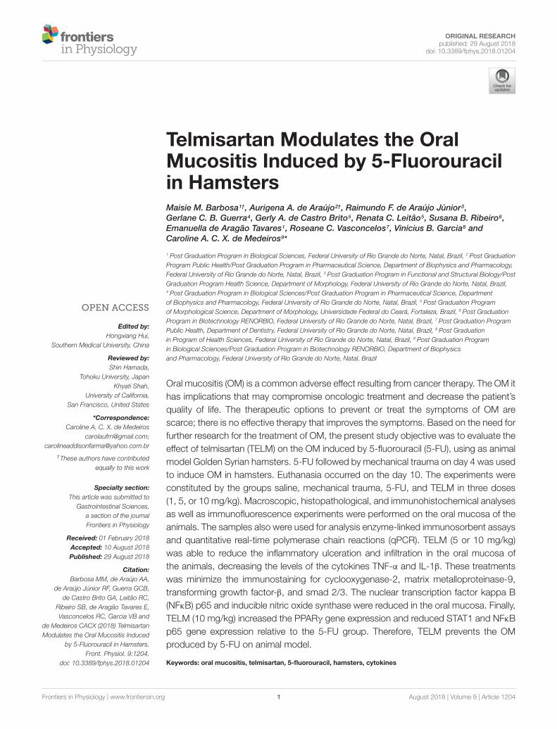
FIGURE 1 | Macroscopic analysis and scores of the oral mucosa of hamsterswith oral mucositis (OM) induced by 5-fluorouracil (5-FU) and MT. The salinegroup consisted of animals with healthy oral mucosa (A). The mechanicaltrauma (MT) group demonstrated vasodilation and erythema in the jugalmucosa (B). The 5-FU group had changes in the oral mucosa, erythema,vasodilatation, erosion, and extensive ulcers (C). TELM at 1 mg/kg did notimprove the OM (D). The groups treated with TELM 5 (E), or 10 mg/kg (F)had mild vasodilation and superficial erosion in the oral mucosa, with noevidence of ulcers. The scores are represented with the standard error of themean (n = 5). ∗p < 0.05 vs. the saline group, ∗∗p < 0.05 vs. the 5-FU group(Kruskal–Wallis test and Dunn’s multiple comparison test).
Frontiers in Physiology | www.frontiersin.org 2 August 2018 | Volume 9 | Article 1204

fphys-09-01204 August 28, 2018 Time: 10:35 # 3
Barbosa et al. Telmisartan in Oral Mucositis
(MT) on the 4th day of the experiment under anesthesia with2% xylazine (10 mg/kg) and 10% ketamine (80 mg/kg) (i.p.), asadapted from de Araujo et al. (2015).
In according of the laboratory protocol, the MT wasperformed by exposing the right oral mucosa of the animaland the perforating the grooves with the tip of an 18 mmgauge needle across the oral mucosa, with the aim of enhancingOM and reproducing the clinical signs of chronic irritationthat occur in patients during chewing. On the 10th dayof the experimental model, after anesthesia with thiopental
(100 mg/kg, i.p.) combined with 2% lidocaine i.p., they hamsterswere euthanized, and specimens of the oral mucosa were retiredfor further analysis (Medeiros et al., 2011; de Araujo et al., 2015).
Experimental GroupsThe animals (n = 5 per group) were treated as follows: saline,without OM and treated with oral saline (p.o.); MT: the animalswere subjected to MT but not 5-FU and treated with saline (p.o.);5-FU: the animals received 5-FU and MT and were treated withsaline (p.o.); TELM 1, 5, or 10: the animals received 5-FU and MT
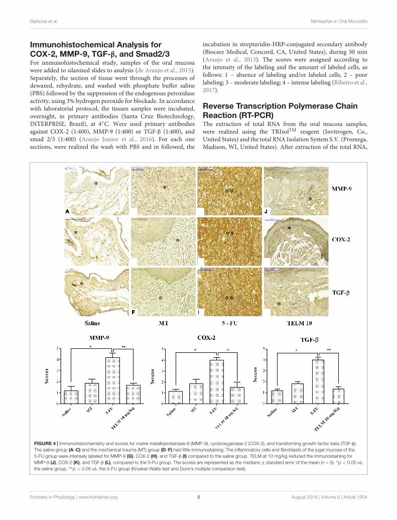
FIGURE 2 | Histopathological analysis and scores of the oral mucosa of hamsters with oral mucositis (OM) induced by 5-fluorouracil (5-FU). The saline groupconsisted of animals with healthy oral mucosa (A). The mechanical trauma (MT) group demonstrated areas of epithelial hyperplasia exhibiting foci of moderateexocytosis (→), small-caliber blood vessels (I), and inflammatory cells dispersed by connective tissue (B). The 5-FU group had alterations in the oral mucosaincluding intense inflammatory infiltrates (∗∗∗), erosion and extensive ulcers (C), and presence of purulent exudate, with areas suggestive of necrosis (F). TELM at1 mg/kg did not improve the OM and presented ulcerated areas and moderate inflammatory infiltrate (∗∗) (D), however, TELM 5 (E) or 10 mg/kg (F) showedvasodilatation and discrete cell infiltration (∗), the absence of hemorrhage, edema, ulcers, and abscesses. The scores are presented as the medians ± standard errorof the mean (n = 5). ∗p < 0.05 vs. the saline group, ∗∗p < 0.05 vs. the 5-FU group (Kruskal–Wallis test and Dunn’s multiple comparison test).
Frontiers in Physiology | www.frontiersin.org 3 August 2018 | Volume 9 | Article 1204
fphys-09-01204 August 28, 2018 Time: 10:35 # 4
Barbosa et al. Telmisartan in Oral Mucositis
and were treated with TELM at doses 1, 5, or 10 mg/kg, orally(Araujo et al., 2013; Guerra et al., 2015). The TELM used was thecommercial formulation (Micardis R©). The tablet was dissolved insaline and administered 30 min prior to 5-FU, during the 10 daysof the experimental model. The control groups received saline(p.o.) for 10 days.
Macroscopic AnalysisMacroscopic analysis of the oral mucosa of the animalswas performed as described by Sonis et al. (2000). Briefly,the inflammatory characteristics observed in the experimentalmodel, such as erythema, erosion, vasodilatation, epithelialulceration and abscess, were evaluated and classified accordingas value of the assigned score: 0 – completely healthy oral mucosawithout erosion or vasodilation; 1 – presence of erythema, butno evidence of erosion of the oral mucosa; 2 – severe erythema,vasodilatation, and superficial erosion; 3 – ulcer formation on oneor more mucosal surfaces but affecting no more than 25% of thecheek surface area, as well as severe erythema and vasodilatation;4 – ulcers with a cumulative involvement of approximately 50%of the surface area of the cheek pouch and; 5 – virtually completeulceration of the oral mucosa (Sonis et al., 2000; Lopes et al., 2010;Ribeiro et al., 2017).
Histopathological AnalysisThe samples of oral mucosa were initially fixed in 10% bufferedformalin and then dehydrated and processed, followed byparaffin embedding. For study histopathological, were obtainedsections of 5 µm thickness for hematoxylin-eosin staining. Theresections were analyzed by light microscopy. The criteria analyzedwere included infiltration of inflammatory cells, vasodilatation,presence of hemorrhagic areas, edema, ulcerations, andabscesses were independently scored following the followingclassification: 1 – normal epithelium and connective tissuewithout vasodilation, absence or discrete cellular infiltrationor absence of hemorrhagic areas, ulcerations or abscesses;2 – discrete areas of vasodilation or re-epithelialization, mildinflammatory infiltration with mononuclear prevalence andabsence of hemorrhagic areas, edema, ulcerations, or abscesses;3 – moderate vasodilatation, areas of epithelial degeneration,inflammatory infiltration with neutrophil prevalence, presenceof hemorrhagic areas, edema and eventual ulceration, andabsence of abscesses; 4 – severe vasodilation and inflammatoryinfiltration with neutrophils (Leitao et al., 2007, 2008; Watanabeet al., 2009; Medeiros et al., 2011; Skeff et al., 2014; de Araujoet al., 2015; Ribeiro et al., 2017).
IL-1β and TNF-α Cytokine AssaysSamples of the oral mucosa were homogenized and processedbased on the methodology of Kendall et al. (1983). In resume,the microplates were incubated overnight for 16 h by 4◦Cwith antibodies against IL-1β and TNF-α, in according to themanufacturer’s protocol. Afterward, the plates were washed, andthe wells were blocked, followed by addition of the samples andthe standards in several dilutions, which were incubated for 2 h.The plates were washed three times with solutions containingbovine serum albumin (BSA), and then biotinylated anti-IL-1β
or anti-TNF-α sheep polyclonal antibodies (diluted 1:1,000 in 1%BSA assay buffer) were added. Immediately after incubation atroom temperature for 1 h, the plates were then washed again, and50 mL of HRP-streptavidin (diluted 1:5,000) were added to eachwell. The O-phenylenediamine color reagent (50 mL) was added15 min later, and the plates were incubated in the dark at 37◦Cfor 15–20 min (Lima-Junior et al., 2012). The enzymatic reactionwas quenched using H2SO4, and the absorbance was measuredby means of UV-VIS spectrophotometry. These absorbance weredetermined at 490 nm and the results were presented as pg/mL(Lima-Junior et al., 2012; Araujo et al., 2013).
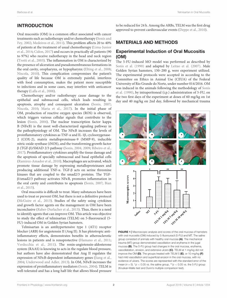
FIGURE 3 | Levels of tumor necrosis factor (TNF)-α and interleukin (IL)-1β
cytokines in the oral mucosa of hamsters with oral mucositis (OM). The salinegroup consists of animals without OM. The mechanical trauma (MT) groupconsisted of hamsters that received excoriations of the oral mucosa, without5-fluorouracil (5-FU). The 5-FU group received 5-FU, was subjected to MTand was treated with saline i.p. The TELM groups received 5-FU, weresubjected to MT, and received TELM i.p. at one of three doses (1, 5, or10 mg/kg). The results are presented as the mean ± standard error of themean (n = 5). ∗p < 0.05 vs. the saline group, ∗∗p < 0.05 vs. the 5-FU group(analysis of variance with Tukey’s post-test).
Frontiers in Physiology | www.frontiersin.org 4 August 2018 | Volume 9 | Article 1204
fphys-09-01204 August 28, 2018 Time: 10:35 # 5
Barbosa et al. Telmisartan in Oral Mucositis
Immunohistochemical Analysis forCOX-2, MMP-9, TGF-β, and Smad2/3For immunohistochemical study, samples of the oral mucosawere added to silanized slides to analysis (de Araujo et al., 2015).Separately, the section of tissue went through the processes ofdewaxed, rehydrate, and washed with phosphate buffer saline(PBS) followed by the suppression of the endogenous peroxidaseactivity, using 3% hydrogen peroxide for blockade. In accordancewith laboratorial protocol, the tissues samples were incubated,overnight, in primary antibodies (Santa Cruz Biotechnology,INTERPRISE, Brazil), at 4◦C. Were used primary antibodiesagainst COX-2 (1:400), MMP-9 (1:400) or TGF-β (1:400), andsmad 2/3 (1:400) (Araujo Junior et al., 2016). For each onesections, were realized the wash with PBS and in followed, the
incubation in streptavidin-HRP-conjugated secondary antibody(Biocare Medical, Concord, CA, United States), during 30 min(Araujo et al., 2013). The scores were assigned according tothe intensity of the labeling and the amount of labeled cells, asfollows: 1 – absence of labeling and/or labeled cells, 2 – poorlabeling; 3 – moderate labeling; 4 – intense labeling (Ribeiro et al.,2017).
Reverse Transcription Polymerase ChainReaction (RT-PCR)The extraction of total RNA from the oral mucosa samples,were realized using the TRIzolTM reagent (Invitrogen, Co.,United States) and the total RNA Isolation System S.V. (Promega,Madison, WI, United States). After extraction of the total RNA,
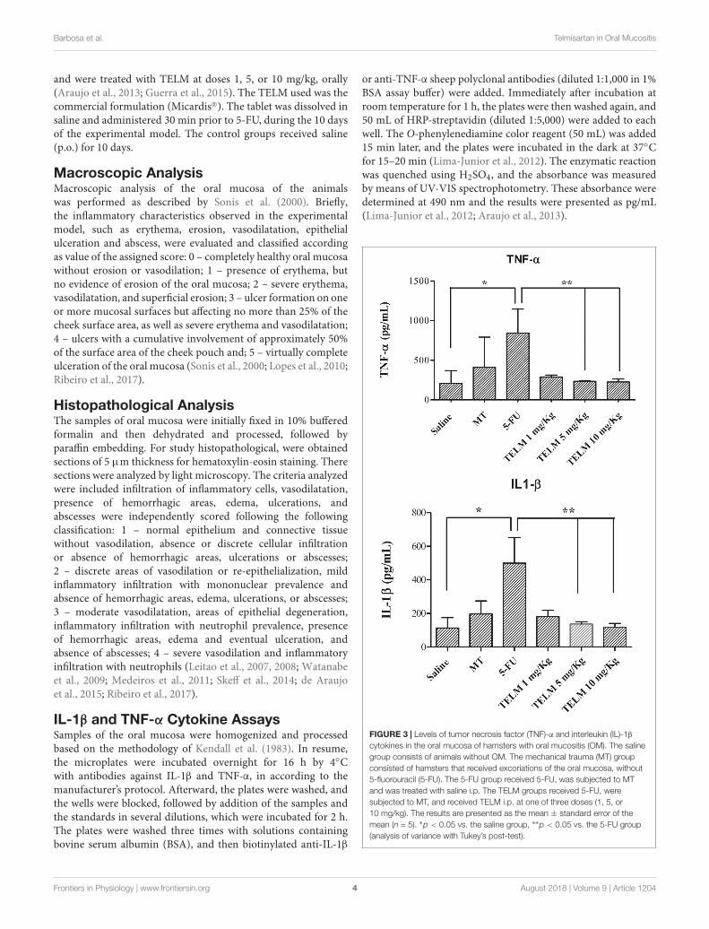
FIGURE 4 | Immunohistochemistry and scores for matrix metalloproteinase-9 (MMP-9), cyclooxygenase-2 (COX-2), and transforming growth factor beta (TGF-β).The saline group (A–C) and the mechanical trauma (MT) group (D–F) had little immunostaining. The inflammatory cells and fibroblasts of the jugal mucosa of the5-FU group were intensely labeled for MMP-9 (G), COX-2 (H), and TGF-β (I) compared to the saline group. TELM at 10 mg/kg reduced the immunostaining forMMP-9 (J), COX-2 (K), and TGF-β (L), compared to the 5-FU group. The scores are represented as the medians ± standard error of the mean (n = 5). ∗p < 0.05 vs.the saline group, ∗∗p < 0.05 vs. the 5-FU group (Kruskal–Wallis test and Dunn’s multiple comparison test).
Frontiers in Physiology | www.frontiersin.org 5 August 2018 | Volume 9 | Article 1204

fphys-09-01204 August 28, 2018 Time: 10:35 # 6
Barbosa et al. Telmisartan in Oral Mucositis
was synthesized the first strand cDNA, from 20 µL of totalRNA, using the ImProm-IITM reverse transcriptase system forRT-PCR (Promega), following the instructions to manufacturer’s(Chomczynski and Sacchi, 2006). The real-time quantitative PCRanalyses of signal transducers and activators of transcription(STAT1), NFκB p65, and PPAR-γ mRNAs were realized usingSYBR Green Mix with the Applied Biosystems R© 7500 FASTsystem (Applied Biosystems, Foster City, CA, United States).The primers were designed using Software Primer ExpressTM
version 3.0.1 (Applied BiosystemsTM) and were as follows: STAT1 (Rattus norvegicus) (Forward: 5′-ATT AAC GAT GAG TTAGTG GAG TGG AA-3′; Reverse: 5′-GGT GGC CCC CCG ATAC-3′); NFκB p65 (Mesocricetus auratus) (Forward: 5′-GAA GAAGCG AGA CCT GGA GCA A-3′; Reverse: 5′-GTT GAT GGTGCT GAG GGA TGC T-3′); PPARγ (M. auratus) (Forward:5′-CAT GAC CAG GGA GTT CCT CAA-3′; Reverse: 5′-TGGGCT CCA TAA AGT CAC CAA-3′); GAPDH (M. auratus)(Forward: 5′-AAC TTT GGC ATC GTG GAA GG-3′; Reverse:5′-GTG GAT GCA GGG ATG ATG TTC-3′). The plates wereprepared with 10 µL final volume in each well (2.0 µL ofcDNA + 8.0 µL of the prepared mixture containing the SYBRMix, nuclease-free water and forward and reverse primers)and were added in the StepOnePlus thermal cycler (AppliedBiosystemsTM, United States) according to the manufacturer’sprotocol. For calculate relative levels obtained in this experiment,
were used the mean Ct values. This value was contributing tocalculate target genes for each of the submitted groups inducedOM for 5-FU, when compared to the negative control group(group without OM). Expression data were standardized usingto the GAPDH reference gene in the formula 2−11Ct (Rao et al.,2013).
Immunofluorescence for iNOS and NFkBp65Immunofluorescence was realized according to the previouslydescribed methodology of Guerra et al. (2016) and Ribeiroet al. (2017). Sections of the oral mucosa from the hamsters,three per group, were dewaxed using xylol and, posteriorly,washed in concentrations different of ethanol and PBS. Inprocess of antigen retrieval, was utilized a 10 mM sodium citratesolution containing 0.05% Tween 20 for a period 40 min at95◦C. Posteriorly, the samples were incubated in Sudan-Black0.1% diluted in 70% ethanol, for 40 min, at room temperatureto decrease the tissue autofluorescence. The slides again wereincubated at 4◦C overnight with mouse monoclonal anti-NFκB(1:200; sc-8008, Santa Cruz Biotechnology) or rat polyclonalanti-iNOS (1:200, sc-8332, Santa Cruz Biotechnology) primaryantibodies and were washed three times for 5 min in PBScontaining 0.2% Triton X-100. Subsequently, the samples were
FIGURE 5 | Immunohistochemistry and scores for Smad 2/3. The saline group and the mechanical trauma (MT) group had little immunostaining. The inflammatorycells and fibroblasts of the jugal mucosa of the 5-FU group were intensely labeled Smad 2/3 compared to the saline group. TELM at 10 mg/kg reduced theimmunostaining for Smad 2/3, compared to the 5-FU group. The scores are represented as the medians ± standard error of the mean (n = 5). ∗p < 0.05 vs. thesaline group, ∗∗p < 0.05 vs. the 5-FU group (Kruskal–Wallis test and Dunn’s multiple comparison test).
Frontiers in Physiology | www.frontiersin.org 6 August 2018 | Volume 9 | Article 1204
fphys-09-01204 August 28, 2018 Time: 10:35 # 7
Barbosa et al. Telmisartan in Oral Mucositis
incubated with an Alexa Fluor 488-conjugated goat anti-rabbitsecondary antibody (1:500 in 1% BSA blocking solution; Dako,Brazil, and United States). Finally, coverslips were appliedusing VECTASHIELD R© mounting medium with DAPI (4′,6-diamidino-2′-phenylindole). Immunofluorescence images wereobtained using a Carl Zeiss Laser Scanning microscope (LSM 710,20× objective, Carl Zeiss, Jena, Germany) (Araujo Junior et al.,2016; de Assis et al., 2016).
Statistical AnalysisThe data are presented as the mean± standard deviation or as themedian± standard deviation. The analysis of variance (ANOVA)and Tukey’s post-test were used to compare the means. Tocompare the medians, the Kruskal–Wallis test followed by Dunn’stest was used. The minimum significance was set at p < 0.05, and
the analyses were performed using the Prism program, version6.0 GraphPad Software, La Jolla, CA, United States.
RESULTS
Macroscopic Aspects andHistopathological AnalysisIn analyze oral mucosa of the animals belonging saline grouphad no macroscopic or histopathological changes (Figure 1A;with score 0 and Figure 2A; score of 1). On MT group animalsshowed some changes. Macroscopic aspects demonstrated inthese group, included presence of erythema and with absence ofmucosal erosion (Figure 1B; value of score equal 1), already in thehistopathological aspects were observed vasodilatation discrete
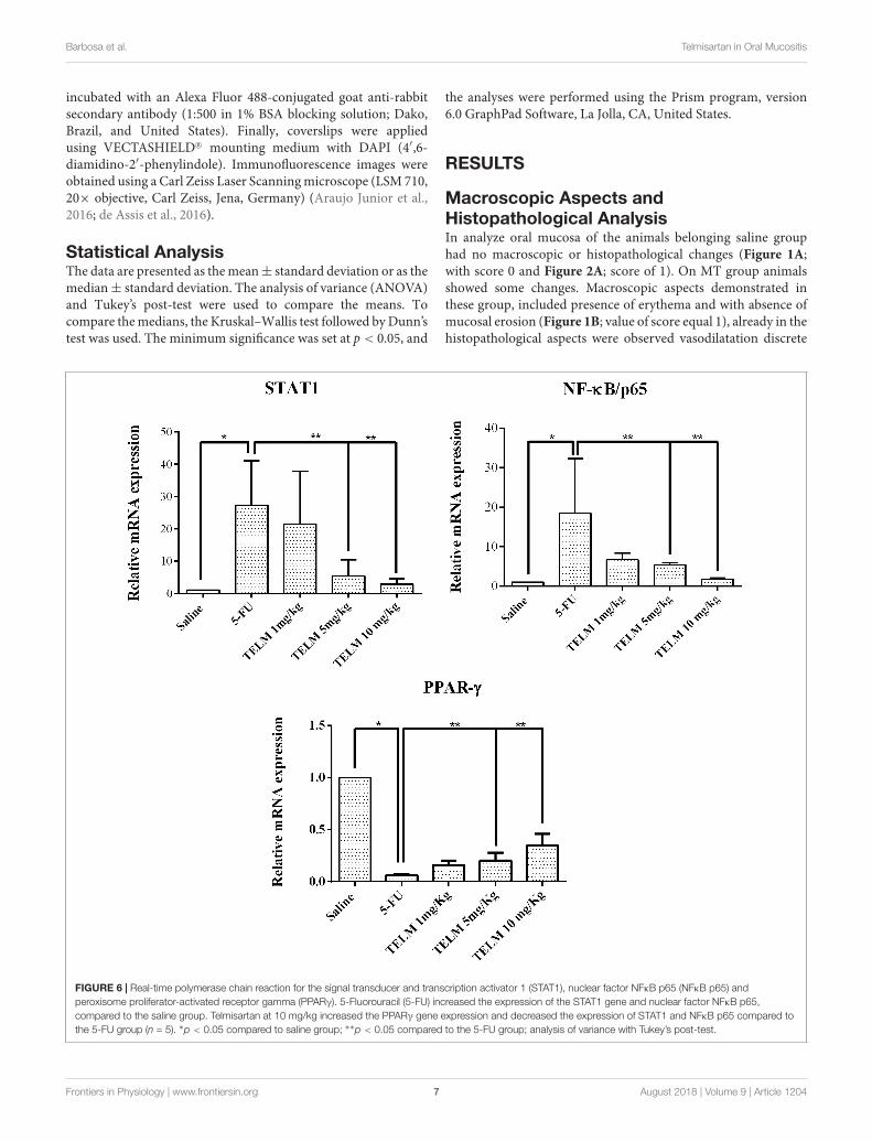
FIGURE 6 | Real-time polymerase chain reaction for the signal transducer and transcription activator 1 (STAT1), nuclear factor NFκB p65 (NFκB p65) andperoxisome proliferator-activated receptor gamma (PPARγ). 5-Fluorouracil (5-FU) increased the expression of the STAT1 gene and nuclear factor NFκB p65,compared to the saline group. Telmisartan at 10 mg/kg increased the PPARγ gene expression and decreased the expression of STAT1 and NFκB p65 compared tothe 5-FU group (n = 5). ∗p < 0.05 compared to saline group; ∗∗p < 0.05 compared to the 5-FU group; analysis of variance with Tukey’s post-test.
Frontiers in Physiology | www.frontiersin.org 7 August 2018 | Volume 9 | Article 1204

fphys-09-01204 August 28, 2018 Time: 10:35 # 8
Barbosa et al. Telmisartan in Oral Mucositis
FIGURE 7 | Immunofluorescence for iNOS; NFκB p65 and mean of the densitometric analysis. The 5-fluorouracil (5-FU) group (C,G) had higher green labeling thanthat of the mechanical trauma (MT) group (B,F) or the saline group (A,E) (p < 0.05; n = 5). The TELM 10 mg/kg group (D,H) had a reduced immunoreactivitycompared to the 5-FU group (n = 5; ∗p < 0.05 vs. the saline group, ∗∗p < 0.05 vs. the 5-FU group (analysis of variance with Tukey’s post-test).
and cell infiltration, areas of re-epithelialization, and absence ofhemorrhage, edema, ulcers, and abscesses (Figure 2B; with scoreof 2). The aspects observed of the 5-FU group demonstratedulcers in approximately 50% of the area of the oral mucosathe animals euthanized (Figure 1C; score of 4, p < 0.05),as others technical features including severe vasodilatation,and inflammatory infiltration with neutrophils, abscesses, andextensive ulcers (Figure 2C; score of 4, p < 0.05). These aspectswere compared to the saline group. TELM at 1 mg/kg didnot prevent the OM lesions, compared to the 5-FU group(Figures 1D, 2D; score of 3). The groups that had been treatedwith TELM at 5 or 10 mg/kg has aspects as discrete vasodilatation,areas of re-epithelialization, discrete cell infiltration, and absenceof hemorrhage, edema, ulcers, and abscesses (Figures 1E,F, 2E,F;score of 2, p< 0.05). These latter groups the observed results werecompared with the experimental 5-FU group.
TNF-α and IL-1β CytokinesThe analyze for cytokines demonstrated the 5-FU group hadhigher values of the TNF-α (Figure 3, p < 0.05) and IL-1β
(Figure 3, p < 0.05), contrary result than with exhibited in the
saline group. TELM at doses of 5 or 10 mg/kg reduced the levelsof TNF-α (Figure 3, p < 0.05) and IL-1β (Figure 3, p < 0.05),when the results obtained in this group were compared to thelevels found in the 5-FU group. The results get by TELM at a doseof 1 mg/kg did not reduce the levels of proinflammatory cytokinescompared to the 5-FU group.
Immunohistochemical Analyses (MMP-9,COX-2, TGF-β, and Smad 2/3)The analyzes realized with the oral mucosa of the animals withOM induced for 5-FU (n = 4), demonstrated intense markingfor proteins MMP-9 (Figure 4G; score of 4, p < 0.05), COX-2(Figure 4H; score of 4, p < 0.05), TGF-β (Figure 4I; score of 4,p< 0.05), and Smad 2/3 (Figure 5, score of 4, p< 0.05), comparedwith samples of the saline group (Figures 4A–F). Inversely, theTELM (10 mg/kg) group showed low immunostaining for MMP-9 (Figure 4J; score of 2, p < 0.05), COX-2 (Figure 4K; score of2, p < 0.05), TGF-β (Figure 4L; score of 2, p < 0.05) and Smad2/3 (Figure 5; score of 2, p < 0.05), also compared with the 5-FUgroup.
Frontiers in Physiology | www.frontiersin.org 8 August 2018 | Volume 9 | Article 1204
fphys-09-01204 August 28, 2018 Time: 10:35 # 9
Barbosa et al. Telmisartan in Oral Mucositis
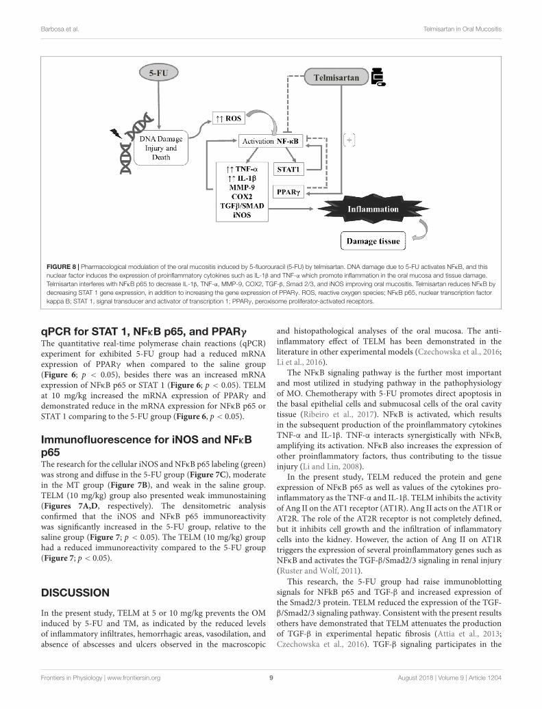
FIGURE 8 | Pharmacological modulation of the oral mucositis induced by 5-fluorouracil (5-FU) by telmisartan. DNA damage due to 5-FU activates NFκB, and thisnuclear factor induces the expression of proinflammatory cytokines such as IL-1β and TNF-α which promote inflammation in the oral mucosa and tissue damage.Telmisartan interferes with NFκB p65 to decrease IL-1β, TNF-α, MMP-9, COX2, TGF-β, Smad 2/3, and iNOS improving oral mucositis. Telmisartan reduces NFκB bydecreasing STAT 1 gene expression, in addition to increasing the gene expression of PPARγ. ROS, reactive oxygen species; NFκB p65, nuclear transcription factorkappa B; STAT 1, signal transducer and activator of transcription 1; PPARγ, peroxisome proliferator-activated receptors.
qPCR for STAT 1, NFκB p65, and PPARγThe quantitative real-time polymerase chain reactions (qPCR)experiment for exhibited 5-FU group had a reduced mRNAexpression of PPARγ when compared to the saline group(Figure 6; p < 0.05), besides there was an increased mRNAexpression of NFκB p65 or STAT 1 (Figure 6; p < 0.05). TELMat 10 mg/kg increased the mRNA expression of PPARγ anddemonstrated reduce in the mRNA expression for NFκB p65 orSTAT 1 comparing to the 5-FU group (Figure 6, p < 0.05).
Immunofluorescence for iNOS and NFκBp65The research for the cellular iNOS and NFκB p65 labeling (green)was strong and diffuse in the 5-FU group (Figure 7C), moderatein the MT group (Figure 7B), and weak in the saline group.TELM (10 mg/kg) group also presented weak immunostaining(Figures 7A,D, respectively). The densitometric analysisconfirmed that the iNOS and NFκB p65 immunoreactivitywas significantly increased in the 5-FU group, relative to thesaline group (Figure 7; p < 0.05). The TELM (10 mg/kg) grouphad a reduced immunoreactivity compared to the 5-FU group(Figure 7; p < 0.05).
DISCUSSION
In the present study, TELM at 5 or 10 mg/kg prevents the OMinduced by 5-FU and TM, as indicated by the reduced levelsof inflammatory infiltrates, hemorrhagic areas, vasodilation, andabsence of abscesses and ulcers observed in the macroscopic
and histopathological analyses of the oral mucosa. The anti-inflammatory effect of TELM has been demonstrated in theliterature in other experimental models (Czechowska et al., 2016;Li et al., 2016).
The NFκB signaling pathway is the further most importantand most utilized in studying pathway in the pathophysiologyof MO. Chemotherapy with 5-FU promotes direct apoptosis inthe basal epithelial cells and submucosal cells of the oral cavitytissue (Ribeiro et al., 2017). NFκB is activated, which resultsin the subsequent production of the proinflammatory cytokinesTNF-α and IL-1β. TNF-α interacts synergistically with NFκB,amplifying its activation. NFκB also increases the expression ofother proinflammatory factors, thus contributing to the tissueinjury (Li and Lin, 2008).
In the present study, TELM reduced the protein and geneexpression of NFκB p65 as well as values of the cytokines pro-inflammatory as the TNF-α and IL-1β. TELM inhibits the activityof Ang II on the AT1 receptor (AT1R). Ang II acts on the AT1R orAT2R. The role of the AT2R receptor is not completely defined,but it inhibits cell growth and the infiltration of inflammatorycells into the kidney. However, the action of Ang II on AT1Rtriggers the expression of several proinflammatory genes such asNFκB and activates the TGF-β/Smad2/3 signaling in renal injury(Ruster and Wolf, 2011).
This research, the 5-FU group had raise immunoblottingsignals for NFkB p65 and TGF-β and increased expression ofthe Smad2/3 protein. TELM reduced the expression of the TGF-β/Smad2/3 signaling pathway. Consistent with the present resultsothers have demonstrated that TELM attenuates the productionof TGF-β in experimental hepatic fibrosis (Attia et al., 2013;Czechowska et al., 2016). TGF-β signaling participates in the
Frontiers in Physiology | www.frontiersin.org 9 August 2018 | Volume 9 | Article 1204
fphys-09-01204 August 28, 2018 Time: 10:35 # 10
Barbosa et al. Telmisartan in Oral Mucositis
pathophysiology of OM by inhibiting epithelial cell growth andcausing apoptosis as well as by promoting the activation of thetranscription factor NFκB (Massagué, 2008). Smad 7 improvesthe OM of patients undergoing chemotherapy or radiotherapy byinhibiting the TGF-β/Smad2/3 and NFkB signaling (Han et al.,2013). Thus, blockade of NFκB and TGF-β signaling may explainpart of the beneficial effect of TELM on OM in hamsters.
The nuclear transcription factor kappa B induces transcriptionof MMP-9, COX-2, and iNOS (Sonis, 2007). TELM at 10 mg/kgreduced the immunostaining for COX2, MMP-9, and iNOSin the oral mucosa of hamsters compared to the 5-FUgroup. MMPs are enzymes that were initially studied in thecontext of their roles in regulating the homeostasis of theextracellular matrix components and maintaining the physiologyof healthy oral mucosa (Clark et al., 2008). However, MMP-9is involved in the chemotherapy-induced OM stages, includinginflammatory cell migration, apoptosis, cytokine activation,and dysregulation of normal cell kinetics, which suggeststhat it participates in the ulcerative phase (Al-Azri et al.,2015). Like the results in present study, others search havedemonstrated that TELM downregulates MMP-9 and COX2(Araujo et al., 2013).
The participation of nitric oxide (NO) in the OM induced by5-FU is consistent with previous reports showing that cytokinesTNF-α and IL-1β stimulate NO production from iNOS (Liu et al.,2005). Other authors have demonstrated that the NO producedby iNOS is associated with the damage and inflammatory eventsof 5-FU-induced OM (Leitao et al., 2007). Consistent with theresults of the current study, TELM decreased the iNOS activity,thus inhibiting the excessive NO generation and inflammatoryresponses in other models of inflammation (Yuan et al., 2015).
Telmisartan at 10 mg/kg increased the gene expressionof PPARγ (peroxisome proliferator-activated receptor-gamma) compared to the 5-FU group. Similarly, others havedemonstrated that TELM increases the gene expression of PPARγ
in an in vitro human umbilical vein endothelial cell model anddecreases the homocysteine-induced inflammatory response inendothelial cells (Xu et al., 2014). The PPARs are a family ofnuclear receptors that regulate the immune and inflammatory
response. Among the ARBs, TELM is the most potent agonist forPPARγ: it activates the receptor to approximately 25–30% of themaximum level reached by full agonists such as pioglitazone androsiglitazone (Janke et al., 2006).
Some investigators have demonstrated that part of the anti-inflammatory effect of PPARγ is mediated by direct inhibition ofthe NFκB p65 (Chung et al., 2000) as well as by the inhibition ofSTAT (Ji et al., 2005). In the current study, TELM inhibited NFκBp65 and reduced the STAT 1 gene expression compared to the 5-FU group. Seven members of the STAT protein family have beendescribed, and STAT1 activates iNOS (Ohmori and Hamilton,2001). 5-FU increased the STAT 1 gene expression and inhibitedthe PPARγ gene expression in the present study. However, studiesof the effects on the expression of these proteins are necessaryto confirm the participation of STAT 1 and the PPARγ gene inthe mechanisms associated with the protective effects of TELMagainst 5-FU-induced OM.
CONCLUSION
TELM prevents the 5-FU-induced OM lesions by reducing theexpression of the proinflammatory mediators TNF-α, IL-1β,TGF-β, Smad 2/3, COX2, MMP9, and iNOS, by inhibiting NFκB,by increasing the PPARγ gene expression, and by reducing theSTAT1 gene expression. These results suggest the contributethese pathways in the protecting outcome that TELM present inOral Mucosa (Figure 8). Though, more researches are needed toelucidate these findings.
AUTHOR CONTRIBUTIONS
CdM: investigation, editing of the manuscript, supervision,and validation. MB and AdA: investigation, preparation of themanuscript, analyzes, methodologies, and validation. RdAJ andGG: investigation and editing of the manuscript and supervision.GdCB and RL: editing and revision of the manuscript. SR, EdAT,RV, and VG: analyzes and methodologies.
REFERENCESAl-Azri, A. R., Gibson, R. J., Bowen, J. M., Stringer, A. M., Keefe, D. M., and Logan,
R. M. (2015). Involvement of matrix metalloproteinases (MMP-3 and MMP-9)in the pathogenesis of irinotecan-induced oral mucositis. J. Oral Pathol. Med.44, 459–467. doi: 10.1111/jop.12255
Araujo, A. A., Souza, T. O., Moura, L. M., Brito, G. A., Aragao, K. S., Araujo, L. S.,et al. (2013). Effect of telmisartan on levels of IL-1, TNF-alpha, down-regulatedCOX-2, MMP-2, MMP-9 and RANKL/RANK in an experimental periodontitismodel. J Clin. Periodontol. 40, 1104–1111. doi: 10.1111/jcpe.12160
Araujo Junior, R. F., Garcia, V. B., Leitao, R. F., Brito, G. A., Miguel Ede, C., Guedes,P. M., et al. (2016). Carvedilol improves inflammatory response, oxidative stressand fibrosis in the alcohol-induced liver injury in rats by regulating kuppfer cellsand hepatic stellate cells. PLoS One 11:e0148868. doi: 10.1371/journal.pone.0148868
Attia, Y. M., Elalkamy, E. F., Hammam, O. A., Mahmoud, S. S., and El-Khatib, A. S. (2013). Telmisartan, an AT1 receptor blocker and a PPARgamma activator, alleviates liver fibrosis induced experimentally by Schistosomamansoni infection. Parasit. Vectors 6:199. doi: 10.1186/1756-3305-6-199
Bian, L., Han, G., Zhao, C. W., Garl, P. J., and Wang, X. J. (2015). The role of Smad7in oral mucositis. Protein Cell. 6, 160–169. doi: 10.1007/s13238-014-0130-4
Chomczynski, P., and Sacchi, N. (2006). The single-step method of RNAisolation by acid guanidinium thiocyanate-phenol-chloroform extraction:twenty-something years on. Nat. Protoc. 1, 581–585. doi: 10.1038/nprot.2006.83
Chung, S. W., Kang, B. Y., Kim, S. H., Pak, Y. K., Cho, D., Trinchieri, G., et al.(2000). Oxidized low density lipoprotein inhibits interleukin-12 productionin lipopolysaccharide-activated mouse macrophages via direct interactionsbetween peroxisome proliferator-activated receptor-gamma and nuclear factor-kappa B. J. Biol. Chem. 275, 32681–32687. doi: 10.1074/jbc.M002577200
Cidon, E. U. (2017). Chemotherapy induced oral mucositis: prevention is possible.Chin. Clin. Oncol. 7:6. doi: 10.21037/cco.2017.10.01
Clark, I. M., Swingler, T. E., Sampieri, C. L., and Edwards, D. R. (2008). Theregulation of matrix metalloproteinases and their inhibitors. Int. J. Biochem. CellBiol. 40, 1362–1378. doi: 10.1016/j.biocel.2007.12.006
Czechowska, G., Celinski, K., Korolczuk, A., Wojcicka, G., Dudka, J., Bojarska, A.,et al. (2016). The effect of the angiotensin II receptor, type 1 receptorantagonists, losartan and telmisartan, on thioacetamide-induced liver fibrosisin rats. J. Physiol. Pharmacol. 67, 575–586.
Frontiers in Physiology | www.frontiersin.org 10 August 2018 | Volume 9 | Article 1204

fphys-09-01204 August 28, 2018 Time: 10:35 # 11
Barbosa et al. Telmisartan in Oral Mucositis
de Araujo, A. A., Varela, H., de Medeiros, C. A., de Castro Brito, G. A., de Lima,K. C., de Moura, L. M., et al. (2015). Azilsartan reduced TNF-alpha and IL-1betalevels, increased IL-10 levels and upregulated VEGF, FGF, KGF, and TGF-alphain an oral mucositis model. PLoS One 10:e0116799. doi: 10.1371/journal.pone.0116799
de Assis, P. O. A., Guerra, G. C. B., de Souza Araújo, D. F., de Araújo, R. F.Jr., Machado, T. A. D. G., de Araújo, A. A., et al. (2016). Intestinal anti-inflammatory activity of goat milk and goat yoghurt in the acetic acid modelof rat colitis. Int. Dairy J. 56, 45–54. doi: 10.1016/j.idairyj.2015.11.002
Deppe, S., Böger, R. H., Weiss, J., and Benndorf, R. A. (2010). Telmisartan: a reviewof its pharmacodynamic and pharmacokinetic properties. Expert Opin. DrugMetab. Toxicol. 6, 863–871. doi: 10.1517/17425255.2010.494597
Elting, L. S., Keefe, D. M., Sonis, S. T., Garden, A. S., Spijkervet, F. K., Barasch, A.,et al. (2008). Patient-reported measurements of oral mucositis in head andneck cancer patients treated with radiotherapy with or without chemotherapy:demonstration of increased frequency, severity, resistance to palliation, andimpact on quality of life. Cancer 113, 2704–2713. doi: 10.1002/cncr.23898
Guerra, G. C., Araujo, A. A., Lira, G. A., Melo, M. N., Souto, K. K., Fernandes, D.,et al. (2015). Telmisartan decreases inflammation by modulating TNF-alpha,IL-10, and RANK/RANKL in a rat model of ulcerative colitis. Pharmacol. Rep.67, 520–526. doi: 10.1016/j.pharep.2014.12.011
Guerra, G. C., de Menezes, M. S., de Araujo, A. A., de Araujo Junior, R. F., andde Medeiros, C. A. (2016). Olmesartan Prevented Intra-articular InflammationInduced by Zymosan in Rats. Biol. Pharm. Bull. 39, 1793–1801. doi: 10.1248/bpb.b16-00296
Hamano, Y., Okude, T., Yokosuka, O., and Ogawa, M. (2011). Attenuation ofimmune-mediated renal injury by Telmisartan, an angiotensin receptor blockerand a selective PPAR-gamma activator. Nephron Extra 1, 78–90. doi: 10.1159/000331704
Han, G., Bian, L., Li, F., Cotrim, A., Wang, D., Lu, J., et al. (2013). Preventive andtherapeutic effects of Smad7 on radiation-induced oral mucositis. Nat. Med. 19,421–428. doi: 10.1038/nm.3118
Janke, J., Schupp, M., Engeli, S., Gorzelniak, K., Boschmann, M., Sauma, L.,et al. (2006). Angiotensin type 1 receptor antagonists induce human in-vitro adipogenesis through peroxisome proliferator-activated receptor-gammaactivation. J. Hypertens. 24, 1809–1816. doi: 10.1097/01.hjh.0000242405.68461.84
Ji, J. D., Kim, H. J., Rho, Y. H., Choi, S. J., Lee, Y. H., Cheon, H. J., et al.(2005). Inhibition of IL-10-induced STAT3 activation by 15-deoxy-Delta12,14-prostaglandin J2. Rheumatology 44, 983–988. doi: 10.1093/rheumatology/keh657
Jiang, B., Xu, S., Hou, X., Pimentel, D. R., and Cohen, R. A. (2004). AngiotensinII differentially regulates interleukin-1-beta-inducible NO synthase (iNOS) andvascular cell adhesion molecule-1 (VCAM-1) expression: role of p38 MAPK.J. Biol. Chem. 279, 20363–20368. doi: 10.1074/jbc.M314172200
Kendall, C., Ionescu-Matiu, I., and Dreesman, G. R. (1983). Utilization ofthe biotin/avidin system to amplify the sensitivity of the enzyme-linkedimmunosorbent assay (ELISA). J. Immunol. Methods 56, 329–339. doi: 10.1016/S0022-1759(83)80022-2
Lalla, R. V., Sonis, S. T., and Peterson, D. E. (2008). Management of oral mucositisin patients who have cancer. Dent. Clin. North Am. 52, 61–77. doi: 10.1016/j.cden.2007.10.002
Leitao, R. F., Ribeiro, R. A., Bellaguarda, E. A., Macedo, F. D., Silva, L. R., Oria,R. B., et al. (2007). Role of nitric oxide on pathogenesis of 5-fluorouracil inducedexperimental oral mucositis in hamster. Cancer Chemother. Pharmacol. 59,603–612. doi: 10.1007/s00280-006-0301-y
Leitao, R. F., Ribeiro, R. A., Lira, A. M., Silva, L. R., Bellaguarda, E. A.,Macedo, F. D., et al. (2008). Glutamine and alanyl-glutamine acceleratethe recovery from 5-fluorouracil-induced experimental oral mucositis inhamster. Cancer Chemother. Pharmacol. 61, 215–222. doi: 10.1007/s00280-007-0463-2
Li, H., and Lin, X. (2008). Positive and negative signaling components involvedin TNFα-induced NF-κB activation. Cytokine 41, 1–8. doi: 10.1016/j.cyto.2007.09.016
Li, H.-B., Li, X., Huo, C.-J., Su, Q., Guo, J., Yuan, Z.-Y., et al. (2016).TLR4/MyD88/NF-κB signaling and PPAR-γ within the paraventricular nucleusare involved in the effects of telmisartan in hypertension. Toxicol. Appl.Pharmacol. 305, 93–102. doi: 10.1016/j.taap.2016.06.014
Lima-Junior, R. C., Figueiredo, A. A., Freitas, H. C., Melo, M. L., Wong, D. V.,Leite, C. A., et al. (2012). Involvement of nitric oxide on the pathogenesisof irinotecan-induced intestinal mucositis: role of cytokines on induciblenitric oxide synthase activation. Cancer Chemother. Pharmacol. 69, 931–942.doi: 10.1007/s00280-011-1780-z
Lima-Junior, R. C., Freitas, H. C., Wong, D. V., Wanderley, C. W., Nunes, L. G.,Leite, L. L., et al. (2014). Targeted inhibition of IL-18 attenuates irinotecan-induced intestinal mucositis in mice. Br. J. Pharmacol. 171, 2335–2350.doi: 10.1111/bph.12584
Liu, X., Buffington, J. A., and Tjalkens, R. B. (2005). NF-kappaB-dependentproduction of nitric oxide by astrocytes mediates apoptosis in differentiatedPC12 neurons following exposure to manganese and cytokines. Brain Res. Mol.Brain Res. 141, 39–47. doi: 10.1016/j.molbrainres.2005.07.017
Lopes, N. N., Plapler, H., Lalla, R. V., Chavantes, M. C., Yoshimura, E. M., da Silva,M. A., et al. (2010). Effects of low-level laser therapy on collagen expressionand neutrophil infiltrate in 5-fluorouracil-induced oral mucositis in hamsters.Lasers Surg. Med. 42, 546–552. doi: 10.1002/lsm.20920
Maria, O. M., Eliopoulos, N., and Muanza, T. (2017). Radiation-induced oralmucositis. Front. Oncol. 7:89. doi: 10.3389/fonc.2017.00089
Massagué, J. (2008). TGFβ in Cancer. Cell 134, 215–230. doi: 10.1016/j.cell.2008.07.001
McGuire, D. B., Fulton, J. S., Park, J., Brown, C. G., Correa, M. E. P., Eilers, J.,et al. (2013). Systematic review of basic oral care for the management of oralmucositis in cancer patients. Support. Care Cancer 21, 3165–3177. doi: 10.1007/s00520-013-1942-0
Medeiros, C. A., Leitao, R. F., Macedo, R. N., Barboza, D. R., Gomes, A. S.,Nogueira, N. A., et al. (2011). Effect of atorvastatin on 5-fluorouracil-inducedexperimental oral mucositis. Cancer Chemother. Pharmacol. 67, 1085–1100.doi: 10.1007/s00280-010-1409-7
Niscola, P. (2010). Mucositis in malignant hematology. Expert Rev. Hematol. 3,57–65. doi: 10.1586/ehm.09.71
Ohmori, Y., and Hamilton, T. A. (2001). Requirement for STAT1 in LPS-inducedgene expression in macrophages. J. Leukoc. Biol. 69, 598–604.
Raber-Durlacher, J. E., von Bultzingslowen, I., Logan, R. M., Bowen, J., Al-Azri,A. R., Everaus, H., et al. (2013). Systematic review of cytokines and growthfactors for the management of oral mucositis in cancer patients. Support. CareCancer 21, 343–355. doi: 10.1007/s00520-012-1594-5
Ramirez-Amador, V., Anaya-Saavedra, G., Crespo-Solis, E., Camacho, E. I.,Gonzalez-Ramirez, I., and Ponce-de-Leon, S. (2010). Prospective evaluationof oral mucositis in acute leukemia patients receiving chemotherapy. Support.Care Cancer 18, 639–646. doi: 10.1007/s00520-009-0708-1
Rao, X., Huang, X., Zhou, Z., and Lin, X. (2013). An improvement of the 2ˆ(-delta delta CT) method for quantitative real-time polymerase chain reactiondata analysis. Biostat. Bioinforma. Biomath. 3, 71–85.
Ribeiro, S. B., de Araujo, A. A., Araujo Junior, R. F., Brito, G. A. C., Leitao,R. C., Barbosa, M. M., et al. (2017). Protective effect of dexamethasone on 5-FU-induced oral mucositis in hamsters. PLoS One 12:e0186511. doi: 10.1371/journal.pone.0186511
Ruster, C., and Wolf, G. (2011). Angiotensin II as a morphogenic cytokinestimulating renal fibrogenesis. J. Am. Soc. Nephrol. JASN 22, 1189–1199.doi: 10.1681/ASN.2010040384
Skeff, M. A., Brito, G. A., de Oliveira, M. G., Braga, C. M., Cavalcante, M. M.,Baldim, V., et al. (2014). S-nitrosoglutathione accelerates recovery from 5-fluorouracil-induced oral mucositis. PLoS One 9:e113378. doi: 10.1371/journal.pone.0113378
Sonis, S. T. (2004). The pathobiology of mucositis. Nat. Rev. Cancer 4, 277–284.doi: 10.1038/nrc1318
Sonis, S. T. (2007). Pathobiology of oral mucositis: novel insights andopportunities. J. Support. Oncol. 5(9 Suppl. 4), 3–11.
Sonis, S. T. (2009). Mucositis: the impact, biology and therapeutic opportunitiesof oral mucositis. Oral Oncol. 45, 1015–1020. doi: 10.1016/j.oraloncology.2009.08.006
Sonis, S. T., and Fey, E. G. (2002). Oral complications of cancer therapy. Oncology16, 680–686; discussion 686, 691–692, 695.
Sonis, S. T., Peterson, R. L., Edwards, L. J., Lucey, C. A., Wang, L., Mason, L., et al.(2000). Defining mechanisms of action of interleukin-11 on the progressionof radiation-induced oral mucositis in hamsters. Oral Oncol. 36, 373–381.doi: 10.1016/S1368-8375(00)00012-9
Frontiers in Physiology | www.frontiersin.org 11 August 2018 | Volume 9 | Article 1204

fphys-09-01204 August 28, 2018 Time: 10:35 # 12
Barbosa et al. Telmisartan in Oral Mucositis
Sonis, S. T., Tracey, C., Shklar, G., Jenson, J., and Florine, D. (1990). An animalmodel for mucositis induced by cancer chemotherapy. Oral Surg. Oral Med.Oral Pathol. 69, 437–443. doi: 10.1016/0030-4220(90)90376-4
Trotti, A., Bellm, L. A., Epstein, J. B., Frame, D., Fuchs, H. J., Gwede, C. K., et al.(2003). Mucositis incidence, severity and associated outcomes in patients withhead and neck cancer receiving radiotherapy with or without chemotherapy: asystematic literature review. Radiother. Oncol. 66, 253–262. doi: 10.1016/S0167-8140(02)00404-8
Underwood, P. C., and Adler, G. K. (2013). The renin angiotensin aldosteronesystem and insulin resistance in humans. Curr. Hypertens. Rep. 15, 59–70.doi: 10.1007/s11906-012-0323-2
Verdecchia, P., Angeli, F., Gentile, G., Mazzotta, G., and Reboldi, G. (2011).Telmisartan for the reduction of cardiovascular morbidity and mortality. ExpertRev. Clin. Pharmacol. 4, 151–161. doi: 10.1586/ecp.10.141
Watanabe, S., Suemaru, K., Yamaguchi, T., Hidaka, N., Sakanaka, M., and Araki, H.(2009). Effect of oral mucosal adhesive films containing ginsenoside Rb1 on5-fluorouracil-induced oral mucositis in hamsters. Eur. J. Pharmacol. 616,281–286. doi: 10.1016/j.ejphar.2009.06.028
Xu, S., Song, H., Huang, M., Wang, K., Xu, C., and Xie, L. (2014). Telmisartaninhibits the proinflammatory effects of homocysteine on human endothelial
cells through activation of the peroxisome proliferator-activated receptor-deltapathway. Int. J. Mol. Med. 34, 828–834. doi: 10.3892/ijmm.2014.1834
Yuan, X., Guo, X., Deng, Y., Zhu, D., Shang, J., and Liu, H. (2015). Chronicintermittent hypoxia-induced neuronal apoptosis in the hippocampus isattenuated by telmisartan through suppression of iNOS/NO and inhibitionof lipid peroxidation and inflammatory responses. Brain Res. 1596, 48–57.doi: 10.1016/j.brainres.2014.11.035
Conflict of Interest Statement: The authors declare that the research wasconducted in the absence of any commercial or financial relationships that couldbe construed as a potential conflict of interest.
Copyright © 2018 Barbosa, de Araújo, de Araújo Júnior, Guerra, de Castro Brito,Leitão, Ribeiro, de Aragão Tavares, Vasconcelos, Garcia and de Medeiros. This is anopen-access article distributed under the terms of the Creative Commons AttributionLicense (CC BY). The use, distribution or reproduction in other forums is permitted,provided the original author(s) and the copyright owner(s) are credited and that theoriginal publication in this journal is cited, in accordance with accepted academicpractice. No use, distribution or reproduction is permitted which does not complywith these terms.
Frontiers in Physiology | www.frontiersin.org 12 August 2018 | Volume 9 | Article 1204