tegafur-induced hyperpigmentation of the tongue
TRANSCRIPT
doi: 10.1111/j.1346-8138.2010.00906.x Journal of Dermatology 2010; 37: 937–938
LETTER TO THE EDITOR
Tegafur-induced hyperpigmentation of the tongue
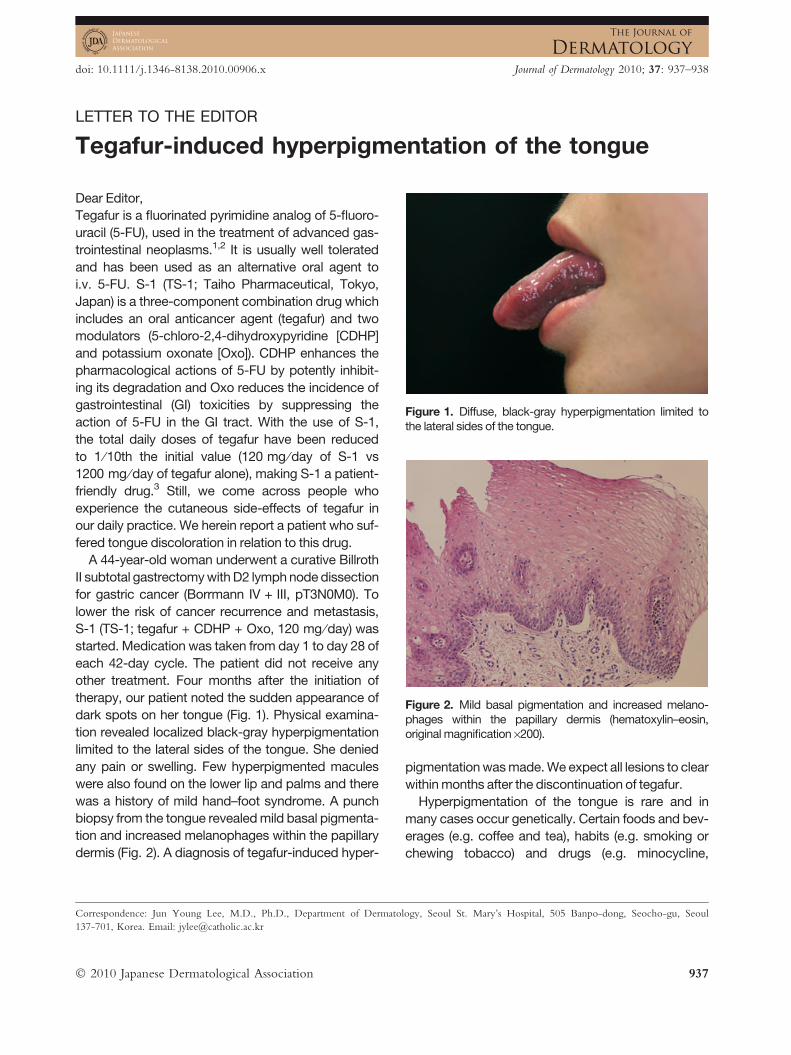
Figure 1. Diffuse, black-gray hyperpigmentation limited tothe lateral sides of the tongue.
Figure 2. Mild basal pigmentation and increased melano-phages within the papillary dermis (hematoxylin–eosin,original magnification ·200).
Dear Editor,
Tegafur is a fluorinated pyrimidine analog of 5-fluoro-
uracil (5-FU), used in the treatment of advanced gas-
trointestinal neoplasms.1,2 It is usually well tolerated
and has been used as an alternative oral agent to
i.v. 5-FU. S-1 (TS-1; Taiho Pharmaceutical, Tokyo,
Japan) is a three-component combination drug which
includes an oral anticancer agent (tegafur) and two
modulators (5-chloro-2,4-dihydroxypyridine [CDHP]
and potassium oxonate [Oxo]). CDHP enhances the
pharmacological actions of 5-FU by potently inhibit-
ing its degradation and Oxo reduces the incidence of
gastrointestinal (GI) toxicities by suppressing the
action of 5-FU in the GI tract. With the use of S-1,
the total daily doses of tegafur have been reduced
to 1 ⁄10th the initial value (120 mg ⁄day of S-1 vs
1200 mg ⁄day of tegafur alone), making S-1 a patient-
friendly drug.3 Still, we come across people who
experience the cutaneous side-effects of tegafur in
our daily practice. We herein report a patient who suf-
fered tongue discoloration in relation to this drug.
A 44-year-old woman underwent a curative Billroth
II subtotal gastrectomy with D2 lymph node dissection
for gastric cancer (Borrmann IV + III, pT3N0M0). To
lower the risk of cancer recurrence and metastasis,
S-1 (TS-1; tegafur + CDHP + Oxo, 120 mg ⁄day) was
started. Medication was taken from day 1 to day 28 of
each 42-day cycle. The patient did not receive any
other treatment. Four months after the initiation of
therapy, our patient noted the sudden appearance of
dark spots on her tongue (Fig. 1). Physical examina-
tion revealed localized black-gray hyperpigmentation
limited to the lateral sides of the tongue. She denied
any pain or swelling. Few hyperpigmented macules
were also found on the lower lip and palms and there
was a history of mild hand–foot syndrome. A punch
biopsy from the tongue revealed mild basal pigmenta-
tion and increased melanophages within the papillary
dermis (Fig. 2). A diagnosis of tegafur-induced hyper-
Correspondence: Jun Young Lee, M.D., Ph.D., Department of Dermatol
137-701, Korea. Email: [email protected]
� 2010 Japanese Dermatological Association
pigmentation was made. We expect all lesions to clear
within months after the discontinuation of tegafur.
Hyperpigmentation of the tongue is rare and in
many cases occur genetically. Certain foods and bev-
erages (e.g. coffee and tea), habits (e.g. smoking or
chewing tobacco) and drugs (e.g. minocycline,
ogy, Seoul St. Mary’s Hospital, 505 Banpo-dong, Seocho-gu, Seoul
937

H.S. Kim et al.
antipsychotics, cytotoxic drugs) are also capable of
inducing tongue discoloration.4 A number of reports
have been made on tegafur-induced mucocutaneous
hyperpigmentation, however, rarely involving the ton-
gue.5 The mechanism of such pigmentation is largely
unknown but post-inflammatory hyperpigmentation
following drug-induced toxicity to keratinocytes has
been suggested as a cause. The abundance of mela-
nophages in our case strongly supports the theory.
Clinicians should be aware of tongue discoloration
associated with tegafur as this drug is being increas-
ingly used in patients with advanced carcinomas.
Like other tegafur-induced cutaneous hyperpigmen-
tation, pigmentation of the tongue is likely to resolve
within months after stopping the drug.5,6
Hei Sung KIM, Young Min PARK,
Hyung Ok KIM, Jun Young LEEDepartment of Dermatology, Seoul St Mary’s Hospital,
The Catholic University of Korea, Seoul, Korea
938
REFERENCES
1 Koizumi W, Kurihara M, Nakano S, Hasegawa K. Phase IIstudy of S-1, a novel oral derivative of 5-fluorouracil,in advanced gastric cancer. Oncology 2000; 58: 191–197.
2 Shirao K, Ohtsu A, Takada H et al. Phase II study of oralS-1 for treatment of metastatic colorectal carcinoma.Cancer 2004; 100: 2355–2361.
3 Shirasaka T. Development history and concept ofan oral anticancer agent S-1 (TS-1� ): its clinical use-fulness and future vistas. Jpn J Clin Oncol 2009; 39:2–15.
4 Scully C. Discolored tongue: a new cause? Br J Dermatol2002; 144: 1293–1294.
5 Rios-Buceta L, Buezo GF, Penas PF, Dauden E, Fernan-dez-Herrera J, Garcia-Diez A. Palmo-plantar erythrodys-aesthesia syndrome and other cutaneous side-effectsafter treatment with tegafur. Acta Derm Venereol 1997;77: 80–81.
6 Llistosella E, Codina A, Alvarez R, Pujol RM, de MorgasJM. Tegafur-induced acral hyperpigmentation. Cutis1991; 48: 205–207.
� 2010 Japanese Dermatological Association