technical approach to laparoscopic examination of the
TRANSCRIPT
Case ReportTechnical Approach to Laparoscopic Examination of the SmallBowel in Gallstone Ileus
Sarah Assali ,1 John Mourany,2 Brendan Jones,2 Lauren Dudas ,2 and Nova Szoka2
1Allegheny Health Network, USA2West Virginia University, USA
Correspondence should be addressed to Sarah Assali; [email protected]
Received 8 June 2020; Revised 11 September 2020; Accepted 29 September 2020; Published 12 October 2020
Academic Editor: Tahsin Colak
Copyright © 2020 Sarah Assali et al. This is an open access article distributed under the Creative Commons Attribution License,which permits unrestricted use, distribution, and reproduction in any medium, provided the original work is properly cited.
Background. Gallstone ileus is an infrequent cause of small bowel obstructions (SBO), accounting for only 0.1-5% of SBOs and 25%of nonstrangulating causes of SBO in the elderly population. There is scant literature available regarding the use of laparoscopy totreat gallstone ileus. Currently, much of the literature available is limited to case reports only. Methods. A complete laparoscopicapproach was utilized to manage a 65-year-old woman with morbid obesity who presented with gallstone ileus. Withregard to our technical approach, we describe the technical approach that facilitates safe laparoscopic examination of theentire small bowel and can be applied to other acute care surgery cases involving small bowel pathology. Results. Thepatient’s postoperative course was complicated by new-onset atrial fibrillation which was treated medically with goodresponse. She was safely discharged on postoperative day 2. Conclusion. Laparoscopy is a feasible option for themanagement of gallstone ileus and can lead to decreased morbidity compared to laparotomy. The technique describedallows for laparoscopic examination of the entire small bowel.
1. Introduction
Gallstone ileus is a condition that results when a biliarycalculus gains entry to the intestinal lumen through abiliary-enteric fistula and causes mechanical bowel obstruc-tion [1, 2]. Most commonly a cholecystoduodenal fistula,the fistula forms as a result of longstanding inflammationfrom cholelithiasis. As the gallstone travels through thesmall bowel, obstruction most commonly occurs in the ter-minal ileum but can be seen at other locations in the gas-trointestinal tract [3]. While 25% to 72% of patients withgallstone ileus have a history of cholelithiasis, only 0.3%to 1.5% of patients with cholelithiasis will develop a gall-stone ileus [1, 4]. Historically, gallstone ileus was thoughtto account for 1% to 5% of all small bowel obstructionsand 25% of nonstrangulating small bowel obstructions inthe elderly population [1, 5]. More recently, a review ofthe Nationwide Inpatient Sample database showed thatgallstone ileus made up 0.095% of all bowel obstructionsand tends to occur with higher prevalence in the elderly,female population [1].
Initial management is directed at fluid resuscitation andresolving electrolyte imbalances followed by surgery [6].Classically, surgery has involved exploratory laparotomywith stone extraction via enterolithotomy [6]. The enterot-omy is created in a longitudinal fashion and closed in a trans-verse fashion to prevent stenosis [7]. More recently,laparoscopy has been shown effective in the treatment of thisdisease [1, 7]. Regardless of operative approach, the remain-der of the bowel should be examined to determine if othergallstones are present. Closure of the cholecystoduodenal fis-tula is not necessary at the initial operation; however, due tothe low prevalence of the disease optimal management of thefistula, it remains uncertain [2, 3, 6].
Recent studies have shown that enterotomy and stoneextraction alone are safe and that enterotomy with fistula clo-sure or bowel resection was associated with higher mortalityrates and greater length of stay [1, 4]. Despite operation,mortality still ranges from 15% to 18% and the process recursin up to 5% to 17% of patients [3, 4, 6].
The purpose of this manuscript is to present the laparo-scopic management of gallstone ileus, including a brief
HindawiCase Reports in SurgeryVolume 2020, Article ID 8852804, 5 pageshttps://doi.org/10.1155/2020/8852804
discussion of the operative technique for laparoscopic exam-ination of the entire length of small bowel in order to demon-strate that this approach can be a safe alternative to opensurgery and may to reduce morbidity, mortality, and hospitallength of stay for this disease.
2. Methods
Written consent was obtained from the patient to use thiscase for educational purposes. The West Virginia Institu-tional Review Board approved this study (Protocol no.2004964936). In this study, we portray the successful use oflaparoscopy in the management of gallstone ileus in the caseof a bariatric patient with multiple comorbidities. The patientwas a 65-year-old woman with a past medical history ofmorbid obesity (BMI 38), obstructive sleep apnea with 2-liter nocturnal oxygen requirement, hypertension, GERD,active tobacco use, and urinary incontinence. Her past sur-gical history included placement of a cholecystostomy tubesix years prior, with subsequent removal. The patient wasan active tobacco user, smoking 1 pack per day, but didnot use alcohol or recreational drugs. She had no knowndrug allergies and was on a single blood pressure medica-tion, a proton pump inhibitor for reflux disease, andDitropan for urinary incontinence.
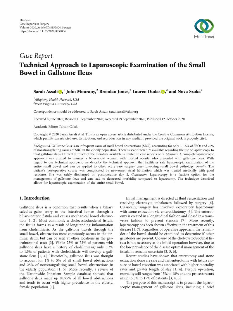
The patient was transferred to our facility from an out-side hospital with a 3-day history of abdominal pain, nausea,vomiting, and obstipation. She was found to have a leukocy-tosis of 20 k/mcL and CT scan findings consistent with pneu-mobilia, a small bowel obstruction, and an ectopic gallstone(Figure 1). She was consented for surgery and taken urgentlyfor operative exploration. The operation was completed in anentirely laparoscopic fashion.
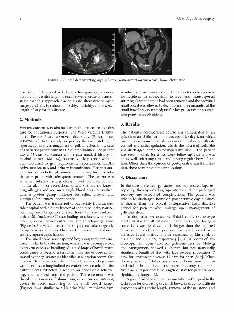
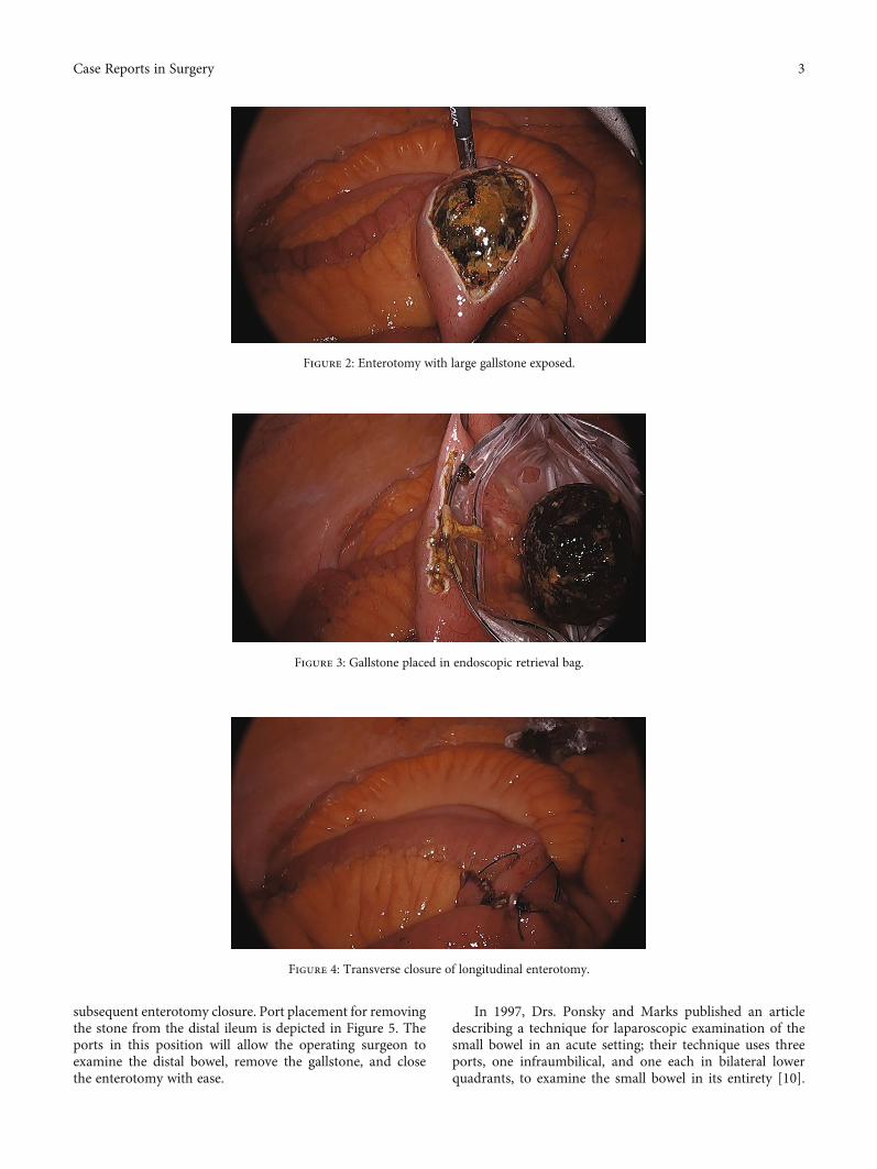
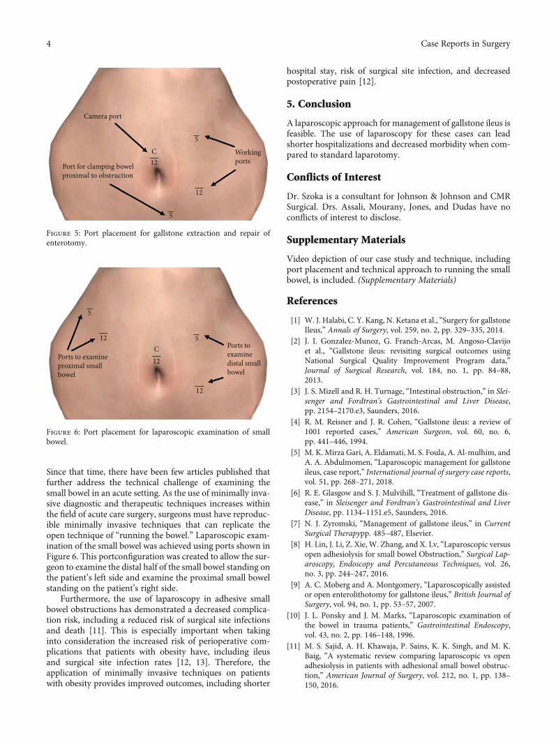
The small bowel was inspected beginning at the terminalileum, distal to the obstruction, where it was decompressed,to prevent excessive handling of dilated loops of bowel whichcould cause iatrogenic enterotomy. The site of obstructioncaused by the gallstone was identified at a location several feetproximal to the terminal ileum. Once the obstructing stonewas identified, a longitudinal enterotomy was made and thegallstone was extracted, placed in an endoscopic retrievalbag, and removed from the patient. The enterotomy wasclosed in a transverse fashion using an endoscopic suturingdevice to avoid narrowing of the small bowel lumen(Figures 2–4), similar to a Heineke-Mikulicz pyloroplasty.
A suturing device was used due to its shorter learning curvefor residents in comparison to free-hand intracorporealsuturing. Once the stone had been removed and the proximalsmall bowel was allowed to decompress, the remainder of thesmall bowel was examined; no further gallstones or obstruc-tion points were identified.
3. Results
The patient’s postoperative course was complicated by anepisode of atrial fibrillation on postoperative day 1, for whichcardiology was consulted. She was treated medically with ratecontrol and anticoagulation, which she tolerated well. Shewas discharged home on postoperative day 2. The patientwas seen in clinic for a two-week follow-up visit and wasdoing well, tolerating a diet, and having regular bowel func-tion. Other than the episode of postoperative atrial fibrilla-tion, there were no other complications.
4. Discussion
In the case presented, gallstone ileus was treated laparos-copically, thereby avoiding laparotomy and the prolongedrecovery and associated complications. The patient wasable to be discharged home on postoperative day 2, whichis shorter than the typical postoperative hospitalizationperiod for patients who undergo open management ofgallstone ileus.
In the series presented by Halabi et al., the averagelength of stay for all patients undergoing surgery for gall-stone ileus was 12 days; this is longer than the reportedlaparoscopic and open postoperative stays noted withadhesive bowel obstructions as measured by Lin et al. of6:4 ± 2:1 and 7:2 ± 2:9, respectively [1, 8]. A review of lap-aroscopic and open cases for gallstone ileus by Mobergand Montgomery showed a shorter, but not statisticallysignificant, length of stay with laparoscopic procedures, 7days for laparoscopic versus 10 days for open [8, 9]. Whencholecystectomy, fistula closure, and/or bowel resection areundertaken in addition to the enterolithotomy, the opera-tive time and postoperative length of stay for patients weresignificantly longer [2].
A great deal of consideration was taken with regard to thetechnique for evaluating the small bowel in order to facilitateinspection of its entire length, removal of the gallstone, and
Figure 1: CT scan demonstrating large gallstone (white arrow) causing a small bowel obstruction.
2 Case Reports in Surgery
subsequent enterotomy closure. Port placement for removingthe stone from the distal ileum is depicted in Figure 5. Theports in this position will allow the operating surgeon toexamine the distal bowel, remove the gallstone, and closethe enterotomy with ease.
In 1997, Drs. Ponsky and Marks published an articledescribing a technique for laparoscopic examination of thesmall bowel in an acute setting; their technique uses threeports, one infraumbilical, and one each in bilateral lowerquadrants, to examine the small bowel in its entirety [10].
Figure 2: Enterotomy with large gallstone exposed.
Figure 3: Gallstone placed in endoscopic retrieval bag.
Figure 4: Transverse closure of longitudinal enterotomy.
3Case Reports in Surgery
Since that time, there have been few articles published thatfurther address the technical challenge of examining thesmall bowel in an acute setting. As the use of minimally inva-sive diagnostic and therapeutic techniques increases withinthe field of acute care surgery, surgeons must have reproduc-ible minimally invasive techniques that can replicate theopen technique of “running the bowel.” Laparoscopic exam-ination of the small bowel was achieved using ports shown inFigure 6. This portconfiguration was created to allow the sur-geon to examine the distal half of the small bowel standing onthe patient’s left side and examine the proximal small bowelstanding on the patient’s right side.
Furthermore, the use of laparoscopy in adhesive smallbowel obstructions has demonstrated a decreased complica-tion risk, including a reduced risk of surgical site infectionsand death [11]. This is especially important when takinginto consideration the increased risk of perioperative com-plications that patients with obesity have, including ileusand surgical site infection rates [12, 13]. Therefore, theapplication of minimally invasive techniques on patientswith obesity provides improved outcomes, including shorter
hospital stay, risk of surgical site infection, and decreasedpostoperative pain [12].
5. Conclusion
A laparoscopic approach for management of gallstone ileus isfeasible. The use of laparoscopy for these cases can leadshorter hospitalizations and decreased morbidity when com-pared to standard laparotomy.
Conflicts of Interest
Dr. Szoka is a consultant for Johnson & Johnson and CMRSurgical. Drs. Assali, Mourany, Jones, and Dudas have noconflicts of interest to disclose.
Supplementary Materials
Video depiction of our case study and technique, includingport placement and technical approach to running the smallbowel, is included. (Supplementary Materials)
References
[1] W. J. Halabi, C. Y. Kang, N. Ketana et al., “Surgery for gallstoneIleus,” Annals of Surgery, vol. 259, no. 2, pp. 329–335, 2014.
[2] J. I. Gonzalez-Munoz, G. Franch-Arcas, M. Angoso-Clavijoet al., “Gallstone ileus: revisiting surgical outcomes usingNational Surgical Quality Improvement Program data,”Journal of Surgical Research, vol. 184, no. 1, pp. 84–88,2013.
[3] J. S. Mizell and R. H. Turnage, “Intestinal obstruction,” in Slei-senger and Fordtran’s Gastrointestinal and Liver Disease,pp. 2154–2170.e3, Saunders, 2016.
[4] R. M. Reisner and J. R. Cohen, “Gallstone ileus: a review of1001 reported cases,” American Surgeon, vol. 60, no. 6,pp. 441–446, 1994.
[5] M. K. Mirza Gari, A. Eldamati, M. S. Foula, A. Al-mulhim, andA. A. Abdulmomen, “Laparoscopic management for gallstoneileus, case report,” International journal of surgery case reports,vol. 51, pp. 268–271, 2018.
[6] R. E. Glasgow and S. J. Mulvihill, “Treatment of gallstone dis-ease,” in Sleisenger and Fordtran’s Gastrointestinal and LiverDisease, pp. 1134–1151.e5, Saunders, 2016.
[7] N. J. Zyromski, “Management of gallstone ileus,” in CurrentSurgical Therapypp. 485–487, Elsevier.
[8] H. Lin, J. Li, Z. Xie, W. Zhang, and X. Lv, “Laparoscopic versusopen adhesiolysis for small bowel Obstruction,” Surgical Lap-aroscopy, Endoscopy and Percutaneous Techniques, vol. 26,no. 3, pp. 244–247, 2016.
[9] A. C. Moberg and A. Montgomery, “Laparoscopically assistedor open enterolithotomy for gallstone ileus,” British Journal ofSurgery, vol. 94, no. 1, pp. 53–57, 2007.
[10] J. L. Ponsky and J. M. Marks, “Laparoscopic examination ofthe bowel in trauma patients,” Gastrointestinal Endoscopy,vol. 43, no. 2, pp. 146–148, 1996.
[11] M. S. Sajid, A. H. Khawaja, P. Sains, K. K. Singh, and M. K.Baig, “A systematic review comparing laparoscopic vs openadhesiolysis in patients with adhesional small bowel obstruc-tion,” American Journal of Surgery, vol. 212, no. 1, pp. 138–150, 2016.
Camera port
Port for clamping bowelproximal to obstruction
Workingports
12
5
C5
12
Figure 5: Port placement for gallstone extraction and repair ofenterotomy.
Ports toexaminedistal smallbowel
Ports to examineproximal smallbowel
5
512
12C
12
Figure 6: Port placement for laparoscopic examination of smallbowel.
4 Case Reports in Surgery

[12] T. Makino, P. J. Shukla, F. Rubino, and J. W. Milsom, “Theimpact of obesity on perioperative outcomes after laparoscopiccolorectal resection,” Annals of Surgery, vol. 255, no. 2,pp. 228–236, 2012.
[13] S. Tsujinaka, F. Konishi, Y. J. Kawamura et al., “Visceral obe-sity predicts surgical outcomes after laparoscopic colectomyfor sigmoid colon cancer,” Diseases of the Colon and Rectum,vol. 51, no. 12, pp. 1757–1767, 2008.
5Case Reports in Surgery