laparoscopic approach as an alternative option in...
TRANSCRIPT

8
Laparoscopic Approach as an Alternative Option in Treatment of Pediatric Inguinal Hernia
B. Haluk Güvenç Professor of Pediatric Surgery,
Turkey
1. Introduction
Inguinal hernia is the most common and prominent surgical entity among all congenital anomalies, constituting more than 15% of total pediatric surgical cases. Inguinal hernia is directly linked to the descent of the developing gonads in children. A patent processus vaginalis, which precedes an inguinal hernia, is a diverticulum of the peritoneum, formed during gonadal descent around the 12th week of gestation. This blind-ending, celomic epithelium layered sac is generally obliterated after the testicular migration is completed. A failure during the obliteration process may develop an inguinal hernia or communicating hydrocele(1-3).
In children, the bowel follows the course of the processus vaginalis through the internal
ring, lateral to the inferior epigastric vessels forming an indirect inguinal hernia. The
diagnosis of hernia can be made during physical examination when organs such as bowel or
ovary protrude into the sac, or can be based on a classic description by the parents or a
referring physician. On the other hand, a communicating hydrocele should also be
considered as a hernia, and repair is indicated regardless of the age. Hydroceles that
develop after birth are more likely to be associated with a patent processus vaginalis that is
less likely to close(3).
The surgical repair of a pediatric inguinal hernia should take place in a timely manner to eliminate any risk of incarceration. The classic open repair is performed through a skin incision, made in the inguinal crease overlying the internal ring. Scarpa’s fascia and the external oblique aponeurosis are opened. In males, the cremasteric fibers are bluntly dissected until the sac can be seen. The sac is then gently separated from the cord structures, divided, dissected to the level of the internal ring, and ligated at this level. In females, the sac is dissected to the level of the internal ring and ligated. Reconstruction of the inguinal ring is not routinely necessary(1, 2).
Data from surgical practice and autopsy studies suggest a strong likelihood of bilateralism
in children with a unilateral presentation. The increase in cost, the unpleasant experience of
additional operative risk and mental trauma for the patient accompanied by the anxiety of
the parents that may result from a metachronous hernia, have led surgeons to adopt a policy
of routine bilateral exploration (4, 5). Bilateral inguinal exploration may be justified in patients
known to be at higher risk for bilateral hernias or at increased operative risk, such as
www.intechopen.com
Advances in Laparoscopic Surgery
120
premature infants or patients with bladder extrophy, ascites, cystic fibrosis,
ventriculoperitoneal shunts, peritoneal dialysis catheters, or connective tissue disorders. In
the meantime, due to fact that the rate of patency decreases with age and the accumulation
of data showing a higher risk of damage to the vas deferens and gonad during negative
exploration, proponents had to restrict bilateral exploration according to the age and sex of
the child and the presenting side(6-15). The author believes that a ‘‘prophylactic’’ contralateral
exploration in children presenting with unilateral hernia is unjustified.
2. The search for contralateral patent processus vaginalis
The introduction of laparoscopic intervention is a milestone in understanding the concept of contemporary pediatric inguinal hernia repair. The debate amongst the proponents and opponents of bilateral inguinal exploration in children, is about to end. The accumulated data concerning the presence and fate of patent processus vaginalis (PPV) about the incidence, relation to age or presenting side and timing of obliteration will be of historic importance in the near future. The true incidence of bilateral hernia however, is still unclear.
Searching through the published data, we may see that the given incidence of a contralateral inguinal hernia is as high as 57% to 85% according to the proponents of bilateral exploration (16, 17). The incidence of a PPV on the other hand, is reported as 80% in the first 2 months of life, steadily decreasing over the next 2 years(18, 19). Nakayama and Rowe stating that in about 60% of infants, a contralateral PPV may accompany a clinically presenting unilateral inguinal hernia report a similar rough estimation. One-third of these anatomic patencies is expected to be obliterated within 2 years, one-third is expected to develop into a subsequent hernia, and the remaining one-third will be clinically silent (20, 21). Published series concerning adult patients does support the estimated incidence. Adult patients without a clinical hernia, present with a surprisingly high number (12 %) of true indirect PPVs, with a defect of around 3 cm seen through the telescope(22-24). Again, the controversy remains; some authors (23) do not recommend prophylactic repair of such an asymptomatic defect, while others do recommend simultaneous repair(22).
Studies investigating the most suitable method in evaluating the contralateral side have been an important topic for many years, since the reported incidence of a developing contralateral hernia is as high as 30%(25). A rather recent meta-analysis by Miltenburg et al. discloses the rate of a metachronous hernia as 7 - 11 %, covering over 13.000 children undergoing unilateral repair(26). Consideration of all processus vaginalis’ as a future clinically significant hernia may attribute to the discrepancy between figures. Contemporary use of ultrasound has revolutionized near precise preoperative diagnosis of a contralateral inguinal hernia when compared to a number of (now historical) tests such as herniography, the Goldstein procedure, and use of Bakes dilators(27). This precision though has been mastered through hard-earned lessons, in search for a less invasive method following introduction of diagnostic laparoscopy in the early 1990’s(28–34). Diagnostic laparoscopic evaluation of the contralateral inguinal ring is regarded as a reliable method, with a sensitivity of 99.4% and a specificity of 99.5%(35). The method is accurate, fast, safe, and effective in reducing negative explorations, avoiding the small risk of incarceration of a metachronous hernia as well as the cost and anxiety of a second operation; it is easily learned and requires no additional capital outlay in hospitals already performing laparoscopic surgery.
www.intechopen.com
Laparoscopic Approach as an Alternative Option in Treatment of Pediatric Inguinal Hernia
121
2.1 Diagnostic laparoscopy
The decisive step of performing a diagnostic laparoscopy aids the surgeon in solving the dilemma, the postoperative complications from an unnecessary inguinal exploration or a missed metachronous hernia. It is a promising step taken forward with respect to the surgical precision achieved through enhanced visualization, magnification and ability to limit collateral damage by minimizing invasion. An additional benefit is the ability to detect all forms of (indirect, direct, combined, recurrent, and incarcerated) hernias(19).
The suggested techniques of diagnostic laparoscopy for evaluating a contralateral patency
mainly include three primary methods. The open infraumbilical approach has the advantage
of permitting a direct view of the internal ring and better correlation of the two sides(28, 30- 33).
To avoid a second incision and eliminate the risk of intra-abdominal visceral injury
associated with trocar placement, the already-opened ipsilateral hernia sac is used in the
second method, referred to as the “nonpuncture” technique(29, 30, 32-34). This technique
requires understanding of the view through an angled telescope, since a peritoneal veil or
the median umbilical fold may obscure the direct view in some patients (36, 37). This
examination is even harder in small babies, as the distance between the two internal rings is
very short. The placement of the trocar is safe, but it is not possible to proceed when the
hernia sac is too narrow or friable. The third technique is the “inline” method, in which a
telescope is introduced through a 14- or 16-gauge catheter placed in line with the
contralateral internal ring and the abdomen is insufflated through the ipsilateral sac. This
technique has the advantage of providing a direct view and enables measurement of the
depth of any processus vaginalis(38, 39). As for measuring the depth, a 2.7 mm 30° angled
Hopkins rod lens is inserted to its maximum depth in the internal ring, and then the shaft of
the scope where it entered the port is marked. A second mark is made after withdrawing the
scope until the tip is at the internal ring. One can proceed with laparoscopy with this
method when the ipsilateral hernia sac is too narrow or friable to insert a trocar. This
technique means a second wound and may carry a risk of injury to the bowel and
abdominal wall vessels. Depending on the chosen technique, the surgeon may utilize a
range of rigid or flexible telescopes from 1.2 mm to 5 mm with an optic range of 0° to 120°.
2.1.1 The surgical method
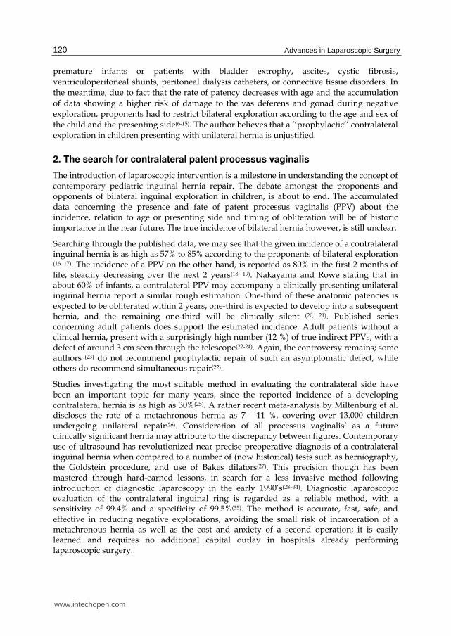
The procedure is performed under general anesthesia. The patient is placed in the supine position on the operating room table, with the abdomen and groin sterilely prepped. The stomach is emptied with a suction catheter and the bladder using Crede maneuver, where older children are asked to urinate prior to entering the operating room. The ipsilateral hernia sac is reached through a skin-crease incision and simply dissected free from the adjacent tissue, the vessels and the spermatic cord and traced up to the internal ring. A reusable trocar is inserted through a longitudinal cut on the sac and secured in place with a suture (Figure 1).
The patient is then placed in the Trendelenburg position and the abdomen insufflated with CO2, preferably at a low flow rate (1 l/min) to a maximum pressure of 8–10 mm Hg. The patency of the contralateral inguinal ring is assessed by the help of a 30-70°, 5 mm laparoscope. The positioning of the contralateral inguinal ring lies laterally to the lateral umbilical fold. The vas deferens in the male and the round ligament in female are traced
www.intechopen.com
Advances in Laparoscopic Surgery
122
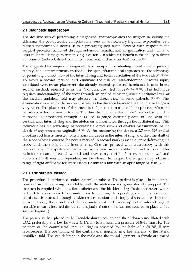
over the pelvic brim to reach the internal inguinal ring. The presence of a significant peritoneal opening, the absence of an identifiable termination of the peritoneal sac, visualization of bubbles internally with external pressure, a hidden opening under a veil of peritoneum, a probing depth of 1.5 cm or concentric peritoneal rings distal to the internal ring are regarded as positive findings of patency (Figure 2).
Fig. 1. Transinguinal approach to the contralateral side in unilateral hernia repair. Following insertion of a reusable trocar through the ipsilateral hernial sac, the port is secured in place with a suture.
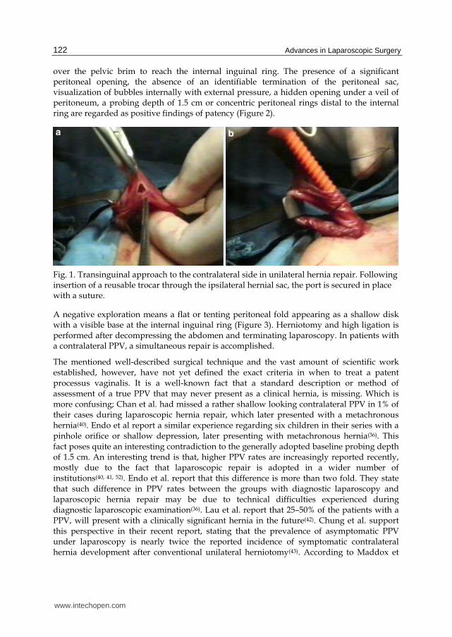
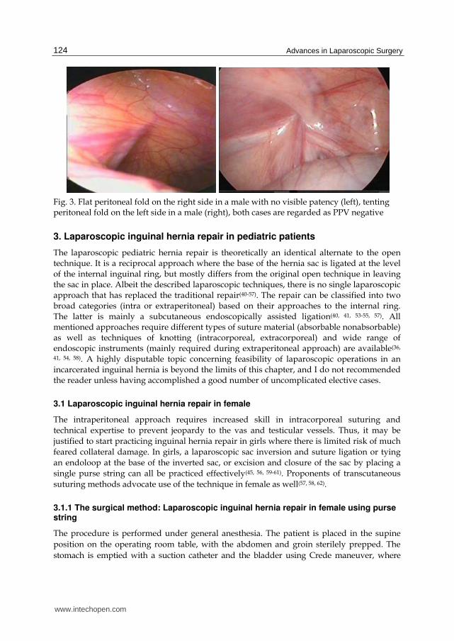
A negative exploration means a flat or tenting peritoneal fold appearing as a shallow disk with a visible base at the internal inguinal ring (Figure 3). Herniotomy and high ligation is performed after decompressing the abdomen and terminating laparoscopy. In patients with a contralateral PPV, a simultaneous repair is accomplished.
The mentioned well-described surgical technique and the vast amount of scientific work established, however, have not yet defined the exact criteria in when to treat a patent processus vaginalis. It is a well-known fact that a standard description or method of assessment of a true PPV that may never present as a clinical hernia, is missing. Which is more confusing; Chan et al. had missed a rather shallow looking contralateral PPV in 1% of their cases during laparoscopic hernia repair, which later presented with a metachronous hernia(40). Endo et al report a similar experience regarding six children in their series with a pinhole orifice or shallow depression, later presenting with metachronous hernia(36). This fact poses quite an interesting contradiction to the generally adopted baseline probing depth of 1.5 cm. An interesting trend is that, higher PPV rates are increasingly reported recently, mostly due to the fact that laparoscopic repair is adopted in a wider number of institutions(40, 41, 52). Endo et al. report that this difference is more than two fold. They state that such difference in PPV rates between the groups with diagnostic laparoscopy and laparoscopic hernia repair may be due to technical difficulties experienced during diagnostic laparoscopic examination(36). Lau et al. report that 25–50% of the patients with a PPV, will present with a clinically significant hernia in the future(42). Chung et al. support this perspective in their recent report, stating that the prevalence of asymptomatic PPV under laparoscopy is nearly twice the reported incidence of symptomatic contralateral hernia development after conventional unilateral herniotomy(43). According to Maddox et
www.intechopen.com
Laparoscopic Approach as an Alternative Option in Treatment of Pediatric Inguinal Hernia
123
al., 6.8% developed a metachronous hernia within a period of 53 months, amongst their study group presenting with 47.5% PPV(44). This figure matches up closely with Miltenburg et al.’s meta-analysis(26).
Fig. 2. Significant peritoneal opening on left side in female (upper left), visualization of bubbles on the left side in a male (upper right), obscure inguinal hernia under peritoneal veil on the right side (lower left), clearly visible peritoneal opening after drawing the veil away (lower right)
Apart from the discrepancy regarding the figures, diagnostic laparoscopy has certainly
achieved its goal. It is obvious that we are sparing an increasing number of children less
than 1 year of age, from routine exploration of their contralateral side. In other words, this
simple examination prevents many unnecessary explorations. On the contrary, an increasing
number of children over two years of age with a PPV were diagnosed and operated. Again,
it is certain that these children will not present with a future hernia in the contralateral side.
www.intechopen.com
Advances in Laparoscopic Surgery
124
Fig. 3. Flat peritoneal fold on the right side in a male with no visible patency (left), tenting peritoneal fold on the left side in a male (right), both cases are regarded as PPV negative
3. Laparoscopic inguinal hernia repair in pediatric patients
The laparoscopic pediatric hernia repair is theoretically an identical alternate to the open technique. It is a reciprocal approach where the base of the hernia sac is ligated at the level of the internal inguinal ring, but mostly differs from the original open technique in leaving the sac in place. Albeit the described laparoscopic techniques, there is no single laparoscopic approach that has replaced the traditional repair(40-57). The repair can be classified into two broad categories (intra or extraperitoneal) based on their approaches to the internal ring. The latter is mainly a subcutaneous endoscopically assisted ligation(40, 41, 53-55, 57). All mentioned approaches require different types of suture material (absorbable nonabsorbable) as well as techniques of knotting (intracorporeal, extracorporeal) and wide range of endoscopic instruments (mainly required during extraperitoneal approach) are available(36,
41, 54, 58). A highly disputable topic concerning feasibility of laparoscopic operations in an incarcerated inguinal hernia is beyond the limits of this chapter, and I do not recommended the reader unless having accomplished a good number of uncomplicated elective cases.
3.1 Laparoscopic inguinal hernia repair in female
The intraperitoneal approach requires increased skill in intracorporeal suturing and
technical expertise to prevent jeopardy to the vas and testicular vessels. Thus, it may be
justified to start practicing inguinal hernia repair in girls where there is limited risk of much
feared collateral damage. In girls, a laparoscopic sac inversion and suture ligation or tying
an endoloop at the base of the inverted sac, or excision and closure of the sac by placing a
single purse string can all be practiced effectively(45, 56, 59-61). Proponents of transcutaneous
suturing methods advocate use of the technique in female as well(57, 58, 62).
3.1.1 The surgical method: Laparoscopic inguinal hernia repair in female using purse string
The procedure is performed under general anesthesia. The patient is placed in the supine
position on the operating room table, with the abdomen and groin sterilely prepped. The
stomach is emptied with a suction catheter and the bladder using Crede maneuver, where
www.intechopen.com
Laparoscopic Approach as an Alternative Option in Treatment of Pediatric Inguinal Hernia
125
older children are asked to urinate prior to entering the operating room. An infraumbilical
incision (open-Hasson technique) or a Veress needle is used in obtaining an abdominal access.
Pneumoperitoneum is established with carbon dioxide according to appropriate age (8–
10mmHg). Initially the abdomen is visualized using a 5-mm, 30° scope with the operating
table positioned in moderate reverse Trendelenburg position. The pelvis is inspected for
anatomical variations of the uterus, ovaries and adnex, and the inguinal rings are evaluated.
Preferably two 2.7-mm working instruments are introduced through two lower abdominal
stab incisions with (or without) the use of ports following detailed anatomic investigation. By
the help of two grasping dissectors, the tip of the hernia sac is grasped and gently inverted into
the abdominal cavity through the inguinal canal. One must be gentle with this blunt traction
maneuver since brutal disruption of the distal attachments of the round ligament to the labia
may lead to edema formation or extraperitoneal hemorrhage, which may render suturing of
the neck of the inverted sac. When near or in the hernia sac, the fallopian tube and/or ovary
are freed by a combination of blunt and sharp dissection. After confirming that the sac is
relatively free of its surrounding attachments it is released back in place to continue with the
purse string. The needle and thread are passed into the abdomen directly through the
abdominal wall. By the help of a needle holder and grasping dissector, a 2-3/0 nonabsorbable
monofilament purse suture is sewn just around the internal ring. In doing this, the suture must
not cut deep in the surrounding tissue, to enable a strong and even strangling force on the
peritoneal covering of the neck of the sac. The final bite is passed through the neck of the
inverted sac. The suture is then secured at the base of the inverted hernia sac. It is advised to
include sac resection after completion of suturing, since most published studies agree upon the
fact that local peritoneal healing aids in preventing recurrences. A similar repair is applied to
the contralateral side when indicated using the same incisions. Operation is terminated by
removing all instruments under direct vision. The fascia and skin are closed with single Vicryl
stitches. Stab incisions may be closed and dressed with SteristripsTM (3M; St. Paul, MN). A
caudal block may additionally be used regarding parental consent. Otherwise, it is preferable
to infiltrate all instrument or port sites prior to skin closure using local anesthetics. (All
incisions are infiltrated with 0.25% or 0.5% bupivacaine solution) (Figures 4 & 5)
3.1.2 The surgical method: Laparoscopic inguinal hernia repair in female using endoloop
The procedure is almost identical to the method as previously described. High ligation of the
inverted hernia may also be accomplished by the help of an Endoloop (Ethicon Inc.,
Somerville, NJ) which is completed by cinching down to the level of internal ring (Figure 6).
According to published reports the average operating time overall is less than 40 minutes for
bilateral repair, which is prolonged in premature infants and cases presenting with a sliding
component such as incarcerated ovary. It is also reported that the simple use of endoloop may
not be safe in patients presenting with large hernias and may require closure of the internal
ring using laparoscopic intracorporeal suturing(56, 59). Additional benefits of this procedure
include diagnosis of androgen insensitivity and other dysgenic situations(45, 59, 60).
The protuberant mass of hernia sac ‘rosebud’ formed by the laparoscopic inversion and
ligation method, is sonographically visible in all cases early after the procedure, has a
characteristic appearance and gradually involutes with time(61).
www.intechopen.com
Advances in Laparoscopic Surgery
126
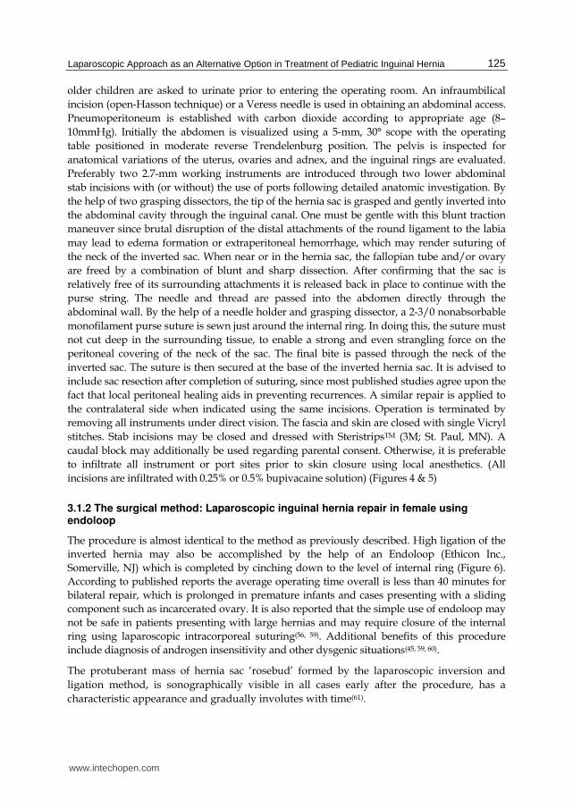
Fig. 4. Laparoscopic female hernia repair on the right side. Following placement of a purse suture around the internal ring, the final bite is passed through the neck of the inverted sac. The suture is than secured at the base.
www.intechopen.com

Laparoscopic Approach as an Alternative Option in Treatment of Pediatric Inguinal Hernia
127
Fig. 5. Left inguinal hernia repair. Grasper is holding the left ovary (Top, left). During initial laparoscopic exploration, a PPV was found on the right side hidden under a peritoneal veil (Top, right). The operation was finalized as a bilateral repair. Looking at the result (bottom, right), we may speculate that this patient would have later come with a metachronous hernia, after a classic left hernia repair.
www.intechopen.com
Advances in Laparoscopic Surgery
128
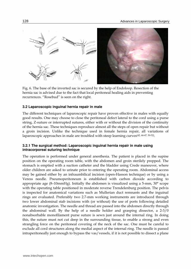
Fig. 6. The base of the inverted sac is secured by the help of Endoloop. Resection of the hernia sac is advised due to the fact that local peritoneal healing aids in preventing recurrences. “Rosebud” is seen on the right.
3.2 Laparoscopic inguinal hernia repair in male
The different techniques of laparoscopic repair have proven effective in males with equally good results. One may choose to close the peritoneal defect lateral to the cord using a purse string, Z-suture or interrupted sutures, either with or without the division of the continuity of the hernia sac. These techniques reproduce almost all the steps of open repair but without a groin incision. Unlike the technique used in female hernia repair, all variations of laparoscopic approaches in male are troubled with steep learning curves(42, 44-47, 50-52).
3.2.1 The surgical method: Laparoscopic inguinal hernia repair in male using intracorporeal suturing technique
The operation is performed under general anesthesia. The patient is placed in the supine
position on the operating room table, with the abdomen and groin sterilely prepped. The
stomach is emptied with a suction catheter and the bladder using Crede maneuver, where
older children are asked to urinate prior to entering the operating room. Abdominal access
may be gained either by an infraumbilical incision (open-Hasson technique) or by using a
Veress needle. Pneumoperitoneum is established with carbon dioxide according to
appropriate age (8–10mmHg). Initially the abdomen is visualized using a 5-mm, 30° scope
with the operating table positioned in moderate reverse Trendelenburg position. The pelvis
is inspected for anatomical variations such as Mullerian duct remnants and the inguinal
rings are evaluated. Preferably two 2.7-mm working instruments are introduced through
two lower abdominal stab incisions with (or without) the use of ports following detailed
anatomic investigation. The needle and thread are passed into the abdomen directly through
the abdominal wall. By the help of a needle holder and grasping dissector, a 2-3/0
nonabsorbable monofilament purse suture is sewn just around the internal ring. In doing
this, the suture must not cut deep in the surrounding tissue, to enable a strong and even
strangling force on the peritoneal covering of the neck of the sac. One must be careful to
exclude all cord structures along the medial aspect of the internal ring. The needle is passed
intraperitoneally just enough to bypass the vas/vessels, if it is not possible to dissect a plane
www.intechopen.com
Laparoscopic Approach as an Alternative Option in Treatment of Pediatric Inguinal Hernia
129
between these structures. The suture is then secured at the base of the internal ring (Figures
7 & 8). Some authors advise to include semi circumferential sac incision on the antero-lateral
aspect of the inguinal ring just distal to the purse string to aid in preventing recurrences.
Operation is terminated by removing all instruments under direct vision. The fascia and
skin are closed with single Vicryl stitches. Stab incisions may be closed and dressed with
SteristripsTM (3M; St. Paul, MN). A caudal block may additionally be used regarding
parental consent. Otherwise, it is preferable to infiltrate all instrument or port sites prior to
skin closure using local anesthetics. (All incisions are infiltrated with 0.25% or 0.5%
bupivacaine solution)
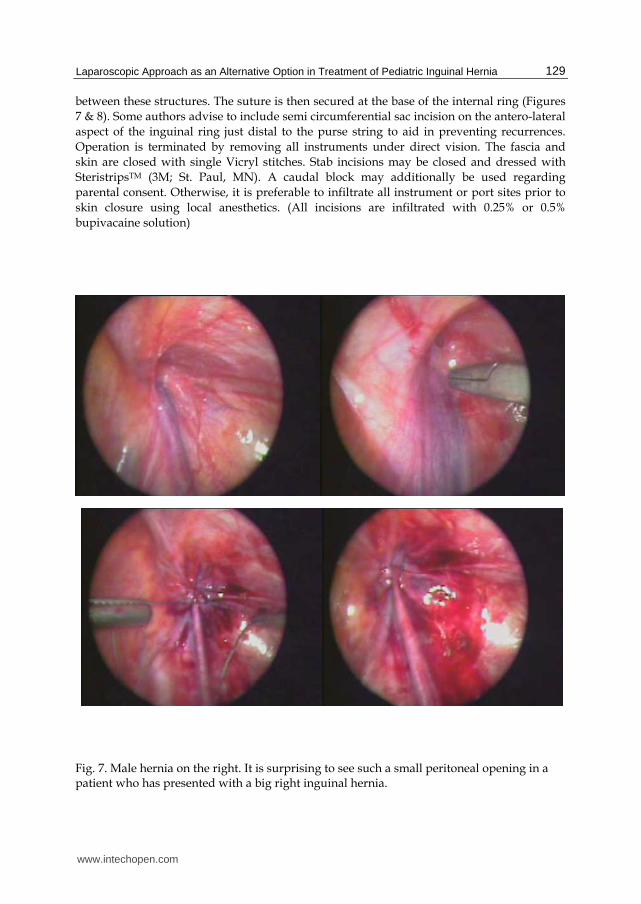
Fig. 7. Male hernia on the right. It is surprising to see such a small peritoneal opening in a patient who has presented with a big right inguinal hernia.
www.intechopen.com
Advances in Laparoscopic Surgery
130
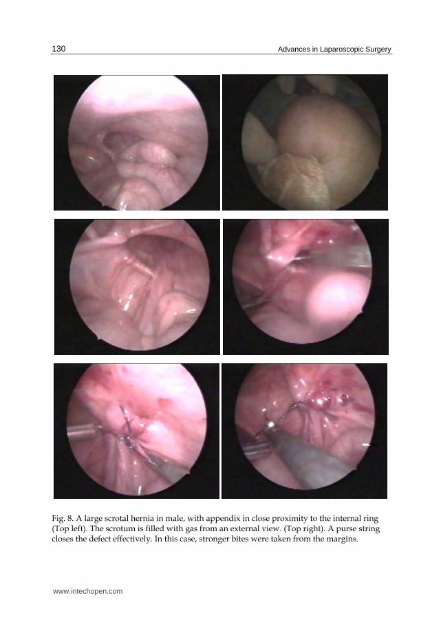
Fig. 8. A large scrotal hernia in male, with appendix in close proximity to the internal ring (Top left). The scrotum is filled with gas from an external view. (Top right). A purse string closes the defect effectively. In this case, stronger bites were taken from the margins.
www.intechopen.com
Laparoscopic Approach as an Alternative Option in Treatment of Pediatric Inguinal Hernia
131
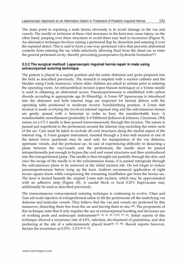
The main point in repairing a male hernia obviously is to avoid damage to the vas and vessels. The needle or inclusion of these vital structures in the knot may cause injury; on the other hand, jumping over these structures to avoid them may lead to recurrence (Figure 9). An alternative technique involves raising a peritoneal flap by dissection and suturing it over the repaired defect. This is said to form a one-way peritoneal valve that prevents abdominal contents from entering the sac while selectively allowing fluid from the distal sac to enter the general peritoneal cavity, thereby preventing postoperative hydrocele formation(63).
3.2.2 The surgical method: Laparoscopic inguinal hernia repair in male using extracorporeal suturing technique
The patient is placed in a supine position and the entire abdomen and groin prepared into the field as described previously. The stomach is emptied with a suction catheter and the bladder using Crede maneuver, where older children are asked to urinate prior to entering the operating room. An infraumbilical incision (open-Hasson technique) or a Veress needle is used in obtaining an abdominal access. Pneumoperitoneum is established with carbon dioxide according to appropriate age (8–10mmHg). A 5-mm 30° laparoscope is introduced into the abdomen and both internal rings are inspected for hernial defects with the operating table positioned in moderate reverse Trendelenburg position. A 2-mm stab incision is made overlying the involved internal inguinal ring and the subcutaneous tissues are gently spread with a hemostat in order to bury the nonabsorbable knot. A nonabsorbable monofilament (preferably 2–0 Ethibond (Johnson & Johnson, Cincinnati, OH) suture on a CT-1 needle is then passed transcutaneously through this incision. The suture is passed just superficial to the peritoneum around the internal ring encircling the entire neck of the sac. Care must be taken to exclude all cord structures along the medial aspect of the internal ring. A 3-mm grasper instrument, inserted through a 2-mm stab incision in one of the lateral lower quadrants may be used only for manipulation of the vas deferens, spermatic vessels, and the peritoneal sac. In case of experiencing difficulty in dissecting a plane between the vas/vessels and the peritoneum, the needle must be passed intraperitoneally just enough to bypass the cord and vessel structures and then reintroduced into the extraperitoneal plane. The needle is then brought out partially through the skin; and once the swage of the needle is in the subcutaneous tissue, it is passed retrograde through the subcutaneous plane to be removed at the initial incision site. Do not forget to reduce pneumoperitoneum before tying up the knot. Authors recommend application of eight secure square knots while compressing the remaining insufflation gas from the hernia sac. The knot is buried beneath the original 2-mm stab incision, which may be approximated with an adhesive strip (Figure 10). A caudal block or local 0.25% bupivacaine may additionally be used as described previously.
The transcutaneous extracorporeal suturing technique is continuing to evolve. Chan and Tam advocate injection of extraperitoneal saline to lift the peritoneum off the underlying vas deferens and testicular vessels. They believe that the vas and vessels are protected by this maneuver, dissecting them free from the sac and leaving them in situ (49). The proponents of this technique state that it only requires the use of extracorporeal knotting and decreases use of working ports and endoscopic instruments(36, 40, 41, 49, 53-55, 57, 62). Initial reports of this technique showed a recurrence rate of 4.8%, infection, development of granuloma, and skin puckering at the site of a subcutaneously placed knot(36, 53, 58). Recent reports however, declare the recurrence as 0.35%- 1.5%(40, 41, 62).
www.intechopen.com
Advances in Laparoscopic Surgery
132
Fig. 9. Male inguinal hernia repair using Z suture technique. Small defect to the lateral needs additional suture (Middle left). Needle holder pointing the weak point where vas and vessels are (Middle right). Peritoneal fold to the left is used to cover the defect with additional suturing (Lower left).
www.intechopen.com
Laparoscopic Approach as an Alternative Option in Treatment of Pediatric Inguinal Hernia
133
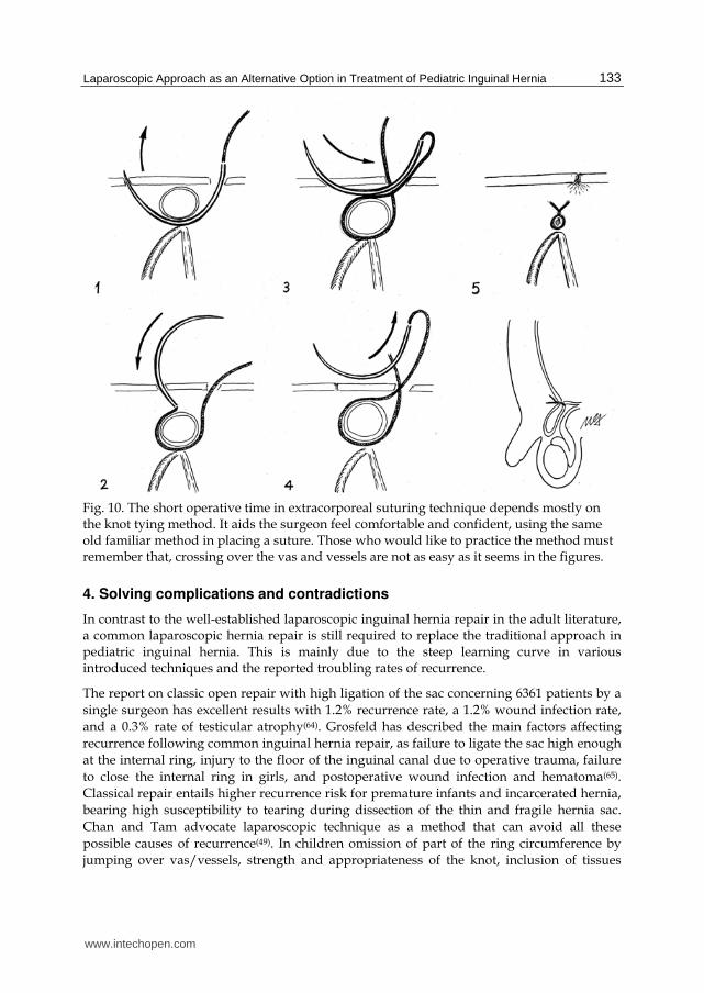
Fig. 10. The short operative time in extracorporeal suturing technique depends mostly on the knot tying method. It aids the surgeon feel comfortable and confident, using the same old familiar method in placing a suture. Those who would like to practice the method must remember that, crossing over the vas and vessels are not as easy as it seems in the figures.
4. Solving complications and contradictions
In contrast to the well-established laparoscopic inguinal hernia repair in the adult literature, a common laparoscopic hernia repair is still required to replace the traditional approach in pediatric inguinal hernia. This is mainly due to the steep learning curve in various introduced techniques and the reported troubling rates of recurrence.
The report on classic open repair with high ligation of the sac concerning 6361 patients by a
single surgeon has excellent results with 1.2% recurrence rate, a 1.2% wound infection rate,
and a 0.3% rate of testicular atrophy(64). Grosfeld has described the main factors affecting
recurrence following common inguinal hernia repair, as failure to ligate the sac high enough
at the internal ring, injury to the floor of the inguinal canal due to operative trauma, failure
to close the internal ring in girls, and postoperative wound infection and hematoma(65).
Classical repair entails higher recurrence risk for premature infants and incarcerated hernia,
bearing high susceptibility to tearing during dissection of the thin and fragile hernia sac.
Chan and Tam advocate laparoscopic technique as a method that can avoid all these
possible causes of recurrence(49). In children omission of part of the ring circumference by
jumping over vas/vessels, strength and appropriateness of the knot, inclusion of tissues
www.intechopen.com
Advances in Laparoscopic Surgery
134
other than peritoneum in the ligature with a propensity for subsequent loosening are
reported factors that may contribute to recurrence. Additional factors are use of absorbable
sutures, an excessively dilated internal ring, and the presence of comorbid conditions (eg,
collagen disorders, malnutrition, or pulmonary disease). Most of the recurrences are noted
within 6 months following the procedure and the most common site of recurrence is along
the medial internal ring at the site of passage of the cord structures(51, 62, 66). The reported
recurrence rates in extracorporeal suturing techniques are given between 0.35–2.8% in which
small spaces are left when crossing over the spermatic cord or the testicular vessels(41, 54, 55, 57,
62). On the contrary, the reported recurrence rates 3.1–4.4% are much higher, in which the
suture material is tied off in a similar way but intracorporeally(47, 52, 67). An intrinsic risk of
recanalization of the vaginal process is mainly believed to result in recurrence. Albeit
continuing search for a well-established approach in male repair, laparoscopic repair is
becoming a promising good alternative to open hernia repair in female children.
Comparable recurrence rates are repeatedly reported in female patients where the hernia sac
is routinely excised(50, 59, 60).
The key to obtain a safe hernia repair relies on the healing process startled by firm ligation of the sac high enough at the internal ring, finally creating a good reperitonealization characterized by smooth and even surface like the palm of our hand. The optimum peritoneal tissue disruption is maintained by means of an essential bisecting force applied on the knot during tying it. This essential force must also warrant a good transfixation that would prevent the suture from migrating distally. A pediatric surgeon learns to feel and keep tactile control of this appropriate suture tension for obtaining an even bisecting and transfixating force. A time consuming, complex cognitive course is required to gain this tactile sense of feeling in hernia repair. The safety of a knot in a laparoscopic procedure relies on its limits in imitating an identical open procedure. What is meant by the “steep learning curve” is the surgeon’s ability to regain expertise and persevere this mentioned, but new tactile feeling. The author believes that published better recurrence rates using extracorporeal suturing technique, depend on this familiar tactile sense of feeling and lower recurrence rates will equally be obtained with increased expertise in intracorporeal approaches. In the meantime, the use of double ligatures may further secure the closure of the hernia sac in intracorporeal approaches as well(41).
Published reports concerning impact of childhood hernia repair on fertility have always been a popular issue; it has forced proponents to restrict bilateral exploration according to the age and sex of the child and the presenting side(6-15). Antonoff et al. has pointed out to the higher risk of an inadvertent injury to vas deferens in the absence of a true hernia(68). Complications that may result in infertility during hernia repair include testicular atrophy, injury to the vas deferens, iatrogenic cryptorchidism, and injury to the fallopian tubes(3, 7, 15,
18, 40, 68-70). A recent survey declares a 5% infertility rate, medically diagnosed in males 50 years after hernia repair(71). Proponents of laparoscopic repair advocate the procedure arguing that the risk of visceral injury should be minimal or less than open surgery, keeping the vas deferens and cord un-touched by limited dissection of the peritoneal layer due to high visual magnifications(72). Theoretically, a laparoscopic approach aids the surgeon in avoiding a wide groin dissection thus reducing extensive inguinal scarring. The operative technique may also save the spermatic cord structures from a redo procedure related injury, should the hernia recur from a previous open repair(62). The reported rare incidence of testicular atrophy in laparoscopic hernia repair is attributed to multiple collateral
www.intechopen.com
Laparoscopic Approach as an Alternative Option in Treatment of Pediatric Inguinal Hernia
135
circulations of the testis, rendering dissection at the internal ring an extremely safe method (73, 74). Albeit reported advantages of pediatric laparoscopic hernia repair, the long-term risk of potential injury to the vas deferens and inguinal vessels should not be underestimated. Turial et al. reported 4% incidence of testicular ascent in babies weighing 5 kg or less, performed in skilled laparoscopic hands(75). Yang et al. due to publishing bias, comment on the necessity of additional randomized controlled trials with standard report format and uniform units in order to investigate the efficiency of laparoscopic hernia repair with increased precision(76).
Other laparoscopy related complications such as; postoperative hydrocele, scrotal edema,
erythema, inguinodynia and wound infections are reported decreasingly. Bharati et al.
postulate that initial fluid accumulation in the distal sac recedes by spontaneous
reabsorbtion and does not require any additional intervention(72). In their recent meta-
analysis report, Yang et al state that incidence of hydrocele, testicular atrophy, postoperative
pain and wound infection show statistical insignificance, concerning laparoscopic vs. open
hernia repair(76).
One must also admit that, laparoscopy carries its own set of complications such as
decreased venous return, hypercapnia, acidosis and air embolism. Recent advancements
in anesthesia and refinements in instruments have revolutionized use of minimal invasive
approach as a safer procedure in pediatric surgical diseases(77). On the other hand,
intraperitoneal approach may additionally mean added risks associated with a violated
peritoneal cavity inheriting specific complications caused by needle or trocar injury to
ovary, bladder, intestines and/or the iliac, inferior epigastric and gonadal vessels. The
burden of these risks is quite heavy to carry when compared to the common inguinal
hernia repair. Laparoscopic approach, on the other hand, may also aid in finding an
unexpected entity (Figure 11).
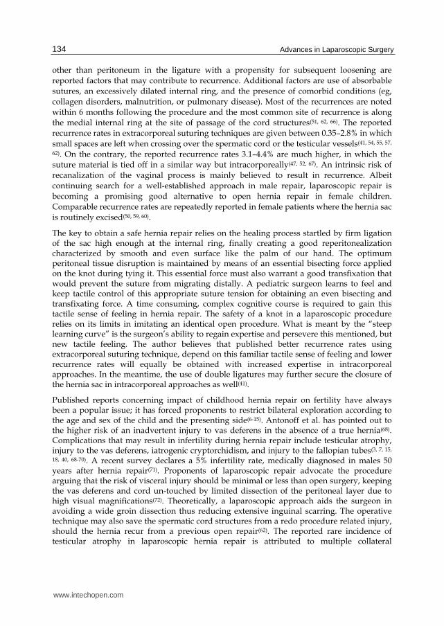
Fig. 11. An iatrogenic hematoma from a puncture in the internal iliac vein (Left). One must refrain from opening the retroperitoneum, since abdominal gas pressure is usually sufficient to stop the oozing. An intracanalicular cyst seen in a male patient, imitating the infamous Nuck’s cyst in females (Right).
www.intechopen.com
Advances in Laparoscopic Surgery
136
As for last but never the least, we have to evaluate the mentioned procedures by means of
cost effectiveness. The long theater time required for the anesthetist in familiarizing with
laparoscopic operations and for assembling all necessary equipment means a longer
operative time, which in turn results in less operations in a day. When coupled with the
high cost of setting up and running a theater with appropriate laparoscopic instrumentation,
it may not be feasible, as it seems from an economic point of view.
5. Conclusion
It is certain that introduction of minimal invasive surgery has revolutionized the classical treatment of pediatric inguinal hernia repair, which has stood the test of time. Those who would think to commence are advised to do so from the initial step, diagnostic laparoscopy. Simple diagnostic laparoscopic examination enables surgical precision through enhanced visualization, magnification and ability to limit collateral damage by minimizing invasion. The reported incidence of a missed metachronous hernia following laparoscopic inspection is given as 1.1%, a figure far less than the expected traditional rate of a metachronous hernia(26, 36, 40). We may conclude that, a certain number of children will be saved from an unnecessary contralateral exploration or a future hernia by using this simple technique. We have to keep in mind; standards of management of a contralateral processus vaginalis awaits consensus.
Laparoscopic hernia repair is proven to allow easier access and excellent visual exposure to the detection and repair of contralateral patencies. The technique entails minimal manipulation of the vas deferens and testicular vessels during hernia repair, with suggested benefits of smaller scars, shorter bilateral operation times and better chance of repair of recurrent hernias through fresh tissue. Reported series however, still declare risk of higher recurrence and testicular ascent rates even in the most experienced hands. Again, it may be justified to start practicing inguinal hernia repair in girls where there is limited risk of much feared collateral damage. Albeit mentioned benefits, laparoscopic repair has potential risks attributable to surgeon’s experience and variations in the chosen technique.
It is an important ethical duty for us to present the odds and evens and discuss the potential risks of each surgical approach with the family and have their consent during the decision making process.
6. Acknowledgment
The author would like to extend his gratitude to Mr. Mehmet Ali Gürsoy M.D. for drawing the illustrations within the text.
7. References
[1] Glick PL, Boulanger SC. Inguinal hernias and hydroceles. In: O'Neill JA, Rowe MI, Grosfeld JC, et al, editors. Pediatric surgery, 5th ed. St Louis, Mo: Mosby; 2007. p. 1172-92
[2] Weber TR, Tracy TF, Keller MS. Groin hernias and hydroceles. In: Ashcraft KW, Holcomb GW, Murphy JP, editors. Pediatric surgery. Philadelphia, Pa: Elsevier Saunders; 2005. p. 697-705
www.intechopen.com
Laparoscopic Approach as an Alternative Option in Treatment of Pediatric Inguinal Hernia
137
[3] Brandt ML. Pediatric Hernias. Surg Clin N Am 2008; 88: 27–43 [4] Kiesewetter WB, Parenzan L. When should hernia in the infant be treated bilaterally?
JAMA 1959; 171: 287–90. [5] Gilbert M, Clatworthy HW Jr. Bilateral operations for inguinal hernia and hydrocele in
infancy and childhood. Am. J. Surg.1959; 97: 255–9. [6] Shandling B, Janik JS. The vulnerability of the vas deferens. J Pediatr Surg 1981;16(4):
461–4. [7] Janik JS, Shandling B. The vulnerability of the vas deferens (II): the case against routine
bilateral inguinal exploration. J Pediatr Surg 1982;17(5):585–8. [8] Abasiyanik A, Güvenç H, Yavuzer D, Peker O, Ince U. The effect of iatrogenic vas
deferens injury on fertility in an experimental rat model. J Pediatr Surg 1997;32(8):1144–6.
[9] McGregor DB, Halverson K, McVay CB. The unilateral pediatric inguinal hernia: should the contralateral side be explored? J. Pediatr. Surg. 1980; 15: 313–7
[10] Given JP, Rubin SZ. Occurrence of contralateral hernia following unilateral repair in a pediatric hospital. J. Pediatr. Surg. 1989; 24: 963–5
[11] Tackett LD, Breuer CK, Luks FI, Caldamone AA, Breuer JG, DeLuca FG, Caesar RE, Efthemiou E, Wesselhoeft CW Jr. Incidence of contralateral inguinal hernia: a prospective analysis. J. Pediatr. Surg. 1999; 34: 684–7
[12] Ballantyne A, Jawaheer G, Munro FD. Contralateral groin exploration is not justified in infants with a unilateral inguinal hernia. Br. J. Surg. 2001; 85: 720–3
[13] Shabbir J, Moore A, O’Sullivan JB. Contralateral groin exploration is not justified in infants with a unilateral inguinal hernia. Ir. J. Med. Sci. 2003; 172: 18–9
[14] Ceylan H, Karakök M, Güldür E, Cengiz B, Bağci C, Mir E. Temporary stretch of the testicular pedicle may damage the vas deferens and the testis. J Pediatr Surg 2003;38(10):1530–3.
[15] Marulaiah M, Atkinson J, Kukkady A, Brown S, Samarakkody U. Is contralateral exploration necessary in preterm infants with unilateral inguinal hernia? J Pediatr Surg 2006;41(12):2004–7.
[16] Sparkman RS. Bilateral exploration in inguinal hernia in juvenile patients. Surgery 1962;51:393–406.
[17] Moss RL, Hatch EI. Inguinal hernia repair in early infancy. Am J Surg 1991;161:596–599 [18] Surana R, Puri P. Fate of patent processus vaginalis: A case against routine contralateral
exploration for unilateral inguinal hernia in children. Pediatr Surg Int 1993;8:412–414
[19] Tackett LD, Breuer CK, Luks FI, Caldamone AA, Breuer JG, DeLuca FG, Caesar RE, Efthemiou E, Wesselhoeft CW Jr. Incidence of contralateral inguinal hernia: a prospective analysis. J. Pediatr. Surg. 1999; 34: 684–7
[20] Nakayama DK, Rowe MI. Inguinal hernia and the acute scrotum. Pediatr Rev 1989;11:87-93
[21] Rowe MI, Copelson LW, Clatworthy HW. The patent processus vaginalis and the inguinal hernia. J Pediatr Surg 1969;4:102-7
[22] Panton NM, Panton RJ. Laparoscopic hernia repair. Am J Surg 1994;167:535–537 [23] Watson DS, Sharp KW, Vasquez JM, Richards WO. Incidence of inguinal hernias during
laparoscopy. South Med J 1994;87:23–25
www.intechopen.com
Advances in Laparoscopic Surgery
138
[24] van Veen RN, van Wessem KJ, Halm JA, Simons MP, Plaisier PW, Jeekel J, Lange JF. Patent processus vaginalis in the adult as a risk factor for the occurrence of indirect inguinal hernia. Surg Endosc 2007;21(2):202–5
[25] Duckett J. W. Treatment of congenital inguinal hernia. Ann Surg. 1952 June; 135(6): 879–884
[26] Miltenburg DM, Nuchtern JG, Jaksic T, Kozinetz CA, Brandt ML. Meta-analysis of the risk of metachronous hernia in infants and children. Am J Surg 1997;174(6):741–4
[27] Erez I, Rathause V, Vacian I, Zohar E, Hoppenstein D, Werner M, Lazar L, Freud E.. Preoperative ultrasound and intraoperative findings of inguinal hernias in children: a prospective study of 642 children. J Pediatr Surg 2002;37(6):865–8
[28] Lobe TE, Schropp KP. Inguinal hernia in pediatrics: initial experience with laparoscopic inguinal exploration of the asymptomatic contralateral side. J. Laparoendosc. Surg. 1992; 2: 135–40
[29] Chu CC, Chou CY, Hsu TM, Yang TH, Ma CP, Cywes S. Intraoperative laparoscopy in unilateral hernia repair to detect a contralateral patent processus vaginalis. Pediatr Surg Int 1993;8:385-8
[30] Wolf SA, Hopkins JW. Laparoscopic incidence of contralateral patent processus vaginalis in boys with clinical unilateral hernias. J. Pediatr. Surg. 1994; 29: 1118–20
[31] Grossmann PA, Wolf SA, Hopkins JW, Paradise NF. The efficacy of laparoscopic examination of the internal inguinal ring in children. J. Pediatr. Surg. 1995; 30: 214–8
[32] Holcomb GW III, Morgan WM III, Brock JW III. Laparoscopic evaluation for contralateral patent processus vaginalis: part II. J. Pediatr. Surg. 1996; 31: 1170–3
[33] Yerkes EB, Brock JW 3rd, Holcomb GW 3rd, Morgan WM 3rd. Laparoscopic evaluation for a contralateral patent processus vaginalis: part III. Urology 1998; 51: 480–3
[34] Guvenc BH. Diagnostic laparoscopic evaluation of the contralateral internal inguinal ring: the search for a prospective hernia. Pediatr Endosurg Innov Techn 2001;5:259-65
[35] Miltenburg DM, Nuchtern JG, Jaksic T, Kozinetiz C, Brandt ML. Laparoscopic evaluation of the pediatric inguinal hernia: A meta-analysis. J Pediatr Surg 1998;33:874–879
[36] Endo M, Watanabe T, Nakano M, Yoshida F, Ukiyama E. Laparoscopic completely extraperitoneal repair of inguinal hernia in children: a single-institute experience with 1,257 repairs compared with cut-down herniorrhaphy. Surg Endosc. 2009 Aug;23(8):1706-12.
[37] Sözübir S, Ekingen G, Senel U, Kahraman H, Güvenç BH. A continuous debate on contralateral processus vaginalis: evaluation technique and approach to patency. Hernia 2006; 10: 74–8.
[38] Fuenfer MM, Pitts RM, Georgeson KE. Laparoscopic exploration of the contralateral groin in children: An improved technique. J Laparoendosc Surg 1996 (suppl 1):S1–S4.
[39] Owings EP, Georgeson KE. A new technique for laparoscopic exploration to find contralateral patent processus vaginalis. Surg Endosc 2000;14:114–116.
[40] Chan KL, Chan YH, Tam PKH. Towards a near-zero recurrence rate in laparoscopic inguinal hernia repair for pediatric patients of all ages. J Pediatr Surg 2007;42:1993-7.
[41] Tam YH, Lee KH, Sihoe JD, Chan KW, Wong PY, Cheung ST, Mou JW. Laparoscopic hernia repair in children by the hook method: a single-center series of 433 consecutive patients Journal of Pediatric Surgery 2009; 44, 1502–1505.
www.intechopen.com

Laparoscopic Approach as an Alternative Option in Treatment of Pediatric Inguinal Hernia
139
[42] Lau ST, Lee YH, Caty MG. Current management of hernias and hydroceles. Semin. Pediatr. Surg. 2007; 16: 50–7.
[43] Maddox MM, Smith DP. A long-term prospective analysis of pediatric unilateral inguinal hernias: should laparoscopy or anything else influence the management of the contralateral side? J. Pediatr. Urol. 2008; 4: 141–5
[44] Chung KLY, Leung MWY, Chao NSY, Wong BPY, Kwok WK, Liu K KW. Laparoscopic repair on asymptomatic contralateral patent processus vaginalis in children with unilateral inguinal hernia: A centre experience and review of the literature Surgical Practice 2011; 15: 12–15
[45] Montupet P, Esposito C. Laparoscopic treatment of congenital inguinal hernia in children. J Pediatr Surg 1999;34(3):420–3.
[46] Tan HL. Laparoscopic repair of inguinal hernias in children. J. Pediatr. Surg. 2001; 36: 833
[47] Schier F, Montupet P, Esposito C Laparoscopic inguinal herniorrhaphy in children. A three-center experience with 933 repairs. J Pediatr Surg 2002; 37:395–397
[48] Yip KF, Tam PK, Li MK. Laparoscopic flip-flap hernioplasty: an innovative technique for pediatric hernia surgery. Surg Endosc 2004;18(7):1126–9
[49] Chan KL, Tam PK. Technical refinements in laparoscopic repair of childhood inguinal hernia. Surg Endosc 2004; 18:957–960
[50] Becmeur F, Philippe P, Lemandat-Schultz A, Moog R, Grandadam S, Lieber A, Toledano D. A continuous series of 96 laparoscopic inguinal hernia repairs in children by a new technique. Surg Endosc 2004; 18:1738–1741
[51] Chinnaswamy P, Malladi V, Jani KV, Parthasarthi R, Shetty RA, Kavalakat AJ, Prakash A. Laparoscopic inguinal hernia repair in children. JSLS 2005; 9:393–398
[52] Schier F. Laparoscopic inguinal hernia repair: a prospective personal series of 542 children. J Pediatr Surg 2006; 41:1081–1084
[53] Ozgediz D, Roayaie K, Lee H, Nobuhara KK, Farmer DL, Bratton B, Harrison MR. Subcutaneous endoscopically assisted ligation (SEAL) of the internal ring for repair of inguinal hernias in children: report of a new technique and early results. Surg Endosc 2007;21(8):1327–31
[54] Spurbeck WW, Prasad R, Lobe TE. Two-year experience with minimally invasive herniorrhaphy in children. Surg Endosc 2005; 19:551–553
[55] Patkowski D, Czernik J, Chrzan R, Jaworski W, Apoznanski W. Percutaneous internal ring suturing: a simple minimally invasive technique for inguinal hernia repair in children. J Laparoendosc Adv Surg Tech 2006; 16:513–517
[56] El-Gohary MA. Laparoscopic ligation of inguinal of inguinal hernia in girls. Pediatr Endosurg Innov Techn 1997;1:185-7
[57] Takehara H, Yakabe S, Kameoka K. Laparoscopic percutaneous extraperitoneal closure for inguinal hernia in children: clinical outcome of 972 repairs done in 3 pediatric surgical institutions. J Pediatr Surg 2006; 41:1999–2003
[58] Prasad R, Lovvorn HN 3rd, Wadie GM, Lobe TE. Early experience with minimally invasive inguinal herniorrhaphy in children. J Pediatr Surg 2003;38:1055-8
[59] Lipskar AM, Soffer SZ, Glick RD, Rosen NG, Levitt MA, Hong AR. Laparoscopic inguinal hernia inversion and ligation in female children: a review of 173 consecutive cases at a single institution. Journal of Pediatric Surgery (2010) 45, 1370–1374
www.intechopen.com

Advances in Laparoscopic Surgery
140
[60] Guner YS, Emami CN, Chokshi NK, Wang K, Shin CE. Inversion herniotomy: a laparoscopic technique for female inguinal hernia repair. J Laparoendosc Adv Surg Tech A. 2010 Jun;20(5):481-4
[61] Akansel G, Guvenc BH, Ekingen G, Sozubir S, Tuzlaci A, Inan N. Ultrasonographic findings after laparoscopic repair of paediatric female inguinal hernias: the 'vanishing rosebud'. Pediatr Radiol. 2003 Oct;33(10):693-6
[62] Dutta S, Albanese C. Transcutaneous laparoscopic hernia repair in children: a prospective review of 275 hernia repairs with minimum 2-year follow-up. Surg Endosc 2009; 23:103–107
[63] Yip KF, Tam PK, Li MK. Laparoscopic flip-flap hernioplasty: an innovative technique for pediatric hernia surgery. Surg Endosc 2004;18(7):1126–9
[64] Ein SH, Njere I, Ein A. Six thousand three hundred sixty-one pediatric inguinal hernias: a 35-year review. J Pediatr Surg 2006;41(5):980–6
[65] Grosfeld JL, Minnick K, Shedd F, West KW, Rescoria FJ, Vane DW. Inguinal hernia in children: factors affecting recurrence in 62 cases. 1991; J Pediatr Surg 26:283–287
[66] Kastenberg Z, Bruzoni M, Dutta S. A modification of the laparoscopic transcutaneous inguinal hernia repair to achieve transfixation ligature of the hernia sac Journal of Pediatric Surgery 2011; 46, 1658
[67] Chinnaswamy P, Malladi V, Jani KV, Parthasarthi R, Shetty RA, Kavalakat AJ, Prakash A. Laparoscopic inguinal hernia repair in children. JSLS 2005; 9:393–398
[68] Antonoff MB, Kreykes NS, Saltzman DA, Acton RD. American Academy of Pediatrics section on surgery hernia survey revisited. J Pediatr Surg 2005;40(6):1009–14
[69] Hansen KA, Eyster KM. Infertility: an unusual complication of inguinal herniorrhaphy. Fertil Steril 2006;86(1):217
[70] Matsuda T, Muguruma K, Hiura Y, Okuno H, Shichiri Y, Yoshida O. Seminal tract obstruction caused by childhood inguinal herniorrhaphy: results of microsurgical reanastomosis. J Urol 1998;159(3):837–40
[71] Zendejas B, Zarroug AE, Erben YM, Holley CT, Farley DR. Impact of childhood inguinal hernia repair in adulthood: 50 years of follow-up. J Am Coll Surg. 2010 Dec;211(6):762-8
[72] Bharathi RS, Arora M, Baskaran V. Minimal access surgery of pediatric inguinal hernias: a review. Surg Endosc 2008;22:1751-62
[73] Barqawi A, Furness III P, Koyle M. Laparoscopic Palomo varicocelectomy in the adolescent is safe after previous ipsilateral inguinal surgery. BJU Int 2002;89:269-72
[74] Riccabona M, Oswald J, Koen M, Lusuardi L, Radmayr C, Bartsch G. Optimizing the operative treatment of boys with varicocele: sequential comparison of 4 techniques. J Urol 2003;169:666-8
[75] Turial S, Enders J, Krause K, Schier F. Laparoscopic inguinal herniorrhaphy in babies weighing 5 kg or less. Surg Endosc. 2011 Jan;25(1):72-8
[76] Yang C, Zhang H, Pu J, Mei H, Zheng L, Tong Q. Laparoscopic vs open herniorrhaphy in the management of pediatric inguinal hernia: a systemic review and meta-analysis. Journal of Pediatric Surgery 2011; 46, 1824–1834
[77] Ozdamar D, Güvenç BH, Toker K, Solak M, Ekingen G. Comparison of the effect of LMA and ETT on ventilation and intragastric pressure in pediatric laparoscopic procedures. Minerva Anestesiol. 2010; Aug;76(8):592-9
www.intechopen.com
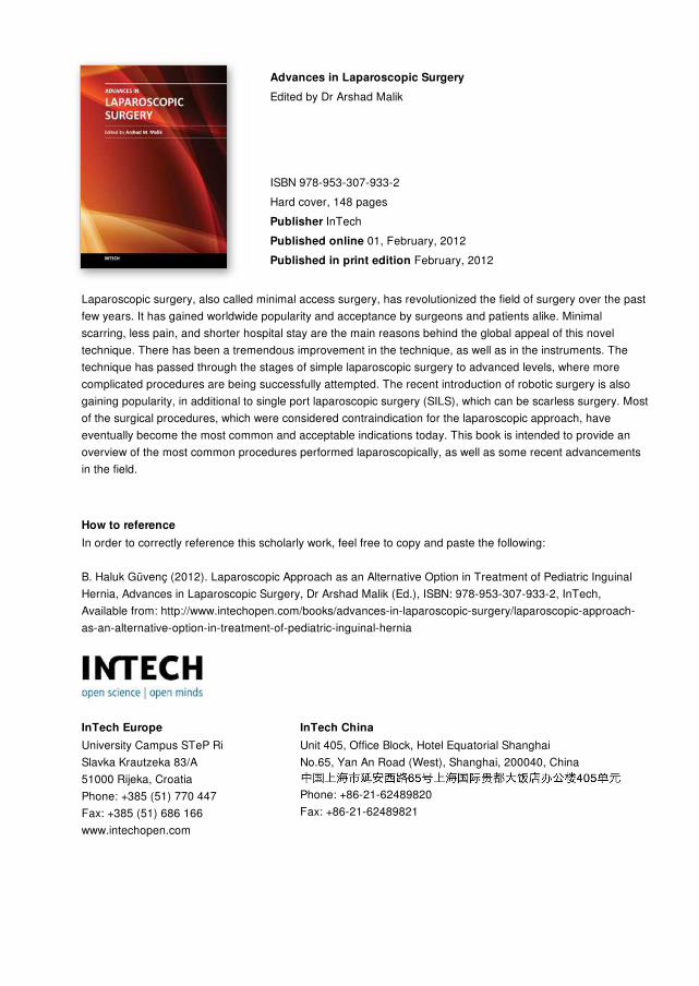
Advances in Laparoscopic SurgeryEdited by Dr Arshad Malik
ISBN 978-953-307-933-2Hard cover, 148 pagesPublisher InTechPublished online 01, February, 2012Published in print edition February, 2012
InTech EuropeUniversity Campus STeP Ri Slavka Krautzeka 83/A 51000 Rijeka, Croatia Phone: +385 (51) 770 447 Fax: +385 (51) 686 166www.intechopen.com
InTech ChinaUnit 405, Office Block, Hotel Equatorial Shanghai No.65, Yan An Road (West), Shanghai, 200040, China
Phone: +86-21-62489820 Fax: +86-21-62489821
Laparoscopic surgery, also called minimal access surgery, has revolutionized the field of surgery over the pastfew years. It has gained worldwide popularity and acceptance by surgeons and patients alike. Minimalscarring, less pain, and shorter hospital stay are the main reasons behind the global appeal of this noveltechnique. There has been a tremendous improvement in the technique, as well as in the instruments. Thetechnique has passed through the stages of simple laparoscopic surgery to advanced levels, where morecomplicated procedures are being successfully attempted. The recent introduction of robotic surgery is alsogaining popularity, in additional to single port laparoscopic surgery (SILS), which can be scarless surgery. Mostof the surgical procedures, which were considered contraindication for the laparoscopic approach, haveeventually become the most common and acceptable indications today. This book is intended to provide anoverview of the most common procedures performed laparoscopically, as well as some recent advancementsin the field.
How to referenceIn order to correctly reference this scholarly work, feel free to copy and paste the following:
B. Haluk Güvenç (2012). Laparoscopic Approach as an Alternative Option in Treatment of Pediatric InguinalHernia, Advances in Laparoscopic Surgery, Dr Arshad Malik (Ed.), ISBN: 978-953-307-933-2, InTech,Available from: http://www.intechopen.com/books/advances-in-laparoscopic-surgery/laparoscopic-approach-as-an-alternative-option-in-treatment-of-pediatric-inguinal-hernia