team effort - arkansas medical society · arkansas medical society, p.o. box 55088, little rock,...
TRANSCRIPT

NUMBER 4 SEPTEMBER 2014 • 49
Vol.111 • No. 4 SEPTEMBER 2014
El Dorado Clinic Takes a Colorful Approach to Medicine
TEAM EFFORT

50 • THE JOURNAL OF THE ARKANSAS MEDICAL SOCIETY VOLUME 111
We don’t provide health care.We help make it better.
advancing
practiceshealthy
• Practice workflow redesign
• Care planning and documentation
• Health information technology assistance
• Patient centered medical home
• Patient engagement and patient portals strategies
• HIPPA security risk assessment
• Health education tools and communications campaigns
afmc.org | [email protected] | 877-650-2362 | 501-212-8600
A R K A N S A S F O U N D AT I O N F O R M E D I C A L C A R E
AFMC’s statewide network of experiencedprovider specialists and representatives can help you with:

How I Manage ChronicLymphocytic Leukemia in 2014
Volume 111 • Number 4 September 2014
ON THE COVER
Winner of the ASAE Excellence in Communications Award
Feature Articles
A Closer Look at Quality
Join us to stay updated on health care news in Arkansas.
facebook.com/ArkMedSoc ArkMed.orgtwitter.com/ArkMedSoc
Established 1890. Owned and edited by the Arkansas Medical Society and published under the direction of the Board of Trustees.
Advertising Information: Penny Henderson, (501) 224-8967 or [email protected]. #10 Corporate Hill Drive, Suite 300, Little Rock, Arkansas 72205.
Postmaster: Send address changes to: The Journal of the Arkansas Medical Society, P.O. Box 55088, Little Rock, Arkansas 72215-5088.
Subscription rate: $30.00 annually for domestic; $40.00, foreign. Single issue $3.00.
The Journal of the Arkansas Medical Society (ISNN 0004-1858) is published monthly, except twice in the month of August by the Arkansas Medical Society, #10 Corporate Hill Drive, Suite 300, Little Rock, Arkansas 72205. (501) 224-8967.
Printed by The Ovid Bell Press Inc., Fulton, Missouri 65251. Periodicals postage is paid at Little Rock, Arkansas, and at additional mailing offices.
Articles and advertisements published in The Journal are for the interest of its readers and do not represent the official position or endorsement of The Journal or the Arkansas Medical Society. The Journal reserves the right to make the final decision on all content and advertisements.
© Copyright 2014 by the Arkansas Medical Society.
www.ArkMed.org
56
P E O P L E + E V E N T S 70
60
COMMENTARY
54LAURA SISTERHEN, MD WHAT HAVE WE DONE FOR YOU LATELY? 52H. SCOTT SMITH, JD DIRECTOR OF GOVERNMENTAL AFFAIRS
STUDYCASE 66Neutrophilic Leukemoid Reaction in a Patient with High Grade Sarcoma
by Sunita Parajuli, MD; Latha Achanta, MD, MPH;Robert H. Hopkins, Jr., MD, FACP, FAAP; Ginell Post, MD
by CASEY L. PENN
SCIENTIFIC ARTICLE
by James E. McDonald, MD; Linda A. Deloney, EdD; Kedar Jambhekar, MD
Pulmonary arterial hypertension: Part 1: A review for an internist
62
68
El Dorado Clinic Takes a Colorful Approach to Medicine
TEAM EFFORT
by Ahmed Alwbari, MD1; Issam Makhoul, MD1
1Hematology/Oncology Division – University of Arkansas for Medical Sciences
HOW I TREAT SPECIAL SERIES
NUMBER 4 SEPTEMBER 2014 • 51
I love all 14 of the Pixar films and have just starting reading a book about Pixar called “Creativity, Inc.” written by Ed Catmull, Pixar President and co-founder. He has some great stories to
tell. Early on, he states that a key component to Pixar’s continuing
success is that the “Story”… and NOT animation, is “King.”
Having worked in legislative advocacy since January of 1996 and
through 14 regular state legislative sessions (along with a VERY in-
teresting fiscal session this year), I can unequivocally say that for
successful advocacy, “Story is King” as well.
Our legislative “issues” are born out of the “stories” that we hear
from you. We try to legislatively address the stories that will have
the biggest positive impact on the physicians of Arkansas and their
patients. But without REAL, compelling stories that impact REAL
physicians and REAL patients, it is very difficult to find the needed
legislative support leading to a good legislative solution.
Leading up to the start of each session (which is now), we search
for those stories but we need your help. If you have a story to tell
that you would like AMS to consider for a legislative remedy, contact
us! We are working on next year’s legislative issues and their stories
now. You all have some of the most compelling stories to be told as
you take care of the sick and injured.
Again, please contact us and let us hear those stories. There’s a good
chance that if you’re experiencing a problem, you are probably NOT
alone, but we need you to step forward. Let us know.
A few of the issues and stories we have been working on since the
end of this year’s fiscal session in April are: telemedicine, prescription
drug abuse, Physician Orders for Life-Sustaining Treatment (POLST)
and the Medicaid Private Option (THE issue of this past fiscal ses-
sion). The Private Option appears to be headed for “Perennial Legis-
lative Battle” status.
To that end, I just returned to the office after an Executive Committee
meeting of the large coalition of organizations supporting the Private
Option. We’re already working on messaging and the anticipated
“vote count” for next session’s Private Option vote in light of primary
election results, and potential general election outcomes. Wise de-
cisions now will hopefully make the path smoother during session.
Other recent discussions with legislators have involved two signifi-
cant scope of practice issues along with an incredibly important in-
surance issue we anticipate needing to address next session. While
your issues and stories are here year-round, the window of time to
address these issues is limited because sessions come and go rather
quickly.
As soon as a regular session finishes, generally in early April of the
odd-numbered years, we move immediately into “election season”
which lasts 18 months until the next general election the following
November. In fiscal session years, like this even-numbered year, the
election “season” takes a timeout for the couple of months during the
fiscal session and resumes in early April for the 4 to 6 week sprint to
the primary elections, the third week in May.
Generally, the legislative “pre-season” really kicks in 6 months or so
before the legislature actually starts in January of those odd-num-
bered years. So, we are currently heading down the backstretch of
the election season, and concurrently starting the 2015 legislative
pre-season.
The remainder of this year brings intensified identification of issues
and the stories needing legislative solutions. Soon, bill drafting will
start to kick-in and visits with legislators regarding some of the sto-
ries will happen on a more frequent basis. However, until the No-
vember election, we will not be able to ascertain precisely who will
even be in the legislature, much less, who will be on key committees.
So, it is important to remember that continuing legislative success
is only possible if we are successful in helping our friends stay in
office. Hardly a day goes by that we don’t either get an invitation to a
candidate fundraiser or receive a call asking for financial assistance.
We keep busy meeting with candidates, attending fundraisers and
making campaign contributions from our political action committee,
ArkMed-PAC.
We need your participation in ArkMed-PAC, just like we need your
stories. Campaigns are expensive, and we want to help as many
of our friends as much as we can. We can help more candidates
as more AMS members contribute to the PAC, so please be on the
lookout for our ArkMed-PAC membership mailing.
Physician support has already helped a number of candidates, and
we are fortunate to have some terrific friends in the Arkansas legisla-
ture and that strong base of legislative support has been amazing in
fighting for the physicians of Arkansas and their patients. With your
continuing assistance during the election season and in identifying
issues and sharing stories in the legislative pre-season, hopefully,
we will expand that base and have another successful legislative
session.
WHAT HAVE WE DONE FOR YOU LATELY?
The Story Is KingH. SCOTT SMITH, JD
DIRECTOR OF GOVERNMENTAL AFFAIRS
52 • THE JOURNAL OF THE ARKANSAS MEDICAL SOCIETY VOLUME 111

Free your mind to think aboutsomething other than med-mal.
Since we’re singularly focused on medical malpractice protection, your mind is free to go other places. LAMMICO is not just insurance. We’re a network of insurance and legal professionals experienced in medical liability claims. A network that closes approximately 90% of all cases without indemnity payment. A network of robust in-person and online Risk Management educational resources to help you avoid a claim in the first place. LAMMICO’s a partner - so that when you insure with us, you’re free to do your job better. And that’s a very peaceful place to be.
Building Enduring Partnerships800.452.2120 www.lammico.com/AMS
LAMMICO is proud to support theArkansas Medical Society’s
Annual Meeting May 2-3, 2014
CLINIC MANAGERS & PHYSICIANS:
PLALLC.COM • 501.603.1751
ARKANSAS-BASED URINALYSIS LAB • OWNED BY PHYSICIANS • CLIA CERTIFIED & COMPLIANT • SUPPLIES PROVIDED • FOUNDED IN 2012 • PERSONALIZED DRUG SCREEN TESTING • ONLINE REPORT ACCESS • ARKANSAS BASED BILLING CO
PHYSICIANS’LABORATORIES
OF AMERICA, LLC
NUMBER 4 SEPTEMBER 2014 • 53
ARKANSAS MEDICAL SOCIETY2014-2015 OFFICERS
Alan Wilson, MD, Crossett President
G. Edward Bryant, MD, West Memphis President-elect
William D. Dedman, MD, CamdenVice President
Omar T. Atiq, MD, Pine BluffImmediate Past President
Chad Rodgers, MD, Little RockSecretary
Frankie Griffin, MD, Van BurenTreasurer
Michael Saitta, MD, Fayetteville Speaker, House of Delegates
Anthony D. Johnson, MD, Little RockVice Speaker, House of Delegates
Gene Shelby, MD, Hot SpringsChairman of the Board of Trustees
David WrotenExecutive Vice President
Nicole RichardsCommunications Coordinator
Jeremy HendersonArt Director
EDITORIAL BOARDFrankie Griffin, MDOrthopedic Surgery
Robert Hopkins, MDPediatrics/Internal Medicine
David Hunton, MDSurgery
Laura Sisterhen, MDPediatrics
Sandra Johnson, MDDermatology
Issam Makhoul, MDOncology
EDITOR EMERITUSAlfred Kahn Jr., MD (1916-2013)

This past May I had the privilege of attending the UAMS College of Medicine Honors Convocation.
Honors Convocation is the annual celebration where graduating medical students are recognized for their distinguished academic achievements. I watched smiling students approach the stage, turn to the audience, and pause for the ceremonial hooding by honored faculty. It is a proud moment for family, friends, and faculty. For me, one of the most poignant moments of the ceremony is the renewal of the Hippocratic Oath by faculty. According to the literature, the vast majority of American medical students pledge an oath upon graduating, and UAMS students are no exception. It is a common bond that we share prior to dispersing into specialized residencies.
I believe in this era of change, uncertainty, increasing pressure to contain cost and ensure quality and accessibility, we need something to guide us. We need a values statement, something that stands the test of time, to help us navigate the complexities of medicine today. In medicine, we need a middle C, a North Star, a constant in rapidly changing times. For many, this can be the oath we took upon graduation.
Although the modernized version varies, the premise of the oath is a promise to practice medicine honestly and to uphold a number of professional ethical standards. The Hippocratic Oath, written in Ionic Greek, dates back to the late 5th century BC. The oath begins with a covenant with the deity Apollo and goes on to address issues such as limits on means and ends, justice, chastity, and confidentiality. The oath has been modified many times and one of the most significant revisions, called the Declaration of Geneva, was written in 1948 by the World Medical Association.
When I graduated from medical school at the University of Tennessee Health Sciences Center in Memphis, I was given a document signed by the Dean that includes the oath that I took when I graduated. It is framed in my office and is a reminder of the commitment I made to myself and to my patients.
I solemnly pledge to consecrate my life to the service of humanity.
I will give respect and gratitude to my deserving teachers.
I will practice medicine with conscience and dignity.
The health and life of my patient will be my first consideration.
I will hold in confidence all that my patient confides in me.
I will maintain the honor and the noble traditions of the medical profession.
My colleagues will be as my family. I will not permit considerations of race, religion, nationality, party politics, or social standing to intervene between my duty and my patient. I will maintain the utmost respect for human life.
Even under threat I will not use my knowledge contrary to the laws of humanity. These promises I make freely and upon my honor.
As a clinical educator, I teach pediatric medicine to students and residents. Some of the things I teach will be outdated in 5-10 years. My hope is that some of what I teach is timeless. Respect for persons as outlined in the Hippocratic Oath will always be the right thing to do. When I’m not certain what course to take or decision to make, is it helpful to apply the wisdom of physicians who have gone before us.
This is the overarching message I heard from College of Medicine leaders at the Honors Convocation. We are in a period of rapid change in healthcare delivery as well as health professions educational reform. This change is driven by the public’s request to improve the patients’ experience of care, improve the health of individuals and populations, and decrease the per capita cost of healthcare. There is so much that is out of our control as individual physicians, but we can focus on what we can do. We can renew our commitment to the oath we took when we graduated.
A Time of RenewalLAURA SISTERHEN, MD
COMMENTARY
54 • THE JOURNAL OF THE ARKANSAS MEDICAL SOCIETY VOLUME 111

NUMBER 4 SEPTEMBER 2014 • 55

Five teams – purple, or-
ange, green, blue, and red
– prepare to take on the
day’s challenges. The at-
mosphere is upbeat and
fans, while loyal to their
own team, root for all sides to win in
this crucial game of improving health
and, ultimately, saving lives.
These physician-led teams are part of SAMA (South Arkansas Medical Associates) HealthCare Services, a one-stop health care facility that is in the process
of making a unique and deliberate transition from reactive to proactive health care, from the status quo to a patient-centered medical home.
SAMA is the recipient of a grant from the Centers for Medicare and Medicaid as part of its Comprehensive Primary Care (CPC) initiative. As The Journal reported last year, 69 Arkansas clinics are participating in this multi-payer initiative (a result of the Affordable Care Act) that fosters collaboration between public and private health care payers and encourages the patient-centered medical home (PCMH) concept. Participating clinics are given a list of milestones to work toward as well as some incentives to aid them in their efforts (http://innovation.cms.gov/initiatives/comprehensive-primary-care-initiative/).
Pioneering a new approach is no anomaly for this El Dorado clinic. “We kind of like to do things first,” confided LPN Nancy New of the changes that have put this small clinic on the nation’s radar as a mod-el of a successful PCMH. Her own career a picture of the clinic’s constantly evolving methods, New had worked as a nurse in the clinic since it opened 15 years ago, but as the most computer-savvy per-son in the clinic, she has also long been the go-to person to help navigate new technology, such as electronic medical records (EMR). That’s why New was recently reassigned as the clinic’s Health In-formatics Coordinator, a role that utilizes her skills and experience. Gary Bevill, MD, one of five SAMA physicians, noted that having a devoted IT person like New has greatly helped the clinic to navigate CPC, EMR and other improvements.
by CASEY L. PENN
El Dorado Clinic Takes a Colorful Approach to Medicine
TEAM EFFORT
Pete Atkinson, MHA
Gary Bevill, MD
James Sheppard, MD
56 • THE JOURNAL OF THE ARKANSAS MEDICAL SOCIETY VOLUME 111

The CPC opportunity came along at a time when SAMA was already well on its way to being a PCMH, according to the clinic’s administrator, Pete Atkinson, MHA. “In health care, not everybody fits into a square box,” said Atkinson, who enrolled the clinic in CPC in August 2012. He and the clinic’s physicians saw the opportunity to build on what they had already done, but to do it in their own way. “We chose CPC over a canned version of PCMH because with CPC, we were given eight goals or milestones [of a PCMH] and then given the freedom to reach those goals as we see fit. Working in El Dorado is nothing like working some-where else. We know our environment, and we know our market.”
Even in a rural market, SAMA is succeeding in reach-ing CPC milestones and is exceeding even its own expectations. “It’s been quite a transformation,” said Atkinson, who described changes to many aspects of the practice, especially patient care and person-nel. “We’ve moved from reactive mode to proactive mode, for the betterment of our patients.”
Previously, SAMA physicians and staff worked un-der one roof but independently; duties were largely disconnected. “They shared the EMR, common ar-eas, and business office staff, but each physician had two nurses and each Advanced Practice Nurse (APN) had one,” said Atkinson, who noted that there was certainly no sharing of nurses.
While the physicians clearly wanted to make sure patients had mammograms, colonoscopies and other preventive services, staff and resources were limited. “In reality, when physicians are seeing 25 to 30 patients a day and handling acute situations, a lot of preventative services fall to the back burner – to a reactive position,” Atkinson explained.
Being reactive is old news – in the past. Today, resources and preventive services stand front and center as part of SAMA’s new model of care, a bla-tant, color-coded, team-based approach that ap-pears to be a fan favorite.
Logistics of ChangeAtkinson described the SAMA health care model – initially sketched out on the back of a pizza box at the end of the first CPC meeting – as a combina-tion of good medical care he had witnessed over time. “I had seen a physician and a nurse practi-tioner working very well together, and I liked it,” he said. With that cooperation in mind, he led the clinic to put together five (so far) self-contained teams. Each includes one physician (team leader), one APN, three nurses and one care coordinator (an LPN by training).
Now, a team’s physician and APN both see acute vis-its and follow-ups. Depending on the circumstances of the visit, they may even pass patients back and forth, each concentrating on a different area of need.
Another change? Within each team, LPNs rotate du-ties weekly (within their team) to equip them for each area. “One will take the phones and the other two will ‘pull’ patients for the providers,” said Atkinson. “Call is taken by each team, and we no longer send all same-day acute visits to on-call provider(s). The idea is that you’re only as strong as your weakest link. Each team’s schedule has a mix of follow-up and acute visits each day which allows patients to see their own team most of the time and improves continuity of care.”
Enter the answer to preventative care and an impor-tant component of a PCMH, the care coordina-tor. Each team’s care coordinator reviews patients’ charts before appointments, manages and checks on referrals, coordinates transition of care from the hospital (inpatient and ER), manages and schedules preventative services and more. “Before CPC, we didn’t have the resources to keep up with preven-tative care services like we wanted,” said Atkinson. “Now, the care coordinators provide things we did not do in the past or things we did not do well. Now, the care coordinator takes care of preventative care, and the physician cares for patients.”
To help develop preventive and long-term, chronic care plans for patients at highest risk, care coordina-
tors now assign a risk level (risk stratification, in CPC terms) to each patient. This helps the patient and the clinic over time. To assign a level, New explained, “They take into account a number of defined condi-tions (i.e., diabetes, hypertension, etc.), if the condi-tion is controlled or not, the number of medications the patient takes and patient hospitalization in the last year.”
“The concept of going to the doctor when you feel well has been foreign to all of us, but this clinic is changing that,” added Atkinson. “Our care coordina-tors are constantly proactive. You may call it pester-ing, [but if you need preventive services,] we’re going to call you, and call you again.”
Increased staff and team members make possible another huge element of SAMA’s new approach, which is patient satisfaction. “I’d been getting com-plaints that people weren’t getting to see their doc-tor – in other words, continuity of care was already an issue as it is in a lot of practices,” said Atkinson, recalling the days before CPC. “Patients come in and see whoever’s available. But patients like to see their doctor. This model, outside of but still in line with CPC, was our attempt to fix that issue … and it’s working.”
Under the old system, clinic physicians shared being on call. On their on-call days, they saw nothing but same-day acute visits for all the doctors in the clinic. APNs, too, saw mostly acute visits, too, and the pa-tients they saw were assigned to the various doctors in the building.
SAMA’s innovative team structure.
NUMBER 4 SEPTEMBER 2014 • 57

Now, by definition, teams work together. “If a pa-tient is sick and they call in when their physician’s on vacation, they’re still going to talk to a member of their own team. In that scenario they’re usually go-ing to see that team’s nurse practitioner,” said Atkin-son. “The patient feels comfortable dealing with the same team all the time, and the team gets to know the patient better. This is good from an efficiency and scheduling standpoint. We know whether that’s going to be a 15-minute visit or a 30-minute visit, where before, we didn’t know the patients well enough.”
“With the team getting to know the patients very well, they can be aware,” he explained, using the example of a patient who is a recent widow. “If Mrs. Smith calls in, we know she’s coming in for a cough, but she also has depression issues, so we need to allow time for that.”
Inevitable Growing PainsSince enrolling in CPC 18 months ago, SAMA has grown from 37 to 57 employees. The first year brought much change and with it, growing pains. “It took time – about 14 months to get all the teams in place. We had a lot of turnover initially – people don’t like change,” said Atkinson, contrasting that with the team in place today. “This is what the staff in place now is used to. That has been nice.”
Challenges have also included additional staff training, some recruiting for APNs, and money, of course. CPC provided much help in the start-up, and that help came with clear expectations. “CPC
gave us the initial investment by way of a per-patient-per-month payment – paid quarterly,” ex-plained Atkinson, stressing that every dollar from CPC was to be used as part of the clinic’s transfor-mation into a PCMH. “It’s roughly $8 per member per month for the healthiest patients and $40 per patient per month for the sickest patients.”
It’s worth noting that, in addition to CPC funds, the new approach gained additional funding – at least on the front end – from within. SAMA physicians stood behind the plan 100%, going so far as to contribute their own money to initial start-up costs while trust-ing the program to pay off over time.
Elaine Butler, Nurse Manager, has been part of the clinic’s efforts to bring a five-year plan to fruition in just 18 months. “It’s been a wild and crazy ride,” said Butler. “We’ve gone from 12 nurses to 22 nurses (four from the original team).”
Making fast strides comes down to focus, Atkinson indicated. “If you focus on your core competencies, the rest will follow. As we got better at what we were doing, people [employees and patients] start-ed coming to us. We have done very little recruiting as a result.”
A Win-Win for AllStaff and patient response, exhibited in a number of ways, has been favorable overall. Since being ac-cepted into CPC (in August 2012), SAMA has billed 2300 new patient visits – 465 just this year.
“It’s kind of fun,” said Atkinson, who pulled the colors idea from his experience being a soccer coach. He uses social media, too, to perpetuate the concept. “When you go back and look at Facebook posts from when we’ve done team vs. team contests, patients like to root for their team. Also, I can post something about Dr. Bevill, without saying anything about the teams, and somebody will inevitably comment ‘Go Team Orange!’”
Team loyalty is an added benefit, but patient ac-ceptance of the new approach is clearly about much more than shirt colors and cheerleading, as pointed out by SAMA’s James Sheppard, MD (“Go Team Blue!”). “Patients like it because they’re getting more attention,” he elaborated. “Their preventative needs are being discussed with them. They like the percep-tion – a true perception – that they’re taken care of a little better.”
Increased support from this team approach allows the physicians to see more patients while still im-proving quality of care. “Before working with an APN, I may have tried to see about 25-30 patients in a day – and felt some guilt about spending less time with each than I might want,” said Dr. Bevill, who added that now, on a regular day, he and his APN together will see closer to 45 patients and are able to give them better care and more follow up.”
As staff has increased, SAMA increased in-house services – another perk for patients – including an on-site lab and radiology and specialty APNs. “Pa-tients are not having to visit multiple locations for care,” explained Atkinson. “It’s great for physicians, too, as results are timely and go directly into our EMR. This allows us to treat patients much faster than if we were sending everything out.
“We have a Pediatric APN and an Adult APN who is a Certified Diabetes Educator (CDE). The Pediatric APN helps with walk-ins and does a majority of the yearly physicals for Medicaid. Our CDE spends a lot of her time doing annual diabetic education.”
Dr. Bevill leans heavily on the diabetes-certified APN in his team’s approach to patients. “As a physician, if I’m seeing a diabetic with a cold, I’ve felt guilty of not spending the time I wanted to spend to look at preventative care and maintenance issues,” said the doctor, echoing Atkinson’s earlier sentiments about time constraints. “When it’s just the physician, there just isn’t time. In this model, it’s easier to take the 10 minutes to deal with the cold, and then let the nurse practitioner spend 45 minutes – whatever is needed – to focus on patient education, etc.”
Initial Team Care Coordinators (left to right): Yolanda Moody, LPN; Amelia Dolden, LPN; Candy Cates, LPN and Britni Jones, LPN.
58 • THE JOURNAL OF THE ARKANSAS MEDICAL SOCIETY VOLUME 111

SAMA has not noticed a backlash from patients about seeing an APN instead of their physician, either, Atkinson pointed out, relaying a helpful, peaceful process the clinic employs when it comes to patient interactions. “Our approach has been, when we introduce the APN, the doctor has been in the room, and vice versa,” he said. “Pa-tients see the mutual respect between the two and learn to feel a kinship with their doctor and his / her team. This approach empowers every member of the team to make things work, so the physicians aren’t bearing the full brunt of patient care.”
Dr. Bevill finds the new approach energizing. Hav-ing practiced medicine since 1985 – 14 of it spent in solo practice – he likes the change. “Health care is changing,” he admitted. “I find that I want to see that through. There are challenges, but it’s encour-aging to focus on my patients. It’s been nice to reach this stage of my practice and find that going to work is not a chore.”
Sustaining a Working ModelChange will continue to come to health care in this country, and for the most part, SAMA’s team is ready. Future plans include recruiting one more physician in 2016, using less paper and increas-
ing attention on medication management and risk stratification.
Definitive results – such as specifics on shared sav-ings – are, for the most part, still a little ways out. Yet, some improvements are apparent without de-tailed statistics. “Part of CPCI is seven-day access. A natural outcome of that is reduced ER visits,” said Atkinson. “We’re seeing better care, and we’ve survived financially as things shift from quantity to quality. Our staff is trained and can adapt to change as it comes.”
CPC still has another two and a half years as a pilot program, but it could well be rolled out nationally. Regardless, SAMA plans to stick with this approach that’s working for them and their patients.
“We are not working in a vacuum with CPC,” con-cedes Atkinson, who realizes that other state and national initiatives exist that can work for or against a small, independent clinic.
“We’re not backed by a large hospital system or university,” he said. “Like any small business, we would have to make some changes because of the loss of revenue; however, our approach from the
beginning was to use this money to build a sus-tainable model. With the additional providers we have put in place, we believe that we have done that in the past 18 months. Having our own ancil-lary services on site helps financially, but we have also found that there is no better marketing than just doing a great job. The team model seems to be ‘selling’ itself because of quality of care as well as the accessibility of our providers.”
Office and Medical space available for lease at The Village at Rahling Road.
Office sites from 1 to 20 acres available throughout Chenal Valley.
Reasons to work in Chenal Valley, a live, work and play community:
• 34 neighborhoods as well as apartment and condominium communities
• Arkansas’ best shopping at The Promenade at Chenal
• Restaurants, banks, Baptist Health, Wal-Mart, Kroger Marketplace, St. Vincent West
• Chenal Country Club and two championship golf courses
chenal.com | 501.821.5555
Grow.
shop
live play work
ADDITIONAL READING:http://www.msnbc.com/msnbc/one-states-health-care-revolution
www.facebook.com/samahealthcare
http://youtu.be/B7LdYzZA4uk
http://www.medpagetoday.com/PracticeManagement/PracticeManagement/44299 http://innovation.cms.gov/initiatives/comprehensive-primary-care-initiative/
NUMBER 4 SEPTEMBER 2014 • 59

Your primary care physician uses an electronic medical record to track your preventive care
needs. You have an office visit soon after your 50th birthday and there’s a prompt to screen for colorectal cancer (CRC). You expected this was coming; you recently read an article in a popular magazine about screening colonoscopy. So many famous people have died from CRC — Vince Lombardi, Joel Siegel, Audrey Hepburn; why would you ignore this potentially preventable condition? CRC is preventable and treatable; how-ever, treatment is most effective if CRC is discovered at a precancerous or early stage. Advanced CRC continues to be associated with poor survival.
Cancer prevention in general has benefited most from early detection. Cervical cancer, breast cancer, and now CRC have all capitulated to early diag-nosis and treatment. Advances in early detection of cervical cancer and breast cancer are well-known and include pap testing, liquid-based cytology, human papilloma virus testing, digital mam-mography and certification programs. Each advance has improved diagnosis accuracy. Not surprisingly, lung cancer is now on our screening radar but that is another topic for another day.
Recent studies suggest that, “all colonoscopies are not created equally.” Certain endoscopists and endoscopy centers perform higher quality colonoscopy exams than others. A 2010 study of gastroenterologists documented significant inter-provider variability in the rate of adenoma detection, cecal intubation and scope withdrawal time.1 Additionally, the short-term risk of development of CRC after a completed colonoscopy has been shown to be related to performing clinician attributes.2,3 Board-certified gastroenterologists and academic medical centers have the lowest rates of colorectal cancer following a negative colonoscopy.
For these reasons, current guide-lines from the American College of Gastroenterology (ACG)4 recom-mend that quality colonoscopy is the preferred screening strategy for CRC. The guideline refers specifi-cally to quality colonoscopy. This implies that there are technical performance issues relative to routine colonoscopy that are critical to the effectiveness of CRC screening.
Other CRC screening procedures have a strong evidence base of ef-fectiveness when compared to no screening. These include fecal occult blood testing, flexible sigmoidoscopy, air contrast barium enema and “virtual
(computerized tomographic) colonos-copy.” Quality colonoscopy outper-forms these modalities and is therefore the primary recommendation.
Given evidence that there is inter-provider variability regarding screening colonoscopy outcomes, one might suspect that the ACG would declare that all colonoscopy should be performed by a board-certified gastroenterologist. That was not the case in the 2008 guideline. The ACG did state that screening colonoscopy should be carried out by “appropriately trained and skilled examiners, who are dedicated to consistent performance of high-quality examinations … [who] employ programmatic measurements to optimize the outcomes through continuous quality improvement processes.” The 2008 ACG guideline went further to specify key performance measures that constitute the definition of quality colonoscopy: n A “split bowel prep” should be
used; i.e., the prep should be given in separate doses and time frames prior to the procedure.
n The report includes verbal and photographic documentation of cecal intubation.
n The report includes documentation of adenoma location and size.
n Adenoma detection rate should be monitored.
Quality colonoscopy and effective screening for colorectal cancer
THE ARKANSAS FOUNDATION FOR MEDICAL CARE INC. (AFMC) IS THE QUALITY IMPROVEMENT ORGANIZATION FOR MEDICARE AND MEDICAID IN ARKANSAS.AFMC WORKS COLLABORATIVELY WITH PROVIDERS, COMMUNITY GROUPS AND OTHER STAKEHOLDERS TO PROMOTE THE QUALITY OF CARE IN ARKANSAS THROUGH EDUCATION AND EVALUATION.
FOR MORE INFORMATION ABOUT AFMC QUALITY IMPROVEMENT PROJECTS, CALL 1-877-375-5700.
A CLOSER LOOK AT QUALITY
EDITORIAL PANELLynda Beth Milligan, MD, FAAFP, CPE, CHCQM; Michael Moody, MD; David Nelsen, MD, MS;
Steven Strode, MD, MEd, MPH; J. Gary Wheeler, MD, MPS
BY DAVID A. NELSEN JR., MD, MS
A C L O S E R L O O K A T Q U A L I T Y
60 • THE JOURNAL OF THE ARKANSAS MEDICAL SOCIETY VOLUME 111

A CLOSER LOOK AT QUALITYA C L O S E R L O O K A T Q U A L I T Y
n Scope withdrawal time should be at least six minutes for intact colons where no procedures or biopsies are performed.
n Polyps should be removed by effective means; polyps greater than 5mm size should be removed by snare instead of forceps.
n Large sessile lesions that require piecemeal resection should be fol-lowed closely.
n Recommended screening and surveillance intervals should be followed for patients who undergo complete examinations with ad-equate preparation (Table 1).
A good quality prep is perhaps the most important attribute of a quality colonoscopy. The ordering physician should review the prep with the patient and strongly encourage compliance. The referring and performing physicians should have clear agreement as to the prep that is used and the required pa-tient education. If a poor prep restricts optimal visualization of the colonic mucosa, then the procedure should be repeated within a year. If prep failure was due to patient factors such as severe or chronic constipation, then a “two-day” prep or additional laxatives (e.g. bisacodyl) may be utilized.
Adenomas are commonly detected during colonoscopy. Studies dem-onstrate that 25 percent of men and 15 percent of women of average risk who are over age 50 will have one or more adenomas detected on routine screening colonoscopy. Endoscopists who experience lower detection rates should undertake quality improvement activities to improve their adenoma detection rate. Scope withdrawal time is perhaps the strongest correlate with adenoma detection. Scope withdrawal time should average six minutes in pa-tients who have no adenomas detected.
The performing physician should generate a report to the referring physician that includes key components and performance metrics for that examination. This includes findings, adequacy of bowel prep, complications, pathology result and recommendations for follow-up. The ordering physician cannot consider the prevention episode to be closed until the report has been received. Follow-up recommendations should be based on the findings and the ongoing CRC risk. Negative exams with a good prep may be repeated in 10 years. Guidelines-based surveillance intervals are presented in Table 1. Guidelines apply primarily to adenomatous polyps, which are considered to be pre-cancerous. The finding of a few isolated hyperplastic polyps is not generally considered to be significant; however, patients who are diagnosed with hyperplastic polyposis syndrome are at increased risk. These patients may have 20 or more hyperplastic polyps in the ascending colon. These polyps may be associated with microsatellite instability as well as inherited cancer syndromes.5
CRC is a serious medical problem that has grave consequences unless detected early. CRC screening research has demonstrated significant reduction in mortality and morbidity. Colonoscopy is a proven modality to detect and manage precancerous colorectal lesions. Successful CRC screening starts with formal preventive care approaches in the primary care office and is more likely to occur when the primary care physician educates and recommends the exam. Successful screening requires a good working relationship between the patient, the referring physician and the performing physician. The performing physician should use quality improvement methodologies to monitor performance. Patient follow-up should be managed according to established guidelines. Adherence to these measures will ensure a successful outcome for all parties. s
Dr. Nelsen is associate professor, Family and Preventive Medicine, University of Arkansas for Medical Sciences.
REFERENCES1. Fletcher RH, Nadel MR, et al. The
Quality of Colonoscopy Services—Responsibilities of Referring Clinicians; J Gen Intern Med. Nov 2010; 25(11): 1230–1234
2. Bressler B , aPszat LF , Chen Z et al. Rates of new or missed colorectal can-cers after colonoscopy and their risk factors: a population-based analysis. Gastroenterology 2007; 132: 96 – 102
3. Singh H , Turner D , Xue L et al. Risk of developing colorectal cancer following a negative colonoscopy examination: evi-dence for a 10-year interval between colo-noscopies. JAMA 2006 ; 295 : 2366 – 73
4. Douglas KR et al. American College of Gastroenterology Guidelines for Colorectal Cancer Screening 2008. Am J Gastroenterol 2009; 104:739 – 750
5. Hyman NH, Anderson P, Blasyk H. Hyperplastic polyposis and the risk of colorectal cancer; Dis Colon Rectum. 2004 Dec;47(12):2101-4
Table 1.Surveillance intervals basedon screening result4
No adenomatous polyps 10 years
1-2 small <10mm tubular adenomas
5-10 years
3-10 tubular adenomas 3 years
>10 tubular adenomas <3 years
1+ villous adenoma(s) 3 years
Adenoma with high grade dysplasia
3 years
Serrated sessile polyp <10mm
5 years
Serrated sessile polyp >10mm
3 years
Serrated polyposis syndrome
1 year
NUMBER 4 SEPTEMBER 2014 • 61

Epidemiology and Pathophysiological fea-tures of Pulmonary Arterial Hypertension:The prevalence of PAH is estimated to be 15 per million with Idiopathic being the predominant type, based on recent evidence from French registry.5 During a 1-year period, 674 confirmed PAH patients were identified in France. Cases of idiopathic, fa-milial, anorexigen, connective tissue diseases, con-genital heart diseases, portal hypertension and HIV associated PAH accounted for 39.2, 3.9, 9.5, 15.3, 11.3, 10.4 and 6.2% respectively. Familial PAH has been reported in 6% -10% of PAH population6. The most common mutation found in 50% -90% of the
familial group is in bone morphogenic protein re-ceptor 2 (BMPR2) which is autosomal dominant7, but has incomplete penetrance. Most cases are di-agnosed between the third and fifth decade with a distinct female predominance especially idiopathic, familial, anorexigen exposure and autoimmune dis-eases. PAH is 0.5% prevalent in patient with HIV in-fection. A systemic screening is warranted in these patients, especially in the era of HAART therapy. Independent of severity of underlying liver disease, 2%-6% of patients with portal hypertension devel-op pulmonary hypertension. Overall, it accounts for 7%-10% of overall PAH population.5
Significant progress has been made in understand-ing the pathogenesis of PAH. Mutations in BMPR2 perturb heterodimerization with its sister receptor BMPR1A to disrupt the ligand-binding function of the protein. Most IPAH patients have reduced BMPR2 protein expression in the pulmonary vas-culature independent of a mutation in the gene. This loss of BMPR2 is associated with increased susceptibility of pulmonary artery endothelial cells (PAEC) to apoptosis and impaired responses to endothelial injury through canonical Wnt signal-ing mechanisms and abnormalities in β-catenin/PPAR (peroxisome proliferator-activated receptor)
Pulmonary arterial hypertension: Part 1: A review for an internist
by James E. McDonald, MD; Linda A. Deloney, EdD; Kedar Jambhekar, MD 1Division of Pulmonary and Critical Care Medicine, University of Arkansas for Medical Sciences
2Division of Pulmonary and Critical Care Medicine, Central Arkansas Veteran Health Care System
SCIENTIFIC ARTICLE
Introduction: Pulmonary hypertension is a devastating chron-ic disease with a poor long-term prognosis and increased morbidity and mortality. A huge vari-ation in the disease course and prognosis may occur based on the underlying etiology.1 The World Health Organization (WHO) has classified pulmonary hypertension into five groups (Table 1) based on their underlying mechanisms, clini-cal context and histopathology.2,3 Disorders in WHO group 1 are characterized as Pulmonary Arterial Hypertension or PAH, whereas disor-ders in groups 2-5 are referred to as simply Pulmonary Hypertension (PH). Current hemody-namic definition for PH based on RHC is a mean pulmonary artery pressure (mPAP) greater than 25 mm Hg at rest. Based on the mean PAP, PH can be further classified into mild, moderate and severe with PAP between 25-35 mm of Hg, 35-45 mm of Hg and more than 45 mm of Hg respectively, although the overall severity is influenced by the degree of impairment in cardiac output late in the disease.
In this review, we will discuss the definition, clinical assessment, diagnostic evaluation and treatment modalities. Our emphasis has been placed on WHO Group 1 disorders or PAH.
Table 1: Updated clinical classification of Pulmonary Hypertension (Dana Point, 2008)4
1. Pulmonary arterial hypertension (PAH) 1.1. Idiopathic (IPAH) 1.2. Heritable
1.2.1. BMPR2 1.2.2. ALK1, endoglin (with or without hereditary hemorrhagic talengiectasia) 1.2.3. Unknown
1.3. Drug and toxin-induced 1.4. Associated with
1.4.1. Connective tissue diseases 1.4.2. HIV infection 1.4.3. Portal hypertension 1.4.4. Congenital heart disease 1.4.5. Schistosomiasis 1.4.6. Chronic hemolytic anemia
1.5. Persistent pulmonary hypertension of the newborn
1’. Pulmonary venoocclusive disease (PVOD) and/or pulmonary capillary hemangiomatosis
2. Pulmonary hypertension with left heart disease 2.1. Systolic dysfunction 2.2. Diastolic dysfunction 2.3. Valvular disease
3. Pulmonary hypertension owing to lung diseases and/or hypoxia
3.1. Chronic obstructive pulmonary disease 3.2. Interstitial lung disease 3.3. Other pulmonary diseases with mixed
obstruction and restriction pattern 3.4. Sleep disordered breathing 3.5. Alveolar hypoventilation disorders 3.6. Chronic exposure to high altitude 3.7. Developmental abnormalities
4. Chronic thromboembolic pulmonary hypertension (CTEPH) 5. Pulmonary hypertension with unclear multifactorial mechanisms
5.1. Hematologic disorders: myeloproliferative disorders, splenectomy
5.2. Systemic disorders: sarcoidosis, pulmonary Langerhans cell histiocytosis, lymphangioleiomyomatosis, neurofibromatosis, vasculitis
5.3. Metabolic disorders: glycogen storage disease, Gaucher disease, thyroid
disorders 5.4. Others: tumoral obstruction, fibrosing
mediastinitis, chronic renal failure on dialysis
62 • THE JOURNAL OF THE ARKANSAS MEDICAL SOCIETY VOLUME 111

gamma regulation of transcription.8-11 Loss of BMPR2 protein also induces pulmonary artery smooth muscle cell (PASMC) proliferation and increased susceptibility to apoptosis as well. Since not all individuals that carry BMPR2 mutations develop PH, other factors, a so-called “second hit,” may be necessary.10,11 These fac-tors include mutations of other members of the trans-forming growth factor receptor family such as ALK1 and endoglin, mutations of genes linked to apoptosis, decreased expression of potassium channels, in-creased production of cytokines and growth factors, inflammation, and overexpression of serotonin trans-porters on PA smooth muscle cells.12 These changes ultimately lead to extensive vascular remodeling with progressive obliteration of the distal pulmonary arter-ies, increased pulmonary vascular resistance, and right heart failure.
An imbalance in the vasoeffectors like nitric oxide (NO), endothelin-1, prostacyclin and thromboxane A2 have also been linked to the underlying pathology. En-dothelin-1, being a potent vasoconstrictor, has direct influence on cardiac hemodynamics. The ET-1 binds to 2 receptor subtypes ETa and ETb with high affinity8. ETa receptors are found in the smooth muscle while ETb receptors are found in both smooth muscle and endothelial cells. In addition to vasoconstriction and proliferation, ET-1 acts on fibroblasts to induce prolif-
eration and fibrosis. It also acts on the endothelium to cause proliferation, vasodilation via NO and PGI2 and vasoconstriction via thromboxane A2.
Nitric oxide is a potent endothelium-derived vaso-dilator.9 It induces vasodilation by acting on smooth muscle cells and inhibits proliferation by increasing production of cyclic guanosine monophosphate via activation of guanylate cyclase. The enzyme, phos-phodiesterase type-5 catalyzes the conversion of cGMP to GMP and therapies targeted to inhibit this enzyme have been one of the treatment strategies.
The PGI2 pathway is activated when PGI2 stimulates the IP (inositol phosphate) receptor leading to in-creased cyclic adenosine monophosphate and result-ing in vasodilatory and antiproliferative effects10. PAH patients are found to have reduced levels of prostacy-clin and reduced expression of PGI2 synthase in the lung.
Other vasoeffectors like serotonin, adrenomedullin, vascular endothelial growth factor and vasointestinal peptide have also been implicated in the pathogenesis of PAH.
Clinical assessmentClinical detection of pulmonary hypertension in-cludes the presence of symptoms of right heart
failure including dyspnea, edema, abdominal distension, and chest pain or syncope episodes. Physical examination may reveal left parasternal lift, an accentuated pulmonary component of sec-ond heart sound, a pan systolic murmur of tricus-pid regurgitation, a diastolic murmur of pulmonary insufficiency, and an RV third sound. Jugular vein distension, hepatomegaly, peripheral edema, as-cites, and cool extremities characterize patients in a more advanced state. Lung sounds are usually normal. The examination may also provide clues as to the cause of PH and findings such as telangiec-tasia, digital ulceration, and sclerodactyly that are seen with scleroderma, while inspiratory crackles may point towards underlying interstitial lung dis-ease. The stigmata of chronic liver disease such as spider angiomata, testicular atrophy, and palmar erythema should be evaluated on physical exam if portopulmonary hypertension is suspected.
Diagnostic Evaluation:PAH (WHO Group 1) represents the type of PH in which the most important advances in the under-standing and treatment have been achieved in the past decade. It is also the group in which PH is the ‘core’ of the clinical problems and may be treated by specific drug therapy. A diagnosis of idiopathic
Healthcare providers count on our dedicated team to comply with the Health Insurance Portability and Accountability Act of 1996 and its amendments. Attorneys Hayden Shurgar (l) & Delanna Padilla (r) review documents, analyze systems & procedures, draft & update policies, as well as train staff on evolving HIPAA regulations to help manage risk & protect your patients.
LITTLE ROCK: 200 W. Capitol Ave., Ste. 2300, Little Rock, AR 72201 | 501.371.0808 EDWIN L. LOWTHER, JR., MANAGING PARTNER NORTHWEST ARKANSAS: 3333 Pinnacle Hills Pkwy., Ste. 510, Rogers, AR 72758 | 479.986.0888
Hayden W. Shurgar Certified HIPAA Professional Certified HIPAA Security Compliance Specialist
P. Delanna Padilla Certified HIPAA Professional Certified HIPAA Security Compliance Specialist
Get a healthy dose of HIPAA compliance
regulatory management.
NUMBER 4 SEPTEMBER 2014 • 63
pulmonary hypertension is made when all the other etiologies are carefully excluded. Therapy that may be effective for PAH may be ineffective or potentially harmful for other forms of PH. Therefore, failure to make an accurate diagnosis of PAH could have could have adverse outcomes.
Once clinically suspected, a thorough physical ex-amination and appropriate laboratory data should be performed. Laboratory evaluation should be directed toward identifying whether the patient has PAH (WHO group 1) or PH (WHO groups 2-5).
Pertinent tests including serology ( ANA, HIV, Scl-70, RF, Hepatitis panel etc), Pulmonary function tests, Echocardiography, CT chest, V/Q scan to deter-mine the underlying etiology should be measured during the initial evaluation for suspicion of PAH. Transthoracic echocardiography is frequently used as a screening test for PAH but there is limited data available with regards to its prognostic value. There has been a lot of debate about whether non invasive measurements like RVSP or PASP reflect the true nature and severity of the disease. When measuring pulmonary pressures by echocardiography, clinical context, the prevalence of pulmonary hypertension in the patient population and other echocardiographic parameters of the right ventricle should be taken into consideration to improve the overall diagnostic ac-curacy. Based on REVEAL registry13, ECHO measured parameters do not correlate with RHC measurements and hence RHC remains the gold standard for diag-nosing PAH. ECHO is a useful tool in the initial evalu-ation and may be useful in follow up.14
Once the diagnosis is confirmed on the basis of ini-tial work up, patients should undergo both right heart catheterization and under certain circumstances, left heart catheterization to measure variables like right
ventricular systolic pressure, mean pulmonary arte-rial pressure, pulmonary capillary wedge pressure, pulmonary vascular resistance, and left ventricular end diastolic pressure in the setting of elevated PCWP (> 15 mm Hg). Measurement of cardiac output by thermodilution technique is recommended in patients with elevated wedge pressure in the absence of frank heart failure. Reversibility testing with vasodilators like adenosine, nitric oxide or epoprostenol should be performed to identify patients in WHO group 1 that may respond to calcium channel blockers. A positive vasodilator response is defined as a >10 mmHg re-duction of mean pulmonary artery pressure (mPAP) down to a mPAP of <40 mmHg, with an unchanged or improved cardiac output. Caution is required in testing patients with known LV dysfunction or sus-pected PVOD since vasodilators can cause pulmo-nary edema.
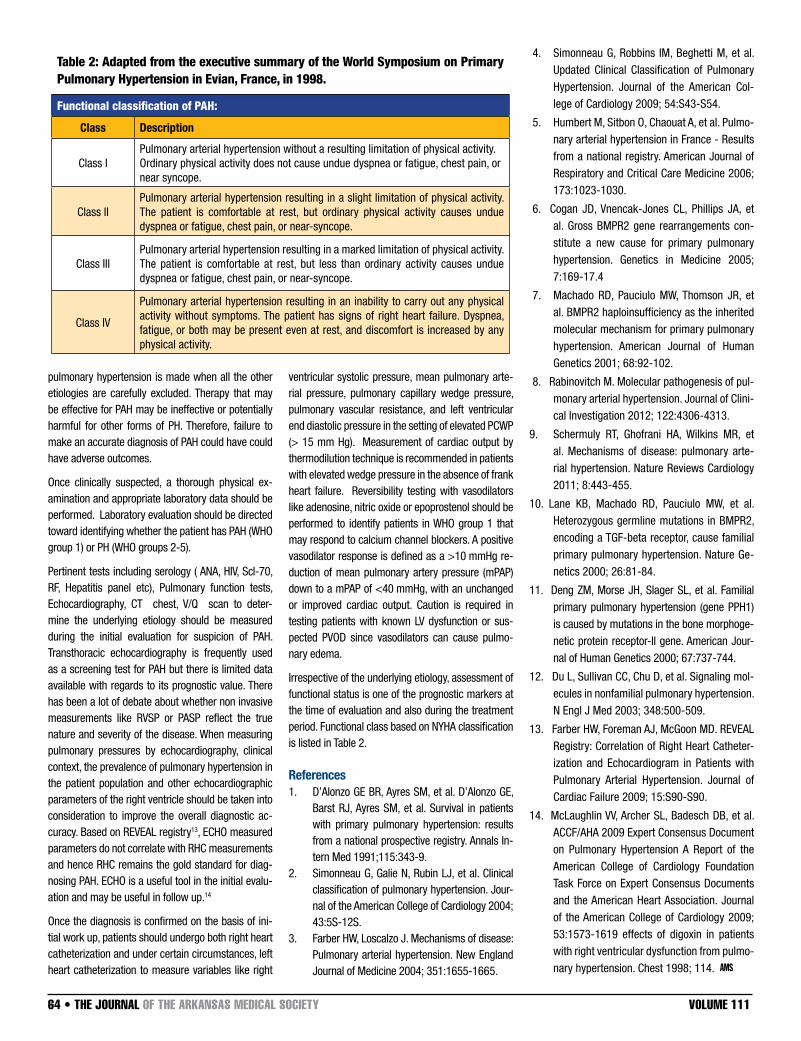
Irrespective of the underlying etiology, assessment of functional status is one of the prognostic markers at the time of evaluation and also during the treatment period. Functional class based on NYHA classification is listed in Table 2.
References1. D’Alonzo GE BR, Ayres SM, et al. D’Alonzo GE,
Barst RJ, Ayres SM, et al. Survival in patients with primary pulmonary hypertension: results from a national prospective registry. Annals In-tern Med 1991;115:343-9.
2. Simonneau G, Galie N, Rubin LJ, et al. Clinical classification of pulmonary hypertension. Jour-nal of the American College of Cardiology 2004; 43:5S-12S.
3. Farber HW, Loscalzo J. Mechanisms of disease: Pulmonary arterial hypertension. New England Journal of Medicine 2004; 351:1655-1665.
4. Simonneau G, Robbins IM, Beghetti M, et al. Updated Clinical Classification of Pulmonary Hypertension. Journal of the American Col-lege of Cardiology 2009; 54:S43-S54.
5. Humbert M, Sitbon O, Chaouat A, et al. Pulmo-nary arterial hypertension in France - Results from a national registry. American Journal of Respiratory and Critical Care Medicine 2006; 173:1023-1030.
6. Cogan JD, Vnencak-Jones CL, Phillips JA, et al. Gross BMPR2 gene rearrangements con-stitute a new cause for primary pulmonary hypertension. Genetics in Medicine 2005; 7:169-17.4
7. Machado RD, Pauciulo MW, Thomson JR, et al. BMPR2 haploinsufficiency as the inherited molecular mechanism for primary pulmonary hypertension. American Journal of Human Genetics 2001; 68:92-102.
8. Rabinovitch M. Molecular pathogenesis of pul-monary arterial hypertension. Journal of Clini-cal Investigation 2012; 122:4306-4313.
9. Schermuly RT, Ghofrani HA, Wilkins MR, et al. Mechanisms of disease: pulmonary arte-rial hypertension. Nature Reviews Cardiology 2011; 8:443-455.
10. Lane KB, Machado RD, Pauciulo MW, et al. Heterozygous germline mutations in BMPR2, encoding a TGF-beta receptor, cause familial primary pulmonary hypertension. Nature Ge-netics 2000; 26:81-84.
11. Deng ZM, Morse JH, Slager SL, et al. Familial primary pulmonary hypertension (gene PPH1) is caused by mutations in the bone morphoge-netic protein receptor-II gene. American Jour-nal of Human Genetics 2000; 67:737-744.
12. Du L, Sullivan CC, Chu D, et al. Signaling mol-ecules in nonfamilial pulmonary hypertension. N Engl J Med 2003; 348:500-509.
13. Farber HW, Foreman AJ, McGoon MD. REVEAL Registry: Correlation of Right Heart Catheter-ization and Echocardiogram in Patients with Pulmonary Arterial Hypertension. Journal of Cardiac Failure 2009; 15:S90-S90.
14. McLaughlin VV, Archer SL, Badesch DB, et al. ACCF/AHA 2009 Expert Consensus Document on Pulmonary Hypertension A Report of the American College of Cardiology Foundation Task Force on Expert Consensus Documents and the American Heart Association. Journal of the American College of Cardiology 2009; 53:1573-1619 effects of digoxin in patients with right ventricular dysfunction from pulmo-nary hypertension. Chest 1998; 114.
Table 2: Adapted from the executive summary of the World Symposium on Primary Pulmonary Hypertension in Evian, France, in 1998.
Functional classification of PAH:
Class Description
Class IPulmonary arterial hypertension without a resulting limitation of physical activity.Ordinary physical activity does not cause undue dyspnea or fatigue, chest pain, ornear syncope.
Class IIPulmonary arterial hypertension resulting in a slight limitation of physical activity. The patient is comfortable at rest, but ordinary physical activity causes undue dyspnea or fatigue, chest pain, or near-syncope.
Class IIIPulmonary arterial hypertension resulting in a marked limitation of physical activity. The patient is comfortable at rest, but less than ordinary activity causes undue dyspnea or fatigue, chest pain, or near-syncope.
Class IV
Pulmonary arterial hypertension resulting in an inability to carry out any physical activity without symptoms. The patient has signs of right heart failure. Dyspnea, fatigue, or both may be present even at rest, and discomfort is increased by any physical activity.
64 • THE JOURNAL OF THE ARKANSAS MEDICAL SOCIETY VOLUME 111

NUMBER 4 SEPTEMBER 2014 • 65
There has never been enough time. Until now.
You have the drive and the motivation to be successful, butgetting the advanced education needed is difficult due to work and family demands. Not anymore. Now you can get the MBA you have always wanted from a quality business school -- no night classes, no missing work. The Weekend MBA meets one Saturday a month in class; the rest is conveniently delivered online. Now you can balance the rest of life’s demands while pursing your degree. Visit ualr.edu/mba or call (501) 683-7490.
COLLEGE OF BUSINESS
UNIVERSITY OF ARKANSAS AT LITTLE ROCK
Weekend MBA

STUDYCASE
Neutrophilic Leukemoid Reaction in a Patient with High Grade Sarcoma
The patient is a 84-year-old Caucasian male with a
past medical history of hypertension and coronary
artery disease and surgical history of a large right
inguinal lipoma which was removed in 2006. In
October, 2011 he was seen in the clinic for a right
scrotal mass that was increasing in size. Ultrasound
showed a mass measuring 12x9x10cm which was
excised 2 months later. Histopathologic evaluation
was consistent with well-differen-
tiated liposarcoma. The WBC count
at that time was 8,000/ul (refer-
ence range: 3-12 K/ul) with a nor-
mal differential cell count. A mild
anemia was noted (HGB = 12.7 g/
dL; reference range = 13.5-17.5 g/
dL). One year later, he presented to
the clinic with left groin pain and in
the course of evaluation was found
to have WBC count of 22,000/ul
with 84.7% neutrophils (reference
range: 40-80%). A CT scan of the
abdomen and pelvis showed no
evidence of recurrence, metastatic
disease or hepatosplenomegaly.
The neutrophilic leukocytosis was
attributed to a urinary infection and
he was treated with antibiotics.
However, he continued to have left
groin pain and was reevaluated in
November 2012. Testing included a
repeat CBC which showed that the
WBC had increased to 30,000/uL
with 92% neutrophils. An extensive
evaluation for infection elsewhere
was negative. A CT scan of the pelvis and thigh was
repeated in January 2013 that showed enlargement
of left iliopsoas muscle with septations consistent
with abscess or pyomyositis and he was admit-
ted to our hospital for further evaluation. His WBC
count remained elevated and ranged from 30,000/
uL to 80,000 /uL with neutrophilic predominance.
Peripheral blood smears confirmed neutrophilia
with left shifted maturation including increased
band forms and no increase in blasts (FIG 1). Bone
marrow biopsy revealed a hypercellular marrow for
age (70% cellularity) with myeloid hyperplasia and
no dysplasia (FIG 2). FISH for BCR/ABL1 and PCR
for JAK2 V617F mutation were negative. Leukocyte
alkaline phosphatase (LAP) score was elevated at
200 (reference range: 22-124), suggestive of a leu-
kemoid reaction. Fine needle aspiration of the left
thigh lesion was suggestive of malignancy and the
tumor was surgically resected. Histologic sections
revealed pleomorphic epithelioid cells with numer-
ous atypical mitotic figures (FIG 3) with no evidence
of a well differentiated component as described for
the liposarcoma of the right scrotum. Imaging stud-
ies showed no evidence of recurrence of the origi-
nal tumor. Based on these findings, the neoplasm
was thought to represent a separate primary high
grade sarcoma rather than a dedifferentiated me-
tastasis. Postoperatively, the WBC count trended
down to 6,400/ul. Approximately
1 month later, he was readmit-
ted to our hospital for intractable
nausea and vomiting and his
white count had again increased
to 48,000/ul. Examination and
testing was consistent with acute
cholecystitis and cholecystectomy
was performed. The WBC count
remained persistently elevated af-
ter cholecystectomy and a repeat
CT of the pelvis and thigh showed
a complex mass in the left groin
measuring 11 x 9.2 x 19cm, high-
ly suggestive of recurrence of the
high grade sarcoma. At this time,
the WBC count had increased to
87,000/ul with 92% neutrophils.
Further work up was declined as
the patient elected to go on hos-
pice, expiring soon afterwards.
DiscussionThe term ‘leukemoid reaction’
refers to a reactive or acquired
leukocytosis in which the WBC ex-
ceeds 50,000/mm3.1 The periph-
by Sunita Parajuli, MD; Latha Achanta, MD, MPH; Robert H. Hopkins, Jr., MD, FACP, FAAP; Ginell Post, MDUniversity of Arkansas for Medical Sciences
Case We report a case of a patient with marked neutrophilia as a manifestation of sarcoma.
CT images of the sarcoma with arrow pointing to the area of interest.
66 • THE JOURNAL OF THE ARKANSAS MEDICAL SOCIETY VOLUME 111

eral blood smear shows neutrophilia with left shifted
granulocytic maturation, including occasional meta-
myelocytes and myelocytes without increased blasts
as seen in acute leukemia. Leukemoid reactions are
benign, but may imitate malignant processes such
as chronic myelogenous leukemia (CML). Acquired
neutrophilias can be seen with severe infection,
acute stress, drugs/hormones, severe tissue dam-
age/trauma, chronic inflammatory conditions or in
association with non-hematopoietic malignancies
including liposarcoma and sarcoma.2 Serum leuko-
cyte alkaline phosphatase (LAP) score is normal or
elevated in leukemoid reactions, but decreased in
CML. Bone marrow pathology in reactive neutrophil-
ic reactions shows a hyper cellular marrow for age
with myeloid hyperplasia and morphologically unre-
markable erythroid and megakaryocytic precursors.
Malignancy-associated leukemoid reactions can be
secondary to increased granulocyte colony stimulat-
ing factor or other cytokines produced by the neo-
plastic cells.3 A paraneoplastic leukemoid reaction
can be seen in patients with various carcinomas
such as lung cancer, melanoma, oropharyngeal car-
cinoma and sarcomas.
Very few cases of sarcomas associated with leuke-
moid reaction have been reported in the literature.
Leukemoid reactions have been associated with
dedifferentiated liposarcoma4, G-CSF secreting lung
sarcoma 5, and spindle cell sarcoma 6 in previous
publications. We would like to make clinicians aware
of the rare association of leukemoid reaction with
sarcoma occurrence and recurrence, as we have
seen in this unfortunate gentleman’s case.
References1. Halkes CJ, Dijstelbloem HM, Eelkman Rooda SJ,
Kramer MH. Extreme leucocytosis: not always leu-
kaemia. Neth J Med. 2007; 65:248–251.[PubMed]
2. Robinson WA. Granulocytosis in neoplasia. Ann N Y
Acad Sci. 1974; 230:212.
3. Hocking W, Goodman J, Golde D. Granulocytosis
associated with tumor cell production of colony-
stimulating activity. Blood. 1983; 61:600–603.
[PubMed
4. Nasser SM, Choudry UH, Nielsen GP, Ott MJ. A leu-
kemoid reaction in a patient with a dedifferenti-
ated liposarcoma. Surgery. 2001; 129:765–767.
[PubMed]
5. Jardin F, Vasse M, Debled M, Dominique S, Cour-
ville P, Callonnec F, et al. Intense paraneoplastic
neutrophilic leukemoid reaction related to a G-
CSF-secreting lung sarcoma. Am J Hematol. 2005;
80:243–245.[PubMed]
6. Snyder MC, Lauter CB. Eosinophilic and neutro-
philic leukemoid reaction in a woman with spindle
cell sarcoma: a case report. J Med Case Reports.
2010; 4:335. [PMC free article][PubMed]
7. Reichard, K. Non-neoplastic granulocytic and
monocytic disorders, excluding neutropenia in
Bone Marrow Pathology: ASCP I Series 3. Foucar
K, Richard K, Czuchlewski D. ASCP Press, Chicago
2010.
Put your business or service in the hands of 4,400 Arkansas
physicians.
For more advertising information, contact Penny Henderson at
501.224.8967 or [email protected]
1-800-455-0581
Little Rock, Arkansas
Medical BoardLegal Issues?
Darren O’Quinn
www.DarrenOQuinn.com
CallPharmacist/Attorney
FIG 1. Wright stained peripheral blood smear showing neutrophilic leukocytosis with primarily mature neutrophils with few band forms. (400X)
FIG 2: The Wright stained aspirate smear shows myeloid predominance (M:E ratio 5:1) with complete maturation. Megakaryocytes show unremarkable morphology. No eosinophila or basophilia is present. (200x)
FIG 3. H and E stained section of the left extremity tumor excised February 2013 showing large pleomorphic cells with atypical mitotic figures. (200x)
NUMBER 4 SEPTEMBER 2014 • 67

BackgroundChronic lymphocytic leukemia (CLL) represents 30% of adult leukemia. It is by far the most common leuke-mia in adults. Table A.
Table A1
Background and clinical presentation
Incidence, 201415,720 (M:9,100; F:6,620)
Death, 2014 4,600 (M and F)
Median age at diagnosis
72 years
Median age at death 79 years
Older >65 70%
Male : female 2:1
Race W>AA>others
Risk factorsFH of CLL, a personal or FH of other NHL
Clinical presentation
Asymptomatic, leukocytosisB symptoms (fever, night sweat, anorexia and weight loss)LAP, splenomegaly,anemia, thrombocytopenia, neutropenia,autoimmune phenomena (ITP or hemolytic anemia)
At the time of CLL diagnosis, the bone marrow is already involved; yet clinical cytopenias, such as anemia, thrombocytopenia and neutropenia, are not typically seen in the early clinical course. When the disease progresses, infiltration of the bone marrow by leukemic cells increases and gradually replaces the normal hematopoiesis resulting in cytopenias. The same process occurs in lymph nodes.2
DiagnosisThe diagnosis of CLL requires the presence of at least 5,000 mature B lymphocytes/μL. Fewer than this number of B cells in the absence of lymphadenopathy is defined as monoclonal B-lymphocytosis.3
The diagnosis of CLL can be made by the detection of a mature B lymphocyte clone on peripheral smear, in the bone marrow or the involved lymph node. How-ever, detection of a clone in the bone marrow is not required for diagnosis.
CLL cells express CD19, dim CD20, dim CD5, CD23, CD43, and CD79a and weakly express surface im-munoglobulin M (IgM) and IgD. CD38 expression is variable and has prognostic significance. Mantle cell lymphoma has a similar expression except it is nega-tive for CD23.3
Table B4
B cell clone characteristics
At least 5,000 mature B lymphocytes/ μL
Lymphoid cells ≤ 55% atypical/immature
Low density of surface Ig with light chain restriction
B-cell surface antigens (CD19, dim CD20, CD23)
dim CD5 surface antigen
Staging:There are two staging systems that are widely accept-ed and used interchangeably in CLL. The Rai staging system is more commonly used in the US.55,6 It incor-porates both clinical and laboratory data to classify patients into low, intermediate and high risk groups. The median survival corresponds uniformly with each group. The Binet staging system7 utilizes similar data and stratifies patients into three groups, A, B and C. It is widely used in Europe. Table C summarizes both staging systems.
Table C. Staging
System Manifestation Median survival
Rai
Low riskLymphocytosis in PB and BM
>10 years
Intermediate risk
Lymphadenopathy, splenomegaly +/− hepatomegaly
7 years
High riskAnemia, thrombocytopenia
0.75-4 years
Binet
A
Fewer than 3 areas of lymphadenopathy; no anemia or thrombocytopenia
12 years
B
More than 3 involved lymph node areas; no anemia or thrombocytopenia
7 years
CHemoglobin < 100 g/L, platelets < 100 × 10 g/L
2-4 years
Prognosis:Molecular profiling 8 helped physicians not only un-derstand the pathogenesis of CLL but also prognosti-cate and anticipate who will benefit from early treat-ment. Several cytogenetic and molecular abnormali-ties have been identified in CLL, namely, CD38, IgVH mutational status and Zap70. Deletions or gains in certain chromosomes may influence the prognosis as well. The most common chromosomal abnormali-ties found in CLL patients are deletions of 13q, 11q, or 17p and trisomy 12.4 11q and 17p deletions carry the worst prognosis with median survivals at 32 and 79 months, respectively as compared to the other abnormalities which offer median survivals exceed-ing 111 months (Table D). 9
Table D.4,8
Poor Prognostic FactorsAdvanced stage at diagnosisAdvanced ageMale sexDiffuse pattern of bone marrow infiltrationShort lymphocyte doubling timeHigh expression of Ki67, p27High serum levels of β2-microglobulin, thymidine kinase, soluble CD23, and TNFα
17p, 11q deletions & complex cytogenetics
unmutated IgVH
High level of CD38 expression
High level of ZAP70 expression
High level of expression of lipoprotein lipase
Altered microRNA expressionPoor response to therapy or short duration of response
How I Manage ChronicLymphocytic Leukemia in 2014
by Ahmed Alwbari, MD1; Issam Makhoul, MD1
1Hematology/Oncology Division – University of Arkansas for Medical Sciences
HOW I TREAT SPECIAL SERIES
68 • THE JOURNAL OF THE ARKANSAS MEDICAL SOCIETY VOLUME 111

Treatment:Asymptomatic CLL patients should be closely moni-tored every 2 to 3 months by history, physical exami-nation and CBC. Institution of chemotherapy should be based on the iwCLL guidelines.10 In a meta-analysis of these studies, there was no statistically significant difference in survival between early versus delayed chemotherapy groups with a trend toward worse survival in the early treatment group.
Physicians should not treat patients solely based on high lymphocyte count alone; they should treat only those with symptomatic disease, bulky progressive adenopathy or marrow failure. 4 Autoimmune-asso-ciated disease should be treated with steroids rather than chemotherapy.11
CLL is not a curable disease. While long-term remis-sion after chemotherapy can occur, it is only a matter of time before the CLL relapses. So the purpose of chemotherapy is to palliate the symptoms and allow for normal hematopoiesis to occur. 4
The chemotherapy of choice for symptomatic pa-tients with good performance status and no poor cy-togenetic features is Fludarabine/Cyclophosphamide/Rituximab (FCR) or Bendamustine/Rituximab (BR). Alemtuzumab-based regimens and allogeneic trans-plant are reserved for patients with 17p or 11q dele-tions. For poor performance status patients in need of treatment, single agent Rituximab, Chlorambucil or a combination of the two are good choices. IVIg should be offered to patients with hypogammglobulinemia with recurrent infections. CLL patients should be fol-lowed for second malignancies, as these are com-mon in this setting.
Recently, several new drugs have shown efficacy in CLL.12 Ibrutinib, an oral Bruton’s tyrosine kinase in-hibitor, proved to be very effective in patients with relapsed CLL with 75% of them being free of pro-gression and 83% being alive at 26 months; some of these displayed durable complete remission. 13 Idelalisib, an oral inhibitor of the delta iso-form of phosphatidylinositol 3-kinase, in combination with Rituximab, showed promising results in relapsed patients with response and one year-overall survival rates of 83% and 92%, respectively. 14 Patients with associated conditions cannot take aggressive che-motherapy and are offered Chlorambucil, Rituximab or the combination. The novel monoclonal anti-CD20 antibody Obinutuzumab showed superior efficacy when combined with Chlorambucil compared to Rituximab and chlorambucil, and there was no ad-ditional toxicity.15 Median progression free survival was 27 months and 16 months, respectively. Inter-estingly, these novel agents showed efficacy even in the unfavorable cytogenetic subsets and their toxicity was mild and manageable.
Resources:Leukemia and Lymphoma Society http://www.lls.org/
American Cancer Society http://www.cancer.org/index
University of Arkansas for Medical Sciences www.uams.edu
Clinical trials http://www.clinicaltrials.gov/ct2/home
1-800-4-CANCER
References:1. Siegel R, Ma J, Zou Z, et al. Cancer statistics, 2014.
CA Cancer J Clin 2014;64: 9-29.
2. Rozman C, Montserrat E, Rodriguez-Fernandez JM, et al. Bone marrow histologic pattern--the best single prognostic parameter in chronic lympho-cytic leukemia: a multivariate survival analysis of 329 cases. Blood 1984;64: 642-648.
3. Hallek M, Cheson BD, Catovsky D, et al. Guidelines for the diagnosis and treatment of chronic lym-phocytic leukemia: a report from the International Workshop on Chronic Lymphocytic Leukemia up-dating the National Cancer Institute-Working Group 1996 guidelines. Blood 2008;111: 5446-5456.
4. Gribben JG. How I treat CLL up front. Blood 2010;115: 187-197.
5. Rai KR, Chiorazzi N. Determining the clinical course and outcome in chronic lymphocytic leukemia. N Engl J Med 2003;348: 1797-1799.
6. Rai KR, Sawitsky A, Cronkite EP, et al. Clinical staging of chronic lymphocytic leukemia. Blood 1975;46: 219-234.
7. Binet JL, Auquier A, Dighiero G, et al. A new prog-nostic classification of chronic lymphocytic leuke-mia derived from a multivariate survival analysis. Cancer 1981;48: 198-206.
8. Gribben JG. Molecular profiling in CLL. Hematology Am Soc Hematol Educ Program 2008: 444-449.
9. Dohner H, Stilgenbauer S, Benner A, et al. Genomic aberrations and survival in chronic lymphocytic leukemia. N Engl J Med 2000;343: 1910-1916.
10. Relf M, LeJeune S, Scott PA, et al. Expression of the angiogenic factors vascular endothelial cell growth factor, acidic and basic fibroblast growth factor, tumor growth factor beta-1, platelet-derived endothelial cell growth factor, placenta growth fac-tor, and pleiotrophin in human primary breast can-cer and its relation to angiogenesis. Cancer Res 1997;57: 963-969.
11. Moreno C, Hodgson K, Ferrer G, et al. Autoimmune cytopenia in chronic lymphocytic leukemia: preva-lence, clinical associations, and prognostic signifi-cance. Blood 2010;116: 4771-4776.
12. Hallek M. Signaling the end of chronic lympho-cytic leukemia: new frontline treatment strategies. Blood 2013;122: 3723-3734.
13. Byrd JC, Furman RR, Coutre SE, et al. Targeting BTK with ibrutinib in relapsed chronic lymphocytic leukemia. N Engl J Med 2013;369: 32-42.
14. Furman RR, Sharman JP, Coutre SE, et al. Idelalisib and rituximab in relapsed chronic lymphocytic leu-kemia. N Engl J Med 2014;370: 997-1007.
15. Goede V, Fischer K, Busch R, et al. Obinutuzumab plus Chlorambucil in Patients with CLL and Coex-isting Conditions. N Engl J Med 2014.
Treatment Algorithm
NUMBER 4 SEPTEMBER 2014 • 69

P E O P L E + E V E N T S 14
AMS Fall Meeting
October 30-31The Lodge at Mount Magazine
All members are invited to discuss issues that could be considered at the 2015 legislative session. Get involved in your association and make a difference!
Basic ICD-9 Coding WorkshopAugust 28, 2014
IIAA BuildingNorth Little Rock
11th Annual AMS Insurance Conferences
October 2Jonesboro
ASU Convocation Center
October 15Springdale
Holiday Inn NWA
November 5 and 6Little Rock
Chenal Country Club
MARK YOURCALENDAR
OBITUARIES
JONESBORO – O.H. Clopton, Jr., MD, 81, passed away June 24, 2014. Dr. Clopton was a graduate
of Rector Public Schools, Murray State University, and the University of Arkansas Medical School. He
completed an internship and medical residency at Baptist Memorial Hospital, Memphis, TN. He served
a two year tour of duty in the United States Air Force, Medical Corp. He and his wife Laura Jean moved
to Jonesboro in 1965, where he started a private practice, Internal Medicine Associates. The clinic later
became Clopton Clinic, in honor of Dr. Clopton. In addition to serving his church, profession and com-
munity, Dr. Clopton held memberships in The Arkansas Medical Society where he was a member of
the Fifty Year Club; American Medical Society; Arkansas Thoracic Society; American Society of Internal
Medicine; Memphis Academy of Internal Medicine and was a teaching assistant for the University of
Arkansas for Medical Sciences. He is survived by his wife of 60 years, Laura Jean Shemwell Clopton,
two daughters: Dr. Claudia L. Clopton of Denver, CO; Dr. Laura D. Bischof, (John) St. Paul, MN; and
several grandchildren.
MABELVALE – Richard Parker Armstrong, MD, 75, passed away June 9, 2014. Dr. Armstrong was born
March 24, 1939, in Toronto, Ontario, Canada. He received his MD degree from University of Manitoba
in Winnipeg, Manitoba, Canada; he served in the Canadian Army Medical Corps; He was currently Vice
President of Medical Affairs for QualChoice, Little Rock, AR. Dr. Armstrong was a member of the Arkan-
sas Medical Society as a Fifty Year Club member, American Academy of Family Physicians, American
Medical Association, American College of Physician Executives, and American College of Utilization
Review Physicians. His distinguished career included, President, Chairman, and Vice President for sev-
eral medical institutions; Medical Director for Prudential Insurance and Vice President of Medical Affairs
for Blue Cross Blue Shield, spanning over 50 years’ service within the Medical Industry.He is survived
by his fiancée Shirley Caldwell of Clarksville, Arkansas; daughter, Cara Bourassa her husband, Jim, of
Tucson, Arizona; daughter Valerie Carr her husband, Steve, of Town and Country, Missouri; and daughter
Claire Kampmann her husband, Thorsten, of Vettweiss, Germany, and grandchildren, Jason Bourassa,
Jack, Charlie, his wife, Ruby and Max Carr.
GROUP MEMBERSHIP PROGRAM FOR CLINICS
AMS group membership eliminates the burden of collecting individual renewal statements
each year with one, itemized statement for your entire clinic. The program also connects
clinic administrators and staff directly with the Society, providing exclusive benefits and
direct access to the information and resources they need. Group memberships are available
at no extra cost to groups with 5 or more physicians and require 100% physician participa-
tion in the Society. If you would like more information on getting your group involved, please
contact [email protected] or apply online at www.arkmed.org/group.
70 • THE JOURNAL OF THE ARKANSAS MEDICAL SOCIETY VOLUME 111

NUMBER 4 SEPTEMBER 2014 • 71

72 • THE JOURNAL OF THE ARKANSAS MEDICAL SOCIETY VOLUME 111
Proudly Serving
forArkansas Physicians
1989-2014
25Years
SVMIC is celebrating its 25th year of providing medical professional liability coverage to physicians in The Natural State.
We are rated “A” (Excellent) by A.M. Best Company and, since 1990, have returned more than $13 million in dividends
to Arkansas physician policyholders. Our Arkansas Advisory Committee is comprised of nine physicians from various
specialties and cities across Arkansas who review claims and make underwriting decisions for Arkansas physicians on
behalf of SVMIC. Three of those members sit on our Board of Directors. Combine that with three professionals based in
Arkansas to directly serve the needs of local policyholders, and it’s clear that SVMIC is the right choice for you. Simply
put, your interests are our interests.
Mutual Interests. Mutually Insured.
Contact Sharon Theriot or Mandy Holmes at [email protected] or call 1-800-342-2239.
Follow us on Twitter @SVMIC www.svmic.com