targeting of gene expression to the trabecular …2755/datastream/obj/... · nerve head (onh) and...
TRANSCRIPT
TARGETING OF GENE EXPRESSION TO THE
TRABECULAR MESHWORK OF GLAUCOMATOUS
BEAGLES BY NON-SELF-COMPLEMENTARY AAV2
By
Annie Oh
A THESIS
Submitted to
Michigan State University
in partial fulfillment of the requirement
for the degree of
Comparative Medicine and Integrative Biology- Master of
Science
2014

ABSTRACT
TARGETING OF GENE EXPRESSION TO THE TRABECULAR MESHWORK OF
GLAUCOMATOUS BEAGLES BY NON-SELF-COMPLEMENTARY AAV2
By
Annie Oh
Glaucoma is a leading cause of irreversible blindness in humans and dogs. Increased
intraocular pressure (IOP) due to abnormal aqueous humor outflow through the trabecular
meshwork (TM) is a major risk factor and is based on genetic predisposition. The purposes of
these studies were to target gene expression to the canine TM and prevent or reverse IOP
elevation in beagle dogs with inherited primary open angle glaucoma (POAG). In these animals,
the disease is caused by a missense mutation in the ADAMTS10 gene. Green fluorescent protein
(GFP) reporter gene expression was successfully targeted to the conventional aqueous humor
outflow pathway of wild type and ADAMTS10-mutant dogs using a non-self-complementary
adeno-associated virus serotype 2 (AAV2) with a single capsid mutation: AAV2(Y444F)-smCBA-
GFP (2 x 10¹⁰
vg/mL to 2 x 10¹²
vg/mL; 50 μL). The triple (Y444,500,730F) and quadruple
(Y444,500,730F + T491V) mutant AAV2s were ineffective. Subsequent gene replacement
therapy was performed in pre-glaucomatous (n=7) and glaucomatous (n=3) ADAMTS10-mutants
with AAV2(Y444F)-smCBA-hADAMTS10 at the highest dose (2 x 10¹²
vg/mL; 50 μL). The IOP
of these animals were monitored weekly for 19 weeks. While the treatment was deemed safe
with no severe adverse effects, a decrease in IOP was not observed. If the transgene was
expressed, a therapeutic effect could probably be achieved by increasing the vector dose and
number of transduced TM cells.

Copyright by
ANNIE OH
2014
iv
ACKNOWLEDGEMENTS
I would like to acknowledge and thank the many people that have helped me throughout
the thesis process.
I first need to acknowledge my mentor, Dr. András Komáromy, for his unending support,
patience, and kindness. He has always been a great inspiration and I can never thank him
enough for all the great opportunities and fun-filled memories. I have learned so much and am
truly grateful for having the chance to work with him.
To Dr. Simon Petersen-Jones and Dr. Joshua Bartoe for their generous support,
mentorship, and knowledge that I will cherish throughout the course of my career. Dr. Vilma
Yuzbasiyna-Gurkan, to whom I owe so much for her mentorship, knowledge, and enthusiasm
during this process. Most importantly, for giving me the opportunity to pursue a Masters degree
in the CMIB program. To my committee members, Dr. Sayoko Moroi, and Dr. Arthur Weber
for their guidance, and input that has furthered my scientific development during my degree.
To the Hauswirth‟s and Boye‟s lab at the University of Florida, for their expertise in
vector technology. To Dr. Gui-shuang Ying and Jiayan Huang at the University of Pennsylvania
for their knowledge in biostatistics.
To everyone in the Komáromy lab – Christine Harman, and Kristin Koehl, for being great
friends and fantastic teachers. Forrest Nussdorfer, Josh Laske, Ron Tsai, and Monica Choo, for
their help during experiments, and of course, all the laughs and joyful memories. To the
graduate students in Dr. Petersen-Jones‟s and Dr. Lorraine Sordillo‟s lab and the ophthalmology
residents, particularly Dr. Laurence Occelli, for helping me with procedures, answering my
v
random questions, and being such a supportive friend during this process, and Dr. Connie Yeh,
for all her kindness, advice, and fun nights out on the town.
Individuals at MSU - Lisa Allen and her staff for taking amazing care of the Vivarium
dogs. And great thanks to Ramona for her enthusiasm and lovely smile. Dr. Victoria Hoelzer-
Maddox for her administrative support that has helped me so much during my time at MSU. Dr.
Melinda Frames at the Center for Advanced Microscopy for taking incredible images for the
figures in the thesis.
Financial support from The Glaucoma Research Foundation, NIH Grants T32OD011167
(Michigan State University), EY021721 (University of Florida), and P30EY021721 (University
of Florida), Research to Prevent Blindness (University of Florida), Foundation Fighting
Blindness (University of Florida), and MSU Faculty Startup Funds.
To the Edward Sheppard and family for the unrestricted gift.
To my family, for all their love and to whom I owe everything.
Most importantly, to the Vivarium dogs that have sacrificed their lives for the
advancement of science. Thank you and may you rest in peace in heaven.
vi
TABLE OF CONTENTS
LIST OF TABLES vi
LIST OF FIGURES vii
KEY TO ABBREVIATIONS viii
CHAPTER 1 – INTRODUCTION 1
REFERENCES 3
CHAPTER 2 – LITERATURE REVIEW 7
Aqueous humor dynamics 7
Aqueous humor outflow pathways 7
Extracellular matrix turnover and outflow resistance 10
Animal models of glaucoma 12
Primary open angle glaucoma in beagle dogs 14
ADAMTS10 gene 17
Viral vectors of gene therapy 18
REFERENCES 21
CHAPTER 3 – MATERIALS AND METHODS 29
Study design 29
Animals 30
AAV constructs 32
Intracameral injections 34
Tonometry 37
Steroid response test 38
Ophthalmic examination 38
Tissue processing/sectioning 39
Immunohistochemistry and image analysis 41
Statistical analysis 42
REFERENCES 43
CHAPTER 4 – RESULTS 45
AAV2(Y444F) targets GFP expression to the canine wt ICA 45
AAV2(Y444F) targets GFP expression to the canine ADAMTS10-mutant ICA 46
Therapeutic effect of AAV2(Y444F)-hADAMTS10 46
Steroid responsiveness of ADAMTS10-mutants and carrier 51
Clinical signs in wt and ADAMTS10-mutants 57
REFERENCES 59
CHAPTER 5 – CONCLUSION & FUTURE STUDIES 61
REFERENCES 69

vii
LIST OF TABLES
Table 2.1 Examples of glaucoma animal models 13
Table 2.2 Top breeds with high prevalence of primary glaucoma (1994 – 2002) 29
15
Table 3.1 Summary of the cohorts 31
Table 4.1 Semi-quantitative analysis of GFP expression 48
Table 4.2 Summary of excluded and abbreviated IOP data 53
Table 4.3 Statistics on IOP data 54
Table 5.1 Positive and negative IHC markers for TM cells 63
viii
LIST OF FIGURES
Figure 2.1 Hybrid aqueous outflow pathways of humans and dogs 9
Figure 3.1 Map of vector plasmids 33
Figure 3.2 Examples of an intracameral injection 36
Figure 3.3 Four quadrants of the anterior segment 40
Figure 4.1 ICA of wt dogs 47
Figure 4.2 ICA of ADAMTS10-mutant dogs 49
Figure 4.3 Posterior pupillary margin of iris of ADAMTS10-mutant dogs 50
Figure 4.4 IOP in ADAMTS10-mutant dogs injected with 52
AAV2(Y444F)-hADAMTS10
Figure 4.5 IOP outcomes of AAV2-GFP 55
Figure 4.6 Effects of steroids on IOP in ADAMTS10-mutant and carrier dogs 56
ix
KEY TO ABBREVIATIONS
Adeno-associated virus AAV
Adeno-associated virus serotype 2 AAV2
Association for Research in Vision and Ophthalmology ARVO
Balanced salt solution BSS
Basic local alignment search tool BLAST
Canine ADAMTS10 cADAMTS10
Confocal scanning laser ophthalmoscopy cSLO
Cross-linked actin networks CLAN
Extracellular matrix ECM
Fibrillin-1 FBN1
Glycosaminoglycan GAG
Generalized estimating equations GEE
Human ADAMTS10 hADAMTS10
Human embryonic kidney 293 variant cells HEK293T
Immunohistochemistry IHC
Inferior-nasal IN (AS2)
Inferior-temporal IT (AS3)
Intraocular pressure IOP
Inverted terminal repeat IRT
Iridocorneal angle ICA
Juxtacanalicular connective tissue JCT
x
Marfan syndrome MS
Matrix metalloprotease MMP
Mega base pairs Mb
Michigan State University MSU
Myocilin MYOC
Non-steroidal anti-inflammatory drug NSAID
Optic nerve head ONH
Optineurin OPTN
Phosphate buffered saline PBS
Primary open angle glaucoma POAG
Retinal ganglion cell RGC
Schlemm‟s canal SC
Superior-nasal SN (AS1)
Superior-temporal ST (AS4)
Truncated hybrid chicken beta actin promoter smCBA
Transforming growth factor-beta TGF-β
Transforming growth factor-beta 2 TGF-β2
Tissue inhibitor of metalloprotease TIMP
Trabecular meshwork TM
Vector genome per milliliter vg/mL
Weill-Marchesani syndrome WMS
Wild type wt

1
CHAPTER 1 - INTRODUCTION
Glaucoma is a common cause of blindness worldwide as an estimated 79.6 million people
will be affected and 11.1 million people will be bilaterally blind by 2020.1 The disease is defined
as an optic neuropathy resulting in vision loss due to structural and functional injury to the optic
nerve head (ONH) and retinal ganglion cells (RGCs).2 Of the different types, primary open
angle glaucoma (POAG) is the most prevalent form.1 Unfortunately there is no cure, and
lifelong monitoring and treatment is required in affected individuals.
POAG is a multifactorial disorder with a largely unknown pathogenesis.3 Elevated
intraocular pressure (IOP) is a major risk factor and the consequence of increased resistance to
outflow at the trabecular meshwork (TM).4, 5
Family history is another major risk factor as a
confirmed first-degree relative raises the probability for an individual to develop POAG by ten
times.6 The disease does not follow a Mendelian inheritance pattern (single gene inheritance),
and its onset and progression is influenced by multiple genes.7 Traditional linkage analysis and
genome wide association studies have identified several genes that are potentially coupled with
POAG and/or IOP. The three well-established POAG genes are MYOC, OPTN, and WDR36.3
TMCO1 and GAS7 are loci reported to be associated with regulating IOP.8, 9
The discovery of
novel genetic variants continues to expand the glaucoma genomic database, and paves the way
for new diagnostics and treatments, such as DNA-based diagnostic testing, personalized
medicine, and ocular gene therapy.10
Successful gene augmentation was previously demonstrated in RPE65-mutant dogs with
an adeno-associated viral (AAV) vector.11, 12
The methods were then translated to human
clinical trials, which led to the restoration of vision in Leber Congenital Amaurosis type 2

2
patients.13-17
Successful proof-of-concept therapies have also been demonstrated in canine
models of achromatopsia,18
and retinitis pigmentosa.19-21
The results from these experiments
have not only placed canines at the forefront of vision research, but exemplify the significant
impact large animal models have in the field of translational medicine.
One of the best characterized and clinically relevant spontaneous animal models for
POAG is the ADAMTS10-mutant beagle dog.22,23
ADAMTS10 encodes a metalloprotease that is
highly expressed in the TM and involved in the formation of extracellular matrix (ECM). 24
The
identification of the underlying G661R missense mutation in this causal gene provides a unique
opportunity to study gene enhancement therapy. Therefore, the purpose of these experiments
was to target gene expression with AAV to the canine TM and prevent or reverse IOP elevation
in beagle dogs with inherited POAG. We hypothesize that introducing the wt ADAMTS10 cDNA
to the TM will rescue the POAG disease phenotype. The significance of our study was to
establish the groundwork for TM-directed gene therapy in a large animal model.

3
REFERENCES

4
REFERENCES
1. Quigley HA, Broman AT. The number of people with glaucoma worldwide in 2010
and 2020. The British Journal of Ophthalmology 2006;90:262-267.
2. Foster PJ, Buhrmann R, Quigley HA, Johnson GJ. The definition and classification of
glaucoma in prevalence surveys. The British Journal of Ophthalmology 2002;86:238-
242.
3. Gemenetzi M, Yang Y, Lotery AJ. Current concepts on primary open-angle glaucoma
genetics: a contribution to disease pathophysiology and future treatment. Eye
2012;26:355-369.
4. Quigley HA. Glaucoma. Lancet 2011;377:1367-1377.
5. Coleman AL, Miglior S. Risk factors for glaucoma onset and progression. Survey of
Ophthalmology 2008;53 Suppl1:S3-10.
6. Wolfs RC, Klaver CC, Ramrattan RS, van Duijn CM, Hofman A, de Jong PT.
Genetic risk of primary open-angle glaucoma. Population-based familial aggregation
study. Archives of Ophthalmology 1998;116:1640-1645.
7. Wiggs JL. Genetic etiologies of glaucoma. Archives of Ophthalmology 2007;125:30-
37.
8. van Koolwijk LM, Ramdas WD, Ikram MK, et al. Common genetic determinants of
intraocular pressure and primary open-angle glaucoma. PLoS Genetics
2012;8:e1002611.
9. Ozel AB, Moroi SE, Reed DM, et al. Genome-wide association study and meta-
analysis of intraocular pressure. Human Genetics 2014;133:41-57.
10. Moroi SE, Raoof DA, Reed DM, Zollner S, Qin Z, Richards JE. Progress toward
personalized medicine for glaucoma. Expert Review of Ophthalmology 2009;4:145-
161.
11. Acland GM, Aguirre GD, Ray J, et al. Gene therapy restores vision in a canine model
of childhood blindness. Nature Genetics 2001;28:92-95.
12. Narfstrom K, Katz ML, Bragadottir R, et al. Functional and structural recovery of the
retina after gene therapy in the RPE65 null mutation dog. Investigative
Ophthalmology & Visual Science 2003;44:1663-1672.
5
13. Bainbridge JW, Smith AJ, Barker SS, et al. Effect of gene therapy on visual function
of Leber‟s congenital amaurosis. The New England Journal of Medicine
2008;358:2231-2239.
14. Maguire AM, Simonelli F, Pierce EA, et al. Safety and efficacy of gene transfer for
Leber's congenital amaurosis. The New England Journal of Medicine 2008;358:2240-
2248.
15. Hauswirth WW, Aleman TS, Kaushal S, et al. Treatment of leber congenital
amaurosis due to RPE65 mutations by ocular subretinal injection of adeno-associated
virus gene vector: short-term results of a phase I trial. Human Gene Therapy
2008;19:979-990.
16. Simonelli F, Maguire AM, Testa F, et al. Gene therapy for Leber's congenital
amaurosis is safe and effective through 1.5 years after vector administration.
Molecular Therapy: The Journal of the American Society of Gene Therapy
2010;18:643-650.
17. Jacobson SG, Cideciyan AV, Ratnakaram R, et al. Gene therapy for leber congenital
amaurosis caused by RPE65 mutations: safety and efficacy in 15 children and adults
followed up to 3 years. Archives of Ophthalmology 2012;130:9-24.
18. Komaromy AM, Alexander JJ, Rowlan JS, et al. Gene therapy rescues cone function
in congenital achromatopsia. Human Molecular Genetics 2010;19:2581-2593.
19. Beltran WA, Cideciyan AV, Lewin AS, et al. Gene therapy rescues photoreceptor
blindness in dogs and paves the way for treating human X-linked retinitis pigmentosa.
Proceedings of the National Academy of Sciences of the United States of America
2012;109:2132-2137.
20. Lheriteau E, Petit L, Weber M, et al. Successful gene therapy in the RPGRIP1-
deficient dog: a large model of cone-rod dystrophy. Molecular Therapy: The Journal
of the American Society of Gene Therapy 2014;22:265-277.
21. Petit L, Lheriteau E, Weber M, et al. Restoration of vision in the pde6beta-deficient
dog, a large animal model of rod-cone dystrophy. Molecular Therapy: The Journal of
the American Society of Gene Therapy 2012;20:2019-2030.
22. Gelatt KN, Gilger BC, Kern TJ. Veterinary Ophthalmology. 5th ed. Ames, Iowa:
Wiley-Blackwell; 2013.
23. Kuchtey J, Olson LM, Rinkoski T, et al. Mapping of the disease locus and
identification of ADAMTS10 as a candidate gene in a canine model of primary open
angle glaucoma. PLoS Genetics 2011;7:e1001306.

6
24. Apte SS. A disintegrin-like and metalloprotease (reprolysin-type) with
thrombospondin type 1 motif (ADAMTS) superfamily: functions and mechanisms.
The Journal of Biological Chemistry 2009;284:31493-31497.
7
CHAPTER 2 - LITERATURE SURVEY
Aqueous humor dynamics
The ciliary body is an anterior continuation of the choroid and is topographically divided
into two regions: the anterior pars plicata and posterior pars plana.1 Ciliary processes in the pars
plicata produce aqueous humor through diffusion, ultrafiltration, and active secretion of solutes.
Diffusion occurs down a concentration gradient across cell membranes, and ultrafiltration
encompasses hydrostatic pressures that force water and water-soluble substances across the
fenestrated ciliary capillary endothelium. These two passive mechanisms create a „reservoir‟ of
plasma ultrafiltrate in the ciliary stroma. Certain ions and substances are then actively secreted
across the nonpigmented ciliary epithelium into the posterior chamber to form aqueous humor.
The Na+/K+ ATPase complex and carbonic anhydrase are two enzymes associated with the
active transport of solutes and are responsible for 80-90% of total fluid formation.2
Once generated, aqueous humor travels from the posterior chamber to the anterior
chamber, providing nutrients and removing waste products for the avascular lens and cornea. It
finally exits the eye through the trabecular and uveoscleral outflow pathways of the ICA.1 In the
healthy eye, moderate aqueous outflow resistance in the ICA is required to generate IOP (10-20
mm Hg) for proper maintenance of globe shape and optics for vision. Thus, tightly controlled
aqueous humor production and outflow drainage are important processes for normal ocular
function.2
Aqueous humor outflow pathways
The main route for aqueous humor exit is through the conventional or trabecular outflow
pathway. In humans, this route is comprised of the TM, Schlemm‟s canal (SC), collector
8
channels, and aqueous veins that lead into the episcleral venous system.3 The SC is a single
circular structure that collects aqueous humor before it enters the bloodstream. A minor pathway
(4-14%) also exists, called the unconventional or uveoscleral outflow pathway. Fluid flows
through the ciliary muscle bundles to the suprachoroidal space before diffusing across the sclera
into the intrascleral venous system.4 The canine conventional pathway (Figure 2.1) is different
because pectinate ligaments secure the iris to the limbal cornea, and instead of having a SC, the
dog possesses multiple vessels collectively known as the angular aqueous venous plexus.5 The
canine uveoscleral pathway is similar to humans, and is responsible for ~15% of total aqueous
humor outflow.1
The TM is critical in the regulation of aqueous humor outflow resistance and generation
of IOP. In humans, it is located in the anterior region of the ICA at the scleral sulcus and
consists of three areas, the uveal meshwork, corneoscleral meshwork, and juxtacanalicular tissue
(JCT).3 The uveal and corneoscleral meshworks are organized and comprised of fenestrated
trabecular beams with large intertrabecular spaces between adjacent sheets.6 In contrast, the JCT
does not form trabecular beams and is mainly composed of a loose network of extracellular
matrix (ECM).7 All three regions are embedded with TM cells that protrude fibroblastlike
processes, and communicate with adjacent cells and those of the inner endothelium of the SC.
These cells also exhibit phagocytic properties that may act to remove cellular debris.3 The inner
endothelial wall of the SC forms giant vacuoles with intra- and paracellular pores which open
into the lumen in response to pressure from aqueous humor flow. These pores control fluid flow
and generate up to10% of the total resistance in normal human eyes depending on the number of
pores present in the inner wall.8, 9

9
Figure 2.1: Hybrid aqueous outflow pathways of humans and dogs. The aqueous humor outflow pathways in the dog have two
main distinctive structures: the pectinate ligaments and the aqueous venous plexus.
10
In canines, the TM is located in the posterior region of the ICA within the ciliary cleft. The tissue
structures of the canine TM outflow pathway, analyzed by transmission and scanning electron
microscopy, are similar to the humans‟ as describe above.1, 5, 10
Extracellular matrix turnover and outflow resistance
The precise location of aqueous humor outflow resistance in normal and glaucomatous
eyes is still under debate.11
It is widely hypothesized that the bulk of resistance in both normal
and glaucomatous eyes occurs in the JCT of the TM; a region that is highly dynamic and
undergoes constant remodeling. Normal homeostatic adjustments of outflow resistance appears
to be triggered by embedded TM cells that sense mechanical stretch from IOP and respond by
secreting matrix metalloproteases (MMPs).12
Studies with mechanical stretching models in vitro
have shown an increase in MMP1, 2, 3, 9, and 14.12, 13
These enzymes selectively breakdown
ECM components including proteoglycans, laminin, fibronectin, collagens (type IV and type VI),
elastin, and osteonectin7 contributing to the expansion of the JCT and thereby permitting
increased aqueous humor outflow.14
Additional proteinases, such as ADAM, ADAMTS, and
tissue plasminogen activator, also play possible roles in modulating outflow resistance.13
Normal ECM homeostasis is tightly controlled by enzymes such as tissue inhibitor
metalloproteases (TIMPs).13
However, reduced or disorganized ECM turnover rate may lead to
increased aqueous humor outflow resistance as seen in POAG. Abnormal ECM remodeling
mainly results in an accumulation of excess material and fibrosis of the TM.6 These
accumulations are normally present in older individuals, but there is a significantly greater
amount of fibrosis and certain components, such as proteoglycans, in the glaucomatous TM.15
11
Transforming growth factor beta 2 (TGF-β2) is a profibrotic cytokine that is normally
secreted into aqueous humor by TM cells, ciliary body epithelium, and the lens, to promote
ocular immune privilege.16-18
In glaucomatous patients, excessive levels of this cytokine have
been consistently identified.19
Studies with human TM cells in monolayer cell cultures revealed
that exogenous perfusion of TGF-β2 leads to the increased synthesis and expression of a broad
variety of proteins, including collagens, elastin, fibronectin, laminin, and myocilin, in addition to
plasminogen activator inhibitor (PAI-1), which inhibits MMPs.16
It also induces the synthesis of
ECM cross-linking enzymes such as tissue transglutaminase (TGM2), lysyl oxidase (LOX), and
lysyl oxidase-like proteins.17
Furthermore, anterior eye segment perfusion culture models
treated with TGF-β2 revealed a measurable decrease in fluid outflow resulting in increased
IOP.20
Therefore, excess TGF-β2 may be linked to the pathogenesis of POAG.
Glucocorticoids (GCs) have also been shown to disorganize ECM degradation.
Administration of GCs results in ocular hypertension in approximately 40% of the general
human population.21
These „steroid responders‟ are likewise more prone to develop POAG
compared to nonresponders. Studies have shown that GCs inhibit TM cell phagocytosis, MMP
activity, and subsequently increase the production of fibrillar proteins. Perfusion of human
anterior segments also demonstrated that GCs gradually remodel TM cytoskeleton to form cross-
linked actin networks (CLANs). The purpose of the CLANs in the outflow pathway is still
unknown, but the unusual arrangement may block aqueous humor outflow.22

12
Animal models of glaucoma
In vitro and in vivo studies are crucial in understanding the fundamental aspects of human
glaucoma. In particular, in vivo experiments in animal models have become the key medium for
translational research.23
Several large and small species have been categorized as spontaneous,
induced, or transgenic models of glaucoma (Table 2.1 examples). In spontaneous models, the
disease is inherited or occurs naturally.24
In induced and transgenic models, the animals‟
anatomic structures or genome are altered to exhibit glaucomatous phenotypes.25
In the end, all
glaucoma models aim to simulate elevated and sustained IOP, and/or RGC loss.
There are advantages and disadvantages to every animal model. For example, nonhuman
primates have a well-developed SC, lamina cribrosa, peripapillary sclera and blood supply that is
virtually identical to the humans.26
Unfortunately, nonhuman primates may not be practical for
many research groups because they are expensive, have limited availability, hard to handle, and
require an experienced staff in addition to special housing facilities.27
Even though a colony of
rhesus monkeys was previously described to exhibit spontaneous POAG,28
there is currently no
natural disease model available in nonhuman primates.
The mouse is simple to upkeep, produces large colonies, and has a SC.27, 29
Since their
genomes are thoroughly mapped and easy to manipulate, they have been essential in
understanding the outcomes of wild type (wt) and mutant gene products.23
However, a
disadvantage of the mouse is the size of their eyes, and the poorly developed lamina cribrosa
which lacks choroidal vascular supply.30
Depending on the question at hand, different animal
models provide the tools required for a greater understanding of glaucoma.

13
Table 2.1: Examples of glaucoma animal models.
Species Size Model mode Method References
Monkey Large Spontaneous Inherited 26
Induced Laser photocoagulation of TM 64
Induced Intracameral injection of latex
microspheres
65
Dog Large Spontaneous Inherited 34, 51
Cat Large Spontaneous Inherited
Sheep and
cow
Large Induced Application of glucocorticoids 66, 67
Mice Small Transgenic MYOC mutation 68
Transgenic OPTN mutation 69
Transgenic α1 subunit of collagen Type 1
mutation
70
Rabbit Small Induced Application of glucocorticoids 71
Zebrafish Small Transgenic Lrp2 mutation 72
TM: trabecular meshwork; MYOC: myocilin; OPTN: optineurin; Lrp2: low-density lipoprotein-
related protein 2
14
Primary open angle glaucoma in beagle dogs
Among all animal species studied, dogs have the highest prevalence of primary glaucoma
comparable to the disease frequency in humans. The highest frequency of primary glaucomas are
found in purebred dogs (0.89%) with twenty-two breeds (Table 2.2), including the American
Cocker Spaniel (5.52%), Bassett Hound (5.44%), and Chow Chow (4.70%), having a prevalence
greater than 1%.31
Contrary to the human glaucomas, the closed-angle form occurs more
frequently in the general canine population, while the open angle form is considered to be rare.32
Thanks to a colony of dogs maintained for ~40 years at the University of Florida, POAG in
beagle dogs is one of the best studied and well-established natural/spontaneous animal models
for glaucoma.33
It is an autosomal recessive disorder caused by a missense mutation in
ADAMTS10,34, 35
and has a predictable onset with severe glaucomatous changes occurring later
in life.36
The slow and progressive nature of the disease provides windows of opportunity for
various biochemical and morphologic research.24
Beagle dogs with inherited POAG are also a
valuable animal model because they share many phenotypic characteristics with the human
form.1 Furthermore, the dogs‟ eye size and ocular anatomy make them excellent subjects for
clinical studies.27
Early studies have defined three stages of the disease: early (8-16 months), moderate (13-
30 months), and advanced (2-4 years).37
At 8-16 months of age, pressures begin to rise from the
normal range of 10-20 mmHg, and by 2-4 years, the mean IOPs range from 25-40 mmHg.
Notably, the ICA of POAG beagle dogs is open until the late disease stages.38

15
Table 2.2: Top breeds with high prevalence of primary glaucoma (1994 – 2002).29
Breed % Affected
Overall 0.89%
American Cocker Spaniel 5.52%
Bassett Hound 5.44%
Chow Chow 4.70%
Shar-Pei 4.40%
Boston Terrier 2.88%
Fox Terrier, Wire 2.28%
Norwegian Elkhound 1.98%
Siberian Husky 1.88%
Cairn Terrier 1.82%
Poodle, Miniature 1.68%
Samoyed 1.59%
Bichon Frise 1.59%
Shih Tzu 1.58%
Australian Cattle Dog 1.51%
Akita 1.39%
Jack Russell Terrier 1.37%
English Cocker Spaniel 1.35%
Lhasa Apso 1.33%
Bouvier des Flandres 1.31%
Pekingese 1.22%
Poodle, Toy 1.20%
Beagle 1.10%

16
Analogous to the human form, the exact source of increased aqueous humor outflow
resistance is still unknown. Transmission electron microscopy of normal and affected eyes
revealed a fully differentiated ICA devoid of developmentally abnormalities at 3 months of
age.10
However at 12 months, clustering of fibrils and irregular elastic fibers were observed, and
extracellular debris, such as glycosaminoglycan-like hyaluronidase-resistant material, start to
appear in the TM.39
Increased amounts of myocilin have also been localized to this region, and
aqueous humor levels are increased in glaucomatous dogs.40, 41
The role of myocilin is unknown,
but mutations in this gene account for 3% of human POAG and the variant protein is
hypothesized to decrease aqueous humor outflow facility.42
In glaucomatous dogs, aqueous
humor outflow facility, measured by pneumotonography, gradually decreases from 0.19 ± 0.07
μL/min/mmHg at 3-6 months of age to 0.07 ± 0.05 μL/min/mmHg at 43-48 months of age.1 In
the final stages of the disease, the trabeculae are compressed and disorganized, and the ICA is
clinically narrowed and occasionally closed.10
Consequently, sustained elevated IOP is the main
cause of damage in these animals.
Another feature of glaucoma in beagles is lens zonule pathology. Early in the disease,
stretching and tears of the lens zonules is observed at maximum mydriasis (dilation of the
pupil).43
As the IOP continues to rise, progressive focal disinsertion of these structures leads to
subluxation of the lens and prolapse of the vitreous humor into the anterior chamber.37
Increased
pressures also results in an enlargement of the globe.1 Posterior structures of the eye, specifically
the ONH and RGCs, are also severely affected. In dogs, the normal ONH slightly protrudes into
the vitreous chamber due to myelination of nerve fibers prior to the lamina cribrosa,44
and at 5-6
months of age there is no noteworthy difference between normal and pre-glaucomatous
individuals.45
With progressive elevations in pressure, variable loss of RGC axons and myelin,

17
in addition to the posterior displacement of the lamina cribrosa, results in ONH atrophy and
cupping. 36, 37, 46
Along with demyelination, the mechanical impact on RGC axons results in
reduced axoplasmic flow.46, 47
Consequently, the excessive release of excitotoxic amino acid
glutamate by dying RGCs is suspected to cause further injury and death to neighboring cells.48
ADAMTS10 gene
ADAMTS10 is part of a superfamily of secreted proteases involved in the formation of
ECM.49
A G661R missense mutation in ADAMTS10 was identified as the cause for beagle
POAG. While the exact molecular mechanism still needs to be determined, the gene is highly
expressed in the TM, and the mutation results in a misfolded protein that may be responsible for
the excessive accumulation of ECM material in the TM of affected dogs.34
Following these
results, the „Microfibril hypothesis of glaucoma‟ was proposed which postulates that genetic
mutations in microfibril-associated genes, including Fibrillin-1 (FBN1) and ADAMTS10, lead to
altered connective tissue integrity. The abnormal connective tissue integrity results in
dysregulation of growth factors signaling, most notably TGF-β2.50
In man, mutations in ADAMTS10 as well as related genes ADAMTS17 and FBN1
involved in microfibril metabolism are responsible for diseases such as Weill-Marchesani
syndrome (WMS) and Marfan syndrome (MS). These inherited connective tissue disorders are
characterized by skeletal abnormalities including brachydactyly in WMS and arachnodactyly in
MS. The ocular phenotypes of both syndromes include ectopia lentis and glaucoma.51
Ectopia
lentis is likely due to mutated FBN1 and ADAMTS10 proteins, which are required for lens
zonule formation and maintenance.51, 52
FBN1 is a major component of lens zonule microfibrils
and ADAMTS10 protein accelerates FBN1 microfibril formation in fibroblast culture.53
The

18
glaucoma phenotype is different between WMS and MS. In WMS, the lens may subluxate into
the pupil or anterior chamber and cause secondary acute angle closure glaucoma.51
In contrast,
the lens tends to subluxate posteriorly in MS, and the glaucoma phenotype is an open-angle.54
Interestingly in dogs, there is no systemic phenotype seen with ADAMTS10 and
ADAMTS17 mutations. While the variant ADAMTS17 described in terrier breeds results in
primary lens-luxation with secondary glaucoma,55
the predominant clinical sign in ADAMTS10-
mutant beagles is POAG. Lens zonule dysplasia is also reported in the beagles43
, but is less
prominent compared to POAG. TGF-β2 levels are elevated in aqueous humor of human patients
with POAG.19
However, an increased concentration of this profibrotic cytokine has not yet been
reported in the eyes of mutant beagle dogs. Nevertheless, abnormal microfibril homeostasis in
the TM and dysregulation of TGF-β2 seems to be highly linked to the pathogenesis of POAG in
both humans and dogs.50
Viral vectors of gene therapy
The current treatment of glaucoma consists of slowing the disease progression by
lowering IOP through medical and/or surgical means. In canines, short-term IOP control includes
the administration of a single, or combination of drugs, while long-term control involves the use
of several medications to supplement surgical procedures, such as cyclophotocoagulation. The
most commonly used topical ophthalmic solutions in clinics are dorzolamide (carbonic
anhydrase inhibitor), timolol maleate (beta blocker), and latanoprost (prostaglandin analog).1
Viral mediated gene therapy is a potential treatment option. Since a loss of function
mutation in ADAMTS10 has been identified as the cause for POAG in beagle dogs, this animal
serves as a spontaneous model to determine if TM-targeted gene replacement therapy can rescue

19
the POAG disease phenotype. Currently, the three most commonly studied viral vectors in basic
science and translational research are adenovirus, lentivirus, and AAV, each possessing their
own major advantages and disadvantages.56
Compared to the others, AAV is the most commonly used viral vector for ocular gene
therapy.56
It is a single-stranded DNA parvovirus that infects both nondividing and dividing cells,
and produces persistent transgene expression. More importantly, AAV is an attractive delivery
vehicle because it is non-replicating and elicits a benign immune response making it safe to use
in preclinical and clinical trials. The main disadvantage is that researchers can only package a
small amount of genetic material (~5kb) into the icosahedral, non-enveloped capsid.57, 58
The variety of serotypes and their transgenes has enhanced AAV‟s potential as a therapy
vector. Twelve human serotypes (AAV1-12) have been described, each exhibiting particular
tropisms.59
Natural variations in the amino acid sequences of the three proteins capsids, VP1,
VP2, and VP3, dictates the virus‟ serotype and affinity for certain tissues. AAV‟s genome codes
inverted terminal repeats (IRTs) that are responsible for packaging genomic data into capsids.
Replacing the wt region between the IRTs produces recombinant AAV that can deliver a DNA
sequence of interest.60, 61
Innovations in vector technology have further improved cell tropism, efficiency of
transduction, and transgene expression intensity. Hybrid vectors have enhanced cell-specific
targeting by packaging the serotype 2 into the capsid of other AAVs, such as serotype 5
(AAV2/5).62
Self-complementary constructs result in more robust transgene expression by
eluding DNA second strand synthesis, a rate limiting step for AAV transduction.63, 64
Mutations
of AAV capsid tyrosine threonine residues also elicit improved viral vector nuclear transport and

20
a stronger transgene expression by avoiding phosphorylation and ubiquitination of exposed
tyrosine residues and subsequent proteasome-mediated degradation.65
These modifications have
expanded the realm of AAV targetable ocular tissues and inherited diseases.

21
REFERENCES

22
REFERENCES
1. Gelatt KN, Gilger BC, Kern TJ. Veterinary Ophthalmology. 5th ed. Ames, Iowa:
Wiley-Blackwell; 2013.
2. Goel M, Picciani RG, Lee RK, Bhattacharya SK. Aqueous humor dynamics: a review.
The Open Ophthalmology Journal 2010;4:52-59.
3. Tamm ER. The trabecular meshwork outflow pathways: structural and functional
aspects. Experimental Eye Research 2009;88:648-655.
4. Alm A, Nilsson SF. Uveoscleral outflow--a review. Experimental Eye Research
2009;88:760-768.
5. Samuelson DA. A Reevaluation of the Comparative Anatomy of the Eutherian
Iridocorneal Angle and Associated Ciliary Body Musculature. Veterinary &
Comparative Ophthalmology 1996;6:153-171.
6. Keller KE, Acott TS. The Juxtacanalicular Region of Ocular Trabecular Meshwork:
A Tissue with a Unique Extracellular Matrix and Specialized Function. Journal of
Ocular Biology 2013;1:3.
7. Acott TS, Kelley MJ. Extracellular matrix in the trabecular meshwork. Experimental
Eye Research 2008;86:543-561.
8. Johnson M, Shapiro A, Ethier CR, Kamm RD. Modulation of outflow resistance by
the pores of the inner wall endothelium. Investigative Ophthalmology & Visual
Science 1992;33:1670-1675.
9. Bill A, Svedbergh B. Scanning electron microscopic studies of the trabecular
meshwork and the canal of schlemm-an attempt to localize the main resistance to
outflow of aqueous humor in man. Acta Ophthalmologica 1972;295-320.
10. Samuelson DA, Gum GG, Gelatt KN. Ultrastructural changes in the aqueous outflow
apparatus of beagles with inherited glaucoma. Investigative Ophthalmology & Visual
Science 1989;30:550-561.
11. Johnson M. 'What controls aqueous humour outflow resistance?'. Experimental Eye
Research 2006;82:545-557.
12. Bradley JM, Kelley MJ, Zhu X, Anderssohn AM, Alexander JP, Acott TS. Effects of
mechanical stretching on trabecular matrix metalloproteinases. Investigative
Ophthalmology & Visual Science 2001;42:1505-1513.

23
13. Keller KE, Aga M, Bradley JM, Kelley MJ, Acott TS. Extracellular matrix turnover
and outflow resistance. Experimental Eye Research 2009;88:676-682.
14. Alexander JP, Samples JR, Van Buskirk EM, Acott TS. Expression of matrix
metalloproteinases and inhibitor by human trabecular meshwork. Investigative
Ophthalmology & Visual Science 1991;32:172-180.
15. Tektas OY, Lutjen-Drecoll E. Structural changes of the trabecular meshwork in
different kinds of glaucoma. Experimental Eye Research 2009;88:769-775.
16. Fuchshofer R, Tamm ER. Modulation of extracellular matrix turnover in the
trabecular meshwork. Experimental Eye Research 2009;88:683-688.
17. Wordinger RJ, Sharma T, Clark AF. The role of TGF-beta2 and bone morphogenetic
proteins in the trabecular meshwork and glaucoma. Journal of Ocular Pharmacology
and Therapeutics: The Official Journal of the Association for Ocular Pharmacology
and Therapeutics 2014;30:154-162.
18. Streilein JW. Ocular immune privilege: therapeutic opportunities from an experiment
of nature. Nature Reviews Immunology 2003;3:879-889.
19. Inatani M, Tanihara H, Katsuta H, Honjo M, Kido N, Honda Y. Transforming growth
factor-beta 2 levels in aqueous humor of glaucomatous eyes. Graefe's Archive for
Clinical and Experimental Ophthalmology = Albrecht von Graefes Archiv fur
Klinische und Experimentelle Ophthalmologie 2001;239:109-113.
20. Fleenor DL, Shepard AR, Hellberg PE, Jacobson N, Pang IH, Clark AF. TGFbeta2-
induced changes in human trabecular meshwork: implications for intraocular pressure.
Investigative Ophthalmology & Visual Science 2006;47:226-234.
21. Clark AF, Wordinger RJ. The role of steroids in outflow resistance. Experimental Eye
Research 2009;88:752-759.
22. Clark AF, Brotchie D, Read AT, et al. Dexamethasone alters F-actin architecture and
promotes cross-linked actin network formation in human trabecular meshwork tissue.
Cell Motility and the Cytoskeleton 2005;60:83-95.
23. Zeiss CJ. Translational models of ocular disease. Veterinary Ophthalmology 2013;16
Suppl 1:15-33.
24. Gelatt KN, Brooks DE, Samuelson DA. Comparative glaucomatology. I: The
spontaneous glaucomas. Journal of Glaucoma 1998;7:187-201.
25. Gelatt KN, Brooks DE, Samuelson DA. Comparative glaucomatology. II: The
experimental glaucomas. Journal of Glaucoma 1998;7:282-294.

24
26. Morrison JC, Cepurna Ying Guo WO, Johnson EC. Pathophysiology of human
glaucomatous optic nerve damage: insights from rodent models of glaucoma.
Experimental Eye Research 2011;93:156-164.
27. Bouhenni RA, Dunmire J, Sewell A, Edward DP. Animal models of glaucoma. J
Biomed Biotechnol 2012;2012:692609.
28. Dawson WW, Brooks DE, Hope GM, et al. Primary open angle glaucomas in the
rhesus monkey. The British Journal of Ophthalmology 1993;77:302-310.
29. Rodriguez-Ramos Fernandez J, Dubielzig RR. Ocular comparative anatomy of the
family Rodentia. Veterinary Ophthalmology 2013;16 Suppl 1:94-99.
30. May CA, Lutjen-Drecoll E. Morphology of the murine optic nerve. Investigative
Ophthalmology & Visual Science 2002;43:2206-2212.
31. Gelatt KN, MacKay EO. Prevalence of the breed-related glaucomas in pure-bred dogs
in North America. Veterinary Ophthalmology 2004;7:97-111.
32. Maggs DJ, Miller PE, Ofri R, Slatter DH. Slatter's Fundamentals of Veterinary
Ophthalmology. 5th ed. St. Louis, Mo.: Elsevier; 2013:x, 506 p.
33. Gelatt KN. Familial glaucoma in the Beagle dog. Journal of the American Animal
Hospital Association 1972;23-28.
34. Kuchtey J, Olson LM, Rinkoski T, et al. Mapping of the disease locus and
identification of ADAMTS10 as a candidate gene in a canine model of primary open
angle glaucoma. PLoS Genetics 2011;7:e1001306.
35. Kuchtey J, Kunkel J, Esson D, et al. Screening ADAMTS10 in dog populations
supports Gly661Arg as the glaucoma-causing variant in beagles. Investigative
Ophthalmology & Visual Science 2013;54:1881-1886.
36. Gelatt KN, Gum GG. Inheritance of primary glaucoma in the beagle. American
Journal of Veterinary Research 1981;42:1691-1693.
37. Gelatt KN, Peiffer RL, Jr., Gwin RM, Gum GG, Williams LW. Clinical
manifestations of inherited glaucoma in the beagle. Investigative Ophthalmology &
Visual Science 1977;16:1135-1142.
38. Gelatt KN, Gum GG, Gwin RM, Bromberg NM, Merideth RE, Samuelson DA.
Primary open angle glaucoma: inherited primary open angle glaucoma in the beagle.
The American Journal of Pathology 1981;102:292-295.

25
39. Gum GG, Samuelson DA, Gelatt KN. Effect of hyaluronidase on aqueous outflow
resistance in normotensive and glaucomatous eyes of dogs. American Journal of
Veterinary Research 1992;53:767-770.
40. Hart H, Samuelson DA, Tajwar H, et al. Immunolocalization of myocilin protein in
the anterior eye of normal and primary open-angle glaucomatous dogs. Veterinary
Ophthalmology 2007;10 Suppl 1:28-37.
41. Mackay EO, Kallberg ME, Gelatt KN. Aqueous humor myocilin protein levels in
normal, genetic carriers, and glaucoma Beagles. Veterinary Ophthalmology
2008;11:177-185.
42. Borras T. The effects of myocilin expression on functionally relevant trabecular
meshwork genes: a mini-review. Journal of Ocular Pharmacology and Therapeutics:
The Official Journal of the Association for Ocular Pharmacology and Therapeutics
2014;30:202-212.
43. Teixeira L, Scott E, Iwabe S, Dubielzig RR, Komaromy A. Zonular ligament
dysplasia in beagles with hereditary primary open angle glaucoma (POAG). ARVO
Meeting; 2013.
44. Brooks DE, Komaromy AM, Kallberg ME. Comparative retinal ganglion cell and
optic nerve morphology. Veterinary Ophthalmology 1999;2:3-11.
45. Palko JR, Iwabe S, Pan X, Agarwal G, Komaromy AM, Liu J. Biomechanical
properties and correlation with collagen solubility profile in the posterior sclera of
canine eyes with an ADAMTS10 mutation. Investigative Ophthalmology & Visual
Science 2013;54:2685-2695.
46. Brooks DE, Samuelson DA, Gelatt KN, Smith PJ. Morphologic changes in the lamina
cribrosa of beagles with primary open-angle glaucoma. American Journal of
Veterinary Research 1989;50:936-941.
47. Samuelson DA, Williams L, Gelatt KN, Gum GG, Meredith R. Orthograde rapid
axoplasmic transport and ultrastructural changes of the optic nerve. Part II. Beagles
with primary open-angle glaucoma. Glaucoma 1983;174-184.
48. Brooks DE, Garcia GA, Dreyer EB, Zurakowski D, Franco-Bourland RE. Vitreous
body glutamate concentration in dogs with glaucoma. American Journal of
Veterinary Research 1997;58:864-867.
49. Apte SS. A disintegrin-like and metalloprotease (reprolysin-type) with
thrombospondin type 1 motif (ADAMTS) superfamily: functions and mechanisms.
The Journal of Biological Chemistry 2009;284:31493-31497.
26
50. Kuchtey J, Kuchtey RW. The microfibril hypothesis of glaucoma: implications for
treatment of elevated intraocular pressure. Journal of Ocular pPharmacology and
Therapeutics: The Official Journal of the Association for Ocular Pharmacology and
Therapeutics 2014;30:170-180.
51. Hubmacher D, Apte SS. Genetic and functional linkage between ADAMTS
superfamily proteins and fibrillin-1: a novel mechanism influencing microfibril
assembly and function. Cellular and Molecular Life Sciences: CMLS 2011;68:3137-
3148.
52. Cain SA, Morgan A, Sherratt MJ, Ball SG, Shuttleworth CA, Kielty CM. Proteomic
analysis of fibrillin-rich microfibrils. Proteomics 2006;6:111-122.
53. Kutz WE, Wang LW, Bader HL, et al. ADAMTS10 protein interacts with fibrillin-1
and promotes its deposition in extracellular matrix of cultured fibroblasts. The
Journal of Biological Chemistry 2011;286:17156-17167.
54. Izquierdo NJ, Traboulsi EI, Enger C, Maumenee IH. Glaucoma in the Marfan
syndrome. Transactions of the American Ophthalmological Society 1992;90:111-117;
discussion 118-122.
55. Farias FH, Johnson GS, Taylor JF, et al. An ADAMTS17 splice donor site mutation
in dogs with primary lens luxation. Investigative Ophthalmology & Visual Science
2010;51:4716-4721.
56. Willett K, Bennett J. Immunology of AAV-Mediated Gene Transfer in the Eye.
Frontiers in Immunology 2013;4:261.
57. Flotte TR, Carter BJ. Adeno-associated virus vectors for gene therapy. Gene Therapy
1995;2:357-362.
58. Buning H, Perabo L, Coutelle O, Quadt-Humme S, Hallek M. Recent developments
in adeno-associated virus vector technology. The Journal of Gene Medicine
2008;10:717-733.
59. Schultz BR, Chamberlain JS. Recombinant adeno-associated virus transduction and
integration. Molecular Therapy: The Journal of the American Society of Gene
Therapy 2008;16:1189-1199.
60. Muzyczka N. Use of adeno-associated virus as a general transduction vector for
mammalian cells. Current Topics in Microbiology and Immunology 1992;158:97-129.
61. McLaughlin SK, Collis P, Hermonat PL, Muzyczka N. Adeno-associated virus
general transduction vectors: analysis of proviral structures. Journal of Virology
1988;62:1963-1973.
27
62. Petersen-Jones SM. Viral vectors for targeting the canine retina: a review. Veterinary
Ophthalmology 2012;15 Suppl 2:29-34.
63. Ferrari FK, Samulski T, Shenk T, Samulski RJ. Second-strand synthesis is a rate-
limiting step for efficient transduction by recombinant adeno-associated virus vectors.
Journal of Virology 1996;70:3227-3234.
64. Petersen-Jones SM, Bartoe JT, Fischer AJ, et al. AAV retinal transduction in a large
animal model species: comparison of a self-complementary AAV2/5 with a single-
stranded AAV2/5 vector. Molecular Vision 2009;15:1835-1842.
65. Zhong L, Zhao W, Wu J, et al. A dual role of EGFR protein tyrosine kinase signaling
in ubiquitination of AAV2 capsids and viral second-strand DNA synthesis. Molecular
Therapy: The Journal of the American Society of Gene Therapy 2007;15:1323-1330.
66. Gaasterland D, Kupfer C. Experimental glaucoma in the rhesus monkey. Investigative
Ophthalmology 1974;13:455-457.
67. Weber AJ, Zelenak D. Experimental glaucoma in the primate induced by latex
microspheres. Journal of Neuroscience Methods 2001;111:39-48.
68. Teixeira LB, Buhr KA, Bowie O, et al. Quantifying optic nerve axons in a cat
glaucoma model by a semi-automated targeted counting method. Molecular Vision
2014;20:376-385.
69. Gerometta R, Podos SM, Danias J, Candia OA. Steroid-induced ocular hypertension
in normal sheep. Investigative Ophthalmology & Visual Science 2009;50:669-673.
70. Gerometta R, Podos SM, Candia OA, et al. Steroid-induced ocular hypertension in
normal cattle. Archives of Ophthalmology 2004;122:1492-1497.
71. Zhou Y, Grinchuk O, Tomarev SI. Transgenic mice expressing the Tyr437His mutant
of human myocilin protein develop glaucoma. Investigative Ophthalmology & Visual
Science 2008;49:1932-1939.
72. Chi ZL, Akahori M, Obazawa M, et al. Overexpression of optineurin E50K disrupts
Rab8 interaction and leads to a progressive retinal degeneration in mice. Human
Molecular Genetics 2010;19:2606-2615.
73. Aihara M, Lindsey JD, Weinreb RN. Ocular hypertension in mice with a targeted
type I collagen mutation. Investigative Ophthalmology & Visual Science
2003;44:1581-1585.
74. Filippopoulos T, Danias J, Chen B, Podos SM, Mittag TW. Topographic and
morphologic analyses of retinal ganglion cell loss in old DBA/2NNia mice.
Investigative Ophthalmology & Visual Science 2006;47:1968-1974.

28
75. Ticho U, Lahav M, Berkowitz S, Yoffe P. Ocular changes in rabbits with
corticosteroid-induced ocular hypertension. The British Journal of Ophthalmology
1979;63:646-650.
76. Veth KN, Willer JR, Collery RF, et al. Mutations in zebrafish lrp2 result in adult-
onset ocular pathogenesis that models myopia and other risk factors for glaucoma.
PLoS Genetics 2011;7:e1001310.
29
CHAPTER 3 – MATERIALS AND METHODS
Study Design
This research project encompasses controlled non-randomized experiments. Four cohorts
of dogs were established: three evaluated the efficiency of recombinant AAV, and one examined
drug effects on IOP. In all AAV studies (cohorts 1, 2, and 3), intracameral injection of the vector
was administered to the right eye. The left eye did not receive any type of injection and served
as the control. In cohort 4, both eyes were tested.
Cohort 1 contained wt beagle dogs (n=6) between the ages of 4-8 months with baseline
IOPs between 10-20 mmHg. The aim was to target wt TM cells with AAV-capsid mutant
vectors containing GFP cDNA. Cohort 2 consisted of ADAMTS10-mutant beagle-derived
mongrel dogs (n=4) ~1 year of age with IOPs < 40 mmHg. This experiment investigated the
efficiency of viral vectors in transducing mutant TM cells. In both cohorts, the primary outcome
measure was GFP expression in tissue samples assessed by IHC. Potential adverse effects, such
as immune reactions, were monitored through ophthalmic examinations and variations in IOP.
Cohort 3 comprised of both pre-glaucomatous (n=7) and glaucomatous (n=3)
ADAMTS10-mutant, beagle-derived mongrel dogs. Pre-glaucomatous dogs were < 2 years of
age with IOP < 40 mmHg. Glaucomatous dogs were of any age and had sustained elevated IOP
(> 40 mmHg) in one or both eyes. The goal was to target human wt ADAMTS10 cDNA to TM
cells in mutant beagles, and provide evidence of gene augmentation by lowering and/or
preventing ocular hypertension. The main output measure for this cohort was weekly diurnal
IOPs collected for 19 weeks.

30
Cohort 4 was added later to study with the intention of characterizing the subacute
increase in pressure observed during the preliminary assessment of the post-injection IOP data.
Previous studies reported that prolonged use of topical dexamethasone lead to transient ocular
hypertension in glaucomatous beagles.1 All AAV-treated dogs in our study received a 4-week
post-injection treatment with glucocorticoids in order to prevent potential vector-induced uveitis.
Since the drugs may mask the therapeutic effect initiated by gene replacement therapy, we
created a side-project to monitor the effect of standard glucocorticoid medication on IOP in eyes
not treated with AAV. The experiment included ADAMTS10-mutant (n=3) and carrier (n=1)
beagle-derived mongrel dogs between the ages of 4-8 months with baseline IOP between 10-20
mmHg. The key outcome measure was weekly diurnal pressures analyzed for 12 weeks.
Animals
For cohort 1, normal beagle dogs (n=6 male dogs; median age at injection 4.6 months, range 4.5
– 4.8 months) were obtained from a commercial supplier (Marshal Bioresources, North Rose,
NY, USA; Table 3.1). For cohorts 2, 3, and 4, ADAMTS10-mutant dogs (n=17; 10 males and 7
females; median age at injection 18.2 months, range 7.6 -63.8 months) and one ADAMTS10-
mutant carrier (female; age at injection 7.6 months) were part of a canine POAG colony at MSU
of beagle-derived mongrel dogs carrying the G661R missense mutation (Table 3.1). The
ADAMTS10 genotype was determined by PCR, gel electrophoresis, and Sanger sequencing either
at the MSU Research Technology Support Facility or at a commercial testing laboratory
(OptiGen® LLC, Ithaca, NY, USA). The animals were housed at the Vivarium of the MSU
College of Veterinary Medicine under a 12 hour light:dark cycle.
31
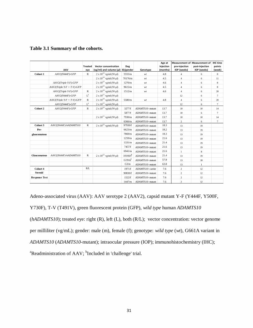
Table 3.1 Summary of the cohorts.
Adeno-associated virus (AAV): AAV serotype 2 (AAV2), capsid mutant Y-F (Y444F, Y500F,
Y730F), T-V (T491V), green fluorescent protein (GFP), wild type human ADAMTS10
(hADAMTS10); treated eye: right (R), left (L), both (R/L); vector concentration: vector genome
per milliliter (vg/mL); gender: male (m), female (f); genotype: wild type (wt), G661A variant in
ADAMTS10 (ADAMTS10-mutant); intraocular pressure (IOP); immunohistochemistry (IHC);
aReadministration of AAV;
bIncluded in 'challenge' trial.
AAV
Treated
eye
Vector concentration
(vg/ml) and volume (µl)
Dog
ID/gender Genotype
Age at
injection
(months)
Measurement of
pre-injection
IOP (weeks)
Measurement of
post-injection
IOP (weeks)
IHC time
points
(week)
Cohort 1 AAV2(Y444F)-GFP R 2 x 10¹²
vg/ml (50 μl) 5555/m wt 4.8 4 6 8
2 x 10¹⁰
vg/ml (50 μl) 70176/m wt 4.5 4 6 11
AAV2(Triple Y-F)-GFP 2 x 10¹²
vg/ml (50 μl) 1270/m wt 4.6 4 6 8
AAV2(Triple Y-F + T-V)-GFP 2 x 10¹²
vg/ml (50 μl) 9615/m wt 4.5 4 6 8
AAV2(Triple Y-F)-GFP R 2 x 10¹⁰
vg/ml (50 μl) 1512/m wt 4.6 4 6 20
AAV2(Y444F)-GFP La
2 x 10¹¹
vg/ml (50 μl) 12 6 7
AAV2(Triple Y-F + T-V)-GFP R 2 x 10¹⁰
vg/ml (50 μl) 5580/m wt 4.8 4 6 20
AAV2(Y444F)-GFP La
2 x 10¹²
vg/ml (50 μl) 12 6 7
Cohort 2 AAV2(Y444F)-GFP R 2 x 10¹²
vg/ml (50 μl) 3277/f ADAMTS10- mutant 13.7 10 10 14
5877/f ADAMTS10- mutant 13.7 10 6 7
2 x 10¹¹
vg/ml (50 μl) 7930/m ADAMTS10- mutant 13.7 10 10 14
6366/m ADAMTS10- mutant 13.7 2 6 7
Cohort 3 AAV2(Y444F)-hADAMTS10 R 2 x 10¹²
vg/ml (50 μl) 97930/f ADAMTS10- mutant 18.3 13 19
Pre- 6623/m ADAMTS10- mutant 18.2 13 19
glaucomatous 7869/m ADAMTS10- mutant 18.3 13 19
1259/m ADAMTS10- mutant 21.6 13 19
1331/m ADAMTS10- mutant 21.4 13 19
7457/f ADAMTS10- mutant 21.6 13 19
6941/m ADAMTS10- mutant 21.6 1 8
Glaucomatous AAV2(Y444F)-hADAMTS10 R 2 x 10¹²
vg/ml (50 μl) 10166/fb
ADAMTS10- mutant 21.4 13 19
G19/mb
ADAMTS10- mutant 57.8 13 18
G3/m ADAMTS10- mutant 63.8 13 1
Cohort 4 R/L 1971/f ADAMTS10- carrier 7.6 2 12
Steroid 90830/f ADAMTS10- mutant 7.6 2 12
Response Test 1522/f ADAMTS10- mutant 7.6 2 12
1447/m ADAMTS10- mutant 7.6 2 12
32
All studies were conducted in compliance with the Association for Research in Vision and
Ophthalmology statement for Use of Animals in Ophthalmic and Vision Research and approved
by the MSU Institutional Animal Care and Use Committee and Institutional Biosafety
Committee.
AAV constructs
The production of the non-self-complementary recombinant AAV serotype 2 vectors was
accomplished by the Hauswirth lab at the University of Florida, Gainesville, FL, USA. The
purification and concentration methods have been previously described.2, 3
Succinctly, site-
directed mutagenesis was performed on AAV helper plasmids containing AAV2 “Cap” to
incorporate mutations to surface exposed tyrosine and/or threonine residues of the capsids. The
vectors were generated by plasmid co-transfection in HEK293T cells. The nuclear and
cytoplasmic fractions was further purified and concentrated by iodixanol (Sigma-Aldrich, St.
Louis, MO, USA) gradient centrifugation and ion exchange column chromatography (HiTrap Sp
Hp 5 mL, GE Healthcare Bio-Sciences, Piscataway, NJ, USA). The vector titer and purity was
established by real-time PCR and silver-stained sodium dodecyl sulfate-polyacrylamide gel
electrophoresis, respectively. Final aliquots were resuspended in BSS (BSS Alcon Laboratories,
Forth Worth, TX, USA) containing 0.014% Tween 20.
The vectors were driven by a ubiquitous smCBA (Figure 3.1). Three types of mutant
vectors were chosen for the study: single (Y444F), triple (Y444,500,730F), and quadruple
(Y444,500,730F + T491V) (Table 3.1). The vectors either carried GFP (cohort 1 and 2) or wt
human ADAMTS10 cDNA (cohort 3).
33
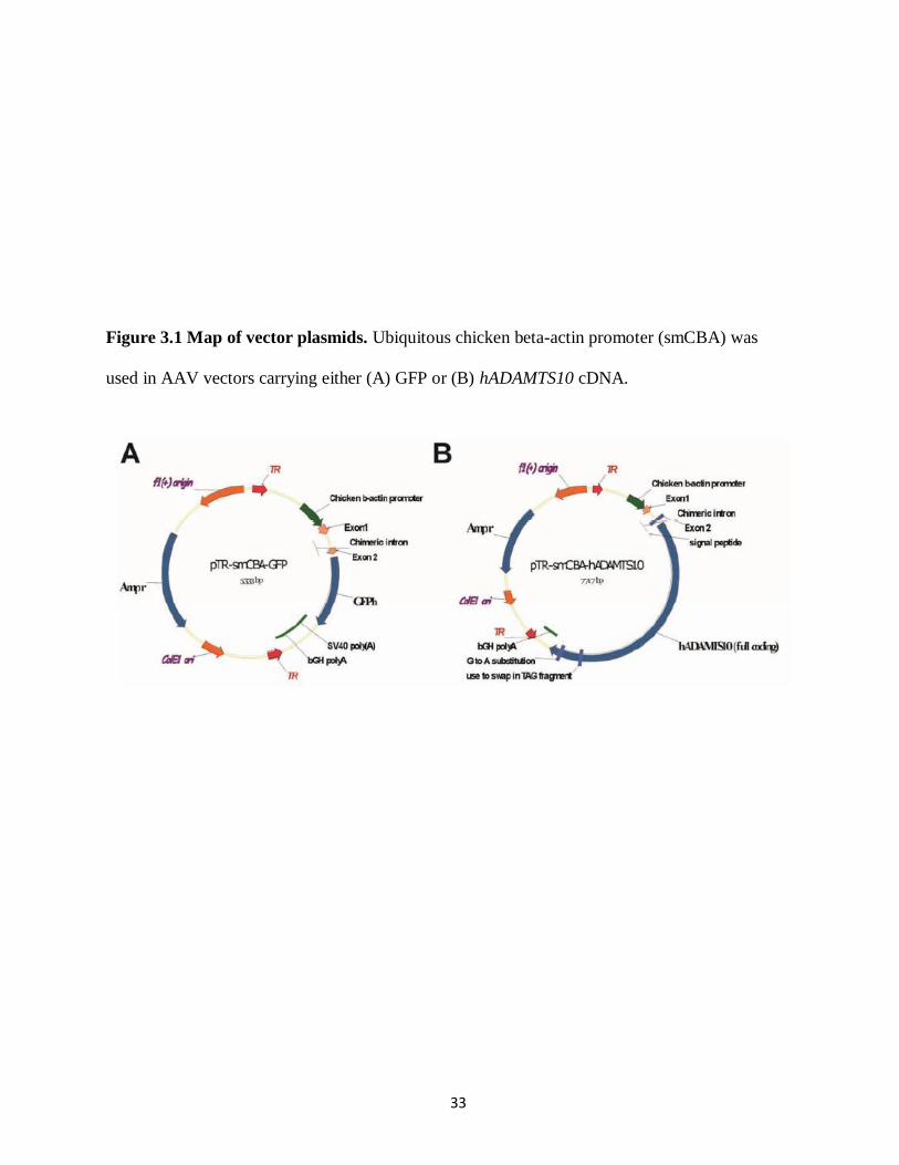
Figure 3.1 Map of vector plasmids. Ubiquitous chicken beta-actin promoter (smCBA) was
used in AAV vectors carrying either (A) GFP or (B) hADAMTS10 cDNA.

34
The hADAMTS10 coding sequence (~3.3 kb) was synthesized according to Genbank accession
number NM_030957 with the addition of consensus „kozak‟ sequence and a silent G to A change
in nucleotide 3150. The full mRNA (~4.3 kb) includes 5‟ and 3 un-translated regions, which
were dropped in order to fit the coding sequence within an AAV vector. The silent substitution
removed an internal Not I restriction enzyme site. The synthetic cDNA was subsequently cloned
into the AAV vector plasmid containing the smCBA promoter following Not I/Sal I digest.
Intracameral injections
The AAV administration procedure remained relatively consistent for each cohort: the
right eye received the vector, the left eye operated as the control, and both eyes received pre- and
post-operative steroids, NSAIDs, and antibiotics as prophylaxis against sterile immune reactions
post-surgery, bacterial infections, and inflammatory responses against the viral vector and/or
transgene. This format eliminated any additional confounding variables that may affect IOP.
Pre-operatively the dogs received prednisone 20 mg (Roxane Laboratories, Inc., Columbus, OH,
USA; 1 mg/kg oral), amoxicillin/clavulanic acid (Clavamox, Zoetis, Florham Park, NJ, USA;
12.5 mg/kg oral), in addition to three drops, 30 minutes apart, of flurbiprofen sodium 0.03%
(Bausch & Lomb Inc., Tampa, FL, USA) and one drop of prednisone acetate 1% (Pacific Pharma,
Irvine, CA, USA) ophthalmic solutions. Post-operatively, both eyes received a subconjunctival
injection of triamcinolone acetonide injectable suspension 4 mg (Kenalog®-40, Bristol-Myers
Squibb Company, Italy), followed by atropine sulfate 1% (Bausch & Lomb Inc., Tampa, FL,
USA) and neomycin and polymyxin B sulfates and dexamethasone (Bausch & Lomb Inc.,
Tampa, FL, USA) ophthalmic ointments. The dogs were given tapering doses of prednisone (1
mg/kg oral; twice daily for 7 days, once daily for 8 days, and every other day for 6 days), and
amoxicillin/clavulanic acid (12.5 mg/kg oral; twice daily for 4 days). Ophthalmic treatment for
35
both eyes included neomycin and polymyxin B sulfates and dexamethasone (eye ointment; twice
daily for 21 days, and once daily for 5 days), and atropine (eye ointment; twice daily for 4 days,
and once daily for 5 days). Note that atropine was excluded from the mutant cohorts because its
long-acting mydriatic properties can further increase in IOP in these dogs.4 The glaucomatous
dogs were also maintained on IOP-reducing medications; dorzolamide hydrochloride-timolol
maleate (Bausch & Lomb Inc., Tampa, FL, USA) and/or latanoprost 0.005% (Greenstone LLC.,
Peapack, NJ, USA) ophthalmic solutions.
The dogs were premedicated with acepromazine maleate injection (Butler Schein Animal
Health, Dublin, OH, USA; 0.2 mg/kg IM), and induced and maintained under anesthesia with
intravenous propofol (PropoFlo™28, Abbott Laboratories, North Chicago, IL, USA; 4mg/kg).
They were positioned in sternal recumbency, and one ocular surface was anesthetized with
proparacaine hydrochloride 0.5% ophthalmic solution (Bausch & Lomb Inc., Tampa, FL, USA)
and aseptically prepped with povidone iodine 10% swabsticks (Dynarex Corporation,
Orangeburg, NY, USA). Vector solutions were diluted with sterile balanced salt solution (BSS,
Alcon Laboratories, Inc., Forth Worth, TX, USA) and drawn into 1cc tuberculin (26 gauge x ½
needle) or insulin syringes (30 gauge x 1/2 inch needle). The superior-temporal limbus of the
eye was visualized by a binocular loupe (EyeMag Pro, Carl Zeiss Inc., Oberkochen, Germany)
with illumination, and the needle was introduced into the anterior chamber at an oblique angle,
parallel to the iris surface for several millimeters with the tip angled (Figure 3.2). Fluid entry of
the AAV preparation (50 μL) was carefully observed. The needle was left in place for 1-10
minutes, before being withdrawn in order to minimize reflux/escape of vector solution. The
injection site was held off for 2 minutes, and fluid leakage was assessed.

36
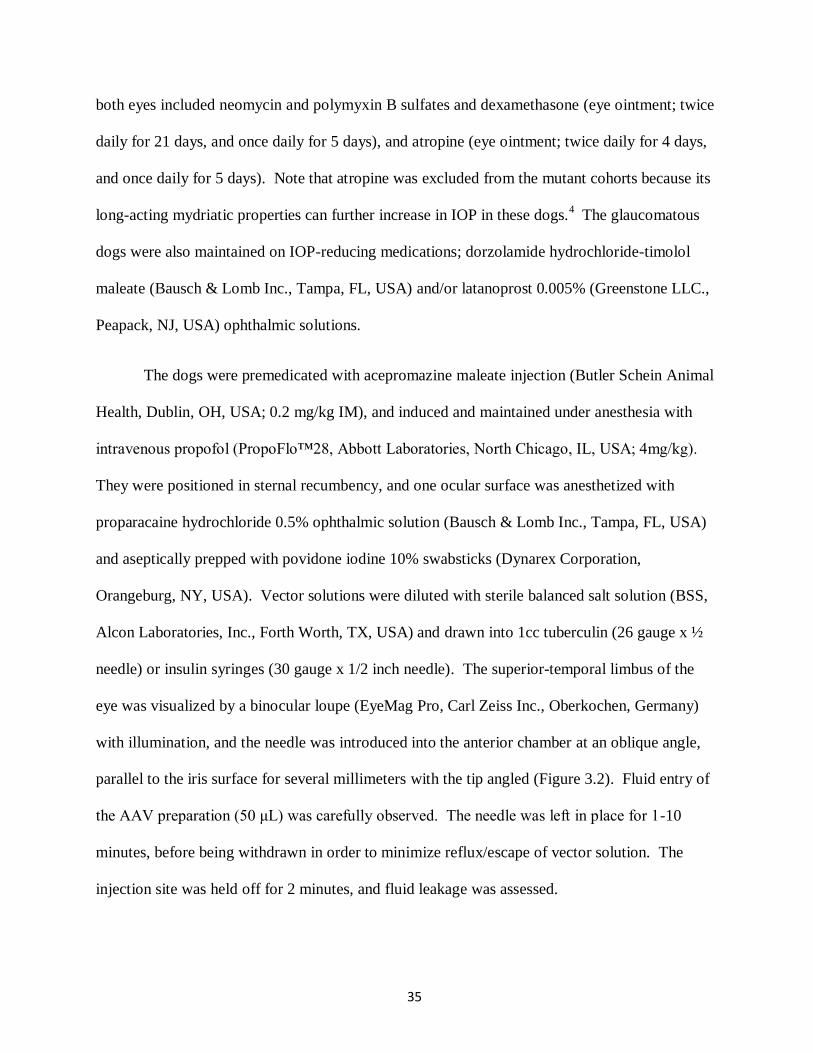
Figure 3.2 Example of an intracameral injection. The animal was under general anesthesia
and the ocular surface was anesthetized with topical proparacaine 0.5% ophthalmic solution.
Toothed Castroviejo suture forceps were used to secure and immobilize the eye. A wire lid
speculum maintained palpebral fissure open. The needle was introduced into the anterior
chamber at the superior-temporal limbus.
37
Cohort 1 received different AAV-capsid mutant vectors carrying GFP at either 2 x 10¹⁰
vg/mL or 2 x 10¹²
vg/mL (Table 3.1). Once it became clear that AAV2(Y444F) was the best
capsid mutant to target the canine TM, two dogs from same study were re-injected in the
contralateral eye three months later (Table 3.1). The objective was to confirm the reporter gene
expression results, evaluate any dosing effect, and assess potential inflammatory reactions from
vector re-administration. AAV2(Y444F)-GFP was administered to cohort 2 at either 2 x 10¹¹
vg/mL or 2 x 10¹²
vg/mL (Table 3.1). Cohort 3 received AAV2(Y444F)-hADAMTS10 at 2 x 10¹²
vg/mL (Table 3.1).
Tonometry
Baseline (pre-injection) and follow-up (post-injection) IOPs were assessed in all groups
(Table 3.1). Diurnal IOPs (8AM, 11AM, 2PM) were collected once a week by one examiner
(AO) with a tonometer. Pressures were assessed throughout the day in order to address the
circadian variations in IOP present in dogs; pressures are higher in the morning compared to the
early evening.5, 6
The first cohort was initially measured with Tono-Pen VETTM
Veterinary
Tonometer applanation (Reichert Inc., Depew, NY, USA), but was later transitioned to
TonovetTM
rebound (Icare Finland, Vantaa, Finland). Applanation and rebound tonometers both
provide consistent IOP measurements in normotensive subjects.7 However, the rebound
tonometer has been proven to be more accurate in ocular hypertensive patients and does not
require topical anesthetics.8 As a result, cohorts 2, 3, and 4 were only assessed with the
TonovetTM
, and differences between tonometers will not be further discussed.
Because any therapeutic effect of the AAV vector was likely masked by the routine use of
IOP-lowering medications, a „challenge‟ trial was performed in two glaucomatous dogs (Table

38
3.1). The purpose was to observe if the AAV treatment alone was sufficient to keep the IOP
controlled. The medications were usually given twice daily (morning and late afternoon). At 2
and 4 months post-injection, the morning regimen was discontinued and IOPs were evaluated
throughout the day.
Steroid response test
To evaluate the confounding effects of steroids, ADAMTS10-mutant dogs (n=3) and an
ADAMTS10-carrier (n=1) were included in a steroid response test (Table 3.1). The dogs were
littermates and of the same age. Both eyes received neomycin and polymyxin B sulfates and
dexamethasone ophthalmic ointment twice a day for 4 weeks. Diurnal IOPs (8AM, 11AM, 2PM)
were measured once a week for 2 weeks prior, 4 weeks during, and 12 weeks after the steroid
treatment.
Ophthalmic examination
Regular ophthalmic examinations were performed pre-injection, immediately post-
injection for 2 days daily, twice per week for 2 weeks, and then weekly to bi-weekly until the
end of each study. Anterior segments were examined for clarity and cells with diffuse and focal
illumination using portable hand-held slit-lamp biomicroscopes (Kowa SL14; Kowa Company,
Tokyo, Japan). Fundic examinations were performed with portable binocular indirect
ophthalmoscopes (Keeler All Pupil II; Keeler Instruments, Broomall, PA, USA) and condensing
lens (Pan Retinal 2.2D; Volk Optical, Mentor, OH, USA). The ICA angle width was evaluated
with gonioscopy: the ocular surface was anesthetized with proparacaine hydrochloride 0.5%
ophthalmic solution and the ICA was imaged with RetCam II (Clarity Medical Systems,
Pleasanton, CA, USA).
39
RetCam II and cSLO were both used to evaluate in vivo GFP expression in the ICA.
With cSLO, the dogs were placed under anesthesia, and one drop of proparacaine hydrochloride
0.5% provided ocular surface anesthesia as a gonioscopic lens (G-4 Goniolaser, Volk, Mentor,
OH, USA) was placed on the corneal surface. The ICA was imaged under Infrared Reflectance
and BluePeak™ blue laser autofluorescence with a 55° lens (Spectralis®, Heidelberg
Engineering, Heidelberg, Germany).
Tissue processing/sectioning
GFP expression was analyzed at multiple time points for cohort 1 and 2 (Table 3.1). The
subjects were euthanized by barbiturate overdose (Fatal Plus, Vortech Pharmaceuticals,
Dearborn, MI, USA). The eyes were enucleated and a 2-mm slit was made along the pars plana
of the globe. Approximately 0.5 mL of 4% paraformaldehyde in PBS was injected intravitreally
through the slit and the globe was then placed in 25 mL of the same solution and stored at 4°C.
Three hours later, the anterior segment was then separated from the posterior portion of the eye
and the vitreous was discarded. The two halves were subsequently placed in 2%
paraformaldehyde and stored at 4°C for 24 hours. The tissues were then transferred to 15% and
30% sucrose in PBS at 4°C for 24 hours each.
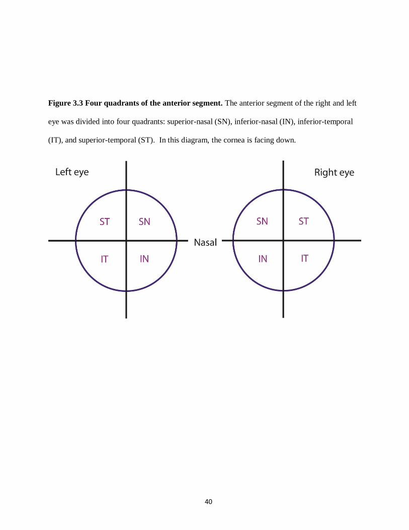
The anterior segment including lens, was cut into 4 even quadrants, superior-nasal (SN),
inferior-nasal (IN), inferior-temporal (IT), and superior-temporal (ST) (Figure 3.3), and
embedded in optical cutting temperature medium (Tissue-Tek OCT, Sakura Finetek USA Inc.,
Torrence, CA, USA), and stored at -80°C. Prior to sectioning, the eyes were kept at -20°C for
~20 minutes.
40
Figure 3.3 Four quadrants of the anterior segment. The anterior segment of the right and left
eye was divided into four quadrants: superior-nasal (SN), inferior-nasal (IN), inferior-temporal
(IT), and superior-temporal (ST). In this diagram, the cornea is facing down.
41
Fourteen micrometer transverse anterior segment cyrosections were collected using a
cryomicrotome (Leica CM3050-S, Leica Microsystems, Buffalo Grove, IL, USA) onto charged
microscope glass slides (Adhesion Superfrost Plus, Brain Research Laboratories, Newton, MA,
USA). The slides were dried for 20 minutes and stored in 4°C.
Immunohistochemistry and image analysis
The same reagents and basic protocol were used for all tissue samples. The slides were
warmed to room temperature, rehydrated for 35 minutes in PBS containing detergent 0.1%
Triton X-100, and then blocked in 5% serum (Normal Goat Serum, 1:20, Jackson
ImmunoResearch Laboratories, Inc., West Grove, PA, USA). The slides were washed with PBS
for 10 minutes. The primary anti-GFP, rabbit polyclonal antibody (AlexaFluor® 594, 1:1000,
Life Technologies, Eugene, OR, USA) was incubated overnight at 4°C. After the slides were
washed for 25 minutes in PBS, they were mounted using antifade reagent with DAPI (ProLong®
Gold, Life Technologies, Eugene, OR, USA) and glass coverslips (Electron Microscopy
Sciences, Hatfield, MO, USA). All slides were stored at 4°C in the dark. Since native GFP was
relatively weak, enhanced GFP with immunolabeling was analyzed. Semi-quantitative analysis
of enhanced GFP was completed with fluorescent microscopy (Eclipse 80i Fluorescent
Microscope, Nikon Instruments Inc., Melville, NY, USA). Each quadrant was graded by a non-
blinded observer (AO) on a scale of 1-5 (1=absent; 2 = weak; 3 = moderate; 4 = strong; 5 = very
strong), and reported values were averaged. Approximately 8 sections per quadrant were
analyzed for the wt group and 16 sections per quadrant were analyzed for the mutant group. The
slides were then imaged with the fluorescent or confocal microscopy (FV1000 Laser Scanning
Confocal Microscope, Olympus America Inc., Center Valley, PA, USA) at x 10, x 20, x 40, and
x 120 magnification.
42
Statistical analysis
In cohort 3, the primary outcome measure was IOP. A power calculation using a two-
sided paired t-test revealed that a sample size of seven ADAMTS10-mutant dogs would provide
90% power to detect a 6-mmHg decrease in IOP of the treated eye compared to the control eye
with a significance level (alpha) of 0.05, assuming the standard deviation of the measured IOPs
is 4 mmHg. To adjust the correlation in IOP from two eyes measured at multiple visits, a
generalized linear model using generalized estimating equation (GEE) Wald test was performed.
IOP was a primary outcome measure for cohort 4 and a secondary outcome measure for cohorts
1 and 2. Even though the sample sizes were small, a generalized linear model using GEE Wald
test was used to estimate the average IOP response.
Selected IOP data points were excluded or abbreviated (Table 4.2). Exclusion criteria
were correspondingly established to decrease variability and increase accuracy of reported data.
Subjects that were added later to their respective cohorts had abbreviated pre-injection pressures,
and data was excluded from those that were euthanized prior to the end of the study. Since IOP-
lowering medications are a confounding variable, pressure records from treated eyes were
excluded from the statistical analysis. Specific omissions are further explained in the results
section.

43
REFERENCES
44
REFERENCES
1. Gelatt KN, Mackay EO. The ocular hypertensive effects of topical 0.1%
dexamethasone in beagles with inherited glaucoma. Journal of Ocular Pharmacology
and Therapeutics: The Official Journal of the Association for Ocular Pharmacology
and Therapeutics 1998;14:57-66.
2. Zolotukhin S, Potter M, Zolotukhin I, et al. Production and purification of serotype 1,
2, and 5 recombinant adeno-associated viral vectors. Methods 2002;28:158-167.
3. Jacobson SG, Acland GM, Aguirre GD, et al. Safety of recombinant adeno-associated
virus type 2-RPE65 vector delivered by ocular subretinal injection. Molecular
Therapy: The Journal of the American Society of Gene Therapy 2006;13:1074-1084.
4. Grozdanic SD, Kecova H, Harper MM, Nilaweera W, Kuehn MH, Kardon RH.
Functional and structural changes in a canine model of hereditary primary angle-
closure glaucoma. Investigative Ophthalmology & Visual Science 2010;51:255-263.
5. Giannetto C, Piccione G, Giudice E. Daytime profile of the intraocular pressure and
tear production in normal dog. Veterinary Ophthalmology 2009;12:302-305.
6. Gelatt KN, Gum GG, Barrie KP, Williams LH. Diurnal variations in intraocular
pressure in normotensive and glaucomatous Beagles. Glaucoma 1981;3:21-24.
7. Leiva M, Naranjo C, Pena MT. Comparison of the rebound tonometer (ICare) to the
applanation tonometer (Tonopen XL) in normotensive dogs. Veterinary
Ophthalmology 2006;9:17-21.
8. McLellan GJ, Kemmerling JP, Kiland JA. Validation of the TonoVet(R) rebound
tonometer in normal and glaucomatous cats. Veterinary Ophthalmology 2013;16:111-
118.
45
CHAPTER 4 - RESULTS
AAV2(Y444F) targets GFP expression to the canine wt ICA
Initial intracameral injection of the single stranded AAV2(Y444F) vector successfully
targeted GFP expression to wt cells located along the aqueous humor outflow pathways (Figure
4.1A). Reporter gene expression was present at both concentrations (2 x 10¹⁰
and 2 x 10¹²
vg/mL;
50 μL); though a modest dosing effect without immunolabeling was observed over the two log
units on subjective assessment. The overall expression was relatively weak with many presumed
cells remaining GFP negative, but native GFP fluorescence was enhanced with immunolabeling
and positive cells were still observed at 8 and 11 weeks post-injection. In vivo GFP fluorescence
was not observed in the ICA by gonioscopy.
Consistent with previous descriptions of TM morphology,1 the cells appeared broad to
spindle shaped with long cytoplasmic processes (Figure 4.1B). The molecular characterization
of these cells is planned for the future; it requires a combination of antibodies for IHC (Table
5.1). GFP expression was detected in all four quadrants with no obvious difference,
demonstrating successful widespread delivery of the vector to the ICA (Table 4.1). Despite
stronger presumed transduction efficiency in murine retinal cells and hepatocytes compared to
AAV2(Y444F), 2-4
positive cells were absent with AAV2(Triple T-F) and AAV2(Y-F + T-V)
capsid based mutant vectors (Figure 4.1C).
Even with a ubiquitous smCBA promoter, GFP expression was rather specific with
almost exclusive fluorescence along the aqueous humor outflow pathways. Nevertheless,
transduction of AAV2(Y444F) was not limited to cells within the ICA as uniform expression
was observed on the anterior border layer of the iris (Figure 4.1A; arrow). Transduced cells of
46
the anterior border of the iris were spindle-shaped, and at times displayed stronger fluorescence
than the cells in the ICA (Figure 4.1D).
AAV2(Y444F) targets GFP expression to the canine ADAMTS10-mutant ICA.
Based on the results with AAV2(Y444F), mutant eyes were injected with AAV2(Y444F)-
hADAMTS10 at the highest dose (2 x 10¹²
vg/mL; 50 μL). Consistent with the findings from the
first cohort, GFP transduction was found along the aqueous humor outflow pathways (Figure
4.2A), observed at both post-injection time points (7 and 14 weeks), and detected in each ICA
quadrant (Table 4.1; Figure 4.2B). In vivo GFP fluorescence in the ICA was also not detected by
gonioscopy.
The morphology of the transduced mutant cells was also similar to the wt cells and
consistent with TM cells (Figure 4.2C). Though, further molecular testing of the cell would be
required to confirm this, as fibroblasts can be misidentified as TM cells. Positive spindle-shaped
cells (Figure 4.2D) were also noted along the length of the anterior border of the iris (Figure
4.2A, arrow). Contrary to the wt canines, GFP expression was consistently present along the
posterior pigmented epithelial layer of the iris (Figure 4.3). Fluorescence was mainly observed
on the pupillary margin.
Therapeutic effect of AAV2(Y444F)-hADAMTS10
A single intracameral injection of AAV2(Y444F)-hADAMTS10 revealed no observable
therapeutic effect on IOP over 19 weeks. Both pre-glaucomatous (n=7) and glaucomatous (n=3)
mutant dogs received the vector at the highest dose (2 x 10¹²
vg/mL; 50 μL). A therapeutic

47
Figure 4.1 ICA of wt dogs. (A) Only AAV2(Y444F)-GFP showed positive native GFP
expression (green). GFP expression was found in the ICA and on the anterior border layer of the
iris (arrow). GFP fluorescence was enhanced with anti-GFP antibody (red). Overlay of images
(yellow) with DAPI stain. Yellow indicates strongest GFP expression, while red means weaker
expression. Un-injected control reveals absence of GFP fluorescence. Calibration bar = 50 µm;
(B) Magnification of transduced cells. Calibration bar = 10 µm; (C) GFP expression was not
detected in the eye injected with AAV2(Triple T-F) or AAV2(Y-F + T-V). Calibration bar = 50
µm; (D) Cells of the anterior border layer of the iris express GFP. Calibration bar = 10 µm.
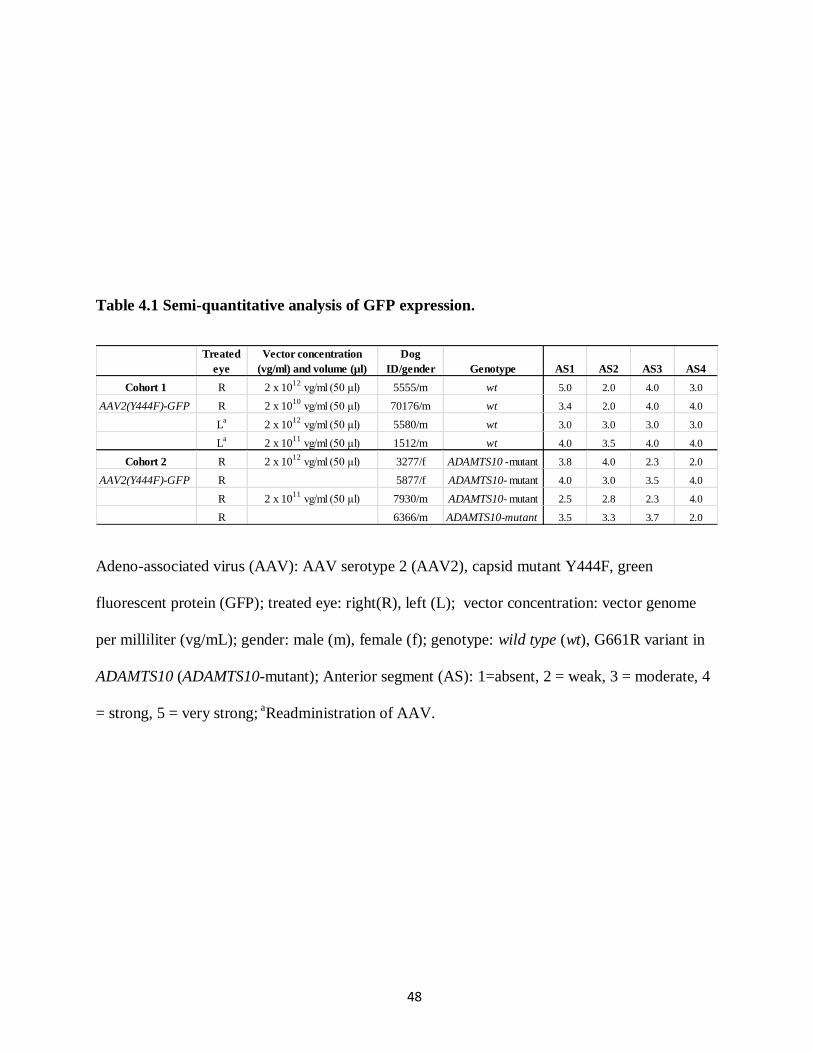
48
Table 4.1 Semi-quantitative analysis of GFP expression.
Adeno-associated virus (AAV): AAV serotype 2 (AAV2), capsid mutant Y444F, green
fluorescent protein (GFP); treated eye: right(R), left (L); vector concentration: vector genome
per milliliter (vg/mL); gender: male (m), female (f); genotype: wild type (wt), G661R variant in
ADAMTS10 (ADAMTS10-mutant); Anterior segment (AS): 1=absent, 2 = weak, 3 = moderate, 4
= strong, 5 = very strong; aReadministration of AAV.
Treated
eye
Vector concentration
(vg/ml) and volume (µl)
Dog
ID/gender Genotype AS1 AS2 AS3 AS4
Cohort 1 R 2 x 10¹²
vg/ml (50 μl) 5555/m wt 5.0 2.0 4.0 3.0
AAV2(Y444F)-GFP R 2 x 10¹⁰
vg/ml (50 μl) 70176/m wt 3.4 2.0 4.0 4.0
La
2 x 10¹²
vg/ml (50 μl) 5580/m wt 3.0 3.0 3.0 3.0
La
2 x 10¹¹
vg/ml (50 μl) 1512/m wt 4.0 3.5 4.0 4.0
Cohort 2 R 2 x 10¹²
vg/ml (50 μl) 3277/f ADAMTS10 -mutant 3.8 4.0 2.3 2.0
AAV2(Y444F)-GFP R 5877/f ADAMTS10- mutant 4.0 3.0 3.5 4.0
R 2 x 10¹¹
vg/ml (50 μl) 7930/m ADAMTS10- mutant 2.5 2.8 2.3 4.0
R 6366/m ADAMTS10-mutant 3.5 3.3 3.7 2.0
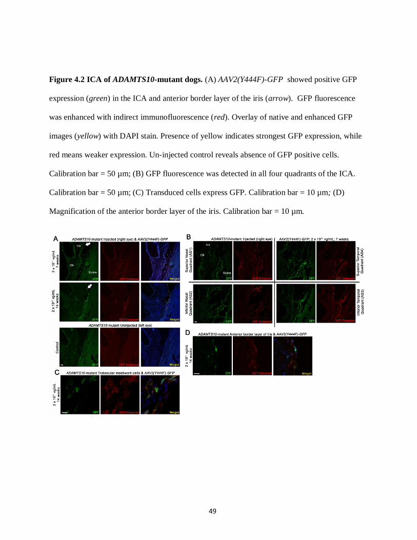
49
Figure 4.2 ICA of ADAMTS10-mutant dogs. (A) AAV2(Y444F)-GFP showed positive GFP
expression (green) in the ICA and anterior border layer of the iris (arrow). GFP fluorescence
was enhanced with indirect immunofluorescence (red). Overlay of native and enhanced GFP
images (yellow) with DAPI stain. Presence of yellow indicates strongest GFP expression, while
red means weaker expression. Un-injected control reveals absence of GFP positive cells.
Calibration bar = 50 µm; (B) GFP fluorescence was detected in all four quadrants of the ICA.
Calibration bar = 50 µm; (C) Transduced cells express GFP. Calibration bar = 10 µm; (D)
Magnification of the anterior border layer of the iris. Calibration bar = 10 µm.
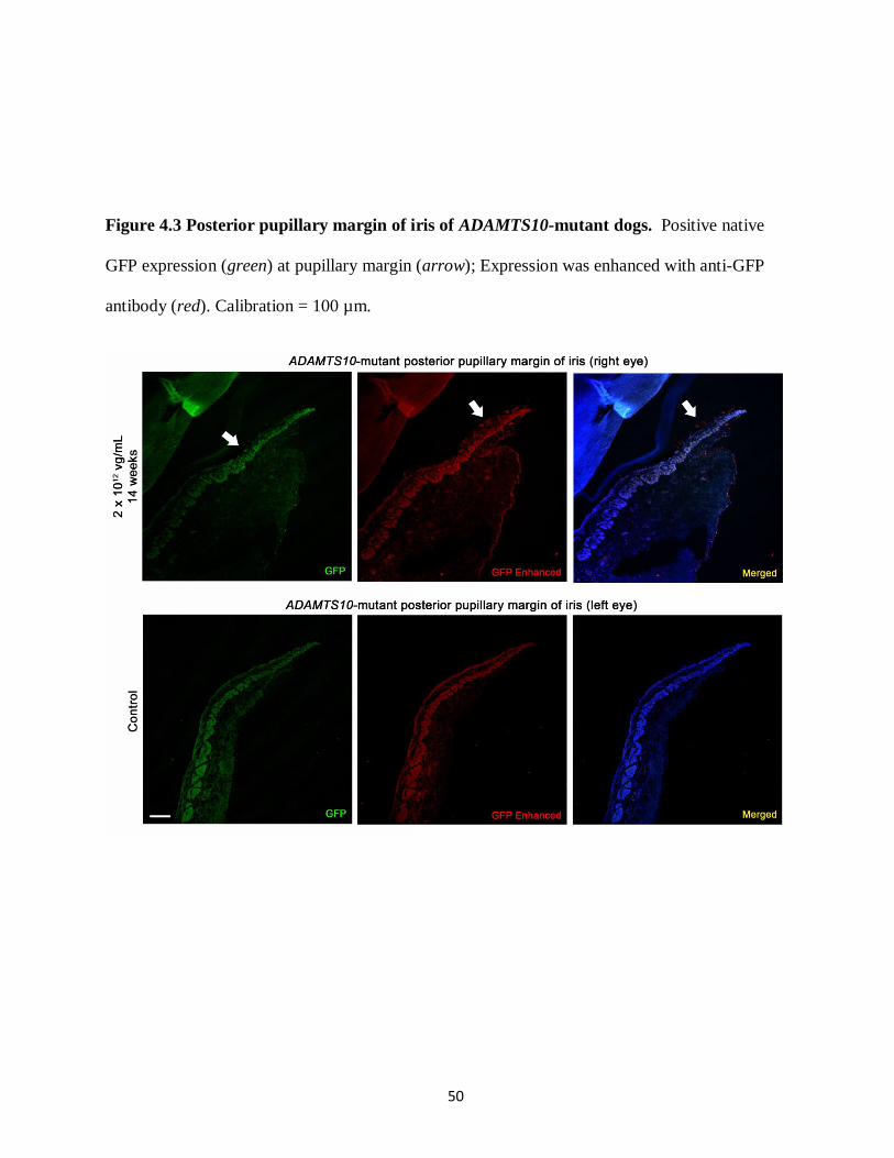
50
Figure 4.3 Posterior pupillary margin of iris of ADAMTS10-mutant dogs. Positive native
GFP expression (green) at pupillary margin (arrow); Expression was enhanced with anti-GFP
antibody (red). Calibration = 100 µm.

51
effect would indicate an absence of progressive ocular hypertension and pressure spikes in the
injected eye, while a gradual increase in pressure is observed in the control.
IOP data (mean ± standard error) collected with a rebound tonometer revealed a steady
bilateral increase in pressures over time in the pre-glaucomatous group (Figure 4.4). IOP
averages between the injected (25.0 ± 1.03 mmHg) and control (24.4 ± 1.15 mmHg) eyes also
showed no significant difference (p = 0.56) with a generalized linear model using GEE Wald test
(Table 4.3). Furthermore one dog developed ocular hypertension 11 weeks post-injection (Table
4.2).
In the glaucomatous eyes, we expected that the AAV treatment alone could keep the IOP
controlled. However in the two dogs that were part of the „challenge‟ trial (Table 4.2), pressures
were above 40 mmHg within ~19 hours from the last instillation of medications (data not shown).
The glaucomatous groups‟ IOPs were excluded from further statistical analysis because they had
been treated with IOP-lowering medications. Additionally, two dogs were euthanized before the
end of the study due to systemic illnesses unrelated to our vector or medical treatments.
Steroid responsiveness of ADAMTS10-mutants and carrier
From preliminary data from cohorts 1 and 2, we assumed that IOP increased with oral
and topical glucocorticoid treatment (Figure 4.5; arrows). Subsequent statistical analysis (Table
4.3) revealed a significant elevation in IOP in both wt (p < 0.0001) and mutant (p = 0.003) dogs.
To investigate the transient elevation, a steroid response test was conducted with topical
dexamethasone in ADAMTS10-mutants (n=3) and an ADAMTS10-carrier (n=1). An acute
increase in pressure was evident within the first week of treatment (Figure 4.6). When the
medications were discontinued, the pressures reverted towards baseline. Statistical analysis
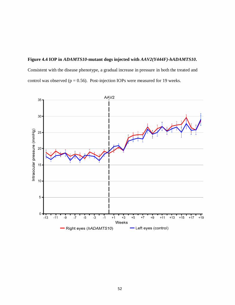
52
Figure 4.4 IOP in ADAMTS10-mutant dogs injected with AAV2(Y444F)-hADAMTS10.
Consistent with the disease phenotype, a gradual increase in pressure in both the treated and
control was observed (p = 0.56). Post-injection IOPs were measured for 19 weeks.

53
Table 4.2 Summary of excluded and abbreviated IOP data.
Adeno-associated virus (AAV): AAV serotype 2 (AAV2), green fluorescent protein (GFP), wild
type human ADAMTS10 (hADAMTS10); eye: right (R), left (L), both (R/L); gender: male (m),
female (f); genotype: wild type (wt), G661R variant in ADAMTS10 (ADAMTS10-mutant);
intraocular pressure (IOP); data: "–" excluded, "/" abbreviated.
AAV capsid mutant vector Eye
Dog
ID/gender Genotype
Cohort 1 AAV2(Y444F)-GFP L 5580/m wt –Excluded data due to acute anterior chamber inflammation in the right eye (24
hours post-injection)
L 1512/m wt – Excluded due to a small sample size
Cohort 2 AAV2(Y444F)-GFP R/L 6366/m ADAMTS10- mutant /Abbreviated pre-injection records because subject was added later to the
study
R/L 3277/f ADAMTS10- mutant /Abbreviated post-injection records because of a delayed onset of anterior
chamber inflammation in the right eye (10 weeks post-injection)
R/L 7930/m ADAMTS10- mutant / Abbreviated due to a small sample size
Cohort 3 AAV2(Y444F)-hADAMTS10 R/L 6941/m ADAMTS10- mutant /Abbreviated pre-injection records because subject was added later to the
study; abbreviated post-injection records due to medications
R/L 10166/f ADAMTS10- mutant –Enrolled in the 'challenge' trial; Excluded data due to IOP-lowering medical
treatment
R/L G19/m ADAMTS10- mutant –Enrolled in the 'challenge' trial; Excluded data due to IOP-lowering medical
treatment; Euthanized (18 weeks post-injection)
R/L G3/m ADAMTS10- mutant – Euthanized (1 week post-injection)
Reasons for excluded or abbreviated IOP data

54
Table 4.3 Statistics on IOP data
Intraocular pressure (IOP); pre-injection IOP: before glucocorticoid treatment; post-injection
IOP: during and after glucocorticoid treatment; mean± standard error.
Cohort Right eye Left eye
P-value comparing right
vs left eye
1 Pre-injection IOP (mmHg) 13.2± 0.31 12.9± 0.41 0.20
Post-injection IOP (mmHg) 18.8± 0.49 18.3± 0.42 0.021
P-value comparing pre- and
post-injection IOP< 0.0001 < 0.0001
2 Pre-injection IOP (mmHg) 19.2± 1.08 18.0± 0.92 0.0005
Post-injection IOP (mmHg) 22.8± 0.59 23.4± 0.79 0.33
P-value comparing pre- and
post-injection IOP0.003 < 0.0001
3 Pre-injection IOP (mmHg) 18.4± 0.49 17.6± 0.52 0.006
Post-injection IOP (mmHg) 25.0± 01.03 24.4± 1.15 0.56
Cohort Both eyes P-value
4 All baseline IOPs (B) 14.2± 0.61
All treatment IOPs (T) 15.6± 0.36
All follow up IOPs (A) 14.8± 0.28
B vs T 0.002
T vs A <0.0001
B vs A 0.16
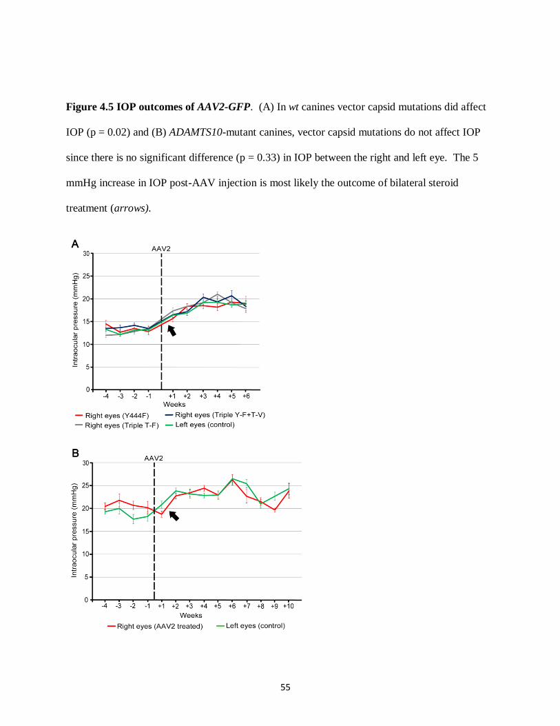
55
Figure 4.5 IOP outcomes of AAV2-GFP. (A) In wt canines vector capsid mutations did affect
IOP (p = 0.02) and (B) ADAMTS10-mutant canines, vector capsid mutations do not affect IOP
since there is no significant difference (p = 0.33) in IOP between the right and left eye. The 5
mmHg increase in IOP post-AAV injection is most likely the outcome of bilateral steroid
treatment (arrows).

56
Figure 4.6 Effect of steroids on IOP in ADAMTS10-mutant and carrier dogs. Topical
dexamethasone led to an increase in IOP within the first week of treatment. At the end of the 4-
weeks, IOP immediately decreased towards baseline.

57
revealed a significant difference between baseline and treatment IOPs, in addition to treatment
and follow-up IOPs (Table 4.3).
Clinical signs in wt and ADAMTS10-mutants
Adverse reactions were absent with the initial injections of AAV in cohort 1. The Tono-
pen VETTM
and TonovetTM
obtained similar pressure measurements, and a line graph of the
values collected showed that AAV-based capsid mutants did not affect IOP (Figure 4.5A).
Interestingly, statistical analysis (Table 4.3) revealed a marginal significant difference in
pressures between injected right (18.8 ± 0.49 mmHg) and control left eyes (18.3 ± 0.42 mmHg)
over 6 weeks in wt dogs (p = 0.02). The difference is too small to be clinically relevant and may
be the result of a systematic measuring error introduced during tonometry (described below). The
two wt dogs (1512 and 5580) that received the second injection of AAV2(Y444F) in the fellow
eye were also monitored for 6 weeks (Table 3.1). Immediately post-injection, one of these two
dogs (5580) developed mild anterior chamber inflammation and ocular hypertension, and was
placed on IOP-lowering medications in addition to the already administered anti-inflammatory
and anti-microbrial drugs. Even though the clinical signs quickly resolved and IOP normalized,
the data for this pair were excluded from the statistics (Table 4.2). Microscopic analysis of the
tissue (5580) revealed positive expression (Table 4.1)
Further adverse reactions were not observed in cohort 2 during the first 6 weeks. After
two dogs were euthanized for IHC analysis, one of the remaining dogs (3277) developed
moderate anterior chamber inflammation in the injected eye 10 weeks post-injection and was
placed on additional medications until euthanasia. In accordance with the exclusion criterion,
these data were omitted (Table 4.2). Fortunately, the complete IOP data were only abbreviated
58
because of the delayed onset in clinical signs. Graphical (Figure 4.5B) and statistical analysis
(Table 4.3) of the injected (22.8 ± 0.59 mmHg) and control pressure (23.4 ± 0.79 mmHg) data
revealed that the AAV2(Y444F) vector had no effect on IOP over 10 weeks (p = 0.33).
Microscopic analysis of this tissue revealed positive GFP expression in all four quadrants (Table
4.1).
Unanticipated significant differences between the right and left eyes were noted in post-
injection IOPs in cohort 1 (p = 0.02), and pre-injection pressures in cohorts 2 (p = 0.0005) and 3
(p = 0.006; Table 4.3). Interestingly, the right eye was regularly elevated across the cohorts. In
cohort 1, the outcome could have been vector related. However, since the contrast between IOP
averages of the right and left eyes were so small, we suspect there was a systematic error during
pressure readings. Our suspicions are further supported because IOP was consistently greater in
the right eye compared to the left at most time points.

59
REFERENCES

60
REFERENCES
1. Fautsch MP, Howell KG, Vrabel AM, Charlesworth MC, Muddiman DC, Johnson
DH. Primary trabecular meshwork cells incubated in human aqueous humor differ
from cells incubated in serum supplements. Investigative Ophthalmology & Visual
Science 2005;46:2848-2856.
2. Aslanidi GV, Rivers AE, Ortiz L, et al. Optimization of the capsid of recombinant
adeno-associated virus 2 (AAV2) vectors: the final threshold? PloS One
2013;8:e59142.
3. Petrs-Silva H, Dinculescu A, Li Q, et al. Novel properties of tyrosine-mutant AAV2
vectors in the mouse retina. Molecular Therapy: The Journal of the American Society
of Gene Therapy 2011;19:293-301.
4. Petrs-Silva H, Dinculescu A, Li Q, et al. High-efficiency transduction of the mouse
retina by tyrosine-mutant AAV serotype vectors. Molecular Therapy: The Journal of
the American Society of Gene Therapy 2009;17:463-471.

61
CHAPTER 5 – CONCLUSION & FUTURE STUDIES
The GFP reporter gene was successfully targeted to the canine wt and mutant ICA with
an AAV vector. These results provided the groundwork for our chief goal of gene replacement
therapy in ADAMTS10-mutant dogs. Canine POAG is an inherited autosomal recessive trait with
an identified loss of function mutation.1-3
Therefore, gene replacement therapy may rescue the
mutant phenotype. While the intracameral administration of AAV in our studies was safe, no
therapeutic effect was observed over 19 weeks at the dose evaluated. Within the treated cohort,
there was no evidence of lowered IOP, or prevention of disease-related pressure spikes.
The study utilized a single-stranded recombinant AAV vector and is the first reported
attempt to target gene expression to a canine ICA. We elected to administer 50 μL of vector
solution at three distinct concentrations: 2 x 10¹⁰
, 2 x 10¹¹
, and 2 x 10¹²
vg/mL. Efficacious
targeting of the ICA was observed with the single mutant (Y444F) vector at all three
concentrations; though a minimal dosing effect was noted over the viral titer range. The
injection technique was effective as all four quadrants of GFP-treated eyes showed transduced
TM cells. There is limited knowledge about the integrity of these cells in ADAMTS10-mutants
dogs. Whether the TM cells are lost in the progression of the disease still remains a pertinent
question, despite the marked loss observed in the ICA of human POAG eyes.4 It also remains to
be seen if transduction with the wt copy of the gene will result in a therapeutic effect. However
the transduced cells‟ morphology and location suggest they are TM cells and that there is no
difference between them in the wt and mutant dogs. To validate these results, further IHC
experiments on wt and mutant tissue samples will be required. Since there are no specific
markers for TM cells, positive and negative labeling with a combination of known antibodies
62
will be pursued (Table 5). Additionally, canine TM cell line cultures are currently under
development. This in vitro model will help verify and potentially improve our technique.
Advancements in AAV-capsid based mutants have allowed us to target the ICA without
the use of self-complementary vectors. Self-complementary constructs have been previously
described to be the most efficient,5 but are limited by their genomic carrying capacity. The
success with conventional AAV now allows larger amounts of genetic material to be delivered to
the tissue of interest. Previous studies with adenovirus in murines,6 and lentivirus in nonhuman
primates have also revealed efficacious transduction of the TM.7 Nonetheless, AAV is the
preferred viral vector in pre-clinical and clinical trials of ocular gene therapy because they
produce a persistent transgene expression without eliciting a strong immune response.8, 9
Thus,
our study has provided additional evidence that AAV, specifically AAV2-capsid based mutants,
can successfully target the TM.
Capsid mutant vectors have an increased nuclear translocation and transduction
efficiency in the host cell because site specific mutagenesis of surface protein residues allows the
vector to avoid proteasome-mediated degradation.10
The vectors chosen for this project were
based on in vitro and in vivo rodent work completed at the University of Florida, Gainesville, FL,
USA.11-13
Based on previous work we expected the triple-mutant (Y444,500,730F) and/or the
quadruple-mutant (Y444,500,730F + T491V) vectors to be the most efficient. On the contrary,
the vector with the single point mutation (Y444F) was the only one successful in transducing TM
cells in the wt and mutant dogs.

63
Table 5.1 Positive and negative IHC markers for TM cells
Targeted antigen Positive or
negative
markers
Function of antigen References
α-smooth muscle actin Positive Cytoskeletal protein 32
Laminin Positive Extracellular matrix basal lamina protein 33
Type IV collagen Positive Extracellular matrix basal lamina protein 34
Desmin Negative Intermediate filament 35
Keratin Negative Intermediate filament 36

64
Administration of AAV was mostly safe at the doses chosen. The anterior chamber,
vitreous cavity, and subretinal space are considered immune privileged sites and respond to
antigens by diminishing the inflammatory reaction through anterior chamber associated immune
deviation.14, 15
However in a minority of dogs, rats, and monkeys, intracameral and subretinal
injection of AAV vectors resulted in a clinically identifiable immune reaction.5, 16
Readministration of viral vectors to the partner eye also has been reported to elicit an immune
response.17-20
In this study, two of the ten dogs developed mild to moderate anterior chamber
inflammation after receiving an AAV carrying the GFP reporter gene. The first dog developed
acute signs after vector readministration to the partner eye. Based on the onset, potential causes
of inflammation include a sterile inflammatory response to the surgical procedure, acute bacterial
endophthalmitis, or an immune reaction to the viral vector.16
We hypothesize that the subject
developed neutralizing antibodies from the initial injection that directly reacted with the viral
vector. The second dog presented with moderate signs 10 weeks post-injection. The delayed
onset suggests that an immune reaction could have developed against the transcribed GFP
protein.21, 22
Caution must be taken in future experiments as modifications to the protocol, such
as increasing the vector dose, may increase the risk of side effects. Positive responses to medical
treatment fortunately indicate that the inflammatory reaction may be controlled in the clinical
setting.
GFP expression was rather specific along the aqueous humor outflow pathways despite
the use of a non-specific promoter. A TM-specific promoter is currently not available, and thus
we also observed positive cells in the anterior border and posterior pupillary margin of the iris.
The tissue specificity presented in our study is unclear, and the mechanisms underlying the tissue
tropisms of different AAV serotypes is not known but hypothesized to be associated with
65
distinctions in cellular uptake and intracellular trafficking.23
Therefore a possible explanation is
that the cells of the aqueous humor outflow pathways and anterior border of the iris express the
same cell surface and co receptors allowing entry of the viral vector and subsequent intracellular
trafficking. Convection currents present in the anterior chamber due to thermal circulation24
may
also contribute by widely distributing the vector and prolonging the vector-host cell interaction
before it exits through the ICA. Interestingly, inner corneal cells were not transduced.
Furthermore, since all aqueous humor is drained through and filtered by the TM, this may
„concentrate‟ the AAV-mediated gene expression to the outflow pathways. As mentioned
previously, further characterization of the transduced cells is necessary.
Even though the expression of GFP in the ICA was present on microscopy, we did not
observe in vivo expression by gonioscopy. This infers that the fluorescence was less prominent
in our dogs compared to previous studies in other species.5, 7
Examination with confocal
microscopy revealed that relatively few TM cells fluoresced while other surrounding cells were
GFP-negative. The BLAST identity between hADAMTS10 and cADAMTS10 protein was 96%,
suggesting the wt human transgene product could function as a substitute in ADAMTS10-mutant
dogs.25
Thus, a low transgene expression may possibly explain the lack of therapeutic effect
seen with AAV2(Y444F)-hADAMTS10. The goal of future trials would then be to increase the
number of TM cells transduced by modifying procedural strategies such as increasing the vector
solution volume, or using higher viral titers (1013
vg/mL). Unfortunately, the latter would be
difficult since our highest concentration was already stock solution from University of Florida.
Additional approaches would be to substitute the steroid therapy with NSAIDs to eliminate
dexamethasone‟s confounding effect on IOP, or to enroll a younger cohort of dogs (< 8 weeks of
age) to target the ICA during its final stages of development and/or much earlier before the onset

66
of the POAG phenotype. Furthermore, atropine could be used to temporarily decrease the
aqueous humor outflow through the ICA to prolong AAV contact time. Quantitative measures of
transduction efficiency, including qRT-PCR (mRNA) and Western blot/IHC (protein) of GFP
and hADAMTS10, will be pursued in the future once the 6-month post-injection observation
period has been concluded.
Initial trials with presumed therapeutic AAV2(Y444F)-hADAMTS10 vector did not show
any effect over 19 weeks. We treated both pre-glaucomatous and glaucomatous mutant dogs,
and the results showed progressive bilateral increase in IOP over time (Figure 4.4) and sustained
IOP > 40 mm Hg, respectively. Based on the GFP study we assume that the wt hADAMTS10
protein was expressed, but the lack of therapeutic effect suggests that a larger number of
transduced TM cells and subsequently transcribed proteins may be required to augment the
phenotype. The presence of hADAMTS10 protein in the treated eyes still needs to be confirmed
by Western blot, IHC of ocular tissue, and ELISA of aqueous humor, in addition to qRT-PCR of
extracted RNA. We also hypothesize that more time is needed for the accumulated ECM to be
removed when the wt ADAMTS10 re-establishes microfibril homeostasis. Therefore the
decision has been made to continue monitoring the IOP of the treated cohort up to 6 months
post-injection. Once future gene therapy trials have been shown to be successful, we will
specifically look at the „timing‟ of excessive ECM material removal. We also want to collect
additional quantitative measurements of conventional aqueous humor outflow by
pneumatonography to support the IOP data. Furthermore, we unexpectedly observed significant
distinctions between the right and left eye in baseline IOPs of cohorts 2 and 3, as well as follow-
up pressures in cohort 1. IOP could be slighted elevated in one eye even though the disease
progresses bilaterally.1 However previous studies have reported no statistical difference when
67
comparing left and right eyes in normal dogs,26
and thus it is unusual that the right eye was
regularly elevated across the cohorts. For that reason, we hypothesize that a systematic error
may have occurred during pressure reading, and the hand/tonometry positioning and/or animal
handling may have affected pressure measurements.
Glucocorticoids are the likely culprit for the transient increase in IOP observed following
AAV administration to both treated and controls eyes as steroids were used in this study in order
to decrease the risk of vector-induced uveitis. Glucocorticoid-induced ocular hypertension is a
well-known phenomenon and has been described in several species including humans27
, rabbits28
,
cats29
, cows30
, and most recently sheep31
. A former study in glaucomatous dogs treated with
topical 0.1% dexamethasone revealed a reversible ~5 mm Hg increase in IOP.32
To the best of
our knowledge this is the first documented report of dexamethasone-induced ocular hypertension
in wt dogs. There is only one other study that unexpectedly observed ocular hypertension in a
heterogeneous group of dogs with cataracts treated one-week preoperatively with topical 1%
prednisolone acetate.33
On the contrary, oral hydrocortisone administration for 5 weeks did not
elevate IOP in clinically normal dogs34
In order to verify the effect of dexamethasone in our
cohorts, we performed a small trial and found significantly higher IOPs recorded for both the
mutant and carrier dogs during the first week of the 4-week treatment. However compared to
cohorts 1 and 2, the increase was not as great and long-lasting. We hypothesize that there may
be a difference between the effects of oral and topical glucocorticoids. In future studies, a larger
sample size should be used to evaluate the influence of steroid delivery on IOP.
In conclusion, conventional AAV2 with capsid mutations transduced the TM in wt and
ADAMTS10-mutant dogs. The dose implemented in this study was reasonably safe, and

68
consistent GFP expression was detected in the aqueous humor outflow pathways and anterior
border of the iris. A therapeutic effect has not yet been observed however upcoming
experiments have been planned to verify and improve our knowledge of the presented outcomes.
Familial aggregation is still a major risk factor in the development of human POAG, and current
and future projects continue to uncover genes linked to the pathogenesis of the disease.
Therefore phenotypic characterization and therapy trials in spontaneous animal models continue
to offer exceptional opportunities for proof-of-principle experiments. The results reported here
have provided a solid foundation for successful TM-directed gene therapy.

69
REFERENCES

70
REFERENCES
1. Gelatt KN, Gum GG, Gwin RM, Bromberg NM, Merideth RE, Samuelson DA.
Primary open angle glaucoma: inherited primary open angle glaucoma in the beagle.
The American Journal of Pathology 1981;102:292-295.
2. Kuchtey J, Olson LM, Rinkoski T, et al. Mapping of the disease locus and
identification of ADAMTS10 as a candidate gene in a canine model of primary open
angle glaucoma. PLoS Genetics 2011;7:e1001306.
3. Kuchtey J, Kunkel J, Esson D, et al. Screening ADAMTS10 in dog populations
supports Gly661Arg as the glaucoma-causing variant in beagles. Investigative
Ophthalmology & Visual Science 2013;54:1881-1886.
4. Tektas OY, Lutjen-Drecoll E. Structural changes of the trabecular meshwork in
different kinds of glaucoma. Experimental Eye Research 2009;88:769-775.
5. Buie LK, Rasmussen CA, Porterfield EC, et al. Self-complementary AAV virus
(scAAV) safe and long-term gene transfer in the trabecular meshwork of living rats
and monkeys. Investigative Ophthalmology & Visual Science 2010;51:236-248.
6. Budenz DL, Bennett J, Alonso L, Maguire A. In vivo gene transfer into murine
corneal endothelial and trabecular meshwork cells. Investigative Ophthalmology &
Visual Science 1995;36:2211-2215.
7. Barraza RA, Rasmussen CA, Loewen N, et al. Prolonged transgene expression with
lentiviral vectors in the aqueous humor outflow pathway of nonhuman primates.
Human Gene Therapy 2009;20:191-200.
8. Buning H, Perabo L, Coutelle O, Quadt-Humme S, Hallek M. Recent developments
in adeno-associated virus vector technology. The Journal of Gene Medicine
2008;10:717-733.
9. Flotte TR, Carter BJ. Adeno-associated virus vectors for gene therapy. Gene Therapy
1995;2:357-362.
10. Zhong L, Li B, Mah CS, et al. Next generation of adeno-associated virus 2 vectors:
point mutations in tyrosines lead to high-efficiency transduction at lower doses.
Proceedings of the National Academy of Sciences of the United States of America
2008;105:7827-7832.
11. Aslanidi GV, Rivers AE, Ortiz L, et al. Optimization of the capsid of recombinant
adeno-associated virus 2 (AAV2) vectors: the final threshold? PloS One
2013;8:e59142.

71
12. Petrs-Silva H, Dinculescu A, Li Q, et al. Novel properties of tyrosine-mutant AAV2
vectors in the mouse retina. Molecular Therapy: The Journal of the American Society
of Gene Therapy 2011;19:293-301.
13. Petrs-Silva H, Dinculescu A, Li Q, et al. High-efficiency transduction of the mouse
retina by tyrosine-mutant AAV serotype vectors. Molecular Therapy: The Journal of
the American Society of Gene Therapy 2009;17:463-471.
14. Streilein JW. Ocular immune privilege: therapeutic opportunities from an experiment
of nature. Nature Reviews Immunology 2003;3:879-889.
15. Willett K, Bennett J. Immunology of AAV-Mediated Gene Transfer in the Eye.
Frontiers in Immunology 2013;4:261.
16. Bainbridge JW, Mistry A, Schlichtenbrede FC, et al. Stable rAAV-mediated
transduction of rod and cone photoreceptors in the canine retina. Gene Therapy
2003;10:1336-1344.
17. Bennett J, Ashtari M, Wellman J, et al. AAV2 gene therapy readministration in three
adults with congenital blindness. Science Translational Medicine 2012;4:120ra115.
18. Hauswirth WW, Aleman TS, Kaushal S, et al. Treatment of leber congenital
amaurosis due to RPE65 mutations by ocular subretinal injection of adeno-associated
virus gene vector: short-term results of a phase I trial. Human Gene Therapy
2008;19:979-990.
19. Amado D, Mingozzi F, Hui D, et al. Safety and efficacy of subretinal
readministration of a viral vector in large animals to treat congenital blindness.
Science Translational Medicine 2010;2:21ra16.
20. Annear MJ, Bartoe JT, Barker SE, et al. Gene therapy in the second eye of RPE65-
deficient dogs improves retinal function. Gene Therapy 2011;18:53-61.
21. Guziewicz KE, Zangerl B, Komaromy AM, et al. Recombinant AAV-mediated
BEST1 transfer to the retinal pigment epithelium: analysis of serotype-dependent
retinal effects. PloS One 2013;8:e75666.
22. Beltran WA, Boye SL, Boye SE, et al. rAAV2/5 gene-targeting to rods:dose-
dependent efficiency and complications associated with different promoters. Gene
Therapy 2010;17:1162-1174.
23. Wu Z, Asokan A, Samulski RJ. Adeno-associated virus serotypes: vector toolkit for
human gene therapy. Molecular Therapy: The Journal of the American Society of
Gene Therapy 2006;14:316-327.

72
24. Gelatt KN, Gilger BC, Kern TJ. Veterinary Ophthalmology. 5th ed. Ames, Iowa:
Wiley-Blackwell; 2013.
25. Altschul SF, Gish W, Miller W, et al. Basic local alignment search tool. Journal of
Molecular Biology 1990;215:403-410.
26. Giannetto C, Piccione G, Giudice E. Daytime profile of the intraocular pressure and
tear production in normal dog. Veterinary Ophthalmology 2009;12:302-305.
27. Kersey JP, Broadway DC. Corticosteroid-induced glaucoma: a review of the literature.
Eye 2006;20:407-416.
28. Ticho U, Lahav M, Berkowitz S, Yoffe P. Ocular changes in rabbits with
corticosteroid-induced ocular hypertension. The British Journal of Ophthalmology
1979;63:646-650.
29. Zhan GL, Miranda OC, Bito LZ. Steroid glaucoma: corticosteroid-induced ocular
hypertension in cats. Experimental Eye Research 1992;54:211-218.
30. Gerometta R, Podos SM, Candia OA, et al. Steroid-induced ocular hypertension in
normal cattle. Archives of Ophthalmology 2004;122:1492-1497.
31. Gerometta R, Podos SM, Danias J, Candia OA. Steroid-induced ocular hypertension
in normal sheep. Investigative Ophthalmology & Visual Science 2009;50:669-673.
32. Gelatt KN, Mackay EO. The ocular hypertensive effects of topical 0.1%
dexamethasone in beagles with inherited glaucoma. Journal of Ocular Pharmacology
and Therapeutics: The Official Journal of the Association for Ocular Pharmacology
and Therapeutics 1998;14:57-66.
33. McLean NJ, Ward DA, Hendrix DV, Vaughn RK. Effects of one-week versus one-
day preoperative treatment with topical 1% prednisolone acetate in dogs undergoing
phacoemulsification. Journal of the American Veterinary Medical Association
2012;240:563-569.
34. Herring IP, Herring ES, Ward DL. Effect of orally administered hydrocortisone on
intraocular pressure in nonglaucomatous dogs. Veterinary Ophthalmology
2004;7:381-384.