targeting chemokines and chemokine receptors in melanoma
TRANSCRIPT

REVIEWpublished: 29 October 2018
doi: 10.3389/fimmu.2018.02480
Frontiers in Immunology | www.frontiersin.org 1 October 2018 | Volume 9 | Article 2480
Edited by:
Brian A. Zabel,
Palo Alto Veterans Institute for
Research, United States
Reviewed by:
Santos Mañes,
Consejo Superior de Investigaciones
Científicas (CSIC), Spain
Jose Luis Rodriguez-Fernandez,
Consejo Superior de Investigaciones
Científicas (CSIC), Spain
*Correspondence:
Nicolas Jacquelot
Laurence Zitvogel
Specialty section:
This article was submitted to
Cytokines and Soluble Mediators in
Immunity,
a section of the journal
Frontiers in Immunology
Received: 30 July 2018
Accepted: 08 October 2018
Published: 29 October 2018
Citation:
Jacquelot N, Duong CPM, Belz GT
and Zitvogel L (2018) Targeting
Chemokines and Chemokine
Receptors in Melanoma and Other
Cancers. Front. Immunol. 9:2480.
doi: 10.3389/fimmu.2018.02480
Targeting Chemokines andChemokine Receptors in Melanomaand Other CancersNicolas Jacquelot 1,2*, Connie P. M. Duong 3,4, Gabrielle T. Belz 1,2 and
Laurence Zitvogel 3,4,5,6*
1Walter and Eliza Hall Institute of Medical Research, Melbourne, VIC, Australia, 2Department of Medical Biology, University of
Melbourne, Melbourne, VIC, Australia, 3Gustave Roussy Comprehensive Cancer Institute, Villejuif, France, 4 INSERM U1015,
Villejuif, France, 5 Faculty of Medicine, Paris Sud/Paris XI University, LeKremlin-Bicêtre, France, 6Center of Clinical
Investigations in Biotherapies of Cancer (CICBT) 1428, Villejuif, France
The tumor microenvironment is highly heterogeneous. It is composed of a diverse array of
immune cells that are recruited continuously into lesions. They are guided into the tumor
through interactions between chemokines and their receptors. A variety of chemokine
receptors are expressed on the surface of both tumor and immune cells rendering them
sensitive to multiple stimuli that can subsequently influence their migration and function.
These features significantly impact tumor fate and are critical in melanoma control
and progression. Indeed, particular chemokine receptors expressed on tumor and
immune cells are strongly associated with patient prognosis. Thus, potential targeting of
chemokine receptors is highly attractive as ameans to quench or eliminate unconstrained
tumor cell growth.
Keywords: chemokine, chemokine receptor, melanoma, immune cell trafficking, cell migration
INTRODUCTION
Patient outcome is dictated by the capacity of immune cells to mount an effective anti-tumorresponse. Migration to, and infiltration of, tumors by immune cells is critical for achieving thisgoal. Elevated tumor immune infiltration is often associated with a favorable prognosis in manymalignancies (1–3) including melanoma (4–6). Although fundamental in the anti-tumor immuneresponse, tumor infiltration by immune cells is a challenging process. Immune cells are guided fromthe circulation to the tumor microenvironment by an evolutionarily conserved and sophisticatedsystem in the form of the chemokine network. Chemokines are cytokines with chemotacticproperties. This superfamily consists of 48 proteins classified into 4 groups (XCL, CCL, CXCL,and CX3CL) based on the position of two cysteine residues in their sequence. They bind to 19Gprotein-coupled seven transmembrane receptors that form either homodimers or heterodimers(7–11). Similar to their ligands, chemokine receptors are classified into 4 groups, namely XCR,CCR, CXCR, and CX3CR. Each receptor can bind to several ligands of the same family andvice versa (Figure 1). Beyond this, atypical chemokine receptors also exist and most act as decoyreceptors that compete for ligand binding but are unable to deliver normal chemokine receptorsignals. They serve as negative regulators during inflammatory responses (12). The expression ofthese receptors and ligands is finely regulated, both spatially and temporally, revealing distinctfunctions at steady-state and during inflammatory responses. Many chemokines are constantlyexpressed and participate in the maintenance of tissue integrity, while some chemokines aretransiently overexpressed or specifically induced in certain conditions (i.e., during inflammatory

Jacquelot et al. Targeting Chemokines in Melanoma
FIGURE 1 | Chemokine receptors and their corresponding ligands. Chemokine receptors (red) influence melanoma tumor cell migration/invasion or immune cell
trafficking to the tumor lesions. The chemokine receptor associated color code is conserved between Figures 1, 2. Images were taken from Servier Medical Art
(https://smart.servier.com) and modified by the authors under the following terms: Creative Commons Attribution 3.0 Unported License.
processes) where they are involved in critical biological functions(i.e., immune cell migration, tissue repair, cell proliferationand angiogenesis) (10, 13, 14). Both immune and non-immunecells express these receptors and ligands, and the impact of thisexpression differs according to cell types. On one hand, selectiveexpression drives the recruitment of specific immune cells into
tumors, subsequently influencing patient prognosis. On the otherhand, overexpression of chemokine receptors on cancer cellsfacilitates tumor dissemination. Collectively, dysregulation ofthis tightly regulated system contributes to tumor escape, andtherefore, appears to be an attractive target in melanoma andother cancers.
Frontiers in Immunology | www.frontiersin.org 2 October 2018 | Volume 9 | Article 2480

Jacquelot et al. Targeting Chemokines in Melanoma
Here, we review the expression of chemokines and chemokinereceptors critically involved in skin migration, their expressionon immune and tumor cells and consequences on dictatingpatient prognosis and, finally, their potential of targeting inmelanoma and other cancers.
MIGRATION TO THE SKIN
The skin forms a physical barrier between an organism andthe environment. It is mainly composed of melanin-producingcells, melanocytes, epithelial cells, keratinocytes, stromal cells,and immune cells that play critical roles both in maintaininghomeostasis with commensals and in rapidly detecting andlimiting pathogen infection and dissemination. Several immunecell types reside in the skin and act as essential sentinels (15).These include memory T cells, Langerhans cells and othertypes of dendritic cells (DC), macrophages, mast cells andinnate lymphoid cells that collectively form a dense networkthat underlies the entire skin surface (15, 16). Localizedat the frontline, keratinocytes are fundamental in protectingus against infections. They express different receptors, calledpattern recognition receptors, specialized in the identification ofconserved motifs across microorganisms (17). Upon detectionof an infection or even after injury, activated keratinocytes startto secrete antimicrobial peptides, pro-inflammatory cytokinesand chemokines (14, 15, 18, 19). In response to this localaccumulation of chemokines and particularly to CXCL8, CXCL1,CCL2, CCL3 and CCL5, CXCR2-expressing monocytes andneutrophils are attracted to the inflammatory site and amplifythis initial response (10, 15). Moreover, neutrophils are alsoattracted to the skin via binding of surface expressed formylpeptide receptor 1, to formylated peptides released by pathogensor dead or dying cells (20). In parallel, skin-resident DCdrive immune responses through their potential to take upantigens. This process induces DC maturation and activationleading to membrane expression of CCR7 and CXCR4. Inaddition, this expression provokes their migration from theskin to the closest skin-draining lymph node (10, 21). Antigen-specific T cells are imprinted with skin-homing moleculesfollowing their engagement with, and activation by, primedDC. These homing molecules include CCR3, CCR4, CCR5,CCR10, CXCR3, and Cutaneous Lymphocyte associated Antigen(CLA), a ligand for E-selectin (22–25). The expression of thesereceptors facilitates T cell migration to the skin through bindingof E-selectin that is expressed selectively on activated skinendothelial cells (22, 26). Moreover, together with skin-residentcells, these endothelial cells also secrete specific chemokines suchas CCL17, CCL20, CCL22 and CCL27, ligand of CCR4, CCR6,and CCR10, respectively, that guide these antigen-specific Tcells specifically to the inflamed skin lesion (15, 27–31). Thismigratory pathway is essential for wound healing after skin injuryand for efficient elimination of infections. In addition, thesechemokine—chemokine receptor interactions are also of extremeimportance in melanoma immunity. Primary tumors localizedin the skin are continuously evolving as a result of the constantinfiltration to, and egress of cells from, the microenvironment.
This is facilitated by the presence of blood and lymphatic vesselsthat guide immune cells to the tumor bed but also enable cancercells to disseminate to various organs. Chemokines and theirreceptors are critically involved in these migratory processes andactively control the specific metastatic melanoma landscape.
SPECIFIC CHEMOKINE RECEPTOREXPRESSION ON MELANOMA CELLS ISASSOCIATED WITH DISTINCTMETASTATIC DISSEMINATION
The formation of secondary lesions involves two major steps.First, tumor cells are guided from the circulation to theirfinal location in response to a chemokine gradient expressedin different organs and then, these newly seeded tumor cellsmust survive and proliferate in these specific environmentssubsequently forming distant metastases (9, 32). In cutaneousmelanoma, as a result of a specific chemokine receptor expressionpattern, melanoma cells disseminate in an organ-specific mannerthat forms secondary lesions preferentially in draining lymphnodes, lung, liver, gut and brain (Figure 2) (33, 34). To determinethe role of key chemokine receptors in tumor cell migrationin melanoma, many of the mouse studies described herehave used the prototypic mouse melanoma model, B16, or itshighly metastatic subclone B16F10 (35, 36). The combinationof preclinical studies and retrospective assessment of humanmelanoma samples for chemokine receptor expression have shedlight on a finely controlled process that notably involves CCR4,CCR6, CCR7, CCR9, CCR10, CXCR3, CXCR4, and CXCR7expression.
CCR4–CCL17/CCL22 AxisSeveral lines of evidence evoked by Klein et al. (37) tendto associate CCR4 expression with increased brain melanomametastases (37). Endothelial cells, astrocytes and microglia cellswere shown to express high levels of CCR4 ligands, CCL17 andCCL22 (37) that likely attract CCR4+ cells. In vitro incubation ofmicroglia cells with conditioned media from brain metastasizingmelanoma cells increased CCR4 ligand secretion. Furthermore,CCR4 is more highly expressed on melanoma brain metastasesthan on paired-primary melanoma tumors (37) (Figure 2). Kleinet al. (37) have further studied whether CCR4 overexpressionin melanoma cells favor brain metastasis formation. In vitro,CCR4 overexpression enhanced cell viability and migration inresponse to astrocyte-conditioned media and to recombinantCCL17. This migration is partially abrogated by the concomitantuse of an anti-CCL17 antibody. In vivo, CCR4 overexpressionpromoted primary tumor growth and enhanced brain metastasesformation in immunocompromised nude mice. Importantly,mice inoculated with CCR4high expressing tumor cells andtreated with a CCR4 antagonist had a significant reduction ofprimary tumor growth associated with a decrease of the presenceof brain micrometastases (37). Collectively these results suggestthat CCR4 overexpression on melanoma tumors might enhancetheir potential to metastasize to the brain (Table 1, Figure 2).
Frontiers in Immunology | www.frontiersin.org 3 October 2018 | Volume 9 | Article 2480

Jacquelot et al. Targeting Chemokines in Melanoma
FIGURE 2 | Organ-specific melanoma metastases according to tissue/melanoma specific chemokine/chemokine receptor expression. Images were taken from
Servier Medical Art (https://smart.servier.com) and modified by the authors under the following terms: Creative Commons Attribution 3.0 Unported License.
CCR6–CCL20 AxisCCR6 is expressed on melanoma cell lines and enhances theirmigration and proliferation in response to stimulation by itsligand, CCL20 (38). Importantly, CCR6 expression is detected ontumor cells from primary melanomas, lymph node, skin, colon,and brain metastases. Despite high expression on tumor cells,CCR6 positivity is not associated with patient outcome. However,CCL20 administration in CCR6+ tumor bearing mice increasedtumor weight and numbers of spontaneous lung metastases
(38) (Table 1, Figure 2) suggesting the potential involvement ofCCR6 in lung metastasis formation. Interestingly, Fusi et al. (53)have evaluated the presence of CCR6 expression on circulatingtumor cells collected from metastatic carcinoma (N = 28)and melanoma (N = 21) patients. Positive CCR6 expression oncirculating tumor cells, evaluated on the whole cohort, was notfound to be associated with the presence of lung metastases (53).However, this chemokine receptor might be regulated differentlyaccording to tumor type. Thus, further studies are required to
Frontiers in Immunology | www.frontiersin.org 4 October 2018 | Volume 9 | Article 2480

Jacquelot et al. Targeting Chemokines in Melanoma
TABLE 1 | Expression of chemokine receptors at the surface of melanoma cells involved in tumor progression.
Chemokine
Receptor
Roles in tumor
development/progression
Clinical association Cohort details Statistical analyses References
CCR4 Favor tumor cell viability, migration,
primary tumor growth, and brain
metastases formation
Not known In vitro and preclinical
models
(37)
CCR6 Enhanced tumor cell migration,
proliferation, tumor growth, and lung
metastasis formation
Not associated with
patient outcome*
40 primary melanomas Log-rank and Cox regression (38)
CCR7 Associated with regional lymph node
metastases
Poor prognosis Preclinical model and 38
primary human samples
Log rank test—P = 0.009 (39, 40)
CCR9 Expressed on tumor cells localized in the
small intestine–Sensitive to CCL25
stimulation
Not associated with
patient outcome* or not
assessed
38 primary samples Log rank test (40–42)
CCR10 Associated with an increase of regional
lymph node metastases, metastatic
sentinel lymph node, thickening of primary
lesions and poor T cell density
Shorter progression
free survival
40 primary lesions and 38
primary melanoma samples
Spearman correlation and
Log rank test–P = 0.002
(40, 43, 44)
CXCR3 Associated with thick primary lesions, the
absence of lymphocytic infiltration and the
presence of distant metastases—Increase
in cell adhesion, migration, and invasion of
CXCR3 expressing melanoma cells lines
upon stimulation.
Not associated with
patient outcome*
Primary melanomas and 9
Lymph node metastases
χ2, Mann-Whitney U and
Kruskal Wallis tests—Log-rank
test and Cox regression
(45–48)
CXCR4 Associated with the presence of
ulceration, thicker lesions—Induce tumor
cell proliferation, migration, and
invasion—Associated with liver and lung
metastases
Reduced disease-free
and overall survival
Primary melanomas and
metastatic samples
χ2 2-sided test—Log-rank test
and Cox regression
(47, 49–52)
*Complementary analyses on larger cohorts are warranted.
understand the impact of tumoral CCR6 expression in metastaticdissemination and how this chemokine receptor might influencemelanoma outcome.
CCR7–CCL19/CCL21 AxisKuhnelt-Leddihn et al. have shown that 6 out of 38 primarymelanoma tumors evaluated presented with high CCR7expression (40), a chemokine receptor involved in leukocytetrafficking to secondary lymphoid organs in response to the localproduction of CCL19 and CCL21 (Table 1, Figure 2). CCR7 hasalso been found on circulating tumor cells and human metastaticmelanoma cell lines (51, 53). Treatment of metastatic melanoma-derived cell lines with histone deacetylase inhibitor anddemethylating agents demonstrated that this increase in CCR7expression is associated with the enhanced migratory responsesto CCL21 stimulation (54). Interestingly, CCL21 expression isdecreased in invaded lymph node compared to non-invadedlymph node (55) that may suggest an escape mechanismto avoid tumor immune infiltration, specifically by CCR7expressing T cells and DC (10, 56). In mice, overexpressionof CCR7 in B16 melanoma cells increased metastasis to thelymph node and neutralizing its ligand, CCL21, using a specificantibody blocked this metastatic process (39), highlightingthe importance of this CCR7/CCL21 axis in the metastasis tothe regional lymph node. Overexpression of CCL21 in tumorcells induce a tolerogenic microenvironment associated witha production of Transforming Growth Factor-β (TGF-β) that
favors the recruitment of regulatory T cells (Tregs) and myeloidderiving suppressor cells (MDSC) (57). More importantly, highexpression of CCR7 by melanoma cells is associated with a worsepatient outcome (40) (Table 1).
CCR9–CCL25 AxisCCR9 is a chemokine receptor involved in the migration of Tcells and other immune cells to its ligand, CCL25, which is highlyexpressed in the small intestine (58). Melanoma tumor cells thathave metastasized to the small intestine have been shown toexpress CCR9 (41, 42) (Table 1, Figure 2). Importantly, CCR9+
melanoma cell lines derived from small intestinal metastasesare responsive to CCL25 (41, 42). CCR9 expression has beenalso reported on circulating tumor cells (53). Unfortunately, theassociation between CCR9 expression on circulating tumor cellsand small intestine metastases has not been assessed. Moreover,after screening a panel of 38 primary melanoma tumors, CCR9expressionwas not found to be associated with patient’s prognosisdespite being highly expressed in one third of lesions (40).Collectively, these results suggest that CCR9 expression at thesurface of melanoma cells may be essential for the migratoryprocess to the gut (Figure 2).
CCR10–CCL27 AxisCCR10 is expressed on melanoma cells in primary tumor lesions(40, 43). Using a preclinical model of melanoma, overexpressionof CCR10 in B16 tumor cells protected them from the host
Frontiers in Immunology | www.frontiersin.org 5 October 2018 | Volume 9 | Article 2480

Jacquelot et al. Targeting Chemokines in Melanoma
immune responses leading to an increase in tumor size andincreased regional lymph node metastases (43). Incubatingtumor cells with a neutralizing antibody for CCL27, one of theligands of CCR10, prevented tumor formation (43). These resultsindicate that CCR10 may play an important role in sustainingtumor viability, protecting cells from immune responses andfavoring metastases formation to the regional draining lymphnode in response to CCL27. In humans, high CCR10 expressionmay be associated with a shorter progression free survival (40)(Table 1). Strikingly, patients with metastatic sentinel lymphnodes had higher levels of CCR10 expression on primary tumorcells than patients with negative sentinel lymph node (44). Thisobservation further supports the probable role of this chemokinereceptor in regional lymph node dissemination (Figure 2).Moreover, high CCR10 expression was associated with thickprimary lesions and negatively correlated with intratumoral Tcell density (44) (Table 1). Altogether, CCR10 overexpressionon melanoma cells is associated with the possible presence ofregional lymph node metastases (Figure 2) accompanied by animmune negative climate.
CXCR3–CXCL9/CXCL10 AxisCXCR3 expression on primary lesion tumor cells is positivelyassociated with deleterious clinical parameters includingthickening of primary lesions, absence of lymphocyticinfiltration, and presence of distant metastases (47, 48) but,surprisingly, is not correlated with patient outcomes (48).Nonetheless, high CXCR3 expression evaluated on 40 primarymelanoma tumors tended to be associated with poor disease-freeand overall survivals (48). CXCR3 positive tumor cells are alsofound in invaded lymph nodes (Figure 2) and together withother metastatic locations including the kidney, ovary andpleura (45, 59). Interestingly, tumor endothelial cells facilitatemelanoma migration through their production of CXCL9 (andCXCL10). This results in endothelial barrier disruption andtransendothelial migration (59) (Figure 2). In addition, invitro stimulation of melanoma cell lines with CXCL9 inducedcytoskeletal rearrangements, cell adhesion and migration (45),that favor cell trafficking and metastasis. Similarly, in vitroincubation of the mouse melanoma cell line B16F10 withCXCR3 ligands significantly enhanced migration and invasion ofthese cells (46). Conversely, specific downregulation of CXCR3in subcutaneous injected B16F10 tumor cells reduced theirmetastatic capabilities to invade the tumor draining lymph node(46). Mouse melanoma tumor cells incubated with the CXCR3ligand, CXCL9, exhibited greater viability than the control cells(Table 1), thus demonstrating that CXCR3 imparts a selectiveadvantage to tumor cells most likely allowing them to competemore effectively for oxygen and nutrient availability in thecompetitive tumor microenvironment (60–62).
CXCR4/CXCR7–CXCL12 AxisIn primary skin tumors, cancer cells express CXCR4, achemokine receptor involved in bone marrow homing and cellretention (10). Importantly, high CXCR4 expression is associatedwith the presence of tumor ulceration and thicker lesions, as wellas shorter disease-free survival, time to metastasis and overall
survival (47, 63) (Table 1). Tumoral CXCR4 expression has alsobeen detected on circulating tumor cells (53) as well as in liver,lung, and nodal metastases (49, 51). Using melanoma cell lines,Scala et al. demonstrated that these cells express functionalCXCR4, as in vitro stimulation with CXCL12 in serum freemedia increased their proliferation that was abrogated with theconcomitant use of a CXCR4 inhibitor, AMD3100 (51). TheB16 mouse melanoma cell line constitutively expresses CXCR4.This increased the cell migration, invasion and proliferation inresponse to the binding its ligand, CXCL12 (52). Importantly,CXCL12 stimulation induced cell adhesion to liver sinusoidalendothelial cells and in vivo, B16 liver metastases are oftenlocalized to CXCL12 expressing liver sinusoidal endothelialcells. Mendt and Cardier (52) have shown that stimulationof B16 cells with CXCL12 prior in vivo injection increasedthe number of liver metastases (52). Several lines of evidencetend to also involve the CXCR4-CXCL12 pathway in lungmetastasis formation. Firstly, high CXCL12 concentrations arefound in lungs (64). Secondly, overexpression of CXCR4 in B16cells enhanced lung nodules formation (49, 50, 65) (Table 1).Thirdly, the use of specific CXCR4 inhibitors, T22 or a dimericform of CXCL12, reduced lung metastases formation andinhibited the growth of primary melanoma tumors (49, 66,67). However, CXCR4 expression on circulating tumor cellswas not found preferentially associated with liver metastasesor with lung metastases in metastatic carcinoma or melanomapatients (53).
CXCL12 also binds to its high-affinity receptor CXCR7, anatypical chemokine receptor also known as ACKR3. CXCR7is expressed on normal human epidermal melanocytes (68)and primary melanoma tumors (63, 69). The role andfunctions of CXCR7 in cell migration/chemotaxis is stillcontroversial (70). In neuroblastoma cell lines, overexpression ofCXCR7 was shown to limit cell growth and CXCR4/CXCL12-mediated chemotaxis (71). In contrast, some studies havedemonstrated that CXCR7 expression favors hepatocellularcarcinoma cell proliferation, migration and VEGF production(72), transendothelial migration of cancer cells (73, 74), andtumor cell migration by forming heterodimers with CXCR4 (75).Using the M14 melanoma cell line that expresses functionalCXCR7, Li et al. have demonstrated that in vitro incubationof M14 cells with CXCL12 induced cell migration, which wasspecifically reduced following abrogation of CXCR7 expression(69). Furthermore, downregulation of CXCR7 expression in themelanoma cell line decreased the growth of the xenotransplantedtumor. However, the expression of CXCR4 was not reported inthis study. The full deletion of CXCR4 in M14 cells togetherwith themodulation of CXCR7 expression are warranted in orderto definitively determine the impact of this atypical chemokinereceptor on M14 cell growth and migration. Furthermore, itsexpression on melanoma metastases and its association withpatient prognosis remain to be determined. Altogether, CXCR4is involved in the metastatic spreading of melanoma cells andtherefore may influence patient outcomes. Based on pre-clinicalresults, it is also tempting to say that tumoral CXCR4 expressionis more preferentially associated with lung and liver metastases(Table 1, Figure 2). However, additional studies are warranted
Frontiers in Immunology | www.frontiersin.org 6 October 2018 | Volume 9 | Article 2480

Jacquelot et al. Targeting Chemokines in Melanoma
to determine the involvement of the CXCR4/CXCR7 -CXCL12axis in favoring organ-specific metastasis formation as reportedin breast or colorectal cancer (76–79).
In the past 20 years, numerous studies have demonstratedthe pivotal role of these chemokine receptors in melanomadissemination and how this coordinated chemokine receptorexpression on the surface of melanoma cells is preferentiallyassociated with specific organ metastases (9, 50, 80). CCR10,CCR7, and CXCR3 are found mainly involved in regionalmetastases formation while CCR9 is often associated with theintestine, CCR6 or CXCR4 are preferentially implicated in theformation of lung and liver lesions. CCR4 does however seem tobe associated with brain metastases, which considerably impactspatient prognosis (81) (Table 1, Figure 2). Collectively, tumorcells eventually use these chemokines and chemokine receptorsto their own advantage to be guided through the body to invadedistant organs and create secondary lesions.
CHEMOKINE RECEPTOR EXPRESSIONON IMMUNE CELLS – DECISIVE ROLES INMELANOMA LESION INFILTRATION ANDTUMOR FATE
Tumor immune cell infiltration is critical in dictatingmelanoma patient outcome (82–84). Specific expression ofchemokine/chemokine receptors and integrins is fundamental tothis process and is involved in the guidance and tissue retentionof immune cells. Transcriptomic analyses of 569 cutaneoussamples and 120 melanoma metastases have demonstrated thepositive association of 12 chemokines (CCL2, CCL3, CCL4,CCL5, CCL8, CCL18, CCL19, CCL21, CXCL9, CXCL10,CXCL11, and CXCL13) with the presence of tertiary lymphoidstructures, ectopic lymph node-like structures containingantigen presenting cells, B cells and T cells (85). This chemokinesignature was associated with a favorable prognosis irrespectiveof tumor localization. This has been further validated inpatients harboring primary tumors that contain peritumoralmatured DC in combination with activated T lymphocytes (86).Furthermore, Harlin et al. found that a restricted signature of sixchemokines, CCL2, CCL3, CCL4, CCL5, CXCL9, and CXCL10,were preferentially expressed in melanoma metastases thatwere highly infiltrated by T cells (87). Importantly, high geneexpression of Cxcl2, Cxcl9, Cxcl10, and Ccl5 together with Ifnγ ,Stat1, and Irf1 expression have been associated with the efficacyof MAGE-A3 vaccination (88) and with clinical responses toCTLA-4 blockade (89). Collectively, chemokines profoundlyaffect tumor immune cell composition and melanoma responsesirrespective of tumor location. To date, the evaluation of thesechemokines are not yet considered in daily clinical practice butthey are likely to be essential to more accurately evaluate theprognosis of melanoma patients and/or therapeutic responses.Immune cell trafficking occurs after specific interactions betweenchemokines with their receptors that guide the immune cells totheir final location. Thus, this expression is extremely importantand dictates the tumormicroenvironment diversity, considerablyinfluencing melanoma evolution.
CCR4–CCL2 AxisIn human and mouse melanomas, the presence of Foxp3+ cells,mainly Tregs, in primary and metastatic tumors was associatedwith a poor prognosis (83, 90–93). Effector and regulatory Tcells both express CCR4 but Foxp3+ Tregs expressed higherlevels of CCR4 than their Foxp3− effector T cell counterparts.Salerno et al. (94) have described the accumulation of CCR4+
effector CD4+ T cells, but not CD8+ T cells, in skin andbowel melanoma metastases (94). Given the large proportionof Tregs within the CD4+ population in tumor lesions, it istempting to associate the presence of CCR4+ effector cellsto Tregs. These cells migrated to the tumor bed in responseto CCL2 accumulation (95–97). The use of an anti-CCR4antibody in vitro efficiently reduced Tregs numbers enablingthe induction of cancer/testis antigen-specific T cell responses(97) (Table 2). In pre-clinical models, the use of an anti-CD25 antibody, or Foxp3DTR (Diphtheria Toxin Receptor)mice where Foxp3-expressing cells can be inducibly deletedfollowing diphtheria toxin injection, delayed tumor growth (100).However, in transgenic mouse melanoma models, the removalof Tregs was not sufficient to induce clinical improvements (96)suggesting that other immunosuppressive pathways are actingin concert to suppress anti-tumor immune functions. Moreover,in a therapeutic setting, anti-CD25 antibody injection did notreduce Treg proportions in tumors (96) potentially explaining theabsence of clinical activity from the treatment.
CCR5–CCL3/CCL4/CCL5 AxisThe relationship between CCR5 expression on immune cellsand tumor fate is not clear. In humans, little is known aboutthe impact of CCR5 expression on immune cells and itsassociation with patient outcomes. High CCR5 expression hasbeen found on the surface of tumor infiltrating T cells (94).Interestingly, stage IV melanoma patients carrying a 32-bp –deletion polymorphism in the Ccr5 gene, rendering this proteinnon-functional, have decreased survival following interferontreatment, interleukin-2 administration, or vaccination (101)suggesting a potential benefit of CCR5 expression in these specificsettings. However, the use of CCR5-deficient mice, blockadeantibody or CCR5-Ig fusion protein that acts as a decoy receptorneutralizing the CCR5 ligands, led to delayed tumor growthand increased the survival of these animals compared withcontrol groups (102–104). Thus, CCR5 expression appears tobe deleterious in pre-clinical models. CCR5 is highly expressedon tumor infiltrating CD8+ T cells, conventional and regulatoryCD4+ T cells (102), and on the surface of MDSC (104).Importantly, CCR5+ MDSC displayed a more suppressivephenotype than their CCR5− counterparts, expressing higherlevels of Arginase 1 and producing more reactive oxygen species.The CCR5 ligands, CCL3, CCL4, and CCL5, are producedby intratumoral and circulating MDSC (102), acting in anautocrine manner on CCR5+ cells. Clinical improvementsobserved in CCR5-deficient mice or using CCR5 blockade wereassociated with a reduction of Tregs (102) and MDSC infiltration(103) together with a decrease of their immunosuppressiveactivities (104). In these models, conventional CD4+ andCD8+ T cell infiltration were maintained suggesting that CCR5
Frontiers in Immunology | www.frontiersin.org 7 October 2018 | Volume 9 | Article 2480
Jacquelot et al. Targeting Chemokines in Melanoma
TABLE 2 | Expression of chemokine and chemokine receptors by immune cells associated with melanoma control or progression.
Chemokine
receptor
Immune cell expression Roles in melanoma
development/progression
Cohort details Statistical
analyses
References
CCR2 Tumor macrophages and
MDSC
Neutralization decreased tumor
macrophage accumulations associated
with a reduction of tumor angiogenesis
and tumor growth
Preclinical studies (98, 99)
CCR4 Blood and tumor Tregs Depletion enhanced anti tumor
immune responses. Controversial using
the spontaneous Ret melanoma model.
In vitro and preclinical
studies
(96, 97, 100)
CCR5 Blood and tumor Tregs and
MDSC
CCR5132 polymorphism in patients
receiving immunotherapy associated with
decreased survival
Immunosuppression -Neutralization
resulted in increased survival of tumor
bearing mice
139 stage IV patients
Preclinical studies
Log-rank test and
Cox regression–
P = 0.002
(101–104)
CCR6 Blood and tumor
pDC—Blood CD8+ T cells
Higher expression in melanoma
patients—circulating effector
CCR6+CD8+ T cells and CCL20
expressed by tumor-associated
macrophages conveyed a dismal
prognosis
40 primary
melanomas−57 stage
III-IV patients
Log rank test and
Cox regression
(38, 105, 106)
CCR9 Blood CD8+ TNaive Associated with increased overall survival 57 stage III-IV patients Log-rank test and
Cox regression–
P = 0.0036
(Stage-adjusted)
(106)
CCR10 Blood CD4+ TEM Associated with worse survival 57 stage III-IV patients Log-rank test and
Cox regression–
P = 0.0189
(Stage-adjusted)
(106)
CXCR2 Tumor MDSC and
neutrophils
Accumulation of tumor CXCR2+ MDSC
and neutrophils. CXCR2 neutralization
reduced tumor growth
Preclinical studies (107, 108)
CXCR3 Blood and tumor CD4+ and
CD8+ TEM
Critical in intratumoral T cell
trafficking—Associated with clinical benefit
Preclinical
studies–Stage III-IV
patients
Log-rank test, χ2
and Cox
regression
(87, 106, 109, 110)
CXCR4 Blood CD45RA+CD4+ T
cells
Associated with prolonged disease free
survival
195 stage I-III patients Log-rank test and
Cox regression–
P = 0.0091
(111)
TEM: Effector memory T cells.
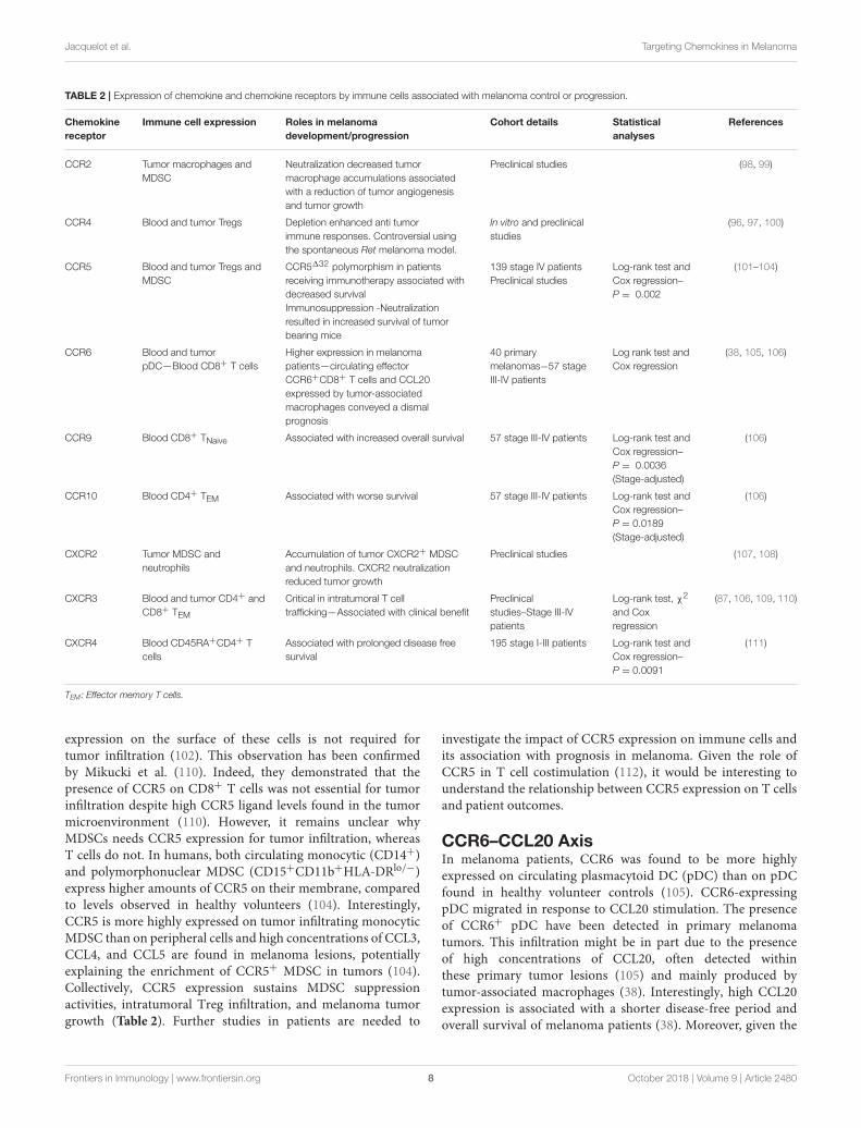
expression on the surface of these cells is not required fortumor infiltration (102). This observation has been confirmedby Mikucki et al. (110). Indeed, they demonstrated that thepresence of CCR5 on CD8+ T cells was not essential for tumorinfiltration despite high CCR5 ligand levels found in the tumormicroenvironment (110). However, it remains unclear whyMDSCs needs CCR5 expression for tumor infiltration, whereasT cells do not. In humans, both circulating monocytic (CD14+)and polymorphonuclear MDSC (CD15+CD11b+HLA-DRlo/−)express higher amounts of CCR5 on their membrane, comparedto levels observed in healthy volunteers (104). Interestingly,CCR5 is more highly expressed on tumor infiltrating monocyticMDSC than on peripheral cells and high concentrations of CCL3,CCL4, and CCL5 are found in melanoma lesions, potentiallyexplaining the enrichment of CCR5+ MDSC in tumors (104).Collectively, CCR5 expression sustains MDSC suppressionactivities, intratumoral Treg infiltration, and melanoma tumorgrowth (Table 2). Further studies in patients are needed to
investigate the impact of CCR5 expression on immune cells andits association with prognosis in melanoma. Given the role ofCCR5 in T cell costimulation (112), it would be interesting tounderstand the relationship between CCR5 expression on T cellsand patient outcomes.
CCR6–CCL20 AxisIn melanoma patients, CCR6 was found to be more highlyexpressed on circulating plasmacytoid DC (pDC) than on pDCfound in healthy volunteer controls (105). CCR6-expressingpDC migrated in response to CCL20 stimulation. The presenceof CCR6+ pDC have been detected in primary melanomatumors. This infiltration might be in part due to the presenceof high concentrations of CCL20, often detected withinthese primary tumor lesions (105) and mainly produced bytumor-associated macrophages (38). Interestingly, high CCL20expression is associated with a shorter disease-free period andoverall survival of melanoma patients (38). Moreover, given the
Frontiers in Immunology | www.frontiersin.org 8 October 2018 | Volume 9 | Article 2480

Jacquelot et al. Targeting Chemokines in Melanoma
negative prognostic value conveyed by tumor-infiltrating pDCin melanoma (113), CCR6 is likely to also be associated withpoor patient outcome. However, this needs to be explored furtherand to validated the prognostic value of CCR6+pDC in themelanoma tumor microenvironment. We have found that a lowproportion of circulating effector memory CD8+CCR6+ T cellswas associated with a better overall survival in stage IVmelanoma(106). Collectively, it seems that both CCL20 and CCR6 immunecell expression in multiple cell types are associated with a poorpatient outcome (Table 2).
CCR9–CCL25 AxisCCR9 is expressed at the membrane of several immune cellsubsets and is mostly associated with gut homing with theexception of immature T cells in transit from the bone marrowto the thymus (114). Further CCR9+ cell populations includeintestinal infiltrating T cells (115), gut pDC (116), and smallintestinal IgA producing plasma cells (117). Unfortunately, todate, the role of CCR9 expression on immune cells in melanomaand other cancers is poorly understood. We have investigated theimpact of CCR9 expression on the membrane of circulating Tcells in stage IV melanoma patients. Interestingly, high CCR9expression on naïve circulating CD8T cells is associated witha favorable prognosis (106) (Table 2). In mice, we have foundtumor infiltrating T cells that express CCR9 and importantly,blockade of its ligand, CCL25, in a sarcoma model, led toincreased tumor growth. This is associated with a reduction ofCD4+ T cell infiltration. Moreover, in this tumor model, highlevels of CCL25 were found in the tumor microenvironmentand these levels were much higher than the levels found in thegut (106) providing a possible explanation for the recruitmentof these CCR9+ T cells to the tumor bed. Further studies arewarranted to validate this positive impact of CCR9 expression onT cells in this pathology.
CCR10–CCL27 AxisCCR10 is one of the chemokine receptors that specifically guidethe migration of immune cells to the skin in response to the localproduction and accumulation of CCL27. In contrast to benignlesions where CCL27 is expressed at low levels, many primarymelanoma lesions express substantial amounts of this chemokine(44). CCL27 expression is correlated with T lymphocyte density,but unexpectedly, higher chemokine expression is associatedwith lower T cell infiltrate (44). This suggests that despite thelocal accumulation of CCL27, CCR10-expressing T cells areunable to infiltrate CCL27-expressing melanoma lesions andthese T cells are therefore restricted to circulate in the periphery.Supporting this hypothesis, in our own work we have shownthat in stage IV patients, the accumulation of circulating effectormemory CCR10 expressing CD4+ T cells was associated withshorter overall survival (106). With the exception of these twostudies, little is known about the impact of CCR10 expressionon immune cells and prognosis. However, it seems that CCL27tumor concentration was not associated with T cell accumulationand thus their peripheral increase was associated with a poorprognosis (Table 2).
CXCR3–CXCL9/CXCL10 AxisHigh expression of CXCR3, on melanoma infiltrating T cellstogether with the recruitment of effector memory CD8+ Tcells has been associated with a better patient outcome (87,89, 109, 118) (Table 2). Mullins et al. (109) reported that highCXCR3 expression on antigen specific CD8+CD45RO+ T cellsis associated with a favorable prognosis in stage III patientsbut fail to do so in patients with distant metastases (109). Wehave found that high CXCR3 expression on circulating effectormemory CD4+ T cells is associated with an enhancement of stageIII-IV patient survival, irrespective of tumor lesion location andpatient stages (106). Mikucki et al. (110) have demonstrated thecritical requirement of CXCR3 expression on mouse CD8+ Tcells for cell adhesion to, and migration through, the endothelialbarrier to infiltrate tumor lesions (110). Furthermore, CXCR3 isassociated with Th1/Tc1 polarization and anti-tumor functions(119, 120). Interestingly, therapy such as peptide vaccinationin Montanide Adjuvant led to the upregulation of CXCR3expression on circulating tumor antigen-specific T cells (121)but Hailemichael et al. have shown that most of these CXCR3+
T cells induced by the vaccination are retained to the site ofvaccine administration (122). Despite this potential inductionof CXCR3 expression, CXCR3+ T cells are unlikely to reachmelanoma lesions in this context. Furthermore, we have foundthat in stage III/IV patients, CXCR3 is poorly expressed on T cellscompared with expression levels observed in healthy volunteers(106). This last observation suggests that (i) CXCR3 is potentiallydownregulated due to a negative feedback loop of cell regulationfollowing STAT3 activation or (ii) these CXCR3+ T cells, whichare underrepresented in the periphery, are actually localized tomelanoma lesions. Currently, there is little evidence to supporteither of these two hypotheses. In favor of CXCR3-regulatedexpression, Yue et al. (123) found that STAT3 expression andsignaling mediated CXCR3 downregulation on CD8+ T cellsthus inhibiting intratumoral CD8+ T cell accumulation andimpacting anti-tumor functions (123). At steady-state, CXCR3is tightly regulated at the surface of T cells and downregulationof its expression with or without ligand binding is finelycontrolled by a regulatory feedback mechanism to preserve cellsfrom over activation (124) and this may even be exacerbatedin a pro-inflammatory context. Moreover, we have previouslyfound an enrichment of CXCR3-expressing CD4+ T cells inmetastatic lymph nodes compared with circulating T cells (106)perhaps explaining the differences found in the blood betweenmelanoma patients and healthy volunteers. In tumor lesions,CXCR3 expression might be sustained by the presence of pro-inflammatory molecules such as IFNγ that has been shown tosustain Tbx21 expression and subsequently TBET to positivelyregulate CXCR3 expression at the surface of T cells (125, 126).Together, these studies highlight that the expression of CXCR3on the surface of T cells is finely regulated and is essentialto melanoma infiltration and tumor control. Furthermore,high tumor expression of CXCR3 ligands together with highexpression of CXCR3 on T cells are both associated with afavorable prognosis in melanoma (Table 2). Thus, strategiesenhancing CXCR3 ligand production or CXCR3 expression on
Frontiers in Immunology | www.frontiersin.org 9 October 2018 | Volume 9 | Article 2480

Jacquelot et al. Targeting Chemokines in Melanoma
effector and memory T cells, but not melanoma cells, is highlydesirable.
CXCR4–CXCL12 AxisThe CXCR4-CXCL12 axis is required for the developmentand survival of mice as complete deletion of CXCR4 isembryonically lethal (127, 128). This axis plays an essential rolein haematopoiesis and cerebellar development, bone marrowimmune cell retention and thymic homing (10, 127, 128). Tostudy the role of CXCR4 expression on non-tumor cells andits association with melanoma progression, D’alterio et al. (64)have used CXCR4 heterozygous mice where they intravenouslyinjected CXCR4 expressing B16 melanoma cells. The partial lossof host-CXCR4 expression reduced lung metastases formationthat is accompanied by a decrease of CXCL12 concentrationtogether with Ly6G+ cell accumulation in lung tissues (64).Similar results have been found in wild type mice treated witha CXCR4 antagonist, Plerixafor (AMD3100) (64). In stage I-III melanoma patients, high expression of CXCR4 in circulatingCD4+CD45RA+ was associated with prolonged disease freesurvival (Table 2). Moreover, the presence of CXCR4 expressingCD4+CD45RA+ T cells correlated with absence of primarytumor ulceration (111).
DO CHEMOKINE RECEPTOREXPRESSION ON IMMUNE CELLSREFLECT THE METASTATICDISSEMINATION OF MELANOMA?
This question was first raised by Salerno et al. (94). Theystudied whether the expression of organ-specific chemokinereceptors and integrins on the surface of T cells differs accordingto the metastatic site (94). This included the evaluation ofCCR4, CCR5, CCR7, CCR9, CXCR3, CLA, and tissue retentionintegrins on the surface of CD4+ and CD8+ T cells by flowcytometry. This group found limited evidence that tissue site-specific chemokine receptor expression was associated with thesite of metastatic location with the exception of CCR9, whichwas found to be preferentially expressed on T cells that infiltratesmall intestine metastases. Expectedly, the expression of tissueretention integrins was higher on tumor infiltrating T cells thanon circulating T cells suggesting a specific maintenance of a poolof intratumoral effector and memory T cells in melanoma lesions(94). This lack of site-specific expression of chemokine receptorson infiltrating T cells might be due in part by an absence ofinfiltration of these site-specific chemokine receptor-expressingcells. Thus, these cells may be maintained in the circulation.Salerno et al. (94) found that CCR4, CCR5, and CLA arehighly expressed on circulating T cells (94). However, how thisexpression differs from healthy volunteers and to what extent thisperipheral expression correlates with site-specific metastases anddictates patient’s prognosis were, at this stage, unknown. Withthis in mind, we retrospectively evaluated the surface expressionof nine chemokine receptors and integrins on circulating andtumor infiltrating T cells collected from stage III-IV patients(106). These included the expression of CCR6, CCR7, CCR9,
CCR10, CXCR3, CXCR4, CXCR5, CLA, and CD103. Moreover,we studied the expression of the chemoattractant receptor-homologous molecule expressed on Th2 cells, CRTH2, knownfor its involvement in Th2 polarization and responses (129, 130).When comparing these expression levels to those found oncirculating T cells from healthy volunteers, patients with a lowerexpression of CXCR3 and CCR6 on effector/memory circulatingT cells had preferential metastases to the skin and lymph nodesand a decrease of CCR9, together with CXCR4 and CXCR5expression on both CD4+ and CD8+ T cells, which was anindicator of the presence of pulmonary lesions (Table 3). Inaddition, multi-metastatic patients with a broad disseminationof disease displayed an increase of chemokine receptor/integrinexpression on naïve T lymphocytes, specifically CCR10, CD103,and CRTH2 (Table 3). This disseminated localization was alsoassociated with a loss of CXCR3 on effector/memory T cellsand a decrease in CXCR4 and CCR9 expression on CD4effector and terminal effector T cells (Table 3). Collectively,these results indicated that the expression pattern of chemokinereceptors/integrins on the surface of circulating T cells potentiallymirror the metastatic spreading in melanoma patients (106).
Interestingly, CD103 expression on naïve T cells was stronglyassociated with livermetastases (106) suggesting that this integrinmight play a role in binding T cells to this organ. CD103expression is a feature of tissue resident memory T lymphocytes(134) and many T lymphocytes that reside in the gut (115)or the liver (135) express this integrin. Its ligand, E-cadherin,is naturally expressed on hepatocytes (136), and notably inthe interlobular bile duct epithelia (137). Shimizu et al. havedemonstrated that CD103-expressing CD4+ and CD8+ T cellsaccumulated in the liver and these cells harbored a particularphenotype with a decrease of TCRαβ expression (135). Asobserved in hepatocellular carcinoma (136, 138), a decrease ofE-cadherin expression during epithelial-mesenchymal transitionof liver metastasis on the surface of hepatocytes is associatedwith an increase of its soluble form in the serum (139)potentially favoring the circulation of CD103+ T cells and theiraccumulation in the blood of melanoma patients harboring livermetastases (Table 3).
Further retrospective and prospective investigations arewarranted to support the clinical relevance of differencesin expression of chemokines and chemokine receptors inmelanoma. Their evaluation would likely benefit patients in theearly detection of metastases and in targeting specific subsets ofT cells to favor their migration to desired organs and to targetthese metastases. Strategies to modulate their expression andfunctions are needed in order to ameliorate patient prognosis andtherapeutic outcomes.
POTENTIAL FOR TARGETING
Chemokines and their receptors have dual roles in melanomaand other cancers. On one hand, they promote immune cellrecruitment necessary for tumor control (e.g., CXCL9/10/11and CXCR3). On the other hand, they are involved in tumorescape and metastases formation by (i) selectively guiding tumor
Frontiers in Immunology | www.frontiersin.org 10 October 2018 | Volume 9 | Article 2480

Jacquelot et al. Targeting Chemokines in Melanoma
TABLE 3 | Chemokine receptors expression at the surface of peripheral immune T cells mirrors the melanoma metastatic dissemination.
Melanoma Stage Tumor lesion localization Chemokine receptors and integrins involved
Stage III Regional cutaneous and lymph node metastases Decrease of CCR6 and CXCR3 expressions on effector/memory
peripheral T cells
Stage IV Regional cutaneous and lymph node metastases + lung
metastases
Reduction of CCR9, CXCR4, and CXCR5 expression on circulating T cells
Stage IV Multi-disseminated disease with or without lung
involvement
Increase expression of CCR10, CD103*, and CRTH2 on naïve
peripheral T cells—Loss of CXCR3 and CCR6 expression on effector and
memory circulating T cells—Decrease of CXCR4 and CCR9 expression
on effector and terminal effector blood T cells
Chemokine receptors expression was retrospectively evaluated on circulating blood T cells collected from 57 stage III–IV melanoma patients (131–133).
*Elevated expression of CD103 on naïve T cells is correlated with the presence of liver metastases.
cells toward specific organs, which subsequently form secondarylesions (e.g., CCR7 or CXCR4), (ii) favoring the recruitmentof immunosuppressive cells (e.g., CCR5) and, (iii) influencingtumor vasculature associated with tumor dissemination (e.g.,CXCL10 and CXCR3) (140, 141). Thus, targeting these moleculesis of particular interest in melanoma and other cancers as anapproach to limit tumor development and to considerably reduceits metastatic spreading. However, the design of selective drugswill need to specifically target tumor cells, the immune system, orboth compartments.
Many small molecule antagonists and therapeutic antibodieshave been developed (142) but so far, this has led to only amoderate improvement in various diseases. As a consequence,only 3 targeting agents have been approved to treat patients,or are in phase III clinical trials. These include a blockingCCR4 antibody, Mogamulizumab, approved in Japan to treatrefractory adult T-cell leukemia, peripheral T cell lymphoma andcutaneous T cell lymphoma (142), an anti-CCR5 antibody testedin graft-vs.-host disease and human immunodeficiency virus-1(143) and an anti-CXCR4 antibody evaluated in lymphoma andmultiple myeloma (144). Thirty-seven additional compounds arecurrently being tested targeting CCR1, CCR2, CCR3, CCR4,CCR5, CCR9, CXCR1, CXCR2, CXCR4, and CX3C1 (142, 145,146). In a small study (147), metastatic colorectal cancer patientswith CCR5+ liver metastases were treated with a small moleculethat antagonizes CCR5, Maraviroc, with encouraging results.Therefore, further evaluation in a larger cohort is warranted todetermine the benefits and toxicity of this approach.
In melanoma, CXCR4 inhibition with AMD11070 abrogatedtumor cell migration in response to CXCL12 stimulation (148).Similarly, the CXCR4 antagonist, AMD3100, prevents thedevelopment of squamous cell carcinomas under chronic UVexposure. Mechanistically, UV radiation induced CXCL12expression in the skin and this was responsible for attractingCXCR4+ mast cells. Thus, blocking the CXCR4-CXCL12pathway using this antagonist reduced mast cell infiltrationinto the skin, tumors and draining lymph nodes, andthis subsequently prevents immune suppression and tumordevelopment (149). Given the involvement of CXCR4 in tumorcell migration to many different organs, oral administrationof CXCR4 inhibitors could be particularly efficient. Moreover,CXCR4 is also involved in the recruitment of suppressiveimmune cells, such as mast cells in the tumor microenvironment.
CCR9 blockade using an antibody significantly reduced thetumor cell migration in response to CCL25 stimulation (42).Interestingly, a new mouse anti-human CCR9 antibody wasdeveloped by Somovilla-Crespo et al. showing promising resultsin blocking the growth of human CCR9+ leukemia cells inNSG mice (150). Similarly, the use of the CCR9 antagonistCCX8037 could also specifically interfere with small intestinaldissemination. However, we have shown that the blockade ofCCL25 in a sarcoma model inoculated in immunocompetentmice was detrimental and notably, resulted in increasing thetumor growth (106). Further investigations are required todetermine the impact of such drugs on both leukocyte traffickingand tumor cell spreading (151) to avoid unexpected off-targeteffects.
Neonatal skin exposed to UVB induced an IFNγ genesignature response frommelanocytes including CCL8 expression(99). Thus accumulation of CCL8 drives the recruitmentof CCR2+ macrophages that were shown to promotemelanomagenesis. The blockade of IFNγ using a specificantibody or the use of CCR2 deficient mice, which weresubjected to UVB exposure, have decreased of macrophagesinfiltration in the skin and reduced tumor volume (99). Similarly,the overexpression of a dominant negative version of CCL2, anon-functional protein that competes with the native form forbinding to CCR2, in melanoma tumor bearing mice specificallyreduced tumor associated macrophage infiltration that isassociated with a decrease of tumor angiogenesis and tumorgrowth (98). Interestingly, mice inoculated with B16F10 tumorsengineered to express GM-CSF harbored an accumulation ofmonocytic CCR2+ MDSC compared to non-GM-CSF expressingtumors. This accumulation of MDSC in melanoma lesions wasassociated with a reduction of CD8+ T cell infiltration and anincrease in tumor burden (152). Although vaccination withirradiated B16 cells producing GM-CSF was shown to favorimmune responses to immunotherapies in preclinical melanomamodels (153, 154), in this setting, this cytokine seemed toplay a negative role in antitumor immune surveillance. CCR2appears to be an attractive target in melanoma and potentiallyin other tumor types and a CCR2 antibody, plozalizumab, iscurrently being tested in phase I clinical trial (NCT02723006) incombination with an immune checkpoint blocker, nivolumab.
CRTH2 associated with Th2 responses would be anattractive target in melanoma as this chemokine expression
Frontiers in Immunology | www.frontiersin.org 11 October 2018 | Volume 9 | Article 2480

Jacquelot et al. Targeting Chemokines in Melanoma
is increased in patients with a multi-metastatic disease (Table 3).CRTH2 is also expressed on eosinophils, basophils, and somemonocytes/macrophages (155), immune subsets which allconvey a distinct prognosis in melanoma (84, 156). Initiallydesigned for targeting CRTH2+ T cells involved in respiratorydiseases (157, 158), CRTH2 antagonists could be indicatedin multi-metastatic melanoma patients with high CRTH2expression.
SX-682 (Syntrix Biosystems, Inc) is a selective and potentCXCR1/2 antagonist. CXCR1/2 is expressed on melanoma cells,MDSC and neutrophils and sustains tumor immunosuppression,tumor growth, angiogenesis and tumor dissemination inresponse to CXCL1, CXCL2 or CXCL8 (107, 108, 159–164)(Tables 1, 2). In melanoma, MDSC accumulated both in tumorlesions and in periphery, correlating with tumor stage. Thisfeature has been associated with a negative prognostic value (84).Furthermore, this compound has been evaluated in combinationto anti-CTLA-4 and anti-PD-1 co-blockade in an elegant mousemodel of prostate cancer (165). In this model, the authorsdemonstrated the crucial role of MDSC in sustaining cancerprogression. The combination of immune checkpoint inhibitorsand SX-682 resulted in decreased prostate mass, lymph nodeand lung metastases (165). This inhibitor is currently beingevaluated in stage III/IV melanoma patients in combinationwith an anti-PD1 antibody, Pembrolizumab (NCT03161431).This phase I study aims to evaluate the tolerability and safetyprofile of SX-682 together with the response rate, tumorresponse duration, progression free and overall survival ofthe combination. Interestingly, another CXCR1/2 inhibitor,Ladarixin, was shown to significantly reduce human melanomacell motility and to induce apoptosis in vitro. In vivo treatmentof melanoma xenografts with Ladarixin reduced tumor growth,polarized intratumoral macrophages to M1 phenotype, andinhibited angiogenesis (166). Inhibition of CXCR1/2 appears tobe very promising as it targets both melanoma and immune cells,reducing tumor burden alone or in combination with immunecheckpoint blockers.
Modulation of chemokine receptor expression on the surfaceof chimeric antigen receptor (CAR) T or NK cells priorto infusion is promising as this would enhance their tumorinfiltration and potentially improve therapeutic results. CX3CR1genetically modified T cells transferred into CX3CL1 producingcolorectal adenocarcinoma tumor bearing mice displayedenhanced tumor infiltration and anti-tumor responses (167).Moreover, significant reduction in tumor size and completeremission have been observed with CCR2b-GD2-CAR T cellsand CXCR4-EGFRvIII-CAR NK cells infused in mice bearingCCL2 producing GD2 neuroblastoma or CXCL12 secretingEGFRvIII glioblastoma cells, respectively (168, 169). Similarly,genetically engineered CCR2 expression on CAR T cells directedto the tumor antigen mesothelin increased tumor cell infiltrationand anti-tumor responses against large and established tumorsinoculated in severe immunodeficient mice (170). To date,CAR specific cells genetically engineered to express particularchemokine receptor have only been tested in preclinical models.Despite having shown impressive anti-tumor responses againstprimary tumors, it will be challenging to find a chemokine that
is highly, specifically and commonly expressed across differenttumor microenvironments, found in multi metastatic patients inorder to efficiently eradicate all disseminated lesions.
CONCLUSION AND PERSPECTIVES
Chemokines and chemokine receptors are key moleculesinvolved in cell migration, proliferation and survival that arecritical in maintaining tissue homeostasis. Melanoma cellsoverexpress many chemokine receptors that are likely involvedin cancer progression and metastasis. Thus, modulation ofchemokines and chemokine receptors appears to be an attractivetarget in cancer therapy. However, targeting them is a doubleedged sword, as treatments will not only affect immune cellmigration to tumor lesions or tumor dissemination but also inthe long term, impact immune cell development and polarization(e.g., CXCR4). This may partly explain why there is low numberof approved drugs targeting chemokines and their receptors intreating chronic diseases, such as cancer. How can we overcomethis? In the era of personalized medicine, designing bispecificantibodies that can specifically target a chemokine receptor and atumor antigen, which are both expressed on the surface of cancercells is highly attractive. However, antigen escape due to theemergence of tumor variants, which do not express the targetedantigen, are likely to emerge, rendering the treatment ineffective.Another promising area of research is to combine chemokinereceptor blockers with anti-PD-1 or anti-CTLA-4 antibodies tofurther improve the clinical activity of these antibodies and thusfurther increase patient survival (171). Together, this would leadto reduced tumor infiltration by immunosuppressive cells asTregs or MDSCs and subsequently, induce anti-tumor immunityby releasing the immunosuppressive brakes. Another approachwould be to use engineered antibodies to target privilegedmetastatic sites. The therapeutic management of brainmetastasesin melanoma and other cancers is challenging, as the brain isprotected by a highly selective blood-brain barrier impermeableto many cells, in particular, immune cells. In melanoma, abispecific antibody could be designed to target CCR4 and ananobody, that selectively binds to human cerebromicrovascularendothelial cells. This attached nanobody is then internalizedand able to transmigrate across the endothelial barrier (146).As a proof of principle, a bispecific antibody specific for themetabotropic glutamate receptor 1, expressed in the brain, andalso carrying a specific nanobody was able to translocate acrossthe endothelial layer into the brain and regulate physiologicalfunctions (172).
Given the association between the accumulation of certainchemokines in tumor lesions and the presence of tertiarylymphoid structures, it would be interesting to reinstatechemokine expression in “cold” tumors to favor the emergence ofectopic-like lymphoid organs that are positively associated withimmune cell activation and patient survival. Several strategies arecurrently being tested, aiming to modulate anti-tumor responsesthrough the induction of tertiary lymphoid structures (173).
Collectively, chemokine and chemokine receptors areessential for guiding immune cells to tumor lesions, however
Frontiers in Immunology | www.frontiersin.org 12 October 2018 | Volume 9 | Article 2480
Jacquelot et al. Targeting Chemokines in Melanoma
melanoma cells often harness these molecules to disseminateto distant organs. Given their broad expression profileand potential side effects, drugs targeting these moleculesmust be carefully designed. Novel technologies have nowrendered this challenge possible with the developmentof compounds that specifically affect a desired target(145, 146). Many chemokine receptor antagonists arecurrently being tested in melanoma and other malignancies,if successful, these treatments will diversify the oncologicarmamentarium currently available therefore increasing possibletherapeutic combinations and ultimately improving patientoutcome.
AUTHOR CONTRIBUTIONS
NJ wrote the initial draft. CD, GB, and LZ made substantialcontributions to discussions of the content. All authors reviewedand/or edited the manuscript prior submission.
ACKNOWLEDGMENTS
We are grateful to our colleagues for helpful discussions thathave led to improve the quality and the content of this review.
NJ has received a postdoctoral fellowship from the FoundationARC pour la recherche sur le cancer. This work has beensupported by the National Health and Medical Research Council(Australia)(APP1135898, 1054925), Victorian State GovernmentOperational Infrastructure Support and Australian GovernmentNHMRC Independent Research Institute Infrastructure Supportscheme (GB), the Ligue contre le Cancer (équipe labelisée);Agence Nationale de la Recherche (ANR)–Projets blancs; ANRunder the frame of E-Rare-2, the ERA-Net for Research onRare Diseases; Association pour la recherche sur le cancer(ARC); Cancéropôle Ile-de-France; Institut National du Cancer(INCa); Institut Universitaire de France; Foundation pour laRecherche Médicale (FRM); a donation by Elior, Lombard OdierFoundation, Seerave Foundation, Swiss Bridge Foundation;the European Commission (ArtForce); the European ResearchCouncil (ERC); Foundation Carrefour; Institut National duCancer (INCa); Inserm (HTE); Institut Universitaire de France;the LeDucq Foundation; the LabEx Immuno-Oncology; theRHU Torino Lumière; the SIRIC Stratified Oncology Cell DNARepair and Tumor Immune Elimination (SOCRATE); the SIRICCancer Research and Personalized Medicine (CARPEM); theParis Alliance of Cancer Research Institutes (PACRI) and byphilanthropia (Mrs E. Badinter and Mrs N. Meyer) (LZ).
REFERENCES
1. Zhang L, Conejo-Garcia JR, Katsaros D, Gimotty PA, Massobrio M,
Regnani G, et al. Intratumoral T cells, recurrence, and survival in epithelial
ovarian cancer. N Engl J Med. (2003) 348:203–13. doi: 10.1056/NEJMoa
020177
2. Galon J, Costes A, Sanchez-Cabo F, Kirilovsky A, Mlecnik B, Lagorce-
Pages C, et al. Type, density, and location of immune cells within human
colorectal tumors predict clinical outcome. Science (2006) 313:1960–4.
doi: 10.1126/science.1129139
3. Rusakiewicz S, Semeraro M, Sarabi M, Desbois M, Locher C, Mendez
R, et al. Immune infiltrates are prognostic factors in localized
gastrointestinal stromal tumors. Cancer Res. (2013) 73:3499–510.
doi: 10.1158/0008-5472.CAN-13-0371
4. Clemente CG, Mihm MC Jr, Bufalino R, Zurrida S, Collini P, Cascinelli N.
Prognostic value of tumor infiltrating lymphocytes in the vertical growth
phase of primary cutaneous melanoma. Cancer (1996) 77:1303–10.
5. Mihm MCJr, Clemente CG, Cascinelli N. Tumor infiltrating lymphocytes
in lymph node melanoma metastases: a histopathologic prognostic
indicator and an expression of local immune response. Lab Invest. (1996)
74:43–7.
6. Tuthill RJ, Unger JM, Liu PY, Flaherty LE, Sondak VK, Southwest Oncology
G. Risk assessment in localized primary cutaneous melanoma: a Southwest
Oncology Group study evaluating nine factors and a test of the Clark
logistic regression prediction model. Am J Clin Pathol. (2002) 118:504–11.
doi: 10.1309/WBF7-N8KH-71KT-RVQ9
7. Mellado M, Rodriguez-Frade JM, Manes S, Martinez AC. Chemokine
signaling and functional responses: the role of receptor dimerization
and TK pathway activation. Annu Rev Immunol. (2001) 19:397–421.
doi: 10.1146/annurev.immunol.19.1.397
8. Mellado M, Rodriguez-Frade JM, Vila-Coro AJ, Fernandez S, Martin De
Ana A, Jones DR, et al. Chemokine receptor homo- or heterodimerization
activates distinct signaling pathways. EMBO J. (2001) 20:2497–507.
doi: 10.1093/emboj/20.10.2497
9. Zlotnik A, Burkhardt AM, Homey B. Homeostatic chemokine receptors
and organ-specific metastasis. Nat Rev Immunol. (2011) 11:597–606.
doi: 10.1038/nri3049
10. Griffith JW, Sokol CL, Luster AD. Chemokines and chemokine receptors:
positioning cells for host defense and immunity. Annu Rev Immunol. (2014)
32:659–702. doi: 10.1146/annurev-immunol-032713-120145
11. Bachelerie F, Ben-Baruch A, Charo IF, Combadiere C, Farber JM, Förster
R, et al. Chemokine Receptors. 2018 ed. (2018). IUPHAR/BPS Guide to
Pharmacology.
12. Nibbs RJ, Graham GJ. Immune regulation by atypical chemokine receptors.
Nat Rev Immunol. (2013) 13:815–29. doi: 10.1038/nri3544
13. Bosisio D, Salvi V, Gagliostro V, Sozzani S. Angiogenic and
antiangiogenic chemokines. Chem Immunol Allergy (2014) 99:89–104.
doi: 10.1159/000353317
14. Bunemann E, Hoff NP, Buhren BA, Wiesner U, Meller S, Bolke E, et al.
Chemokine ligand-receptor interactions critically regulate cutaneous wound
healing. Eur J Med Res. (2018) 23:4. doi: 10.1186/s40001-017-0299-0
15. Nestle FO, DiMeglio P, Qin JZ, Nickoloff BJ. Skin immune sentinels in health
and disease. Nat Rev Immunol. (2009) 9:679–91. doi: 10.1038/nri2622
16. Tikoo S, Jain R, Kurz AR, Weninger W. The lymphoid cell network in the
skin. Immunol Cell Biol. (2018) 96:485–96. doi: 10.1111/imcb.12026
17. Lebre MC, Van Der Aar AM, Van Baarsen L, Van Capel TM, Schuitemaker
JH, Kapsenberg ML, et al. Human keratinocytes express functional Toll-
like receptor 3, 4, 5, and 9. J Invest Dermatol. (2007) 127:331–41.
doi: 10.1038/sj.jid.5700530
18. Bos JD, Kapsenberg ML. The skin immune system: progress
in cutaneous biology. Immunol Today (1993) 14:75–8.
doi: 10.1016/0167-5699(93)90062-P
19. Handfield C, Kwock J, Macleod AS. Innate antiviral immunity in the skin.
Trends Immunol. (2018) 39:328–40. doi: 10.1016/j.it.2018.02.003
20. Dorward DA, Lucas CD, Chapman GB, Haslett C, Dhaliwal K, Rossi
AG. The role of formylated peptides and formyl peptide receptor 1 in
governing neutrophil function during acute inflammation. Am J Pathol.
(2015) 185:1172–84. doi: 10.1016/j.ajpath.2015.01.020
21. Stutte S, Quast T, Gerbitzki N, Savinko T, Novak N, Reifenberger J, et al.
Requirement of CCL17 for CCR7- and CXCR4-dependent migration of
cutaneous dendritic cells. Proc Natl Acad Sci USA. (2010) 107:8736–41.
doi: 10.1073/pnas.0906126107
22. Berg EL, Yoshino T, Rott LS, Robinson MK, Warnock RA, Kishimoto TK,
et al. The cutaneous lymphocyte antigen is a skin lymphocyte homing
Frontiers in Immunology | www.frontiersin.org 13 October 2018 | Volume 9 | Article 2480

Jacquelot et al. Targeting Chemokines in Melanoma
receptor for the vascular lectin endothelial cell-leukocyte adhesion molecule
1. J Exp Med. (1991) 174:1461–6. doi: 10.1084/jem.174.6.1461
23. Dudda JC, Simon JC, Martin S. Dendritic cell immunization route
determines CD8+ T cell trafficking to inflamed skin: role for tissue
microenvironment and dendritic cells in establishment of T cell-homing
subsets. J Immunol. (2004) 172:857–63. doi: 10.4049/jimmunol.172.2.857
24. Mora JR, Cheng G, Picarella D, Briskin M, Buchanan N, Von Andrian UH.
Reciprocal and dynamic control of CD8T cell homing by dendritic cells from
skin- and gut-associated lymphoid tissues. J Exp Med. (2005) 201:303–16.
doi: 10.1084/jem.20041645
25. Ferguson AR, Engelhard VH. CD8T cells activated in distinct
lymphoid organs differentially express adhesion proteins and coexpress
multiple chemokine receptors. J Immunol. (2010) 184:4079–86.
doi: 10.4049/jimmunol.0901903
26. Brinkman CC, Rouhani SJ, Srinivasan N, Engelhard VH. Peripheral tissue
homing receptors enable T cell entry into lymph nodes and affect the
anatomical distribution of memory cells. J Immunol. (2013) 191:2412–25.
doi: 10.4049/jimmunol.1300651
27. Campbell JJ, Haraldsen G, Pan J, Rottman J, Qin S, Ponath P, et al. The
chemokine receptor CCR4 in vascular recognition by cutaneous but not
intestinal memory T cells. Nature (1999) 400:776–80. doi: 10.1038/23495
28. Homey B, Wang W, Soto H, Buchanan ME, Wiesenborn A, Catron
D, et al. Cutting edge: the orphan chemokine receptor G protein-
coupled receptor-2 (GPR-2, CCR10) binds the skin-associated
chemokine CCL27 (CTACK/ALP/ILC). J Immunol. (2000) 164:3465–70.
doi: 10.4049/jimmunol.164.7.3465
29. Reiss Y, Proudfoot AE, Power CA, Campbell JJ, Butcher EC. CC chemokine
receptor (CCR)4 and the CCR10 ligand cutaneous T cell-attracting
chemokine (CTACK) in lymphocyte trafficking to inflamed skin. J Exp Med.
(2001) 194:1541–7. doi: 10.1084/jem.194.10.1541
30. Homey B, Alenius H, Muller A, Soto H, Bowman EP, Yuan W, et al. CCL27-
CCR10 interactions regulate T cell-mediated skin inflammation. Nat Med.
(2002) 8:157–65. doi: 10.1038/nm0202-157
31. Schmuth M, Neyer S, Rainer C, Grassegger A, Fritsch P, Romani N,
et al. Expression of the C-C chemokine MIP-3 alpha/CCL20 in human
epidermis with impaired permeability barrier function. Exp Dermatol. (2002)
11:135–42. doi: 10.1034/j.1600-0625.2002.110205.x
32. Massague J, Obenauf AC.Metastatic colonization by circulating tumour cells.
Nature (2016) 529:298–306. doi: 10.1038/nature17038
33. Payne AS, Cornelius LA. The role of chemokines in melanoma
tumor growth and metastasis. J Invest Dermatol. (2002) 118:915–22.
doi: 10.1046/j.1523-1747.2002.01725.x
34. Nguyen DX, Bos PD, Massague J. Metastasis: from dissemination to
organ-specific colonization. Nat Rev Cancer (2009) 9:274–84. doi: 10.1038/
nrc2622
35. Overwijk WW, Restifo NP. B16 as a mouse model for human
melanoma. Curr Protoc Immunol. (2001) Chapter 20:Unit 20.1.
doi: 10.1002/0471142735.im2001s39
36. Kuzu OF, Nguyen FD, Noory MA, Sharma A. Current state of animal
(Mouse) modeling in melanoma research. Cancer Growth Metast. (2015)
8:81–94. doi: 10.4137/CGM.S21214
37. Klein A, Sagi-Assif O, Meshel T, Telerman A, Izraely S, Ben-Menachem
S, et al. CCR4 is a determinant of melanoma brain metastasis. Oncotarget
(2017) 8:31079–91. doi: 10.18632/oncotarget.16076
38. Samaniego R, Gutierrez-Gonzalez A, Gutierrez-Seijo A, Sanchez-Gregorio S,
Garcia-Gimenez J, Mercader E, et al. CCL20 Expression by tumor-associated
macrophages predicts progression of human primary cutaneous melanoma.
Cancer Immunol Res. (2018). doi: 10.1158/2326-6066.CIR-17-0198. [Epub
ahead of print].
39. Wiley HE, Gonzalez EB, Maki W, Wu MT, Hwang ST. Expression
of CC chemokine receptor-7 and regional lymph node metastasis
of B16 murine melanoma. J Natl Cancer Inst. (2001) 93:1638–43.
doi: 10.1093/jnci/93.21.1638
40. Kuhnelt-Leddihn L, Muller H, Eisendle K, Zelger B, Weinlich G.
Overexpression of the chemokine receptors CXCR4, CCR7, CCR9, and
CCR10 in human primary cutaneous melanoma: a potential prognostic
value for CCR7 and CCR10? Arch Dermatol Res. (2012) 304:185–93.
doi: 10.1007/s00403-012-1222-8
41. Letsch A, Keilholz U, Schadendorf D, Assfalg G, Asemissen AM, Thiel E, et al.
Functional CCR9 expression is associated with small intestinal metastasis. J
Invest Dermatol. (2004) 122:685–90. doi: 10.1111/j.0022-202X.2004.22315.x
42. Amersi FF, Terando AM, Goto Y, Scolyer RA, Thompson JF, Tran AN, et al.
Activation of CCR9/CCL25 in cutaneous melanoma mediates preferential
metastasis to the small intestine. Clin Cancer Res. (2008) 14:638–45.
doi: 10.1158/1078-0432.CCR-07-2025
43. Murakami T, Cardones AR, Finkelstein SE, Restifo NP, Klaunberg BA,
Nestle FO, et al. Immune evasion by murine melanoma mediated
through CC chemokine receptor-10. J Exp Med. (2003) 198:1337–47.
doi: 10.1084/jem.20030593
44. Simonetti O, Goteri G, Lucarini G, Filosa A, Pieramici T, Rubini C,
et al. Potential role of CCL27 and CCR10 expression in melanoma
progression and immune escape. Eur J Cancer (2006) 42:1181–7.
doi: 10.1016/j.ejca.2006.01.043
45. Robledo MM, Bartolome RA, Longo N, Rodriguez-Frade JM, Mellado M,
Longo I, et al. Expression of functional chemokine receptors CXCR3 and
CXCR4 on human melanoma cells. J Biol Chem. (2001) 276:45098–105.
doi: 10.1074/jbc.M106912200
46. Kawada K, Sonoshita M, Sakashita H, Takabayashi A, Yamaoka Y, Manabe
T, et al. Pivotal role of CXCR3 in melanoma cell metastasis to lymph nodes.
Cancer Res. (2004) 64:4010–7. doi: 10.1158/0008-5472.CAN-03-1757
47. Longo-Imedio MI, Longo N, Trevino I, Lazaro P, Sanchez-Mateos P. Clinical
significance of CXCR3 and CXCR4 expression in primary melanoma. Int J
Cancer (2005) 117:861–5. doi: 10.1002/ijc.21269
48. Monteagudo C, Martin JM, Jorda E, Llombart-Bosch A. CXCR3 chemokine
receptor immunoreactivity in primary cutaneous malignant melanoma:
correlation with clinicopathological prognostic factors. J Clin Pathol. (2007)
60:596–9. doi: 10.1136/jcp.2005.032144
49. Murakami T, Maki W, Cardones AR, Fang H, Tun Kyi A, Nestle FO,
et al. Expression of CXC chemokine receptor-4 enhances the pulmonary
metastatic potential of murine B16 melanoma cells. Cancer Res. (2002)
62:7328–34. Available online at: http://cancerres.aacrjournals.org/content/
62/24/7328.long
50. Murakami T, Cardones AR, Hwang ST. Chemokine receptors
and melanoma metastasis. J Dermatol Sci. (2004) 36:71–8.
doi: 10.1016/j.jdermsci.2004.03.002
51. Scala S, Giuliano P, Ascierto PA, Ierano C, Franco R, Napolitano M, et al.
Human melanoma metastases express functional CXCR4. Clin Cancer Res.
(2006) 12:2427–33. doi: 10.1158/1078-0432.CCR-05-1940
52. Mendt M, Cardier JE. Activation of the CXCR4 chemokine
receptor enhances biological functions associated with B16
melanoma liver metastasis. Melanoma Res. (2017) 27:300–8.
doi: 10.1097/CMR.0000000000000346
53. Fusi A, Liu Z, KummerlenV, Nonnemacher A, Jeske J, Keilholz U. Expression
of chemokine receptors on circulating tumor cells in patients with solid
tumors. J Transl Med. (2012) 10:52. doi: 10.1186/1479-5876-10-52
54. Mori T, Kim J, Yamano T, Takeuchi H, Huang S, Umetani N, et al. Epigenetic
up-regulation of C-C chemokine receptor 7 and C-X-C chemokine
receptor 4 expression in melanoma cells. Cancer Res. (2005) 65:1800–7.
doi: 10.1158/0008-5472.CAN-04-3531
55. Takeuchi H, Fujimoto A, Tanaka M, Yamano T, Hsueh E, Hoon
DS. CCL21 chemokine regulates chemokine receptor CCR7 bearing
malignant melanoma cells. Clin Cancer Res. (2004) 10:2351–8.
doi: 10.1158/1078-0432.CCR-03-0195
56. Gunn MD, Kyuwa S, Tam C, Kakiuchi T, Matsuzawa A, Williams LT, et al.
Mice lacking expression of secondary lymphoid organ chemokine have
defects in lymphocyte homing and dendritic cell localization. J Exp Med.
(1999) 189:451–60. doi: 10.1084/jem.189.3.451
57. Shields JD, Kourtis IC, Tomei AA, Roberts JM, Swartz MA. Induction
of lymphoidlike stroma and immune escape by tumors that express the
chemokine CCL21. Science (2010) 328:749–52. doi: 10.1126/science.11
85837
58. Mora JR, Bono MR, Manjunath N, Weninger W, Cavanagh LL, Rosemblatt
M, et al. Selective imprinting of gut-homing T cells by Peyer’s patch dendritic
cells. Nature (2003) 424:88–93. doi: 10.1038/nature01726
59. Amatschek S, Lucas R, Eger A, Pflueger M, Hundsberger H, Knoll C,
et al. CXCL9 induces chemotaxis, chemorepulsion and endothelial barrier
Frontiers in Immunology | www.frontiersin.org 14 October 2018 | Volume 9 | Article 2480

Jacquelot et al. Targeting Chemokines in Melanoma
disruption through CXCR3-mediated activation of melanoma cells. Br J
Cancer (2011) 104:469–79. doi: 10.1038/sj.bjc.6606056
60. Chang CH, Qiu J, O’sullivan D, Buck MD, Noguchi T, Curtis JD, et al.
Metabolic competition in the tumor microenvironment is a driver of cancer
progression. Cell (2015) 162:1229–41. doi: 10.1016/j.cell.2015.08.016
61. Jenkins MH, Brinckerhoff CE, Mullins DW. CXCR3 signaling in BRAFWT
melanoma increases IL-8 expression and tumorigenicity. PLoS ONE (2015)
10:e0121140. doi: 10.1371/journal.pone.0121140
62. Scharping NE, Menk AV, Moreci RS, Whetstone RD, Dadey RE,Watkins SC,
et al. The tumormicroenvironment represses T cell mitochondrial biogenesis
to drive intratumoral T cell metabolic insufficiency and dysfunction.
Immunity (2016) 45:374–88. doi: 10.1016/j.immuni.2016.07.009
63. Mcconnell AT, Ellis R, Pathy B, Plummer R, Lovat PE, O’boyle G. The
prognostic significance and impact of the CXCR4-CXCR7-CXCL12 axis
in primary cutaneous melanoma. Br J Dermatol. (2016) 175:1210–20.
doi: 10.1111/bjd.14720
64. D’alterio C, Barbieri A, Portella L, Palma G, Polimeno M, Riccio
A, et al. Inhibition of stromal CXCR4 impairs development of
lung metastases. Cancer Immunol Immunother. (2012) 61:1713–20.
doi: 10.1007/s00262-012-1223-7
65. Kim SY, Lee CH,Midura BV, YeungC,Mendoza A,Hong SH, et al. Inhibition
of the CXCR4/CXCL12 chemokine pathway reduces the development
of murine pulmonary metastases. Clin Exp Metastasis (2008) 25:201–11.
doi: 10.1007/s10585-007-9133-3
66. Drury LJ, Ziarek JJ, Gravel S, Veldkamp CT, Takekoshi T, Hwang ST,
et al. Monomeric and dimeric CXCL12 inhibit metastasis through distinct
CXCR4 interactions and signaling pathways. Proc Natl Acad Sci USA. (2011)
108:17655–60. doi: 10.1073/pnas.1101133108
67. Takekoshi T, Ziarek JJ, Volkman BF, Hwang ST. A locked, dimeric CXCL12
variant effectively inhibits pulmonary metastasis of CXCR4-expressing
melanoma cells due to enhanced serum stability. Mol Cancer Ther. (2012)
11:2516–25. doi: 10.1158/1535-7163.MCT-12-0494
68. Lee E, Han J, Kim K, Choi H, Cho EG, Lee TR. CXCR7 mediates SDF1-
induced melanocyte migration. Pigment Cell Melanoma Res. (2013) 26:58–
66. doi: 10.1111/pcmr.12024
69. Li XJ, Liu P, Tian WW, Li ZF, Liu BG, Sun JF. Mechanisms of CXCR7
induction in malignant melanoma development.Oncol Lett. (2017) 14:4106–
14. doi: 10.3892/ol.2017.6720
70. Wang C, Chen W, Shen J. CXCR7 Targeting and its major disease relevance.
Front Pharmacol. (2018) 9:641. doi: 10.3389/fphar.2018.00641
71. Liberman J, Sartelet H, Flahaut M, Muhlethaler-Mottet A, Coulon A,
Nyalendo C, et al. Involvement of the CXCR7/CXCR4/CXCL12 axis in the
malignant progression of human neuroblastoma. PLoSONE (2012) 7:e43665.
doi: 10.1371/journal.pone.0043665
72. Zheng K, Li HY, Su XL, Wang XY, Tian T, Li F, et al. Chemokine
receptor CXCR7 regulates the invasion, angiogenesis and tumor growth of
human hepatocellular carcinoma cells. J Exp Clin Cancer Res. (2010) 29:31.
doi: 10.1186/1756-9966-29-31
73. Zabel BA, Wang Y, Lewen S, Berahovich RD, Penfold ME, Zhang P,
et al. Elucidation of CXCR7-mediated signaling events and inhibition of
CXCR4-mediated tumor cell transendothelial migration by CXCR7 ligands.
J Immunol. (2009) 183:3204–11. doi: 10.4049/jimmunol.0900269
74. Zabel BA, Lewen S, Berahovich RD, Jaen JC, Schall TJ. The novel chemokine
receptor CXCR7 regulates trans-endothelial migration of cancer cells. Mol
Cancer (2011) 10:73. doi: 10.1186/1476-4598-10-73
75. Decaillot FM, Kazmi MA, Lin Y, Ray-Saha S, Sakmar TP, Sachdev
P. CXCR7/CXCR4 heterodimer constitutively recruits beta-arrestin
to enhance cell migration. J Biol Chem. (2011) 286:32188–97.
doi: 10.1074/jbc.M111.277038
76. Mukherjee D, Zhao J. The role of chemokine receptor CXCR4 in breast
cancer metastasis. Am J Cancer Res. (2013) 3:46–57. Available online at:
http://www.ajcr.us/files/ajcr0000162.pdf
77. Gao Y, Li C, Nie M, Lu Y, Lin S, Yuan P, et al. CXCR4 as a novel predictive
biomarker for metastasis and poor prognosis in colorectal cancer. Tumour
Biol. (2014) 35:4171–5. doi: 10.1007/s13277-013-1545-x
78. Yamada S, Shimada M, Utsunomiya T, Morine Y, Imura S, Ikemoto T,
et al. CXC receptor 4 and stromal cell-derived factor 1 in primary tumors
and liver metastases of colorectal cancer. J Surg Res. (2014) 187:107–12.
doi: 10.1016/j.jss.2013.10.030
79. Martinez-Ordonez A, Seoane S, Cabezas P, Eiro N, Sendon-Lago
J, Macia M, et al. Breast cancer metastasis to liver and lung is
facilitated by Pit-1-CXCL12-CXCR4 axis. Oncogene (2018) 37:1430–44.
doi: 10.1038/s41388-017-0036-8
80. Kakinuma T, Hwang ST. Chemokines, chemokine receptors, and cancer
metastasis. J Leukoc Biol. (2006) 79:639–51. doi: 10.1189/jlb.1105633
81. Gershenwald JE, Scolyer RA, Hess KR, Sondak VK, Long GV, Ross MI,
et al. Melanoma staging: evidence-based changes in the American Joint
Committee on Cancer eighth edition cancer staging manual. CA Cancer J
Clin. (2017) 67:472–92. doi: 10.3322/caac.21409
82. Erdag G, Schaefer JT, Smolkin ME, Deacon DH, Shea SM, Dengel LT, et al.
Immunotype and immunohistologic characteristics of tumor-infiltrating
immune cells are associated with clinical outcome in metastatic melanoma.
Cancer Res. (2012) 72:1070–80. doi: 10.1158/0008-5472.CAN-11-3218
83. Jacquelot N, Roberti MP, Enot DP, Rusakiewicz S, Semeraro M, Jegou
S, et al. Immunophenotyping of stage III melanoma reveals parameters
associated with patient prognosis. J Invest Dermatol. (2016) 136:994–1001.
doi: 10.1016/j.jid.2015.12.042
84. Jacquelot N, Pitt JM, Enot DP, Roberti MP, Duong CPM, Rusakiewicz S,
et al. Immune biomarkers for prognosis and prediction of responses to
immune checkpoint blockade in cutaneous melanoma. Oncoimmunology
(2017) 6:e1299303. doi: 10.1080/2162402X.2017.1299303
85. Messina JL, Fenstermacher DA, Eschrich S, Qu X, Berglund AE, Lloyd MC,
et al. 12-Chemokine gene signature identifies lymph node-like structures
in melanoma: potential for patient selection for immunotherapy? Sci Rep.
(2012) 2:765. doi: 10.1038/srep00765
86. Ladanyi A, Kiss J, Somlai B, Gilde K, Fejos Z, Mohos A, et al.
Density of DC-LAMP(+) mature dendritic cells in combination with
activated T lymphocytes infiltrating primary cutaneous melanoma is a
strong independent prognostic factor. Cancer Immunol Immunother. (2007)
56:1459–69. doi: 10.1007/s00262-007-0286-3
87. Harlin H, Meng Y, Peterson AC, Zha Y, Tretiakova M, Slingluff C,
et al. Chemokine expression in melanoma metastases associated
with CD8+ T-cell recruitment. Cancer Res. (2009) 69:3077–85.
doi: 10.1158/0008-5472.CAN-08-2281
88. Ulloa-Montoya F, Louahed J, Dizier B, Gruselle O, Spiessens B,
Lehmann FF, et al. Predictive gene signature in MAGE-A3 antigen-
specific cancer immunotherapy. J Clin Oncol. (2013) 31:2388–95.
doi: 10.1200/JCO.2012.44.3762
89. Ji RR, Chasalow SD, Wang L, Hamid O, Schmidt H, Cogswell J, et al.
An immune-active tumor microenvironment favors clinical response
to ipilimumab. Cancer Immunol Immunother. (2012) 61:1019–31.
doi: 10.1007/s00262-011-1172-6
90. Miracco C, Mourmouras V, Biagioli M, Rubegni P, Mannucci S,
Monciatti I, et al. Utility of tumour-infiltrating CD25+FOXP3+
regulatory T cell evaluation in predicting local recurrence in vertical
growth phase cutaneous melanoma. Oncol Rep. (2007) 18:1115–22.
doi: 10.3892/or.18.5.1115
91. Mourmouras V, Fimiani M, Rubegni P, Epistolato MC, Malagnino V,
Cardone C, et al. Evaluation of tumour-infiltrating CD4+CD25+FOXP3+
regulatory T cells in human cutaneous benign and atypical naevi,
melanomas and melanoma metastases. Br J Dermatol. (2007) 157:531–9.
doi: 10.1111/j.1365-2133.2007.08057.x
92. Ahmadzadeh M, Felipe-Silva A, Heemskerk B, Powell DJJr, Wunderlich
JR, Merino MJ, et al. FOXP3 expression accurately defines the
population of intratumoral regulatory T cells that selectively
accumulate in metastatic melanoma lesions. Blood (2008) 112:4953–60.
doi: 10.1182/blood-2008-06-163048
93. Mohos A, Sebestyen T, Liszkay G, Plotar V, Horvath S, Gaudi I, et al.
Immune cell profile of sentinel lymph nodes in patients with malignant
melanoma - FOXP3+ cell density in cases with positive sentinel node status
is associated with unfavorable clinical outcome. J Transl Med. (2013) 11:43.
doi: 10.1186/1479-5876-11-43
94. Salerno EP, Olson WC, Mcskimming C, Shea S, Slingluff CLJr. T cells in
the human metastatic melanoma microenvironment express site-specific
Frontiers in Immunology | www.frontiersin.org 15 October 2018 | Volume 9 | Article 2480

Jacquelot et al. Targeting Chemokines in Melanoma
homing receptors and retention integrins. Int J Cancer (2014) 134:563–74.
doi: 10.1002/ijc.28391
95. Zhang T, Somasundaram R, Berencsi K, Caputo L, Gimotty P, Rani P, et al.
Migration of cytotoxic T lymphocytes toward melanoma cells in three-
dimensional organotypic culture is dependent on CCL2 and CCR4. Eur J
Immunol. (2006) 36:457–67. doi: 10.1002/eji.200526208
96. Kimpfler S, Sevko A, Ring S, Falk C, Osen W, Frank K, et al. Skin melanoma
development in ret transgenic mice despite the depletion of CD25+Foxp3+
regulatory T cells in lymphoid organs. J Immunol. (2009) 183:6330–7.
doi: 10.4049/jimmunol.0900609
97. Sugiyama D, Nishikawa H, Maeda Y, Nishioka M, Tanemura A, Katayama
I, et al. Anti-CCR4 mAb selectively depletes effector-type FoxP3+CD4+
regulatory T cells, evoking antitumor immune responses in humans. Proc
Natl Acad Sci USA. (2013) 110:17945–50. doi: 10.1073/pnas.1316796110
98. Koga M, Kai H, Egami K, Murohara T, Ikeda A, Yasuoka S, et al. Mutant
MCP-1 therapy inhibits tumor angiogenesis and growth of malignant
melanoma in mice. Biochem Biophys Res Commun. (2008) 365:279–84.
doi: 10.1016/j.bbrc.2007.10.182
99. Zaidi MR, Davis S, Noonan FP, Graff-Cherry C, Hawley TS, Walker RL, et al.
Interferon-gamma links ultraviolet radiation to melanomagenesis in mice.
Nature (2011) 469:548–53. doi: 10.1038/nature09666
100. Shimizu J, Yamazaki S, Sakaguchi S. Induction of tumor immunity by
removing CD25+CD4+ T cells: a common basis between tumor immunity
and autoimmunity. J Immunol. (1999) 163:5211–8.
101. Ugurel S, Schrama D, Keller G, Schadendorf D, Brocker EB, Houben
R, et al. Impact of the CCR5 gene polymorphism on the survival of
metastatic melanoma patients receiving immunotherapy. Cancer Immunol
Immunother. (2008) 57:685–91. doi: 10.1007/s00262-007-0407-z
102. Schlecker E, Stojanovic A, Eisen C, Quack C, Falk CS, Umansky
V, et al. Tumor-infiltrating monocytic myeloid-derived suppressor cells
mediate CCR5-dependent recruitment of regulatory T cells favoring
tumor growth. J Immunol. (2012) 189:5602–11. doi: 10.4049/jimmunol.12
01018
103. Tang Q, Jiang J, Liu J. CCR5 Blockade suppresses melanoma
development through inhibition of IL-6-Stat3 pathway via upregulation
of SOCS3. Inflammation (2015) 38:2049–56. doi: 10.1007/s10753-015-
0186-1
104. Blattner C, Fleming V, Weber R, Himmelhan B, Altevogt P, Gebhardt
C, et al. CCR5(+) Myeloid-derived suppressor cells are enriched
and activated in melanoma lesions. Cancer Res. (2018) 78:157–67.
doi: 10.1158/0008-5472.CAN-17-0348
105. Charles J, Di Domizio J, Salameire D, Bendriss-Vermare N, Aspord
C, Muhammad R, et al. Characterization of circulating dendritic cells
in melanoma: role of CCR6 in plasmacytoid dendritic cell recruitment
to the tumor. J Invest Dermatol. (2010) 130:1646–56. doi: 10.1038/jid.
2010.24
106. Jacquelot N, Enot DP, Flament C, Vimond N, Blattner C, Pitt JM,
et al. Chemokine receptor patterns in lymphocytes mirror metastatic
spreading in melanoma. J Clin Invest. (2016) 126:921–37. doi: 10.1172/JCI
80071
107. Jablonska J, Wu CF, Andzinski L, Leschner S, Weiss S. CXCR2-mediated
tumor-associated neutrophil recruitment is regulated by IFN-beta. Int J
Cancer (2014) 134:1346–58. doi: 10.1002/ijc.28551
108. Bian Z, Shi L, Venkataramani M, Abdelaal AM, Culpepper C, Kidder
K, et al. Tumor conditions induce bone marrow expansion of
granulocytic, but not monocytic, immunosuppressive leukocytes with
increased CXCR2 expression in mice. Eur J Immunol. (2018) 48:532–42.
doi: 10.1002/eji.201746976
109. Mullins IM, Slingluff CL, Lee JK, Garbee CF, Shu J, Anderson SG,
et al. CXC chemokine receptor 3 expression by activated CD8+ T
cells is associated with survival in melanoma patients with stage III
disease. Cancer Res. (2004) 64:7697–701. doi: 10.1158/0008-5472.CAN-
04-2059
110. Mikucki ME, Fisher DT, Matsuzaki J, Skitzki JJ, Gaulin NB, Muhitch JB, et al.
Non-redundant requirement for CXCR3 signalling during tumoricidal T-cell
trafficking across tumour vascular checkpoints.Nat Commun. (2015) 6:7458.
doi: 10.1038/ncomms8458
111. Napolitano M, Ottaiano A, Mauro F, Ierano C, Satriano R, Pacelli R, et al.
CD4(+)CD45RA(+)CXCR4 (+) lymphocytes are inversely associated with
progression in stages I-III melanoma patients. Cancer Immunol Immunother
(2010) 59:511–7. doi: 10.1007/s00262-009-0766-8
112. Molon B, Gri G, Bettella M, Gomez-Mouton C, Lanzavecchia A, Martinez
AC, et al. T cell costimulation by chemokine receptors. Nat Immunol. (2005)
6:465–71. doi: 10.1038/ni1191
113. Jensen TO, Schmidt H, Moller HJ, Donskov F, Hoyer M, Sjoegren P, et al.
Intratumoral neutrophils and plasmacytoid dendritic cells indicate poor
prognosis and are associated with pSTAT3 expression in AJCC stage I/II
melanoma. Cancer (2012) 118:2476–85. doi: 10.1002/cncr.26511
114. Uehara S, Grinberg A, Farber JM, Love PE. A role for CCR9 in T
lymphocyte development and migration. J Immunol. (2002) 168:2811–9.
doi: 10.4049/jimmunol.168.6.2811
115. Mann ER, Bernardo D, English NR, Landy J, Al-Hassi HO, Peake ST, et al.
Compartment-specific immunity in the human gut: properties and functions
of dendritic cells in the colon versus the ileum. Gut (2016) 65:256–70.
doi: 10.1136/gutjnl-2014-307916
116. Wendland M, Czeloth N, Mach N, Malissen B, Kremmer E, Pabst
O, et al. CCR9 is a homing receptor for plasmacytoid dendritic cells
to the small intestine. Proc Natl Acad Sci USA. (2007) 104:6347–52.
doi: 10.1073/pnas.0609180104
117. Pabst O, Ohl L, Wendland M, Wurbel MA, Kremmer E, Malissen B, et al.
Chemokine receptor CCR9 contributes to the localization of plasma cells to
the small intestine. J Exp Med. (2004) 199:411–6. doi: 10.1084/jem.20030996
118. Fridman WH, Zitvogel L, Sautes-Fridman C, Kroemer G. The immune
contexture in cancer prognosis and treatment. Nat Rev Clin Oncol. (2017)
14:717–34. doi: 10.1038/nrclinonc.2017.101
119. Sallusto F, Lenig D, Mackay CR, Lanzavecchia A. Flexible programs
of chemokine receptor expression on human polarized T helper 1
and 2 lymphocytes. J Exp Med. (1998) 187:875–83. doi: 10.1084/jem.1
87.6.875
120. Chheda ZS, Sharma RK, Jala VR, Luster AD, Haribabu B. Chemoattractant
receptors BLT1 and CXCR3 regulate antitumor immunity by facilitating
CD8+ T cell migration into tumors. J Immunol. (2016) 197:2016–26.
doi: 10.4049/jimmunol.1502376
121. Clancy-Thompson E, King LK, Nunnley LD, Mullins IM, Slingluff CLJr,
Mullins DW. Peptide vaccination in Montanide adjuvant induces and GM-
CSF increases CXCR3 and cutaneous lymphocyte antigen expression by
tumor antigen-specific CD8T cells. Cancer Immunol Res. (2013) 1:332–9.
doi: 10.1158/2326-6066.CIR-13-0084
122. Hailemichael Y, Dai Z, Jaffarzad N, Ye Y, Medina MA, Huang XF, et al.
Persistent antigen at vaccination sites induces tumor-specific CD8(+) T
cell sequestration, dysfunction and deletion. Nat Med. (2013) 19:465–72.
doi: 10.1038/nm.3105
123. Yue C, Shen S, Deng J, Priceman SJ, Li W, Huang A, et al. STAT3 in
CD8+ T cells inhibits their tumor accumulation by downregulating
CXCR3/CXCL10 axis. Cancer Immunol Res. (2015) 3:864–70.
doi: 10.1158/2326-6066.CIR-15-0014
124. Meiser A, Mueller A, Wise EL, Mcdonagh EM, Petit SJ, Saran N, et al. The
chemokine receptor CXCR3 is degraded following internalization and is
replenished at the cell surface by de novo synthesis of receptor. J Immunol.
(2008) 180:6713–24. doi: 10.4049/jimmunol.180.10.6713
125. Lighvani AA, Frucht DM, Jankovic D, Yamane H, Aliberti J, Hissong BD,
et al. T-bet is rapidly induced by interferon-gamma in lymphoid andmyeloid
cells. Proc Natl Acad Sci USA. (2001) 98:15137–42. doi: 10.1073/pnas.2615
70598
126. Groom JR, Luster AD. CXCR3 in T cell function. Exp Cell Res. (2011)
317:620–31. doi: 10.1016/j.yexcr.2010.12.017
127. Ma Q, Jones D, Borghesani PR, Segal RA, Nagasawa T, Kishimoto T, et al.
Impaired B-lymphopoiesis, myelopoiesis, and derailed cerebellar neuron
migration in CXCR4- and SDF-1-deficient mice. Proc Natl Acad Sci USA.
(1998) 95:9448–53. doi: 10.1073/pnas.95.16.9448
128. Zou YR, Kottmann AH, Kuroda M, Taniuchi I, Littman DR.
Function of the chemokine receptor CXCR4 in haematopoiesis and
in cerebellar development. Nature (1998) 393:595–9. doi: 10.1038/
31269
Frontiers in Immunology | www.frontiersin.org 16 October 2018 | Volume 9 | Article 2480

Jacquelot et al. Targeting Chemokines in Melanoma
129. Nagata K, Tanaka K, Ogawa K, Kemmotsu K, Imai T, Yoshie O, et al.
Selective expression of a novel surface molecule by human Th2 cells in vivo.
J Immunol. (1999) 162:1278–86.
130. Cosmi L, Annunziato F, Galli MIG, Maggi, RME, Nagata K, Romagnani, S.
CRTH2 is the most reliable marker for the detection of circulating human
type 2 Th and type 2 T cytotoxic cells in health and disease. Eur J Immunol.
(2000) 30:2972–9. doi: 10.1002/1521-4141(200010)30:10<2972::AID-
IMMU2972>3.0.CO;2-#
131. Kruit WH, Van Ojik HH, Brichard VG, Escudier B, Dorval T, Dreno B,
et al. Phase 1/2 study of subcutaneous and intradermal immunization
with a recombinant MAGE-3 protein in patients with detectable
metastatic melanoma. Int J Cancer (2005) 117:596–604. doi: 10.1002/ijc.
21264
132. Pautier P, Locher C, Robert C, Deroussent A, Flament C, Le Cesne A,
et al. Phase I clinical trial combining imatinib mesylate and IL-2 in
refractory cancer patients: IL-2 interferes with the pharmacokinetics of
imatinib mesylate. Oncoimmunology (2013) 2:e23079. doi: 10.4161/onci.
23079
133. Romero AI, Chaput N, Poirier-Colame V, Rusakiewicz S, Jacquelot
N, Chaba K, et al. Regulation of CD4(+)NKG2D(+) Th1 cells in
patients with metastatic melanoma treated with sorafenib: role of
IL-15Ralpha and NKG2D triggering. Cancer Res. (2014) 74:68–80.
doi: 10.1158/0008-5472.CAN-13-1186
134. Gebhardt T, Palendira U, Tscharke DC, Bedoui S. Tissue-resident memory
T cells in tissue homeostasis, persistent infection, and cancer surveillance.
Immunol Rev. (2018) 283:54–76. doi: 10.1111/imr.12650
135. Shimizu Y, Minemura M, Murata H, Hirano K, Nakayama Y, Higuchi
K, et al. Preferential accumulation of CD103+ T cells in human livers;
its association with extrathymic T cells. J Hepatol. (2003) 39:918–24.
doi: 10.1016/S0168-8278(03)00481-1
136. Kozyraki R, Scoazec JY, Flejou JF, D’errico A, Bedossa P, Terris
B, et al. Expression of cadherins and alpha-catenin in primary
epithelial tumors of the liver. Gastroenterology (1996) 110:1137–49.
doi: 10.1053/gast.1996.v110.pm8613003
137. Ihara A, Koizumi H, Hashizume R, Uchikoshi T. Expression of
epithelial cadherin and alpha- and beta-catenins in nontumoral
livers and hepatocellular carcinomas. Hepatology (1996) 23:1441–7.
doi: 10.1053/jhep.1996.v23.pm0008675162
138. Shimoyama Y, Hirohashi S. Cadherin intercellular adhesion molecule
in hepatocellular carcinomas: loss of E-cadherin expression in
an undifferentiated carcinoma. Cancer Lett. (1991) 57:131–5.
doi: 10.1016/0304-3835(91)90206-W
139. KatayamaM,Hirai S, Kamihagi K, Nakagawa K, YasumotoM, Kato I. Soluble
E-cadherin fragments increased in circulation of cancer patients. Br J Cancer
(1994) 69:580–5. doi: 10.1038/bjc.1994.106
140. Strieter RM, Burdick MD, Mestas J, Gomperts B, Keane MP, Belperio JA.
Cancer CXC chemokine networks and tumour angiogenesis. Eur J Cancer
(2006) 42:768–78. doi: 10.1016/j.ejca.2006.01.006
141. Billottet C, Quemener C, Bikfalvi A. CXCR3, a double-edged sword in tumor
progression and angiogenesis. Biochim Biophys Acta (2013) 1836:287–95.
doi: 10.1016/j.bbcan.2013.08.002
142. Vela M, Aris M, Llorente M, Garcia-Sanz JA, Kremer L. Chemokine
receptor-specific antibodies in cancer immunotherapy: achievements
and challenges. Front Immunol. (2015) 6:12. doi: 10.3389/fimmu.2015.
00012
143. Vangelista L, Vento S. The expanding therapeutic perspective of CCR5
blockade. Front Immunol. (2017) 8:1981. doi: 10.3389/fimmu.2017.
01981
144. Wang Y, Xie Y, Oupicky D. Potential of CXCR4/CXCL12 chemokine
axis in cancer drug delivery. Curr Pharmacol Rep. (2016) 2:1–10.
doi: 10.1007/s40495-015-0044-8
145. Hauser AS, Attwood MM, Rask-Andersen M, Schioth HB, Gloriam
DE. Trends in GPCR drug discovery: new agents, targets and
indications. Nat Rev Drug Discov. (2017) 16:829–42. doi: 10.1038/nrd.
2017.178
146. Hutchings CJ, Koglin M, Olson WC, Marshall FH. Opportunities for
therapeutic antibodies directed at G-protein-coupled receptors. Nat Rev
Drug Discov. (2017) 16:787–810. doi: 10.1038/nrd.2017.91
147. Halama N, Zoernig I, Berthel A, Kahlert C, Klupp F, Suarez-Carmona M,
et al. Tumoral immune cell exploitation in colorectal cancer metastases can
be targeted effectively by Anti-CCR5 therapy in cancer patients. Cancer Cell
(2016) 29:587–601. doi: 10.1016/j.ccell.2016.03.005
148. O’boyle G, Swidenbank I, Marshall H, Barker CE, Armstrong J, White
SA, et al. Inhibition of CXCR4-CXCL12 chemotaxis in melanoma by
AMD11070. Br J Cancer (2013) 108:1634–40. doi: 10.1038/bjc.2013.124
149. Sarchio SNE, Scolyer RA, Beaugie C, Mcdonald D, Marsh-Wakefield F,
Halliday GM, et al. Pharmacologically antagonizing the CXCR4-CXCL12
chemokine pathway with AMD3100 inhibits sunlight-induced skin cancer.
J Invest Dermatol. (2014) 134:1091–100. doi: 10.1038/jid.2013.424
150. Somovilla-Crespo B, Martin Monzon MT, Vela M, Corraliza-Gorjon I,
Santamaria S, Garcia-Sanz JA, et al. 92R monoclonal antibody inhibits
human CCR9(+) leukemia cells growth in NSG mice xenografts. Front
Immunol. (2018) 9:77. doi: 10.3389/fimmu.2018.00077
151. Tubo NJ, Wurbel MA, Charvat TT, Schall TJ, Walters MJ, Campbell JJ.
A systemically-administered small molecule antagonist of CCR9 acts as
a tissue-selective inhibitor of lymphocyte trafficking. PLoS ONE (2012)
7:e50498. doi: 10.1371/journal.pone.0050498
152. Lesokhin AM, Hohl TM, Kitano S, Cortez C, Hirschhorn-Cymerman D,
Avogadri F, et al. Monocytic CCR2(+) myeloid-derived suppressor cells
promote immune escape by limiting activated CD8 T-cell infiltration
into the tumor microenvironment. Cancer Res. (2012) 72:876–86.
doi: 10.1158/0008-5472.CAN-11-1792
153. Quezada SA, Peggs KS, Curran MA, Allison JP. CTLA4 blockade and
GM-CSF combination immunotherapy alters the intratumor balance
of effector and regulatory T cells. J Clin Invest. (2006) 116:1935–45.
doi: 10.1172/JCI27745
154. Curran MA, Montalvo W, Yagita H, Allison JP. PD-1 and CTLA-4
combination blockade expands infiltrating T cells and reduces regulatory T
and myeloid cells within B16 melanoma tumors. Proc Natl Acad Sci USA.
(2010) 107:4275–80. doi: 10.1073/pnas.0915174107
155. Radnai B, Sturm EM, Stancic A, Jandl K, Labocha S, Ferreiros N, et al.
Eosinophils contribute to intestinal inflammation via chemoattractant
receptor-homologous molecule expressed on Th2 cells, CRTH2, in
experimental crohn’s disease. J Crohns Colitis. (2016) 10:1087–95.
doi: 10.1093/ecco-jcc/jjw061
156. Varricchi G, Galdiero MR, Loffredo S, Lucarini V, Marone G, Mattei F,
et al. Eosinophils: the unsung heroes in cancer? Oncoimmunology (2018)
7:e1393134. doi: 10.1080/2162402X.2017.1393134
157. Bateman ED, Guerreros AG, Brockhaus F, Holzhauer B, Pethe A, Kay RA,
et al. Fevipiprant, an oral prostaglandin DP2 receptor (CRTh2) antagonist, in
allergic asthma uncontrolled on low-dose inhaled corticosteroids. Eur Respir
J. (2017) 50:1700670. doi: 10.1183/13993003.00670-2017
158. Ratner P, Andrews CP, Hampel FC, Martin B, Mohar DE, Bourrelly
D, et al. Efficacy and safety of setipiprant in seasonal allergic rhinitis:
results from Phase 2 and Phase 3 randomized, double-blind, placebo-
and active-referenced studies. Allergy Asthma Clin Immunol. (2017) 13:18.
doi: 10.1186/s13223-017-0183-z
159. Norgauer J, Metzner B, Schraufstatter I. Expression and growth-promoting
function of the IL-8 receptor beta in human melanoma cells. J Immunol.
(1996) 156:1132–7.
160. Varney ML, Johansson SL, Singh RK. Distinct expression of CXCL8 and its
receptors CXCR1 and CXCR2 and their association with vessel density and
aggressiveness in malignant melanoma.Am J Clin Pathol. (2006) 125:209–16.
doi: 10.1309/VPL5R3JR7F1D6V03
161. Singh S, Nannuru KC, Sadanandam A, Varney ML, Singh RK. CXCR1 and
CXCR2 enhances human melanoma tumourigenesis, growth and invasion.
Br J Cancer (2009) 100:1638–46. doi: 10.1038/sj.bjc.6605055
162. Sharma B, Singh S, Varney ML, Singh RK. Targeting CXCR1/CXCR2
receptor antagonism in malignant melanoma. Expert Opin Ther Targets
(2010) 14:435–42. doi: 10.1517/14728221003652471
163. Miyake M, Goodison S, Urquidi V, Gomes Giacoia E, Rosser CJ. Expression
of CXCL1 in human endothelial cells induces angiogenesis through the
CXCR2 receptor and the ERK1/2 and EGF pathways. Lab Invest. (2013)
93:768–78. doi: 10.1038/labinvest.2013.71
164. Gabellini C, Gomez-Abenza E, Ibanez-Molero S, Tupone MG, Perez-Oliva
AB, De Oliveira S, et al. Interleukin 8 mediates bcl-xL-induced enhancement
Frontiers in Immunology | www.frontiersin.org 17 October 2018 | Volume 9 | Article 2480
Jacquelot et al. Targeting Chemokines in Melanoma
of human melanoma cell dissemination and angiogenesis in a zebrafish
xenograft model. Int J Cancer (2018) 142:584–96. doi: 10.1002/ijc.31075
165. Lu X, Horner JW, Paul E, Shang X, Troncoso P, Deng P, et al. Effective
combinatorial immunotherapy for castration-resistant prostate cancer.
Nature (2017) 543:728–32. doi: 10.1038/nature21676
166. Kemp DM, Pidich A, Larijani M, Jonas R, Lash E, Sato T, et al.
Ladarixin, a dual CXCR1/2 inhibitor, attenuates experimental
melanomas harboring different molecular defects by affecting malignant
cells and tumor microenvironment. Oncotarget (2017) 8:14428–42.
doi: 10.18632/oncotarget.14803
167. Siddiqui I, Erreni M, Van Brakel M, Debets R, Allavena P. Enhanced
recruitment of genetically modified CX3CR1-positive human T cells into
Fractalkine/CX3CL1 expressing tumors: importance of the chemokine
gradient. J Immunother Cancer (2016) 4:21. doi: 10.1186/s40425-016-0
125-1
168. Craddock JA, Lu A, Bear A, Pule M, Brenner MK, Rooney CM, et al.
Enhanced tumor trafficking of GD2 chimeric antigen receptor T cells by
expression of the chemokine receptor CCR2b. J Immunother. (2010) 33:780–
8. doi: 10.1097/CJI.0b013e3181ee6675
169. Muller N, Michen S, Tietze S, Topfer K, Schulte A, Lamszus K, et al.
Engineering NK cells modified with an EGFRvIII-specific chimeric
antigen receptor to overexpress CXCR4 improves immunotherapy
of CXCL12/SDF-1alpha-secreting glioblastoma. J Immunother. (2015)
38:197–210. doi: 10.1097/CJI.0000000000000082
170. Moon EK, Carpenito C, Sun J, Wang LC, Kapoor V, Predina J, et al.
Expression of a functional CCR2 receptor enhances tumor localization
and tumor eradication by retargeted human T cells expressing a
mesothelin-specific chimeric antibody receptor. Clin Cancer Res (2011)
17:4719–30. doi: 10.1158/1078-0432.CCR-11-0351
171. Ugurel S, Rohmel J, Ascierto PA, Flaherty KT, Grob JJ, Hauschild A,
et al. Survival of patients with advanced metastatic melanoma: the
impact of novel therapies-update 2017. Eur J Cancer (2017) 83:247–57.
doi: 10.1016/j.ejca.2017.06.028
172. Webster CI, Caram-Salas N, Haqqani AS, Thom G, Brown L, Rennie
K, et al. Brain penetration, target engagement, and disposition of the
blood-brain barrier-crossing bispecific antibody antagonist of metabotropic
glutamate receptor type 1. FASEB J. (2016) 30:1927–40. doi: 10.1096/fj.2015
00078
173. Sautes-Fridman C, Lawand M, Giraldo NA, Kaplon H, Germain C, Fridman
WH, et al. Tertiary lymphoid structures in cancers: prognostic value,
regulation, and manipulation for therapeutic intervention. Front Immunol.
(2016) 7:407. doi: 10.3389/fimmu.2016.00407
Conflict of Interest Statement: The authors declare that the research was
conducted in the absence of any commercial or financial relationships that could
be construed as a potential conflict of interest.
Copyright © 2018 Jacquelot, Duong, Belz and Zitvogel. This is an open-access article
distributed under the terms of the Creative Commons Attribution License (CC BY).
The use, distribution or reproduction in other forums is permitted, provided the
original author(s) and the copyright owner(s) are credited and that the original
publication in this journal is cited, in accordance with accepted academic practice.
No use, distribution or reproduction is permitted which does not comply with these
terms.
Frontiers in Immunology | www.frontiersin.org 18 October 2018 | Volume 9 | Article 2480

Minerva Access is the Institutional Repository of The University of Melbourne
Author/s:
Jacquelot, N; Duong, CPM; Belz, GT; Zitvogel, L
Title:
Targeting Chemokines and Chemokine Receptors in Melanoma and Other Cancers
Date:
2018-10-29
Citation:
Jacquelot, N., Duong, C. P. M., Belz, G. T. & Zitvogel, L. (2018). Targeting Chemokines and
Chemokine Receptors in Melanoma and Other Cancers. FRONTIERS IN IMMUNOLOGY, 9,
https://doi.org/10.3389/fimmu.2018.02480.
Persistent Link:
http://hdl.handle.net/11343/253144
File Description:
Published version
License:
CC BY