targeted therapy of the xiap&proteasome pathway …edoc.mdc-berlin.de/13072/1/13072oa.pdf ·...
TRANSCRIPT

Targeted therapy of the XIAP/proteasome pathwayovercomes TRAIL-resistance in carcinomaby switching apoptosis signaling to aBax/Bak-independent ‘type I’ mode
B Gillissen1, A Richter1, A Richter1, T Overkamp1, F Essmann2, PG Hemmati1, R Preissner1, C Belka3 and PT Daniel*,1,4
TRAIL is a promising anticancer agent, capable of inducing apoptosis in a wide range of treatment-resistant tumor cells. In ‘typeII’ cells, the death signal triggered by TRAIL requires amplification via the mitochondrial apoptosis pathway. Consequently,deregulation of the intrinsic apoptosis-signaling pathway, for example, by loss of Bax and Bak, confers TRAIL-resistance andlimits its application. Here, we show that despite resistance of Bax/Bak double-deficient cells, TRAIL-treatment resulted incaspase-8 activation and complete processing of the caspase-3 proenzymes. However, active caspase-3 was degraded by theproteasome and not detectable unless the XIAP/proteasome pathway was inhibited. Direct or indirect inhibition of XIAP by RNAi,Mithramycin A or by the SMAC mimetic LBW-242 as well as inhibition of the proteasome by Bortezomib overcomes TRAIL-resistance of Bax/Bak double-deficient tumor cells. Moreover, activation and stabilization of caspase-3 becomes independent ofmitochondrial death signaling, demonstrating that inhibition of the XIAP/proteasome pathway overcomes resistance byconverting ‘type II’ to ‘type I’ cells. Our results further demonstrate that the E3 ubiquitin ligase XIAP is a gatekeeper critical for the‘type II’ phenotype. Pharmacological manipulation of XIAP therefore is a promising strategy to sensitize cells for TRAIL and toovercome TRAIL-resistance in case of central defects in the intrinsic apoptosis-signaling pathway.Cell Death and Disease (2013) 4, e643; doi:10.1038/cddis.2013.67; published online 23 May 2013Subject Category: Cancer
TRAIL (tumor necrosis factor-related apoptosis-inducingligand/Apo2L) is capable of inducing cell death in a widerange of cancers resistant to conventional therapy withoutapparent toxic side effects to normal tissues.1 The expressionof TRAIL-R1 is an independent prognostic parameter, forexample, in colon carcinoma and high expression of TRAIL-R1 correlates with prolonged disease-free survival.2 Further-more, TRAIL and the death ligands CD95L/FasL and TNFasensitize tumor cells for ionizing radiation- and drug-inducedapoptosis3,4 albeit toxicity profiles may hamper (TNFa) oreven preclude (CD95L/FasL) clinical use.
Death ligands initiate receptor oligomerization and forma-tion of the death inducing signaling complex (DISC), resultingin activation of the initiator caspase-8. In type I cells, activecaspase-8 directly mediates sufficient activation of theeffector caspase-3 that triggers execution of apoptosis. Incontrast, in type II cells, the death signal requires amplificationvia activation of the intrinsic cell death pathway5 throughcaspase-8-mediated cleavage and activation of Bid, aBH3-only protein of the Bcl-2 family.6
Thus, proteins of the Bcl-2 family are key regulators of bothmitochondrial and death receptor-mediated apoptosis. Mem-bers of the family show homology in at least one of the fourBcl-2 homology (BH) domains. Anti-apoptotic proteins of thisfamily (Bcl-2, Bcl-xL Bcl-w, Mcl-1 and Bfl-1/A1) are character-ized by the presence of all four BH domains. Pro-apoptoticmembers can be subdivided into the multidomain BH123homologs (Bax and Bak), and the proteins of the BH3-onlysubfamily (Bad, Bim, Puma, Noxa, Nbk/Bik, Bmf, Bnip3, Hrkand Bid).7
Activated, truncated (t)Bid triggers Bax activation to inducemitochondrial membrane permeabilization (MMP) accom-panied by the release of apoptogenic factors from themitochondrial inter-membrane space into the cytosol. One ofthese factors, cytochrome c, associates with APAF-1 and pro-caspase-9 to form the ‘apoptosome’, a platform that facilitatesautocatalytic activation of caspase-9, which in turn triggers theeffector caspases. Another pro-apoptotic factor releasedupon MMP is SMAC/DIABLO.8,9 SMAC potentiates apoptosisby neutralizing cytosolic inhibitor of apoptosis proteins (IAPs).
1Department of Hematology, Oncology and Tumor Immunology, University Medical Center Charite, Campus Berlin-Buch, Humboldt University, Berlin, Germany;2Interfaculty Institute for Biochemistry, University of Tubingen, Hoppe-Seyler-Str. 4, Tubingen, Germany; 3Department of Radiotherapy and Radiation Oncology, LudwigMaximilians University, Munchen, Germany and 4Department of Clinical and Molecular Oncology, Max Delbruck Center for Molecular Medicine, Berlin-Buch, Germany*Corresponding author: PT Daniel, Department of Clinical and Molecular Oncology, University Medical Center Charite, Max Delbruck Center for Molecular Medicine,Lindenberger Weg 80, Berlin 13125, Germany. Tel: þ 49 30 450 553982; Fax: þ 49 30 450 553963; E-mail: [email protected]
Received 03.8.12; revised 07.1.13; accepted 15.1.13; Edited by G Ciliberto
Keywords: TRAIL; XIAP; SMAC; Bax; Bak; apoptosisAbbreviations: Bak, Bcl-2 homologous antagonist/killer; Bax, Bcl-2-associated x protein; Bcl-2, B-cell lymphoma 2; Bcl-xL, long splice variant of Bcl-x; BH3, Bcl-2homology region 3; Bid, BH3-interacting domain death agonist; BZM, bortezomib; CD95/FasL, cluster of differentiation 95/fibroblast-associated ligand; IAP, inhibitor ofapoptosis proteins; Mcl-1, myeloid cell leukemia 1; Mit A, Mithramycin A; MMP, mitochondrial membrane permeabilization; PI, propidium iodide; SMAC/DIABLO, secondmitochondria-derived activator of caspase/direct IAP-binding protein with low pI; TNF, tumor necrosis factor; TRAIL, tumor necrosis factor-related apoptosis-inducingligand; XIAP, X-linked inhibitor of apoptosis
Citation: Cell Death and Disease (2013) 4, e643; doi:10.1038/cddis.2013.67& 2013 Macmillan Publishers Limited All rights reserved 2041-4889/13
www.nature.com/cddis

IAPs, a family of eight human analogs including cIAP1, cIAP2and XIAP, the latter the most potent one,10 prevent inadvertentcaspase activation XIAP does this by binding directly tocaspases through its baculovirus IAP repeat (BIR) domain.Many human tumors express high levels of IAPs includingXIAP and aberrant expression of IAPs has been linked totherapy resistance and poor prognosis.11 In addition to theupregulation of XIAP, deregulation of pro- and anti-apoptoticBcl-2 family proteins is a central mechanism involved inTRAIL-resistance.12,13 We have recently shown that loss ofBax, despite expression of its homolog Bak, confers resistanceto TRAIL-induced apoptosis. However, inhibition of theendogenous Bak antagonist Mcl-1 enables TRAIL to kill cellsvia Bak.14
Here, we show that inhibition of the XIAP/proteasomepathway renders Bax/Bak double-deficient carcinoma cellssensitive to TRAIL. Because mitochondrial amplification of thedeath receptor signal is impossible in these cells, down-regulation of XIAP converts type II into type I cells. Theseresults show that in TRAIL-induced apoptosis, XIAP dictatesthe type II mode of cell death and that switching the cell deathmode is a promising strategy to overcome TRAIL-resistance incells with central defects in mitochondrial apoptosis signaling.
Results
To evaluate strategies to overcome resistance to TRAIL, weaddressed the role of XIAP in a HCT116 colon cancer cell linemodel. We compared the impact of Mcl-1 and XIAP down-regulation on TRAIL-treated isogenic HCT116 wt cells,HCT116 Bax-deficient cells (termed HCT116 Bax� ) andcells devoid of Bax and Bak (termed HCT Bax� /Bak� ).15
Specific loss of protein expression was achieved by knockoutof bax16 and stable shRNA-mediated Bak knockdown17 andwas verified by western blot analysis (Figures 1a and c can becompared because they represent the same membranes).
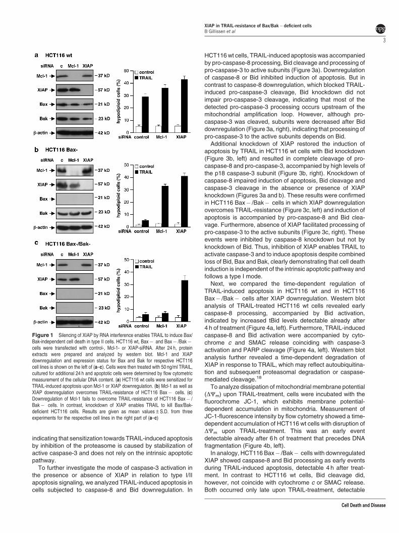
HCT116 cells were transfected with control-, Mcl-1- orXIAP-siRNA and downregulation of the respective proteinswas confirmed by western blot analysis (figure1, left). Upondownregulation of Mcl-1 or XIAP, cells were incubated with50 ng/ml TRAIL for 24 h and induction of apoptosis wasanalyzed by flow cytometric measurement of cells with ahypodiploid DNA content. TRAIL-treatment resulted in apop-totic DNA fragmentation in about 28% of the HCT116 wt cells.Sensitivity toward TRAIL was increased by knockdown ofMcl-1 and even more by XIAP knockdown, about 35 and 42%of the respective cells were apoptotic (Figure 1a, right).
Unlike HCT116 wt cells, Bax-deficient HCT116 cells wereresistant to TRAIL-induced apoptosis. However, Mcl-1 orXIAP downregulation rendered these cells susceptible toTRAIL. Compared with 6% of control cells, TRAIL-inducedapoptosis increased up to 32 and 41% upon Mcl-1 or XIAPdownregulation, respectively (Figure 1b, right).
In contrast, TRAIL-resistance of HCT116 Bax� /Bak� cellscould not be overcome by inhibition of Mcl-1. However,downregulation of XIAP strongly sensitized these double-deficient cells for TRAIL-induced apoptosis. Cells transfectedwith control- or Mcl-1-siRNA and treated with TRAIL showedapoptotic DNA fragmentation in o6% of the cells, whichincreased to 34% upon XIAP downregulation (Figure 1c, right).
Annexin V-FITC/propidium iodide (PI) staining of HCT116wt and HCT116 Bax� /Bak� cells upon TRAIL-treatmentconfirmed that cell death occurs by apoptosis and that TRAIL-resistance of HCT116 Bax� /Bak� cells can be overcome bydownregulation of XIAP (Supplementary Figure S1). Further-more, XIAP downregulation overrides resistance of Bcl-2 orBcl-xL overexpressing HCT116 cells, supporting our findingthat upon TRAIL-treatment XIAP-deficient cells die indepen-dently of a Bcl-2 family protein regulated pathway(Supplementary Figure S2).
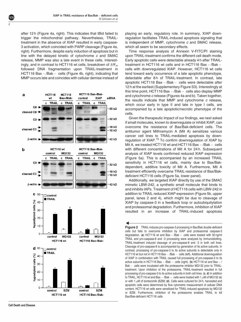
As caspase-8 and -3 are crucial for TRAIL-inducedapoptosis, we next examined to which extent Bax/Bakdeficiency impacts on processing of caspase-8 and -3. Wetreated HCT116 wt and Bax� /Bak� cells with TRAIL andanalyzed the cleavage pattern of these caspases. Pro-caspase-8 and -3 levels are comparable in both cell lines,respectively (Figure 2a, left). Following TRAIL-treatment, pro-caspase-8 is cleaved in both cell lines and processed to itsactive subunits to a similar extent. Interestingly, in both celllines, caspase-8 activation goes along with cleavage of thepro-caspase-3 zymogen, indicating that caspase-8 activationupon TRAIL-treatment is sufficient to cleave pro-caspase-3.Nevertheless, cleavage of pro-caspase-3 is accompanied byprocessing to its active p18/p16 subunits only in TRAIL-sensitive HCT116 wt cells. In contrast, TRAIL-resistantHCT116 Bax� /Bak� cells showed no processing ofcaspase-3 to its active subunits (Figure 2a, left). Thus, incontrast to cleavage of the pro-caspase-3 zymogen, which isindependent of Bax and Bak, processing of pro-caspase-3 toits active subunits relies on an intact intrinsic mitochondrialpathway.
To analyze if the ubiquitin ligase activity of XIAP mediatesdegradation of the caspase-3 subunits we knocked downXIAP. In HCT116 Bax� /Bak� cells transfected with controlsiRNA, TRAIL-treatment resulted in pro-caspase-3 cleavagewithout generation of the active caspase-3. In contrast,downregulation of XIAP in combination with TRAIL causedfull processing of pro-caspase-3 to its active subunits(Figure 2a, right). This indicates that caspase-8 activitytriggered by TRAIL is sufficient to cleave pro-caspase-3 whilecaspase-3 activation is prevented by XIAP-mediated degra-dation of processed caspase-3.
To investigate proteasomal degradation of active caspase-3,we treated HCT116 wt and Bax� /Bak� cells with non-toxicconcentrations of the proteasome inhibitor MG132. InHCT116 wt cells, TRAIL-induced caspase-3 activation isincreased upon additional treatment with MG132 (Figure 2b,left). More importantly, in HCT116 Bax� /Bak� cells, TRAILalone induced the cleavage of pro-caspase-3 without detect-able levels of active subunits, whereas the combinationinduced full caspase-3 processing to its active subunits(Figure 2b, right).
In agreement with increased caspase-3 activation, TRAIL-induced apoptosis was increased in HCT116 wt cells uponadditional inhibition of the proteasome. Moreover, MG132treatment overcame TRAIL-resistance of Bax/Bax� deficientHCT116 cells (Figure 2c). Like MG132, bortezomib (BZM),the first therapeutic proteasome inhibitor, sensitized HCT116wt cells for TRAIL-induced apoptosis and overcame TRAIL-resistance of HCT116 Bax� /Bak� cells (Figure 2d),
XIAP in TRAIL-resistance of Bax/Bak� deficient cellsB Gillissen et al
2
Cell Death and Disease
indicating that sensitization towards TRAIL-induced apoptosisby inhibition of the proteasome is caused by stabilization ofactive caspase-3 and does not rely on the intrinsic apoptoticpathway.
To further investigate the mode of caspase-3 activation inthe presence or absence of XIAP in relation to type I/IIapoptosis signaling, we analyzed TRAIL-induced apoptosis incells subjected to caspase-8 and Bid downregulation. In
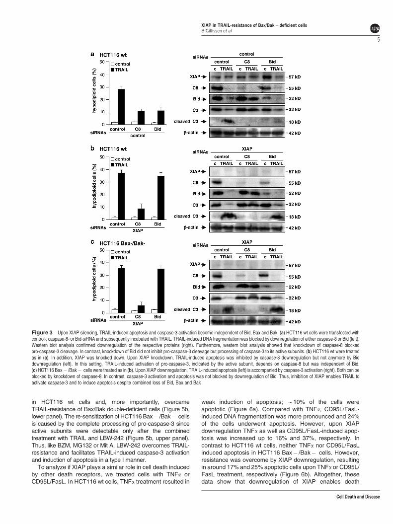
HCT116 wt cells, TRAIL-induced apoptosis was accompaniedby pro-caspase-8 processing, Bid cleavage and processing ofpro-caspase-3 to active subunits (Figure 3a). Downregulationof caspase-8 or Bid inhibited induction of apoptosis. But incontrast to caspase-8 downregulation, which blocked TRAIL-induced pro-caspase-3 cleavage, Bid knockdown did notimpair pro-caspase-3 cleavage, indicating that most of thedetected pro-caspase-3 processing occurs upstream of themitochondrial amplification loop. However, although pro-caspase-3 was cleaved, subunits were decreased after Biddownregulation (Figure 3a, right), indicating that processing ofpro-caspase-3 to the active subunits depends on Bid.
Additional knockdown of XIAP restored the induction ofapoptosis by TRAIL in HCT116 wt cells with Bid knockdown(Figure 3b, left) and resulted in complete cleavage of pro-caspase-8 and pro-caspase-3, accompanied by high levels ofthe p18 caspase-3 subunit (Figure 3b, right). Knockdown ofcaspase-8 impaired induction of apoptosis, Bid cleavage andcaspase-3 cleavage in the absence or presence of XIAPknockdown (Figures 3a and b). These results were confirmedin HCT116 Bax� /Bak� cells in which XIAP downregulationovercomes TRAIL-resistance (Figure 3c, left) and induction ofapoptosis is accompanied by pro-caspase-8 and Bid clea-vage. Furthermore, absence of XIAP facilitated processing ofpro-caspase-3 to the active subunits (Figure 3c, right). Theseevents were inhibited by caspase-8 knockdown but not byknockdown of Bid. Thus, inhibition of XIAP enables TRAIL toactivate caspase-3 and to induce apoptosis despite combinedloss of Bid, Bax and Bak, clearly demonstrating that cell deathinduction is independent of the intrinsic apoptotic pathway andfollows a type I mode.
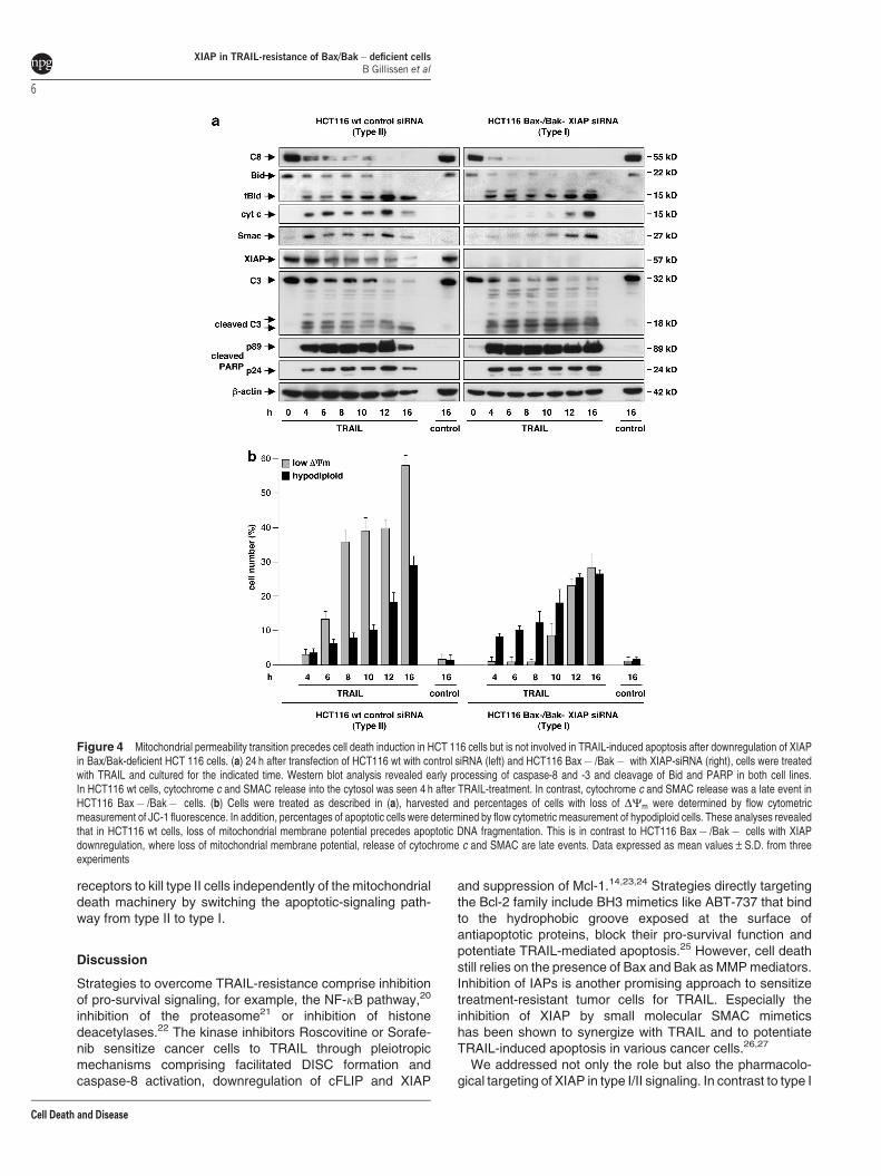
Next, we compared the time-dependent regulation ofTRAIL-induced apoptosis in HCT116 wt and in HCT116Bax� /Bak� cells after XIAP downregulation. Western blotanalysis of TRAIL-treated HCT116 wt cells revealed earlycaspase-8 processing, accompanied by Bid activation,indicated by increased tBid levels detectable already after4 h of treatment (Figure 4a, left). Furthermore, TRAIL-inducedcaspase-8 and Bid activation were accompanied by cyto-chrome c and SMAC release coinciding with caspase-3activation and PARP cleavage (Figure 4a, left). Western blotanalysis further revealed a time-dependent degradation ofXIAP in response to TRAIL, which may reflect autoubiquitina-tion and subsequent proteasomal degradation or caspase-mediated cleavage.18
To analyze dissipation of mitochondrial membrane potential(DCm) upon TRAIL-treatment, cells were incubated with thefluorochrome JC-1, which exhibits membrane potential-dependent accumulation in mitochondria. Measurement ofJC-1-fluorescence intensity by flow cytometry showed a time-dependent accumulation of HCT116 wt cells with disruption ofDCm upon TRAIL-treatment. This was an early eventdetectable already after 6 h of treatment that precedes DNAfragmentation (Figure 4b, left).
In analogy, HCT116 Bax� /Bak� cells with downregulatedXIAP showed caspase-8 and Bid processing as early eventsduring TRAIL-induced apoptosis, detectable 4 h after treat-ment. In contrast to HCT116 wt cells, Bid cleavage did,however, not coincide with cytochrome c or SMAC release.Both occurred only late upon TRAIL-treatment, detectable
Figure 1 Silencing of XIAP by RNA interference enables TRAIL to induce Bax/Bak-independent cell death in type II cells. HCT116 wt, Bax� and Bax� /Bak�cells were transfected with control-, Mcl-1- or XIAP-siRNA. After 24 h, proteinextracts were prepared and analyzed by western blot. Mcl-1 and XIAPdownregulation and expression status for Bax and Bak for respective HCT116cell lines is shown on the left of (a–c). Cells were then treated with 50 ng/ml TRAIL,cultured for additional 24 h and apoptotic cells were determined by flow cytometricmeasurement of the cellular DNA content. (a) HCT116 wt cells were sensitized forTRAIL-induced apoptosis upon Mcl-1 or XIAP downregulation. (b) Mcl-1 as well asXIAP downregulation overcomes TRAIL-resistance of HCT116 Bax� cells. (c)Downregulation of Mcl-1 fails to overcome TRAIL-resistance of HCT116 Bax� /Bak� cells. In contrast, knockdown of XIAP enables TRAIL to kill Bax/Bak-deficient HCT116 cells. Results are given as mean values±S.D. from threeexperiments for the respective cell lines in the right part of (a–c)
XIAP in TRAIL-resistance of Bax/Bak� deficient cellsB Gillissen et al
3
Cell Death and Disease
after 12 h (Figure 4a, right). This indicates that tBid failed totrigger the mitochondrial pathway. Nevertheless, TRAIL-treatment in the absence of XIAP resulted in early caspase-3 activation, which coincided with PARP cleavage (Figure 4a,right). Furthermore, despite early induction of apoptosis but inline with the delayed kinetic of cytochrome c and SMACrelease, MMP was also a late event in these cells. Interest-ingly, and in contrast to HCT116 wt cells, breakdown of DCm
followed DNA fragmentation upon TRAIL-treatment inHCT116 Bax� /Bak� cells (Figure 4b, right), indicating thatMMP occurs late and coincides with cellular demise instead of
playing an early, regulatory role. In summary, XIAP down-regulation facilitates TRAIL-induced apoptosis signaling thatis independent of MMP, cytochrome c and SMAC release,which all seem to be secondary effects.
Time response analysis of Annexin V-FITC/PI stainingupon TRAIL-treatment confirms the different cell death mode.Early apoptotic cells were detectable already 4 h after TRAIL-treatment in HCT116 wt cells and in HCT116 Bax� /Bak�cells with downregulated XIAP. However, HCT116 wt cellstend toward early occurrence of a late apoptotic phenotype,detectable after 8 h of TRAIL-treatment. In contrast, lateapoptotic HCT116 Bax� /Bak� cells were detectable after12 h at the earliest (Supplementary Figure S3). Interestingly atthis time point, HCT116 Bax� /Bak� cells also display MMPand cytochrome c release (Figures 4a and b). Taken together,the results indicate that MMP and cytochrome c release,which occur early in type II and late in type I cells, areaccompanied by a late apoptotic/necrotic phenotype of thecells.
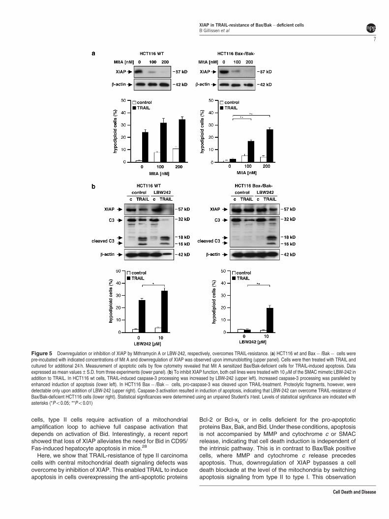
Given the therapeutic impact of our findings, we next askedif small molecules, known to downregulate or inhibit XIAP, canovercome the resistance of Bax/Bak-deficient cells. Theantitumor agent Mithramycin A (Mit A) sensitizes variouscancer cell lines to TRAIL-mediated apoptosis by down-regulation of XIAP.19 To confirm downregulation of XIAP byMit A, we treated HCT116 wt and HCT116 Bax� /Bak� cellswith different concentrations of Mit A for 24 h. Subsequentanalysis of XIAP levels confirmed reduced XIAP expression(Figure 5a). This is accompanied by an increased TRAILsensitivity in HCT116 wt cells, mainly due to Bax/Bak-dependent, additive toxicity of Mit A. Furthermore, Mit Atreatment efficiently overcame TRAIL-resistance of Bax/Bak-deficient HCT116 cells (Figure 5a, lower panel).
Additionally, we targeted XIAP directly by use of the SMACmimetic LBW-242, a synthetic small molecule that binds toand inhibits IAPs. Treatment of HCT116 cells with LBW-242 inaddition to TRAIL reduced XIAP expression (Figure 5b, upperpanel, lanes 2 and 4), which might be due to cleavage ofXIAP by caspase-3 in a feedback loop or autoubiquitylationand proteasomal degradation. Furthermore, inhibition of XIAPresulted in an increase of TRAIL-induced apoptosis
Figure 2 TRAIL-induces pro-caspase-3 processing in Bax/Bak double-deficientcells but fails to overcome inhibition by XIAP and proteasomal caspase-3degradation. (a) HCT116 wt and Bax� /Bak� cells were treated with 50 ng/mlTRAIL and pro-caspase-8 and -3 processing were analyzed by immunoblotting.TRAIL-treatment induced cleavage of pro-caspase-8 and -3 in both cell lines.Cleavage of pro-caspase-8 is accompanied by generation of its active subunits. Incontrast, processing of pro-caspase-3 to its active subunits is detectable only inHCT116 wt but not in HCT116 Bax� /Bak� cells (left). Additional downregulationof XIAP in combination with TRAIL caused full processing of pro-caspase-3 to itsactive subunits in HCT116 Bax� /Bak� cells (right). (b) HCT116 wt and Bax� /Bak� cells were incubated with the proteasome inhibitor MG132 prior to TRAIL-treatment. Upon inhibition of the proteasome, TRAIL-treatment resulted in fullprocessing of pro-caspase-3 to its active subunits in both cell lines. (c, d) In additionto TRAIL, HCT116 wt and Bax� /Bak� cells were treated with 1 mM of MG132 (c)or with 1 mM of bortezomib (BZM) (d). Cells were cultured for 24 h, harvested andapoptotic cells were determined by flow cytometric measurement of cellular DNAcontent. HCT116 wt cells were sensitized for TRAIL-induced apoptosis by MG132or BZM. Furthermore, inhibition of the proteasome enables TRAIL to killBax/Bak-deficient HCT116 cells
XIAP in TRAIL-resistance of Bax/Bak� deficient cellsB Gillissen et al
4
Cell Death and Disease
in HCT116 wt cells and, more importantly, overcameTRAIL-resistance of Bax/Bak double-deficient cells (Figure 5b,lower panel). The re-sensitization of HCT116 Bax� /Bak� cellsis caused by the complete processing of pro-caspase-3 sinceactive subunits were detectable only after the combinedtreatment with TRAIL and LBW-242 (Figure 5b, upper panel).Thus, like BZM, MG132 or Mit A, LBW-242 overcomes TRAIL-resistance and facilitates TRAIL-induced caspase-3 activationand induction of apoptosis in a type I manner.
To analyze if XIAP plays a similar role in cell death inducedby other death receptors, we treated cells with TNFa orCD95L/FasL. In HCT116 wt cells, TNFa treatment resulted in
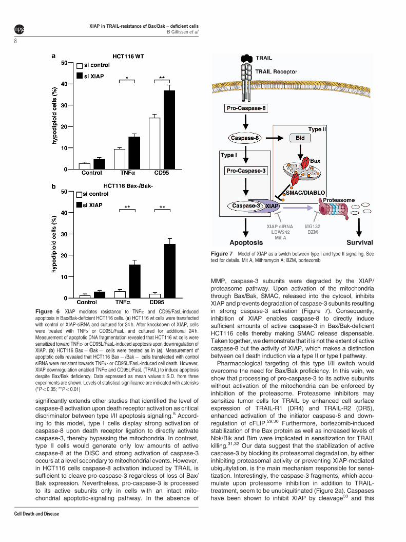
weak induction of apoptosis; B10% of the cells wereapoptotic (Figure 6a). Compared with TNFa, CD95L/FasL-induced DNA fragmentation was more pronounced and 24%of the cells underwent apoptosis. However, upon XIAPdownregulation TNFa as well as CD95L/FasL-induced apop-tosis was increased up to 16% and 37%, respectively. Incontrast to HCT116 wt cells, neither TNFa nor CD95L/FasLinduced apoptosis in HCT116 Bax� /Bak� cells. However,resistance was overcome by XIAP downregulation, resultingin around 17% and 25% apoptotic cells upon TNFa or CD95L/FasL treatment, respectively (Figure 6b). Altogether, thesedata show that downregulation of XIAP enables death
Figure 3 Upon XIAP silencing, TRAIL-induced apoptosis and caspase-3 activation become independent of Bid, Bax and Bak. (a) HCT116 wt cells were transfected withcontrol-, caspase-8- or Bid-siRNA and subsequently incubated with TRAIL. TRAIL-induced DNA fragmentation was blocked by downregulation of either caspase-8 or Bid (left).Western blot analysis confirmed downregulation of the respective proteins (right). Furthermore, western blot analysis showed that knockdown of caspase-8 blockedpro-caspase-3 cleavage. In contrast, knockdown of Bid did not inhibit pro-caspase-3 cleavage but processing of caspase-3 to its active subunits. (b) HCT116 wt were treatedas in (a). In addition, XIAP was knocked down. Upon XIAP knockdown, TRAIL-induced apoptosis was inhibited by caspase-8 downregulation but not anymore by Biddownregulation (left). In this setting, TRAIL-induced activation of pro-caspase-3, indicated by the active subunit, depends on caspase-8 but was independent of Bid.(c) HCT116 Bax� /Bak� cells were treated as in (b). Upon XIAP downregulation, TRAIL-induced apoptosis (left) is accompanied by caspase-3 activation (right). Both can beblocked by knockdown of caspase-8. In contrast, caspase-3 activation and apoptosis was not blocked by downregulation of Bid. Thus, inhibition of XIAP enables TRAIL toactivate caspase-3 and to induce apoptosis despite combined loss of Bid, Bax and Bak
XIAP in TRAIL-resistance of Bax/Bak� deficient cellsB Gillissen et al
5
Cell Death and Disease
receptors to kill type II cells independently of the mitochondrialdeath machinery by switching the apoptotic-signaling path-way from type II to type I.
Discussion
Strategies to overcome TRAIL-resistance comprise inhibitionof pro-survival signaling, for example, the NF-kB pathway,20
inhibition of the proteasome21 or inhibition of histonedeacetylases.22 The kinase inhibitors Roscovitine or Sorafe-nib sensitize cancer cells to TRAIL through pleiotropicmechanisms comprising facilitated DISC formation andcaspase-8 activation, downregulation of cFLIP and XIAP
and suppression of Mcl-1.14,23,24 Strategies directly targetingthe Bcl-2 family include BH3 mimetics like ABT-737 that bindto the hydrophobic groove exposed at the surface ofantiapoptotic proteins, block their pro-survival function andpotentiate TRAIL-mediated apoptosis.25 However, cell deathstill relies on the presence of Bax and Bak as MMP mediators.Inhibition of IAPs is another promising approach to sensitizetreatment-resistant tumor cells for TRAIL. Especially theinhibition of XIAP by small molecular SMAC mimeticshas been shown to synergize with TRAIL and to potentiateTRAIL-induced apoptosis in various cancer cells.26,27
We addressed not only the role but also the pharmacolo-gical targeting of XIAP in type I/II signaling. In contrast to type I
Figure 4 Mitochondrial permeability transition precedes cell death induction in HCT 116 cells but is not involved in TRAIL-induced apoptosis after downregulation of XIAPin Bax/Bak-deficient HCT 116 cells. (a) 24 h after transfection of HCT116 wt with control siRNA (left) and HCT116 Bax� /Bak� with XIAP-siRNA (right), cells were treatedwith TRAIL and cultured for the indicated time. Western blot analysis revealed early processing of caspase-8 and -3 and cleavage of Bid and PARP in both cell lines.In HCT116 wt cells, cytochrome c and SMAC release into the cytosol was seen 4 h after TRAIL-treatment. In contrast, cytochrome c and SMAC release was a late event inHCT116 Bax� /Bak� cells. (b) Cells were treated as described in (a), harvested and percentages of cells with loss of DCm were determined by flow cytometricmeasurement of JC-1 fluorescence. In addition, percentages of apoptotic cells were determined by flow cytometric measurement of hypodiploid cells. These analyses revealedthat in HCT116 wt cells, loss of mitochondrial membrane potential precedes apoptotic DNA fragmentation. This is in contrast to HCT116 Bax� /Bak� cells with XIAPdownregulation, where loss of mitochondrial membrane potential, release of cytochrome c and SMAC are late events. Data expressed as mean values±S.D. from threeexperiments
XIAP in TRAIL-resistance of Bax/Bak� deficient cellsB Gillissen et al
6
Cell Death and Disease
cells, type II cells require activation of a mitochondrialamplification loop to achieve full caspase activation thatdepends on activation of Bid. Interestingly, a recent reportshowed that loss of XIAP alleviates the need for Bid in CD95/Fas-induced hepatocyte apoptosis in mice.28
Here, we show that TRAIL-resistance of type II carcinomacells with central mitochondrial death signaling defects wasovercome by inhibition of XIAP. This enabled TRAIL to induceapoptosis in cells overexpressing the anti-apoptotic proteins
Bcl-2 or Bcl-xL or in cells deficient for the pro-apoptoticproteins Bax, Bak, and Bid. Under these conditions, apoptosisis not accompanied by MMP and cytochrome c or SMACrelease, indicating that cell death induction is independent ofthe intrinsic pathway. This is in contrast to Bax/Bak positivecells, where MMP and cytochrome c release precedesapoptosis. Thus, downregulation of XIAP bypasses a celldeath blockade at the level of the mitochondria by switchingapoptosis signaling from type II to type I. This observation
Figure 5 Downregulation or inhibition of XIAP by Mithramycin A or LBW-242, respectively, overcomes TRAIL-resistance. (a) HCT116 wt and Bax� /Bak� cells werepre-incubated with indicated concentrations of Mit A and downregulation of XIAP was observed upon immunoblotting (upper panel). Cells were then treated with TRAIL andcultured for additional 24 h. Measurement of apoptotic cells by flow cytometry revealed that Mit A sensitized Bax/Bak-deficient cells for TRAIL-induced apoptosis. Dataexpressed as mean values±S.D. from three experiments (lower panel). (b) To inhibit XIAP function, both cell lines were treated with 10mM of the SMAC mimetic LBW-242 inaddition to TRAIL. In HCT116 wt cells, TRAIL-induced caspase-3 processing was increased by LBW-242 (upper left). Increased caspase-3 processing was paralleled byenhanced induction of apoptosis (lower left). In HCT116 Bax� /Bak� cells, pro-caspase-3 was cleaved upon TRAIL-treatment. Proteolytic fragments, however, weredetectable only upon addition of LBW-242 (upper right). Caspase-3 activation resulted in induction of apoptosis, indicating that LBW-242 can overcome TRAIL-resistance ofBax/Bak-deficient HCT116 cells (lower right). Statistical significances were determined using an unpaired Student’s t-test. Levels of statistical significance are indicated withasterisks (*Po0.05; **Po0.01)
XIAP in TRAIL-resistance of Bax/Bak� deficient cellsB Gillissen et al
7
Cell Death and Disease
significantly extends other studies that identified the level ofcaspase-8 activation upon death receptor activation as criticaldiscriminator between type I/II apoptosis signaling.5 Accord-ing to this model, type I cells display strong activation ofcaspase-8 upon death receptor ligation to directly activatecaspase-3, thereby bypassing the mitochondria. In contrast,type II cells would generate only low amounts of activecaspase-8 at the DISC and strong activation of caspase-3occurs at a level secondary to mitochondrial events. However,in HCT116 cells caspase-8 activation induced by TRAIL issufficient to cleave pro-caspase-3 regardless of loss of Bax/Bak expression. Nevertheless, pro-caspase-3 is processedto its active subunits only in cells with an intact mito-chondrial apoptotic-signaling pathway. In the absence of
MMP, caspase-3 subunits were degraded by the XIAP/proteasome pathway. Upon activation of the mitochondriathrough Bax/Bak, SMAC, released into the cytosol, inhibitsXIAP and prevents degradation of caspase-3 subunits resultingin strong caspase-3 activation (Figure 7). Consequently,inhibition of XIAP enables caspase-8 to directly inducesufficient amounts of active caspase-3 in Bax/Bak-deficientHCT116 cells thereby making SMAC release dispensable.Taken together, we demonstrate that it is not the extent of activecaspase-8 but the activity of XIAP, which makes a distinctionbetween cell death induction via a type II or type I pathway.
Pharmacological targeting of this type I/II switch wouldovercome the need for Bax/Bak proficiency. In this vein, weshow that processing of pro-caspase-3 to its active subunitswithout activation of the mitochondria can be enforced byinhibition of the proteasome. Proteasome inhibitors maysensitize tumor cells for TRAIL by enhanced cell surfaceexpression of TRAIL-R1 (DR4) and TRAIL-R2 (DR5),enhanced activation of the initiator caspase-8 and down-regulation of cFLIP.29,30 Furthermore, bortezomib-inducedstabilization of the Bax protein as well as increased levels ofNbk/Bik and Bim were implicated in sensitization for TRAILkilling.31,32 Our data suggest that the stabilization of activecaspase-3 by blocking its proteasomal degradation, by eitherinhibiting proteasomal activity or preventing XIAP-mediatedubiquitylation, is the main mechanism responsible for sensi-tization. Interestingly, the caspase-3 fragments, which accu-mulate upon proteasome inhibition in addition to TRAIL-treatment, seem to be unubiquitinated (Figure 2a). Caspaseshave been shown to inhibit XIAP by cleavage33 and this
Figure 6 XIAP mediates resistance to TNFa and CD95/FasL-inducedapoptosis in Bax/Bak-deficient HCT116 cells. (a) HCT116 wt cells were transfectedwith control or XIAP-siRNA and cultured for 24 h. After knockdown of XIAP, cellswere treated with TNFa or CD95L/FasL and cultured for additional 24 h.Measurement of apoptotic DNA fragmentation revealed that HCT116 wt cells weresensitized toward TNFa- or CD95L/FasL-induced apoptosis upon downregulation ofXIAP. (b) HCT116 Bax� /Bak� cells were treated as in (a). Measurement ofapoptotic cells revealed that HCT116 Bax� /Bak� cells transfected with controlsiRNA were resistant towards TNFa- or CD95L/FasL-induced cell death. However,XIAP downregulation enabled TNFa and CD95L/FasL (TRAIL) to induce apoptosisdespite Bax/Bak deficiency. Data expressed as mean values±S.D. from threeexperiments are shown. Levels of statistical significance are indicated with asterisks(*Po0.05; **Po0.01)
Figure 7 Model of XIAP as a switch between type I and type II signaling. Seetext for details. Mit A, Mithramycin A; BZM, bortezomib
XIAP in TRAIL-resistance of Bax/Bak� deficient cellsB Gillissen et al
8
Cell Death and Disease

feedback loop might be responsible for the accumulation ofunubiquitinated active caspase-3 upon inhibition of theproteasome. However, inhibition of the proteasome or XIAPresult in sufficient generation of active caspase-3 and mediatea switch from a type II to a type I mode of cell death. Thisnotion is supported by experiments where we inhibited XIAPfunction by the use of the synthetic small molecule IAPinhibitor LBW-242 that mimics the activity of SMAC. LBW-242is a promising compound to overcome drug resistance.34,35
Here, we show that LBW-242 overcomes resistance of Bax/Bak-deficient cancer cells and re-sensitizes these cells toTRAIL. Loss of both Bax or Bak expression has been reportedfor a number of tumors.36–39 Whereas loss of Bax is a frequentevent in human cancer, for example, in colon cancers having amicrosatellite mutator phenotype, Bak expression persists inmost cancers and loss of both is a rare event. Notably, loss ofBax, despite expression of Bak, is sufficient for tumor cells toacquire TRAIL-resistance. In addition, the intrinsic apoptoticpathway of tumor cells can be blocked by other mechanisms,for example, upregulation of Bcl-xL (which efficiently inhibitsboth, Bax and Bak) or endogenous Bak-inhibitors Mcl-1 orVDAC2 in Bax-deficient carcinoma.14,15,40 The data pre-sented in this study indicate that use of SMAC mimetics is asuitable strategy to antagonize therapy resistance caused byall these central defects in the intrinsic apoptosis machineryand delineates the combined use of TRAIL together withSMAC mimetics as a useful strategy. Altogether, these datashow that targeting of XIAP enables death receptor signalingto kill type II cells via a type I pathway and define XIAP as acrucial decision point between these two cell death pathways.
Materials and MethodsCell culture. HCT116 wild-type cells and the isogenic knockout sublineHCT116-Bax� /� were kindly provided by Dr. Bert Vogelstein, Johns HopkinsCancer Center, Baltimore, MD, USA. The stable knockdown of Bak shRNA wasachieved in HCT116-Bax k.o. cells yielding Bax� /Bak� cells.17 Mocktransfectants showed an identical apoptosis sensitivity as compared withHCT116 parental cells (HCT116 wt). Cells were grown in DMEM mediumsupplemented with 10% fetal calf serum, 100 000 U/l penicillin and 0.1 g/lstreptomycin at 37 1C with 5% CO2 in a fully humidified atmosphere. Media andculture reagents were from Invitrogen (Karlsruhe, Germany).
Antibodies and reagents. SMAC/Diablo mAb (#2954), C-8 (1C12) mAb(#9746) and XIAP mAb (3B6) (#2045) were purchased from Cell SignalingTechnology, Inc. (Boston, MA, USA). Bcl-2 antibody (NCL-bcl-2) was purchasedfrom Novocastra Laboratories (Newcastle, UK). The anti-Bak Ab (clone TC102) wasfrom Calbiochem (Darmstadt, Germany). Anti-Bax Ab (clone YTH-2D2) waspurchased from Trevigen (Gaithersburg, MD, USA) and anti-Mcl-1H-260 was fromSanta Cruz Biotechnology (Santa Cruz, CA, USA). Anti-Bcl-x Ab was from BDBiosciences Pharmingen (San Diego, CA, USA). Anti-caspase-8 (clone 12F5) wasfrom Alexis (Grunberg, Germany). The anti-C-3 Ab and anti-Bid Ab were from R&DSystems GmbH (Wiesbaden-Nordenstadt, Germany), anti-actin Ab was from Sigma-Aldrich (Taufkirchen, Germany). Secondary anti-rabbit, anti-goat and anti-mousehorseradish peroxidase-conjugated antibodies were from Promega (Mannheim,Germany) or Southern Biotechnology Associates (Birmingham, AL, USA). RNase Awas from Roth (Karlsruhe, Germany). The recombinant human TRAIL was fromR&D Systems GmbH (Wiesbaden-Nordenstadt, Germany). TNFa and Super-FasLigand were from Alexis (Grunberg, Germany). MG132 was purchased fromSigma-Aldrich, Mit A was from Genaxxon BioScience GmbH (Ulm, Germany). TheLBW-242 SMAC mimetic was kindly provided by Novartis (Basel, Switzerland).
RNA interference. On-target plus XIAP, Mcl-1, caspase-8, Bid and controlsiRNA were purchased from Dharmacon (Lafayette, LA, USA). Transfection of thecells was carried out by use of DharmaFECT Transfection Reagent according to
the manufacturer’s instructions. Downregulation of the respective proteins wasconfirmed by immunoblotting 24 h after transfection.
Immunoblotting. After trypsination, cells were washed twice with ice-coldPBS and lysed in 10 mM Tris-HCl pH 7.5, 137 mM NaCl, 1% Triton X-100, 2 mMEDTA, 1mM pepstatin, 1 mM leupeptin and 0.1 mM phenylmethyl sulfonylfluoride(PMSF). Protein concentration was determined using the bicinchoninic acid assay.Equal amounts of protein were separated by SDS-PAGE, electroblotted andvisualized as described.41 For analysis of cytochrome c and SMAC release,cytosolic extracts were prepared according to a method described previously.41
Briefly, after induction of apoptosis, cells were harvested in PBS, equilibrated inhypotonic buffer (20 mM HEPES pH 7.4, 10 mM KCl, 2 mM MgCl2, 1 mM EDTA)supplemented with 0.1 mM PMSF and 0.75 mg/ml digitonin (Sigma-Aldrich) andincubated on ice for 3 min. Debris was pelleted by centrifugation at 10 000� g at4 1C for 5 min and the supernatant was subjected to western blot analysis.
Measurement of mitochondrial permeability transition. Cells wereharvested and collected by centrifugation at 300� g at 4 1C for 5 min.Mitochondrial permeability transition was determined by staining the cells withJC-1 (5,50,6,60-tetrachloro-1,10,3,30-tetraethyl-benzimidazolylcarbocyanin iodide;Molecular Probes, Leiden, The Netherlands), a cationic dye that exhibitsmembrane potential-dependent accumulation in mitochondria, as described.41
Mitochondrial permeability transition was then quantified by flow cytometricdetermination of cells with decreased red fluorescence, that is, with mitochondriadisplaying a reduced membrane potential (DCm).
Measurement of apoptotic cell death by flow cytometry. DNAfragmentation was measured as described.15 Briefly, cells were collected bycentrifugation at 300� g for 5 min, washed with PBS at 4 1C, and fixed in PBS/2%(vol/vol) formaldehyde on ice for 30 min. After fixation, cells were incubated withethanol/PBS (2 : 1, vol/vol) for 15 min, pelleted, and resuspended in PBScontaining 40mg/ml RNase A. After incubation for 30 min at 37 1C, cells werepelleted and finally resuspended in PBS containing 50 mg/ml PI. DNAfragmentation was quantified by flow cytometric determination of hypodiploidDNA. Data were collected and analyzed using a FACScan (Becton Dickinson,Heidelberg, Germany) equipped with the CELLQuest software. Data are given in% hypoploidy (subG1), which reflects the number of apoptotic cells. Alternatively,cell death was determined by staining cells with Annexin V-fluoresceinisothiocyanate (FITC) and counterstaining with PI.15 Briefly, cells were washedtwice with cold PBS and resuspended in 10 mM N-[2-hydroxyethyl]piperazin-N0-3[propansulfonicacid]/NaOH, pH 7.4, 140 mM NaCl, 2.5 mM CaCl2 at 1� 106
cells/ml. Next, 5ml of Annexin V-FITC (BD PharMingen, Heidelberg, Germany)and 10ml PI (20 g/ml, Sigma-Aldrich) were added. Analyses were performed usinga FACScan (Becton Dickinson) and CELLQuest analysis software. Results aregiven in % of cells.
Conflict of InterestThe authors declare no conflict of interest.
Acknowledgements. This study was supported by the DeutscheForschungsgemeinschaft, BMBF-MedSys and the Deutsche Krebshilfe. We thankNovartis, Basel, for kindly providing the SMAC mimetic LBW-242.
1. Gonzalvez F, Ashkenazi A. New insights into apoptosis signaling by Apo2L/TRAIL.Oncogene 2010; 29: 4752–4765.
2. Strater J, Hinz U, Walczak H, Mechtersheimer G, Koretz K, Herfarth C et al. Expression ofTRAIL and TRAIL receptors in colon carcinoma: TRAIL-R1 is an independent prognosticparameter. Clin Cancer Res 2002; 8: 3734–3740.
3. Schmelz K, Wieder T, Tamm I, Muller A, Essmann F, Geilen CC et al. Tumor necrosisfactor alpha sensitizes malignant cells to chemotherapeutic drugs via the mitochondrialapoptosis pathway independently of caspase-8 and NF-kappaB. Oncogene 2004; 23:6743–6759.
4. Marini P, Schmid A, Jendrossek V, Faltin H, Daniel PT, Budach W et al. Irradiationspecifically sensitises solid tumour cell lines to TRAIL mediated apoptosis. BMC Cancer2005; 5: 5.
5. Scaffidi C, Fulda S, Srinivasan A, Friesen C, Li F, Tomaselli KJ et al. Two CD95 (APO-1/Fas) signaling pathways. EMBO J 1998; 17: 1675–1687.
XIAP in TRAIL-resistance of Bax/Bak� deficient cellsB Gillissen et al
9
Cell Death and Disease
6. Li H, Zhu H, Xu CJ, Yuan J. Cleavage of BID by caspase 8 mediates the mitochondrialdamage in the Fas pathway of apoptosis. Cell 1998; 94: 491–501.
7. van Delft MF, Huang DC. How the Bcl-2 family of proteins interact to regulate apoptosis.Cell Res 2006; 16: 203–213.
8. Verhagen AM, Ekert PG, Pakusch M, Silke J, Connolly LM, Reid GE et al. Identification ofDIABLO, a mammalian protein that promotes apoptosis by binding to and antagonizing IAPproteins. Cell 2000; 102: 43–53.
9. Du C, Fang M, Li Y, Li L, Wang X. Smac, a mitochondrial protein that promotescytochrome c-dependent caspase activation by eliminating IAP inhibition. Cell 2000; 102:33–42.
10. Deveraux QL, Reed JC. IAP family proteins—suppressors of apoptosis. Genes Dev 1999;13: 239–252.
11. LaCasse EC, Mahoney DJ, Cheung HH, Plenchette S, Baird S, Korneluk RG. IAP-targetedtherapies for cancer. Oncogene 2008; 27: 6252–6275.
12. LeBlanc H, Lawrence D, Varfolomeev E, Totpal K, Morlan J, Schow P et al. Tumor-cellresistance to death receptor—induced apoptosis through mutational inactivation of theproapoptotic Bcl-2 homolog Bax. Nat Med 2002; 8: 274–281.
13. Taniai M, Grambihler A, Higuchi H, Werneburg N, Bronk SF, Farrugia DJ et al. Mcl-1mediates tumor necrosis factor-related apoptosis-inducing ligand resistance in humancholangiocarcinoma cells. Cancer Res 2004; 64: 3517–3524.
14. Gillissen B, Wendt J, Richter A, Richter A, Muer A, Overkamp T et al. Endogenous Bakinhibitors Mcl-1 and Bcl-xL: differential impact on TRAIL resistance in Bax� deficientcarcinoma. J Cell Biol 2010; 188: 851–862.
15. Gillissen B, Essmann F, Hemmati PG, Richter A, Richter A, Oztop I et al. Mcl-1determines the Bax dependency of Nbk/Bik-induced apoptosis. J Cell Biol 2007; 179:701–715.
16. Zhang L, Yu J, Park BH, Kinzler KW, Vogelstein B. Role of BAX in the apoptotic responseto anticancer agents. Science 2000; 290: 989–992.
17. Subramanian T, Chinnadurai G. Pro-apoptotic activity of transiently expressed BCL-2occurs independent of BAX and BAK. J Cell Biochem 2003; 89: 1102–1114.
18. Hornle M, Peters N, Thayaparasingham B, Vorsmann H, Kashkar H, Kulms D. Caspase-3cleaves XIAP in a positive feedback loop to sensitize melanoma cells to TRAIL-inducedapoptosis. Oncogene 2010; 30: 575–587.
19. Lee TJ, Jung EM, Lee JT, Kim S, Park JW, Choi KS et al. Mithramycin A sensitizes cancercells to TRAIL-mediated apoptosis by down-regulation of XIAP gene promoter through Sp1sites. Mol Cancer Ther 2006; 5: 2737–2746.
20. Keane MM, Rubinstein Y, Cuello M, Ettenberg SA, Banerjee P, Nau MM et al. Inhibition ofNF-kappaB activity enhances TRAIL mediated apoptosis in breast cancer cell lines. BreastCancer Res Treat 2000; 64: 211–219.
21. Chen KF, Yeh PY, Hsu C, Hsu CH, Lu YS, Hsieh HP et al. Bortezomib overcomes tumornecrosis factor-related apoptosis-inducing ligand resistance in hepatocellular carcinomacells in part through the inhibition of the phosphatidylinositol 3-kinase/Akt pathway. J BiolChem 2009; 284: 11121–11133.
22. Inoue S, Mai A, Dyer MJ, Cohen GM. Inhibition of histone deacetylase class I but not classII is critical for the sensitization of leukemic cells to tumor necrosis factor-related apoptosis-inducing ligand-induced apoptosis. Cancer Res 2006; 66: 6785–6792.
23. Ricci MS, Kim SH, Ogi K, Plastaras JP, Ling J, Wang W et al. Reduction of TRAIL-inducedMcl-1 and cIAP2 by c-Myc or sorafenib sensitizes resistant human cancer cells to TRAIL-induced death. Cancer Cell 2007; 12: 66–80.
24. Ortiz-Ferron G, Yerbes R, Eramo A, Lopez-Perez AI, De Maria R, Lopez-Rivas A.Roscovitine sensitizes breast cancer cells to TRAIL-induced apoptosis through apleiotropic mechanism. Cell Res 2008; 18: 664–676.
25. Huang S, Sinicrope FA. BH3 mimetic ABT-737 potentiates TRAIL-mediated apoptoticsignaling by unsequestering Bim and Bak in human pancreatic cancer cells. Cancer Res2008; 68: 2944–2951.
26. Schimmer AD, Welsh K, Pinilla C, Wang Z, Krajewska M, Bonneau MJ et al. Small-molecule antagonists of apoptosis suppressor XIAP exhibit broad antitumor activity.Cancer Cell 2004; 5: 25–35.
27. Vogler M, Walczak H, Stadel D, Haas TL, Genze F, Jovanovic M et al. Small molecule XIAPinhibitors enhance TRAIL-induced apoptosis and antitumor activity in preclinical models ofpancreatic carcinoma. Cancer Res 2009; 69: 2425–2434.
28. Jost PJ, Grabow S, Gray D, McKenzie MD, Nachbur U, Huang DC et al. XIAP discriminatesbetween type I and type II FAS-induced apoptosis. Nature 2009; 460: 1035–1039.
29. He Q, Huang Y, Sheikh MS. Proteasome inhibitor MG132 upregulates death receptor 5and cooperates with Apo2L/TRAIL to induce apoptosis in Bax� proficient and -deficientcells. Oncogene 2004; 23: 2554–2558.
30. Brooks AD, Jacobsen KM, Li W, Shanker A, Sayers TJ. Bortezomib sensitizes human renalcell carcinomas to TRAIL apoptosis through increased activation of caspase-8 in the death-inducing signaling complex. Mol Cancer Res 2010; 8: 729–738.
31. Liu FT, Agrawal SG, Gribben JG, Ye H, Du MQ, Newland AC et al. Bortezomib blocksBax degradation in malignant B cells during treatment with TRAIL. Blood 2008; 111:2797–2805.
32. Nikrad M, Johnson T, Puthalalath H, Coultas L, Adams J, Kraft AS. The proteasomeinhibitor bortezomib sensitizes cells to killing by death receptor ligand TRAIL via BH3-onlyproteins Bik and Bim. Mol Cancer Ther 2005; 4: 443–449.
33. Deveraux QL, Leo E, Stennicke HR, Welsh K, Salvesen GS, Reed JC. Cleavage of humaninhibitor of apoptosis protein XIAP results in fragments with distinct specificities forcaspases. EMBO J 1999; 18: 5242–5251.
34. Hundsdoerfer P, Dietrich I, Schmelz K, Eckert C, Henze G. XIAP expression is post-transcriptionally upregulated in childhood ALL and is associated with glucocorticoidresponse in T-cell ALL. Pediatr Blood Cancer 2010; 55: 260–266.
35. Chauhan D, Neri P, Velankar M, Podar K, Hideshima T, Fulciniti M et al. Targetingmitochondrial factor Smac/DIABLO as therapy for multiple myeloma (MM). Blood 2007;109: 1220–1227.
36. Sturm I, Kohne CH, Wolff G, Petrowsky H, Hillebrand T, Hauptmann S et al. Analysis of thep53/BAX pathway in colorectal cancer: low BAX is a negative prognostic factor in patientswith resected liver metastases. J Clin Oncol 1999; 17: 1364–1374.
37. Kondo S, Shinomura Y, Miyazaki Y, Kiyohara T, Tsutsui S, Kitamura S et al.Mutations of the bak gene in human gastric and colorectal cancers. Cancer Res 2000; 60:4328–4330.
38. Krajewska M, Moss SF, Krajewski S, Song K, Holt PR, Reed JC. Elevated expression ofBcl-X and reduced Bak in primary colorectal adenocarcinomas. Cancer Res 1996; 56:2422–2427.
39. Mrozek A, Petrowsky H, Sturm I, Kraus J, Hermann S, Hauptmann S et al. Combined p53/Bax mutation results in extremely poor prognosis in gastric carcinoma with lowmicrosatellite instability. Cell Death Differ 2003; 10: 461–467.
40. Plotz M, Gillissen B, Hossini AM, Daniel PT, Eberle J. Disruption of the VDAC2-Bakinteraction by Bcl-x(S) mediates efficient induction of apoptosis in melanoma cells.Cell Death Differ 2012; 19: 1928–1938.
41. von Haefen C, Wieder T, Essmann F, Schulze-Osthoff K, Dorken B, Daniel PT. Paclitaxel-induced apoptosis in BJAB cells proceeds via a death receptor-independent, caspases-3/-8-driven mitochondrial amplification loop. Oncogene 2003; 22: 2236–2247.
Cell Death and Disease is an open-access journalpublished by Nature Publishing Group. This work is
licensed under a Creative Commons Attribution-NonCommercial-NoDerivs 3.0 Unported License. To view a copy of this license, visithttp://creativecommons.org/licenses/by-nc-nd/3.0/
Supplementary Information accompanies this paper on Cell Death and Disease website (http://www.nature.com/cddis)
XIAP in TRAIL-resistance of Bax/Bak� deficient cellsB Gillissen et al
10
Cell Death and Disease