t he b iology of i g e and the b asis of a llergic d isease
TRANSCRIPT

11 Feb 2003 17:8 AR AR180-IY21-18.tex AR180-IY21-18.sgm LaTeX2e(2002/01/18)P1: IKH10.1146/annurev.immunol.21.120601.141103
Annu. Rev. Immunol. 2003. 21:579–628doi: 10.1146/annurev.immunol.21.120601.141103
Copyright c© 2003 by Annual Reviews. All rights reservedFirst published online as a Review in Advance on December 18, 2002
THE BIOLOGY OF IGE AND THE BASIS
OF ALLERGIC DISEASE
Hannah J. Gould, Brian J. Sutton, Andrew J. Beavil,Rebecca L. Beavil, Natalie McCloskey, Heather A. Coker,David Fear, and Lyn SmurthwaiteThe Randall Centre, King’s College London, London SE1 1UL, United Kingdom;email: [email protected]
Key Words IgE, IgE receptors, immediate hypersensitivity, allergy, mucosalimmunity
■ Abstract Allergic individuals exposed to minute quantities of allergen experiencean immediate response. Immediate hypersensitivity reflects the permanent sensitizationof mucosal mast cells by allergen-specific IgE antibodies bound to their high-affinityreceptors (FcεRI). A combination of factors contributes to such long-lasting sensiti-zation of the mast cells. They include the homing of mast cells to mucosal tissues,the local synthesis of IgE, the induction of FcεRI expression on mast cells by IgE,the consequent downregulation of FcγR (through an insufficiency of the commonγ -chains), and the exceptionally slow dissociation of IgE from FcεRI. To understandthe mechanism of the immediate hypersensitivity phenomenon, we need explanationsof why IgE antibodies are synthesized in preference to IgG in mucosal tissues andwhy the IgE is so tenaciously retained on mast cell–surface receptors. There is nowcompelling evidence that the microenvironment of mucosal tissues of allergic diseasefavors class switching to IgE; and the exceptionally high affinity of IgE for FcεRI cannow be interpreted in terms of the recently determined crystal structures of IgE-FcεRIand IgG-FcγR complexes. The rate of local IgE synthesis can easily compensate forthe rate of the antibody dissociation from its receptors on mucosal mast cells. Effec-tive mechanisms ensure that allergic reactions are confined to mucosal tissues, therebyminimizing the risk of systemic anaphylaxis.
OVERVIEW
An allergic reaction is initiated when an antigen crosslinks immunoglobulin E(IgE) antibodies bound to their high-affinity Fc receptor (FcεRI) on tissue mastcells or blood basophils (1). The immediate reaction, taking effect within minutesof allergen provocation, results in the release of mediators that lead to symptomscharacteristic of the target organ. A late-phase response associated with the influxof T cells, monocytes, and eosinophils may ensue some hours later. Hayfever,
0732-0582/03/0407-0579$14.00 579
Ann
u. R
ev. I
mm
unol
. 200
3.21
:579
-628
. Dow
nloa
ded
from
ww
w.a
nnua
lrev
iew
s.or
gby
Col
orad
o St
ate
Uni
vers
ity o
n 10
/04/
13. F
or p
erso
nal u
se o
nly.

11 Feb 2003 17:8 AR AR180-IY21-18.tex AR180-IY21-18.sgm LaTeX2e(2002/01/18)P1: IKH
580 GOULD ET AL.
asthma, reactions to food, and eczema are the most common allergic responses,caused by mast cell activation in mucosal tissues of, respectively, the nose, lung,gut, and skin. Blood basophils are the IgE effector cells in the rare, but moredangerous, manifestation of systemic anaphylaxis.
IgG antibodies mediate hypersensitivity reactions in animal models but havenever been found to display this activity in humans. A part, at least, of the expla-nation for this fundamental divergence lies in the different physical properties ofIgE and IgG, the relevant features of which can now be discerned from recentlydetermined high-resolution crystal structures of the Fc, Fc receptor (FcR), andFc:FcR complexes. These structures afford, in particular, an explanation for theuniquely high affinity of the IgE-FcεRI complex.
Why are allergic reactions confined to particular target organs? Clearly thehoming propensity of mast cells to mucosal tissues (2, 3) is essential. The tenacityof IgE binding to FcεRI on mast cells is important, but an additional importantfactor is the persistent local synthesis and secretion of IgE. This synthesis andsecretion maintains the sensitization of the mast cells by replacing IgE lost bydegradation or by dissociation and diffusion into the circulation or secretions. Inaddition the mast cell mediators induce changes in the local vasculature that resultin the influx and activation of inflammatory cells, effectively targeting the latephase response to sites of allergen provocation.
The microenvironment of mucosal tissues favors the synthesis of IgE at theexpense of IgG. IgE regulation of FcεRI expression on mast cells couples theexpression of receptor to that of IgE, and thus local synthesis and secretion ofIgE leads to the upregulation of FcεRI on neighboring mast cells. Competitionbetween FcεRI and the IgG receptor (FcγR) in the mast cell for their commonγ -chain may then lead to a concerted downregulation of FcγR.
The basis for local IgE synthesis can be inferred in outline. The expressionof IgE requires heavy-chain switching from IgM, often by way of IgG, to IgEby somatic recombination of the germline genes in B cells. Class switching islinked to cell division, and the IgE switch requires more cycles than the switchto IgG. Cells may drop out at any stage of this process by terminal differen-tiation of the B cells into immunoglobulin-secreting plasma cells or by apop-tosis. Local conditions in the mucosal tissues of allergic individuals evidentlyfavor class switching to IgE, and the rate at which it is synthesized and se-creted can be calculated to overcome the loss of bound antibody from the mastcells.
IgE and IgE receptors (FcεRI and the low-affinity receptor, FcεRII/CD23)participate in the afferent phase of the immune response to allergens. Langerhanscells in epidermal tissues internalize and process allergens from the environmentand transport peptide-MHC class II antigen complexes to the local lymphoid tissuesto amplify the immune response and reimplant memory of the allergen for futureresponses. Restriction of these activities to local lymphoid and mucosal tissuesat sites of exposure to antigen constitutes an essential safeguard against systemicanaphylaxis.
Ann
u. R
ev. I
mm
unol
. 200
3.21
:579
-628
. Dow
nloa
ded
from
ww
w.a
nnua
lrev
iew
s.or
gby
Col
orad
o St
ate
Uni
vers
ity o
n 10
/04/
13. F
or p
erso
nal u
se o
nly.

11 Feb 2003 17:8 AR AR180-IY21-18.tex AR180-IY21-18.sgm LaTeX2e(2002/01/18)P1: IKH
IGE AND ALLERGIC DISEASE 581
PROTEINS OF THE IgE NETWORK
IgE
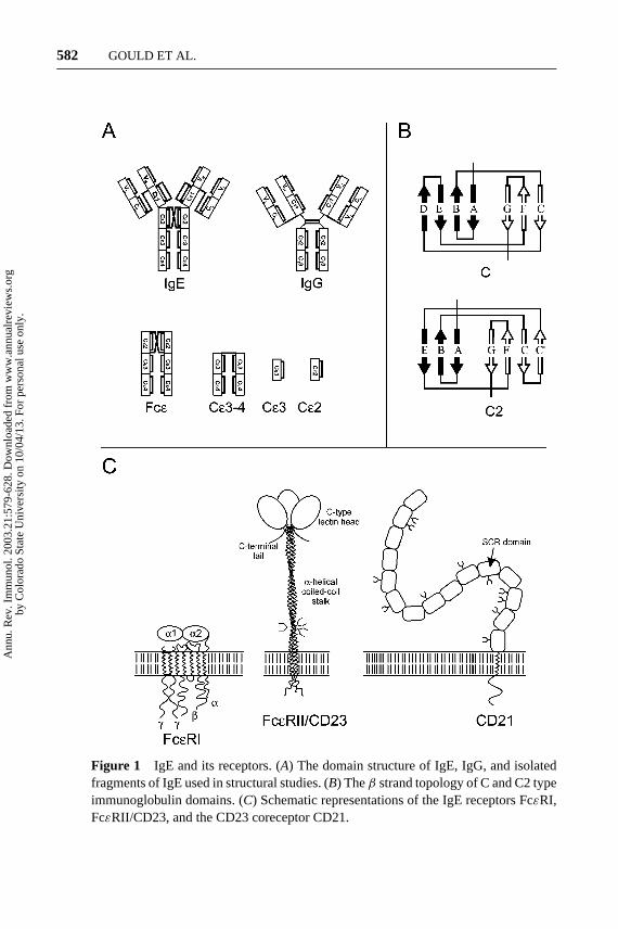
There are nine antibody classes (isotypes) in humans: IgM, IgD, IgG1, IgG2, IgG3,IgG4, IgA1, IgA2, and IgE. All have a similar structure consisting of heavy (H) andlight (L) chains with variable (V) and constant (C) regions made up of Ig domains,as shown in Figure 1A. H-chains differ in the number of CH domains, with threein IgD, IgG, and IgA, and four in IgM and IgE. Ig domains each contain about 110amino acids and comprise aβ-sheet “sandwich” with three and fourβ-strands inthe C-type topology (4) (Figure 1B). The V regions of the L- and H-chains makeup a pair of identical antigen-binding sites. These, together with the adjacent CHdomain pair, constitute the Fab region of the antibody. The remaining Ig domainspair off to create the Fc region of the antibody, which contains the FcR binding sites.
IgE is characterized byε H-chains, which contain one variable (V) H-chain(VH) and four constant region (Cε1-4) domains (Figure 1A). IgD, IgG, and IgAhave a flexible hinge in place of the CH2 domain in IgM and IgE, but at leastone inter-heavy-chain disulfide bond is conserved between CH2 and the hinge.Because of the missing Ig domain, the CH2 and CH3 domains of IgD, IgG, andIgA are homologous, respectively, to CH3 and CH4 in IgM and IgE. The extradomain (Cε2) in IgE is a critical determinant in its distinctive physical propertiesand isotype-specific functions (see Crystal and NMR Structures, below).
The V regions expressed in a B cell determine its antigen specificity, and thosein the whole B cell population determine the individual’s antibody repertoire atany given time. The VH repertoire of IgE in allergic individuals, however, differsfrom that of other antibody classes. This may reflect the action of allergens assuperantigens (see Somatic Hypermutation of IgE VH Regions, below).
Mature B cells begin by expressing IgM but may switch to another antibodyclass or sequentially to two or more other antibody classes upon antigen activation(see H-Chain Class Switching to IgE, below). Antibody classes lend diversity tothe effector functions by enabling the antibody to bind to specific FcR, distributedunequally among the various effector cell types, concentrated in different microen-vironments. The half-life of IgE in serum is 3 days, compared with 20 days forIgG, but much of the IgE is sequestered in tissues (5). Thus immune surveillanceby IgG occurs primarily in the circulation, IgE in tissues, and IgA in secretions.
IgE is the least abundant antibody class in serum, with a concentration of∼150 ng/ml, compared with 10 mg/ml for IgG in the circulation of normal(“nonatopic”) individuals. Serum IgE concentrations reflect the number of cir-culating B cells committed to IgE synthesis (6, 7). In certain parasitic diseases andin the hyper-IgE syndrome, serum IgE concentrations may be up to three ordersof magnitude higher than normal without signs of allergic disease (8).
IgE concentrations in the circulation may reach over 10 times the normal levelin “atopic” individuals, who have increased risk of developing allergies. Allergen-specific IgE antibody concentrations are generally more closely correlated with
Ann
u. R
ev. I
mm
unol
. 200
3.21
:579
-628
. Dow
nloa
ded
from
ww
w.a
nnua
lrev
iew
s.or
gby
Col
orad
o St
ate
Uni
vers
ity o
n 10
/04/
13. F
or p
erso
nal u
se o
nly.
11 Feb 2003 17:8 AR AR180-IY21-18.tex AR180-IY21-18.sgm LaTeX2e(2002/01/18)P1: IKH
582 GOULD ET AL.
Figure 1 IgE and its receptors. (A) The domain structure of IgE, IgG, and isolatedfragments of IgE used in structural studies. (B) Theβ strand topology of C and C2 typeimmunoglobulin domains. (C) Schematic representations of the IgE receptors FcεRI,FcεRII/CD23, and the CD23 coreceptor CD21.
Ann
u. R
ev. I
mm
unol
. 200
3.21
:579
-628
. Dow
nloa
ded
from
ww
w.a
nnua
lrev
iew
s.or
gby
Col
orad
o St
ate
Uni
vers
ity o
n 10
/04/
13. F
or p
erso
nal u
se o
nly.

11 Feb 2003 17:8 AR AR180-IY21-18.tex AR180-IY21-18.sgm LaTeX2e(2002/01/18)P1: IKH
IGE AND ALLERGIC DISEASE 583
symptoms and may be over 1000 times higher than the minimum level of detec-tion (∼0.6 ng/ml) found in the majority of healthy subjects (9). However, in someindividuals with hayfever or asthma IgE antibodies are detectable only in secre-tions from the target organ. This may reflect the occurrence of local IgE antibodysynthesis against autoantigens in the target organ (see Local Regulation of IgE,below).
Antibodies are expressed in a developmental stage–dependent manner asmembrane-bound (mIg) and secreted (Ig) forms, arising from differential splicingof a common mRNA precursor. The mIg is associated withα- andβ-chains, whichtransduce signals from the antigen (10). Signal transduction by mIgE may playan important role in isotype determination (see Multiple Functions of CD23, andH-Chain Class Switching to IgE, below).
FcεRI
FcεRI, the high-affinity IgE receptor, is abundant (200,000 molecules/cell) in mastcell and basophil membranes and is expressed at much lower levels in Langerhanscell, monocyte, platelet, and eosinophil membranes (see Effector Functions ofFcεRI, below). FcεRI is expressed as anαβγ 2 heterotetramer in mast cells andbasophils (and possibly eosinophils), and as anαγ 2 heterotrimer in monocytes andplatelets (Figure 1C).
FcεRI shares a number of attributes with most other FcRs (11, 12). Like FcεRI,FcγRI, FcγRIII, and FcαR areα(β)γ 2 heterotrimers or heterotetramers. Theα-chains (FcRα) are type I integral membrane proteins and contain the Fc bindingsites in their extracellular (N-terminal) region, whereas theβ- andγ -chains exertmainly cytoplasmic functions, acting in cell signaling. (FcγRII is not associatedwith γ or β chains but contains its own cytoplasmic signaling sequence.) Theectodomains of theα-chains contain two (FcγRII, FcγRIII, FcεRI, and FcαR)or three (FcγRI) Ig-like domains of∼80 amino acids with the C2 topology(Figure 1B) (see, however, Crystal and NMR Structures, below). The Ig-like do-mains are designatedα1,α2, andα3 (FcγRI) reading from the N-terminus, with ahigh degree of homology between corresponding domains. These receptors containa single transmembrane segment of∼20 amino acids and cytoplasmic sequences ofvarying length or, in the case of FcγRIIIB, a glycosylphosphatidylinositol anchor.
FcεRII/CD23 and Its Coreceptor, CD21
Unlike FcεRI and the other FcR, the low-affinity IgE receptor, FcεRII/CD23, is nota member of the Ig superfamily. CD23 (as this receptor is better known) is a type IIintegral membrane protein with a C-type (calcium-dependent) lectin domain at thedistal, C-terminal end of the extracellular sequence (Figure 1C). It is thus relatedto the C-type lectin superfamily, which includes several adhesion molecules andcarbohydrate pattern recognition receptors (13, 14).
The lectin domain is separated from the cell membrane by a heptad hydropho-bic repeating sequence, which forms a three-stranded, 15-nm-longα-helical
Ann
u. R
ev. I
mm
unol
. 200
3.21
:579
-628
. Dow
nloa
ded
from
ww
w.a
nnua
lrev
iew
s.or
gby
Col
orad
o St
ate
Uni
vers
ity o
n 10
/04/
13. F
or p
erso
nal u
se o
nly.

19 Feb 2003 16:40 AR AR180-IY21-18.tex AR180-IY21-18.sgm LaTeX2e(2002/01/18)P1: IKH
584 GOULD ET AL.
coiled-coil stalk (sometimes called a “leucine zipper”). A C-terminal “tail” ex-tends from the lectin domain. Two CD23 sequences, CD23a and CD23b, differby seven and six amino acids, respectively, in their cytoplasmic N-termini andcontain different signaling motifs that modify their functions (see Multiple Func-tions of CD23, below). CD23a is expressed in antigen-activated B cells, whereasCD23b expression is induced in a wide range of cells by IL-4. Messenger RNAs(mRNAs) for CD23a and CD23b are transcribed from different promoters inthe same gene and the short first exons are spliced to the common codingsequence.
The lectin domain and/or tail contains the binding sites for all known ligandsof CD23 including IgE, complement receptors CR2, CR3, and CR4 (also termedCD21, CD18-CD11b, and CD18-CD11c, respectively), and vitronectin. Calciumions must be lodged in the conserved carbohydrate recognition sites to maintain thenative fold of the lectin domain and for recognition of carbohydrate substituentsin the complement receptors. CD23 recognizes only protein epitopes in IgE. Thethree-dimensional structure of the CD23 lectin domain has been modeled on theknown structures of homologous proteins (15, 16) (see Figure 4A).
CD23 exists in the cell membrane as an equilibrium mixture of a 45-kDamonomer and a trimer; the formation of trimers leads to a 10-fold higher affinityfor IgE (17, 18). Trimeric CD23 is cleaved at a specific site in the stalk to release asoluble fragment (sCD23) of 37 kDa, containing the lectin domain with an intacttail and a large part of the stalk. Further proteolysis of sCD23 at specific sites yieldsa relatively stable 16-kDa fragment, comprising the lectin domain and the proxi-mal part of the C-terminal tail (see Multiple Functions of CD23, and Specificationof IgE, below).
CD21 (coreceptor for CD23) plays a role in B cell survival and the specificationof IgE (see Multiple Functions of CD23, and Specification of IgE, below). CD21 isa type I integral membrane protein containing in its extracellular sequence 15–16domains homologous to the short consensus repeat, found in several other proteinsof the complement cascade (Figure 1C). The other ligands for CD21 are C3d (andother fragments of the C3 component of complement), Epstein Barr virus, andα-interferon. Crystal structures have been determined for a fragment containingthe two N-terminal domains of CD21 and its complex with C3d (19).
PHYSICAL PROPERTIES OF IgE
Shape and Flexibility of IgE
The image of a bent IgE molecule that has dominated the literature for over 10 yearswas originally advanced by Baird, Holowka, and co-workers, based on fluorescenceenergy transfer experiments (20). Donor and acceptor probes were attached tothe two ends of a chimeric IgE, which bound FcεRI with the full affinity. Thedistance between the C-terminus and the antigen-combining site emerged as 71A, compared with the 175A expected for a planar Y-shaped structure modeled on
Ann
u. R
ev. I
mm
unol
. 200
3.21
:579
-628
. Dow
nloa
ded
from
ww
w.a
nnua
lrev
iew
s.or
gby
Col
orad
o St
ate
Uni
vers
ity o
n 10
/04/
13. F
or p
erso
nal u
se o
nly.
11 Feb 2003 17:8 AR AR180-IY21-18.tex AR180-IY21-18.sgm LaTeX2e(2002/01/18)P1: IKH
IGE AND ALLERGIC DISEASE 585
IgG1 (21). IgE was thus inferred to be highly bent. The distances between theseflourophores, located in the antigen combining site or the C-terminus, and the cellmembrane were found to be greater than 100A or 50 A, respectively. To satisfythese dimensions the receptor must also be bent in its complex with IgE on thecell membrane.
IgE was depicted as smoothly curved in the linker regions between Cε1 andCε4, but the distance data were insufficient to define with any precision the anglesbetween the domains. X-ray and neutron scattering profiles corresponded to acompact structure, compatible with only a few configurations, some of whichcould be eliminated by reference to other data. The best fit was a model in whichthe local 2-fold of the Cε3-4 domains is perpendicular to that of the Cε2 domains(22). The bend between Cε2 and Cε3 is more acute in the crystal structure ofFcε than in the models deduced from the solution studies (see Crystal and NMRStructures, below), which may mean that the Cε2-3 linker is flexible and thatthe crystal structure represents one extreme of the angular range available to thejunction between the Cε2 and Cε3 domains. The solution data in this case wouldreflect the mean of a distribution of conformations (23). Fluorescence anisotropydecay measurements showed that IgE is less flexible than IgG in solution, and stillless so in the Fcε-FcεRI complex (23).
IgM, the only other mammalian Ig possessing a domain homologous to Cε2,forms a pentamer, linked by a J chain and disulfide bonds. Pentameric IgM isplanar as judged by electron microscopy (24) and X-ray scattering (25). Sur-prisingly, it forms a table-like structure when it binds to a multivalent antigen,the top formed by the Cµ3-4 region of the pentamer and the legs by the Cµ2-Fab elements, attached to the multivalent antigen (24). Thus the 90◦ angle be-tween Cµ2 and Cµ3 reproduces the orientation of the corresponding domainsof IgE.
Interaction with Receptors
The complex between IgE or Fcε and FcεRI is bimolecular (26). It is characterizedby an association constant of Ka of 1010 M−1, contrasting with 105 M−1 for IgG1binding to FcγRIII (27) and 108 M−1 for IgG1 binding to FcγRI (11). This excep-tionally high affinity is mainly a reflection of the very slow dissociation rate witha half-life of about 20 h for both IgE and Fcε on the receptor (28). The residencetime on mast cells in tissues is further extended to∼14 days by restricted diffusionand rebinding to cell receptors (29, 29a).
The binding of IgE to CD23 on the cell exhibits two phases corresponding toaffinities of 2–7× 10⁷ M−1 and 2–7× 10⁶ M−1 (for murine CD23). These areinterpreted in terms of a monomer-trimer equilibrium (17). Two molecules of themonomeric 16-kDa CD23 fragment bind to the Cε3-4 fragment of IgE with similaraffinities (Ka∼ 105 M−1) but different thermodynamic parameters (30). Accord-ingly, one IgE molecule may crosslink two molecules of the membrane-boundCD23 trimers, and several IgE molecules may form linear or cyclic oligomers.
Ann
u. R
ev. I
mm
unol
. 200
3.21
:579
-628
. Dow
nloa
ded
from
ww
w.a
nnua
lrev
iew
s.or
gby
Col
orad
o St
ate
Uni
vers
ity o
n 10
/04/
13. F
or p
erso
nal u
se o
nly.

19 Feb 2003 16:45 AR AR180-IY21-18.tex AR180-IY21-18.sgm LaTeX2e(2002/01/18)P1: IKH
586 GOULD ET AL.
CRYSTAL AND NMR STRUCTURES
From Models to Structures
Models for the three-dimensional structures of IgE-Fc (Fcε), FcεRIα, and CD23have been constructed based on homologies with proteins of known structure(1). Although crystal structures of Fcγ and IgGs have been available for someyears, the lack of suitable crystals has long impeded determination of the Fcε,FcRα, and both Fcε:FcRα and Fcγ :FcγR structures. Recently these problems havebeen overcome, and high-resolution structures for FcεRI, FcγRII, and FcRγ IIIα-chains, Cε3-4, Fcε, and the two complexes, Cε3-4:FcεRIα and Fcγ :FcγRIIIα,have been obtained (Table 1). We give below a summary of these results and theirsignificance.
Cε3-4
The structure of this Fc subfragment (31) shows, as predicted (32, 33), that theCε3 and Cε4 domains resemble Cγ2 and Cγ3 of IgG (21). Like Fcγ struc-tures, the molecule also exhibits twofold (dyad) symmetry (Figure 2A). The anglebetween the Cε3 domains is more acute than that between the Cγ2 domains;
TABLE 1 Crystal structures of IgE and IgG antibodies and their Fc receptor complexes
Structure Protein data bank code Resolution (A) Reference
Fcε 1LS0 2.60 45
Cε3-4 1FP5 2.30 31
FcεRIα 1F2Q 2.40 35
FcεRIα 1J86-9 3.20–4.10 36
Cε3-4/FcεRIα 1F6A 3.50 37
Fcγ1 1FC1 2.90 21
IgG1 1HZH 2.7 276
IgG1∗ 1IGY 3.20 277
IgG2a∗ 1IGT 2.80 278
FcγRIIaα 1H9V 3.00 279
FcγRIIaα 1FCG 2.00 38
FcγRIIbα 2FCB 1.74 39
FcγRIIIbα 1E4J 2.50 41
FcγRIIIbα 1FNL 1.80 40
Fcγ1/FcγRIIIbα 1E4K 3.20 41
Fcγ1/FcγRIIIbα 1IIS,1IIX 3.00, 3.50 43
∗murine protein.
Ann
u. R
ev. I
mm
unol
. 200
3.21
:579
-628
. Dow
nloa
ded
from
ww
w.a
nnua
lrev
iew
s.or
gby
Col
orad
o St
ate
Uni
vers
ity o
n 10
/04/
13. F
or p
erso
nal u
se o
nly.

19 Feb 2003 16:45 AR AR180-IY21-18.tex AR180-IY21-18.sgm LaTeX2e(2002/01/18)P1: IKH
IGE AND ALLERGIC DISEASE 587
Cε3-4 thus has a more compact structure than Fcγ . This difference results from adisplacement propagated from a “hinge” within Cε3, not between Cε3 and Cε4,as might be expected (31). Carbohydrate chains attached to the pair of conservedasparagine residues (N394 in Cε3) mask hydrophobic residues corresponding tothose protected by carbohydrate in Fcγ (at N297 in Cγ2).
FcεRIα
Since the review of this structure by Garman and co-workers (34, 35) more detailshave been brought to light by X-ray results of other crystal forms (36), includingits structure in the complex with Cε3-4 (37). The extracellular region of FcεRIαcontains two Ig-like domains,α1 andα2; these form an arch that fits into thebinding site in IgE (Figure 2B). Contact residues on the receptor are located inα2and the linker region betweenα1 andα2. All the FcRα structures that have beendetermined (Table 1) are broadly similar both in terms of their three-dimensionalstructure (34, 36, 38–40) and their mode of interaction with their cognate Fcdomains (37, 41–43). Comparison of the several crystal forms of FcεRIα revealedone region of substantial structural variability, the segment of chain connectingthe C and E strands inα2. At one extreme it takes the same form as a C′ strandalongside the C strand in the conventional C2-type topology, and at the other itflips across to the otherβ-sheet to make hydrogen bonds with the E strand, in amanner similar to the D strand in C-type domains (Figure 1B).
Cε3-4:FcεRIα
The crystal structure of Cε3-4:FcεRIα revealed a 1:1 complex, in which the re-ceptor engages with both Cε2-3 linker regions and both Cα3 domains (Figure 2B)(37, 44). Two structural features explain the 1:1 stoichiometry of the Cε3-4:FcεRIcomplex. First, the binding of FcεRIα at the pseudo-dyad axis between the twoCε3 domains obstructs the entry of a second receptor molecule. Second, inducedconformational distortion leading to local asymmetry at the contact region disruptsthe arrangement of residues in the receptor-binding site on the other face of theCε3-4.
The two contact sites, one on each Cε3 domain, are quite different. The twochains and the sites they contain have been designated 1 and 2 (37) but A andB in the homologous Fcγ :FcγRIII complexes (41, 43). Here we refer to site 1in chain A and site 2 in chain B, following Wan et al. (45). Formation of thehigh-affinity Cε3-4:FcεRIα complex occludes a contact surface of 1830A2, 860A2 at site 1 and 970A2 at site 2 (37). The residues involved are shown in Figure3A, from which it can be seen that sites 1 and 2 on IgE are made up of partlyoverlapping sets of contact residues. Of the 15 FcεRIα contact residues, 7 arearomatic and, of these, 5 are surface-exposed tryptophans. The large number ofaromatic side chains and large occluded surface area undoubtedly contribute tothe exceptionally high stability of the complex. In site 1, Y131 of the receptorprojects into a pocket in the Fcε formed by the BC and FG loops of Cε3 and the
Ann
u. R
ev. I
mm
unol
. 200
3.21
:579
-628
. Dow
nloa
ded
from
ww
w.a
nnua
lrev
iew
s.or
gby
Col
orad
o St
ate
Uni
vers
ity o
n 10
/04/
13. F
or p
erso
nal u
se o
nly.

19 Feb 2003 16:45 AR AR180-IY21-18.tex AR180-IY21-18.sgm LaTeX2e(2002/01/18)P1: IKH
588 GOULD ET AL.
Cε2-3 linker, while in site 2, P426 from Cε3 intercalates between receptor residuesW87 and W110, forming a “proline sandwich.” This interaction is conserved in theFcγ1:FcγRIIIα complex and across all the Fcγ1:FcγRIII complexes (Figure 3A).
The contact residues in sites 1 and 2 are only partially accessible in the uncom-plexed Cε3-4 (31). Thus, formation of the complex clearly demands a conforma-tional change in the Cε3-4 polypeptide, involving separation of the Cε3 domains,by movement of the “hinge” within Cε3. This may explain why mutations in theregion of the hinge (e.g., in the AB loop) affect FcεRI binding even though theyare far from the receptor in the complex (46).
Fcε
The crystal structure of Fcε (45) exposed the Cε2 domain pair to view for the firsttime. The Cε2 domain pair is folded back asymmetrically onto the Cε3 and Cε4domains in an acutely bent configuration (Figure 2C,D). This both bears out theearlier evidence for bent IgE and Fcε (20, 22) and accounts for the interaction seenin solution between Cε2 monomers and Cε3-4 (28).
The Cε2 domains display a novel interdomain interface, quite different frompredictions based upon other C domain pairs (32, 33). In contrast to the typicalC interface as between Cε4:Cε4, which is predominantly hydrophobic (2326A2,63% nonpolar), the Cε2:Cε2 interface is much smaller and mainly hydrophilic(1760A2, 25% nonpolar), with nine trapped water molecules. The Cε2 domains(Cε2A and Cε2B) are linked by two interchain disulfide bonds between cysteineresidues C241 and C328. The configuration of these bonds is of interest becausethey were first modeled as crossed (C241A-C328B, C241B-C328A) (33) and laterparallel (C241A-C241B and C328A-C328B) to conform to a biochemical analysis(32). They are in fact crossed in the Fcε crystal structure (Figure 2C,D). The Cε3-4construct used for the crystal structure analyses (31, 44) contained only C328 andis linked by a C328-C328 bridge. The flexibility of the linker region, however, mayallow the two Cε3 domains freedom to adopt their preferred relative orientation.
The bent Fcε results in an asymmetric disposition of the Cε2 domains. Cε2 ofchain B packs against the Cε3 domain of chain A and even makes contacts with theCε4 domain of this chain (Figure 2C,D). Cε2A, however, makes few contacts withCε3B and none with either Cε4 domain. These interactions distort the symmetryof the Cε3 domains, resulting in a structure that is more asymmetric with respect tothese domains than the Cε3-4 fragment in either the free or receptor-bound state.Figure 3B compares the Cε3 and Cε4 domains of Fcε with the isolated Cε3-4.The figure shows that Cε3A (against which Cε2B packs) adopts the more or less“closed” conformation seen in uncomplexed Cε3-4, whereas that of Cε3B is closeto the “open” configuration of Cε3-4 in its complex. As a result, in the free Fcεstructure, only site 2 in chain B is available for receptor binding.
Thus, the free receptor molecule can dock onto site 2 on Cε3B of Fcε butnot site 1, as seen in Figure 4B. In fact, for this to happen the free receptorstructure with its CE connection in theα2 domain must exist as a D ratherthan a C′ strand (see above) to avoid steric interference from Cε3A. For the
Ann
u. R
ev. I
mm
unol
. 200
3.21
:579
-628
. Dow
nloa
ded
from
ww
w.a
nnua
lrev
iew
s.or
gby
Col
orad
o St
ate
Uni
vers
ity o
n 10
/04/
13. F
or p
erso
nal u
se o
nly.

11 Feb 2003 17:8 AR AR180-IY21-18.tex AR180-IY21-18.sgm LaTeX2e(2002/01/18)P1: IKH
IGE AND ALLERGIC DISEASE 589
receptor to dock on site 1 also, and bind in the same way as in the Cε3-4:FcεRIαcomplex, not only must the Cε3A domain be in the open state, but the D strand ofthe CE region of the receptorα2 domain must adopt the C′ strand conformationbeside strand C. Because the change in Cε3A requires a displacement of Cε2B,the structure of Fcε implies that a conformational change in the Cε2 domains mustalso accompany receptor binding.
Cε2:FcεRIα
The conformation of the monomeric Cε2 domain in solution has been solved byNMR spectroscopy (28). Disulfide bridging was precluded by mutations C241Sand C328S. The monomer did not self-associate even at a concentration of 3 mM,probably because of the relatively hydrophilic interdomain interface. It did, how-ever, show a weak interaction (Kd∼ 200µM) with FcεRIα, the first indication ofa direct interaction with Cε2. The contact residues are found on a patch of the Cε2sequence (Figure 4Bi). In the modeled complex (45), with the receptor attachedat site 2 in Fcε, these residues make no contact with FcεRIα (Figure 4Bi), butmovement of the Cε2 domains away from Cε3-4 (shown schematically in Figure4Bii) would bring them into apposition and allow the Cε3A domain to adopt theopen conformation required for receptor binding at site 1.
The Cε2 domains in Fcε and IgE have a significant influence on the kineticsand affinity of the interaction. Cε3-4 fragments bind to the receptor with faster on-and off-rates than Fcε and IgE and a 10-fold lower affinity (Ka= 1.6× 10⁹ M−1 vs.2.7× 108 M−1, respectively) (28). Additional contacts with the receptor and the(slow) conformational change may account for the slow dissociation of IgE fromthe complex. Furthermore, initial contact at site 2 (depicted in Figure 4Bi), fol-lowed by the conformational change to permit contact at site 1, affords a structuralexplanation for the reported biphasic nature of the binding kinetics (46–48).
Comparison of IgG-FcR and IgE-FcεRI Interactions
The structures of the IgE- and IgG-receptor complexes are quite similar. Bothare 1:1 complexes and therefore differ from the 2:1 stoichiometry of the otherIg receptor interactions, including those with FcRn, C1q, bacterial Ig-bindingproteins A and G, and rheumatoid factors in which the receptor binds to symmetricsites on opposite sides of Fcγ (49). CD23 also displays a 2:1 stoichiometry ofbinding to Cε3-4 (30). The effective univalency of the Ig is a safeguard againstthe catastrophic eventuality of receptor crosslinking by single Ig molecules withconsequent activation of cells in the absence of antigen. Such protection is afundamental requirement of the adaptive immune system.
Common features in the manner of Fc-FcRα binding extend to the strandsand loops that engage between receptor and ligand. Contact residues at site 2(Figure 3A), which are mainly hydrophobic, are more extensively conserved. Ithas been suggested that the greater variation in site 1 provides a “fine-tuning” ofaffinities for the different classes and subclasses of antibody (44).
Ann
u. R
ev. I
mm
unol
. 200
3.21
:579
-628
. Dow
nloa
ded
from
ww
w.a
nnua
lrev
iew
s.or
gby
Col
orad
o St
ate
Uni
vers
ity o
n 10
/04/
13. F
or p
erso
nal u
se o
nly.

19 Feb 2003 16:45 AR AR180-IY21-18.tex AR180-IY21-18.sgm LaTeX2e(2002/01/18)P1: IKH
590 GOULD ET AL.
EFFECTOR FUNCTIONS OF FcεRI
Allergic Disease
The acute early phase of the allergic reaction reflects the actions of mast cell medi-ators, some of which are preformed and stored in cytoplasmic granules, awaitingrelease upon allergen provocation, whereas others result from the activation ofenzymes. The well-known products of activated mast cells include histamine,serotonin, lipid mediators (prostaglandins, leukotrienes), proteases (tryptase, chy-mase), chemokines (eotaxin, RANTES), and cytokines. Among the latter are TNF-α, GM-CSF, macrophage inflammatory protein-1α, and a number of “T helper (Th)2-cell type” cytokines, such as interleukins IL-3, IL-4, IL-5, IL-6, IL-9, IL-10, andIL-13.
The activities of allergen-activated mast cells are said to “orchestrate the allergicresponse” (50). The local effects (e.g., in skin in atopic dermatitis or eczema)include (a) enhanced local vascular permeability, leading to leakage of plasmaprotein, such as fibrinogen (resulting in local deposition of crosslinked fibrin andtissue swelling); (b) increased cutaneous blood flow, with intravascular trappingof red cells, owing to arteriolar dilation; (c) increased loss of intravascular fluidfrom postcapillary venules to produce erythema (giving the name of IgE); (d) andother effects such as itching, owing to stimulation of cutaneous sensory nerves byhistamines (3, 51, 52).
Local variations on this theme characterize allergic reactions in other aller-gic diseases, resulting in different signs and symptoms (53, 54). Chronic asthmahas additional complications associated with remodeling of airway tissues, no-tably smooth muscle hypertrophy and mucus cell hyperplasia, to cause persistentwheezing and congestion.
IL-4 released by mast cells upregulates VLA-4 on the local epithelium, whichleads to the recruitment of VCAM-1-expressing T cells, basophils, eosinophils, andmonocytes to sites of allergen provocation. IL-3 is an autocrine growth factor formast cells and basophils. Eotaxin and RANTES attract T cells and eosinophils intothe tissue, where the latter proliferate in response to IL-5. In turn these cells generatemore inflammatory mediators to create a cascade of inflammatory reactions (the“mast cell-leukocyte-cytokine cascade”) manifested in the late phase of the allergicresponse.
Allergen activation of mucosal mast cells may also result in the enhancementof local IgE synthesis. Crosslinking of FcεRI leads to the expression of CD40-ligand (CD40-L), which may foster interaction with local cells expressing CD40,notably dendritic cells and B cells. Contact with resident B cells and secretionof IL-4 and IL-13 induces class switching to IgE (50, 55) and may constitutea positive feedback mechanism for local IgE synthesis (Figure 5) (see H-ChainClass Switching to IgE, and Compartmentation, below).
Immediate hypersensitivity is classically associated with IgE, FcεRI, and mastcells in mucosal tissues. In situ assays reveal that FcεRI is copiously expressed on
Ann
u. R
ev. I
mm
unol
. 200
3.21
:579
-628
. Dow
nloa
ded
from
ww
w.a
nnua
lrev
iew
s.or
gby
Col
orad
o St
ate
Uni
vers
ity o
n 10
/04/
13. F
or p
erso
nal u
se o
nly.

11 Feb 2003 17:8 AR AR180-IY21-18.tex AR180-IY21-18.sgm LaTeX2e(2002/01/18)P1: IKH
IGE AND ALLERGIC DISEASE 591
mucosal mast cells in the nose and lung and is further upregulated in subjects withallergic rhinitis and asthma (56). FcεRI is upregulated by IgE and IL-4 (57–69).The elevated expression of FcεRI on tissue mast cells may therefore be secondaryto the enrichment of the tissues in IgE and IL-4 compared with the circulation.
FcεRI is upregulated in part by IgE, which prevents protease digestion at thecell surface (63, 70), and in part by IL-4, which may act through STAT6 sites in theα-chain promoter (D. Fear, unpublished results). Monomeric IgE binding to FcεRIhas a further unanticipated effect: It promotes the survival of the cells (71–73a).The accretion of new mast cells from the circulation and local proliferation, theincreased longevity of these cells, and the upregulation of FcεRI all contribute tothe abundance of this receptor in tissues. The effector function of mast cells is sig-nificantly enhanced, because they undergo degranulation at lower concentrationsof antigen and release larger amounts of preformed mediators, lipid products, andcytokines in response to IgE-mediated activation (57, 59, 63, 67, 69).
Association with theγ -chain dimer is required for FcεRI and FcγRIII to beexpressed on the surface of mast cells. The quantity ofγ -chain is limiting inmast cells, and consequently the enhancement of FcεRI expression by IgE andIL-4 is accompanied by suppression of FcγRIII expression; it follows that IgGantibody–dependent anaphylaxis is also suppressed (74). FcεRI signaling path-ways and mechanisms of mast cell activation have been intensively studied andcomprehensively reviewed (75). The pathophysiological effects of the individualmast cell mediators in allergic disease are also well known (53, 54).
Allergic Disease: Is IgE Essential?
This question has been addressed in two recent studies of mouse models, both ofwhich conclude that IgG is just as effective, if not more so, in triggering anaphylac-tic reactions. In the first of these studies systemic anaphylaxis in FcεRIα-deficientmice was induced by a single intraperitoneal injection of ovalbumin, followed threeweeks later by a second injection of the antigen (76). Mice lacking FcεRIαwere noless susceptible than wild-type mice. Passive immunization with anti-DNP IgG1and challenge with DNP-haptenated human serum albumin (DNP-HSA) yieldedsimilar results, except that mast cell degranulation was not observed in either theFcεRIα-deficient or wild-type mice.
In the second study the reactions of mice deficient in IgE, FcεRI, mast cells,FcγRII/III, or macrophages were compared with wild-type mice after a singleintravenous or intraperitoneal injection of goat antimouse IgG (GaMIgG) andchallenge with goat IgG two weeks later (77). IgE, FcεRI, or mast cell ablationhad no effect, whereas FcγRII/III or macrophage ablation prevented systemicanaphylaxis. In this system anaphylaxis was clearly engendered by IgG activationof macrophages via FcγRIII (Fcγ II being unable to function in this manner).Platelet-activating factor was identified as the vasoactive mediator.
Whereas these studies contribute to an understanding of the fundamental bi-ology of IgG and IgE antibody classes in mouse, they do not directly bear on
Ann
u. R
ev. I
mm
unol
. 200
3.21
:579
-628
. Dow
nloa
ded
from
ww
w.a
nnua
lrev
iew
s.or
gby
Col
orad
o St
ate
Uni
vers
ity o
n 10
/04/
13. F
or p
erso
nal u
se o
nly.

11 Feb 2003 17:8 AR AR180-IY21-18.tex AR180-IY21-18.sgm LaTeX2e(2002/01/18)P1: IKH
592 GOULD ET AL.
the pathogenesis of allergic disease in man: In the first place, they focus on theresponse to a single antigen challenge at the minimal time after primary immu-nization, a procedure that bears little relationship to the usual manner in whichallergic responses are elicited. The results in fact are those of a primary immuneresponse. Further, FcεRI is not expressed in effector cells other than mast cellsand basophils in mouse (75); nor is CD23 expressed on cells other than B cellsand follicular dendritic cells. Mouse, unlike human, IgE binds with low affinity toIgG receptors (75), and mouse, unlike human, CD23 does not interact at all withendogenous CD21 (78).
FcεRI-deficient mice are even less analogous to humans, who are not in generaldeficient in expression of this receptor (all too often quite the contrary). WhereasFcγRIII may be under-expressed in allergen-sensitive tissues, in consequence ofthe over-expression of FcεRI on the mast cells, FcγRIII is likely to be over-expressed in the FcεRIα-deficient compared with wild-type mice, allowing abnor-mal function of IgG in anaphylaxis.
The conclusion must be that IgE, FcεRI, and mast cells or basophils are notessential for active or passive systemic anaphylaxis in the absence of FcεRI, anunnatural condition in mammals. All three are required for anaphylactic reactionsin tissues and in particular those of the target organs of allergic disease.
Improved Animal Models and Protocols for theStudy of Allergic Disease
Several approaches have been used to improve the mouse models of allergic dis-ease. In one study using wild-type mice, antigens were delivered by aerosol tosimulate the route of allergen entry into the respiratory tract in man. In a landmarkpaper Gelfand and co-workers showed that nebulized interferon-γ (IFN-γ ) inhibitsthe development of primary and secondary allergic responses in mice immunizedagainst ovalbumin through the airway (79). The effect could be ascribed to thegeneration of Th1 cells, which directed class switching to IgG2a instead of IgE(see H-Chain Class Switching to IgE, below). This treatment averted the danger ofairway hyper-responsiveness and the sensitization of cutaneous mast cells againstovalbumin.
Two lessons can be drawn from this work: (a) Together with similar resultsobtained with two other inhibitors, soluble IL-4 receptor (80) and an anti-IL-5antibody (81), it shows that direct delivery of inhibitors to the site of sensitizationsuppresses not only the primary but also the more important secondary allergicresponse. (b) Topical treatments acting at more than one point in the inflammatorycascade, whether by IFN-γ or soluble IL-4R (which result in Th1 cell polarization;see H-Chain Class Switching to IgE, below) or neutralization of IL-5 (required forthe activation of eosinophils) may restore tolerance to allergens.
Humanization of FcεRIα (82, 83) in a transgenic mouse (hFcεRIα) may alsorender the mouse model more relevant, as demonstrated in a study of intestinalbowel disease, resembling Crohn’s disease in humans (84). hFcεRIαmice expressFcεRI with the same cell-type distribution as humans. It was recently shown that
Ann
u. R
ev. I
mm
unol
. 200
3.21
:579
-628
. Dow
nloa
ded
from
ww
w.a
nnua
lrev
iew
s.or
gby
Col
orad
o St
ate
Uni
vers
ity o
n 10
/04/
13. F
or p
erso
nal u
se o
nly.

11 Feb 2003 17:8 AR AR180-IY21-18.tex AR180-IY21-18.sgm LaTeX2e(2002/01/18)P1: IKH
IGE AND ALLERGIC DISEASE 593
FcεRI is expressed in rat with the same cellular distribution as in humans (85). Thisprobably explains why the rat has often proved a better model than the mouse forhuman allergic disease. The use of airway immunization and hFcεRI mice or genet-ically modified rats should prove advantageous in future studies of allergic disease.
Eosinophils, Platelets, and Monocytes
A hallmark of allergic disease is the infiltration of the tissues with increased num-bers of eosinophils (86). Eosinophils express FcεRI, but most of the protein isconfined to the cytoplasm (87–89); there is little evidence for IgE-dependent func-tion. The migration of eosinophils to the sites of allergen provocation is orches-trated by Th2 cytokines. They are recruited to the tissues by adhesion of VLA-4to VCAM-1, upregulated on the local endothelium by IL-4, and by eotaxin, whichbinds the chemokine CCR3, almost exclusively expressed by eosinophils and ba-sophils. IL-5 promotes their survival and activation in the tissue. The relativelyeosinophil-specific basic proteins, which are stored in granules, are major basicprotein, eosinophil cationic protein, eosinophil peroxidase, and eosinophil-derivedneurotoxins, all of which are toxic to the bronchial epithelial cells and helminthicparasites. Lipid bodies are the most prominent source of leukotrienes. Cytokines,such as GM-CSF, act in an autocrine manner to prolong survival. The activatedcells undergo “piecemeal degranulation” (as do basophils) without cell death, butvigorous allergen stimulation induces cytolysis. In the pathophysiology of asthma,eosinophils may promote cough and airway wall remodeling.
One of the well-documented IgE-dependent activities of eosinophils (90) andplatelets (91) is killing of schistosome parasites (92) [see Parasitic Disease(Schistosomiasis), below]. Monocytes are well-known IgG effector cells inantibody-dependent cell-mediated cytotoxicity; we have recently shown that theycan also act in IgE antibody-dependent phagocyte-mediated killing of tumor cells(93).
The mean number of surface-expressed FcεRI molecules in monocytes fromnormal (nonatopic) individuals is∼3000 per cell (93), compared with 200,000for blood basophils (60, 61). Levels of FcεRI of expression on monocytes arevery heterogeneous, and only a minority of the cells express FcεRI detectableby flow cytometry (93, 94). A higher proportion of HLA-DR+ peripheral-blooddendritic cells express FcεRI than monocytes, as reflected in their relative activitiesin antigen presentation (95) (see Function of FcεRI on Antigen-Presenting Cells,below). Degranulation of eosinophils may be required to release the stored FcεRI.
As in mast cells and basophils, FcεRI is upregulated on monocytes (96, 97), cordblood-derived dendritic/Langerhans cells (98), and eosinophils (99, 100) by incu-bation with either IgE or IL-4. Accordingly, somewhat higher levels ofexpression of the receptor on monocytes were observed in peripheral blood mono-cytes in atopic individuals (96) and in the lung mucosa of atopic and nonatopicasthma patients (56) compared with healthy nonatopic individuals.
FcεRI is expressed in monocytes as anαγ 2 trimer. Transfection of a monocyticcell line with an expression vector for theβ-chain increased the level of expression
Ann
u. R
ev. I
mm
unol
. 200
3.21
:579
-628
. Dow
nloa
ded
from
ww
w.a
nnua
lrev
iew
s.or
gby
Col
orad
o St
ate
Uni
vers
ity o
n 10
/04/
13. F
or p
erso
nal u
se o
nly.

11 Feb 2003 17:8 AR AR180-IY21-18.tex AR180-IY21-18.sgm LaTeX2e(2002/01/18)P1: IKH
594 GOULD ET AL.
by 5–7-fold. Theβ-chain enhances the strength of signal transduction (101) andassists in transporting the receptor to the cell surface (102). Neither incubation ofthe cells with IgE or IL-4, nor expression of theβ-chain in a monocytic cell line,elevated expression to the level seen in mast cells and basophils. The basis, then,of the differences in expression between cell types remains uncertain, althoughupregulation by IgE and IL-4 may occur in the manner indicated. The lower ex-pression may relate to the primary function in antigen presentation, while at thesame time limiting IgE effector functions to minimize damage to host tissues.
Parasitic Disease (Schistosomiasis)
In all animal models and in humans elevated IgE concentrations, including IgEspecific for schistosomes, and blood and tissue eosinophilia are hallmarks of schis-tosomaiasis (103). A specific IgE antibody was protective against schistosomiasisin rats, and adoptive transfer experiments revealed that eosinophils or plateletsbearing cytophilic IgE were crucial (104). Epidemiological studies of endemichelminthic worm infections in humans have demonstrated a correlation betweenthe serum IgE concentration and resistance to infection (105).
Experiments in mice in which either the IgEε-chain or FcεRI α-chain genewere deleted, however, only partially supported an IgE protective response. IgE-deficient mice exhibited increased worm burdens and reduced granulomatous in-flammation in liver following primary infection withSchistosoma mansoni(106);this is consistent with a role for IgE in host protection. However, mice withoutFcεRIα suffered normal worm burdens and increased egg granuloma formation,arguing against a role for FcεRI in host resistance (107). The ambiguous resultsmay be due to some of the same problems that confound the use of mouse modelsof allergic disease (see Allergic Disease: Is IgE Essential?, above).
It has been suggested that the IgE-FcεRI-mast cell complex constitutes onlyone of many IL-4- and IL-13-mediated effector mechanisms in mammals and thatthese cytokines in turn are the agents of only one of a number of strategies thathave evolved for immune defense against extracellular parasites (108). Differentstrategies may then be effective against different parasites, and some may be inef-fective or even harmful. This diversity may broaden the defensive options againsta range of parasites large enough to defy antigenic recognition. Accordingly, IL-13 (though not IL-4), IL-4Rα, and STAT6 were each required for expulsion ofthe gastrointestinal nematode parasiteNippostrongylus brasiliensisin mice (109).Allergic disease may be the legacy of this primitive strategy.
FUNCTION OF FcεRI ON ANTIGEN-PRESENTING CELLS
Most data regarding FcεRI activity come from studies of this receptor in the “pro-fessional” antigen-presenting cells (APC), monocytes, dendritic cells, and Langer-hans cells, although mast cells (110) and eosinophils (92, 111) expressing FcεRIare also capable of presenting antigens to T helper cells (Figure 5).
Ann
u. R
ev. I
mm
unol
. 200
3.21
:579
-628
. Dow
nloa
ded
from
ww
w.a
nnua
lrev
iew
s.or
gby
Col
orad
o St
ate
Uni
vers
ity o
n 10
/04/
13. F
or p
erso
nal u
se o
nly.

11 Feb 2003 17:8 AR AR180-IY21-18.tex AR180-IY21-18.sgm LaTeX2e(2002/01/18)P1: IKH
IGE AND ALLERGIC DISEASE 595
The addition of allergen-specific IgE to monocytes depleted of their endoge-nous IgE brings about a hundred- or thousand-fold increase in efficacy of antigenpresentation to Th cells (112). Peripheral blood dendritic cells, however, not onlyexpress FcεRI at higher levels than monocytes but also prime Th cells with 5–10times greater efficiency: Far fewer dendritic cells are required to attain the sameresponse elicited by monocytes (95).
The biological consequences of IgE antibody-dependent antigen presentationhave been cogently summarized in two recent reviews (113, 114). The IgE de-pendence of antigen presentation implies that only immune responses to allergen,previously mediated by IgE antibodies, will be amplified. In the case of dendriticcells, antigen presentation may also widen the spectrum of allergen epitopes rec-ognized by IgE, because these cells can present antigens to naive Th cells. Thepredicted effect is a lowering of the threshold allergen concentration for subsequentresponses. A similar argument pertains to CD23-facilitated antigen presentation(115) (see next section). Dendritic cells migrate from sites of antigen uptake to lo-cal lymphoid tissue for presentation of antigen to Th cells; their activity is thereforestrictly regional.
APC are a potent source of cytokines (e.g., IL-1, macrophage inflammatoryprotein-1α, TNF-α) and eicosanoids. The crosslinking of FcεRI on dendritic cellsby allergens may therefore modulate or even initiate allergic inflammatory re-sponses in allergen-exposed tissues. For example, IL-1 and TNF-α upregulateVCAM-1 and ICAM-1 on endothelial cells and cause transmigration of inflam-matory cells into inflamed tissue. Thus, APC can contribute to the inflammatorycascade when tissues of allergic individuals are exposed to allergen.
MULTIPLE FUNCTIONS OF CD23
Cleavage by Proteases
The initial cleavage of the membrane-associated CD23 stalk to generate the largest(37-kDa) soluble fragment (sCD23) from the 45-kDa parent polypeptide chainis effected by a membrane-bound metalloprotease (116). Other proteases attacksCD23 at specific sites in the residual stalk sequence, terminating in formationof the 16-kDa fragment, which is also the product of digestion by the house dustmite,Dermatophagoides pteronyssinus, Der p I protease (116a) (see H-Chain ClassSwitching to IgE, below). CD23 and 16-kDa sCD23 may have opposite effects onthe regulation of IgE synthesis (13, 117).
Facilitated Antigen Presentation
CD23a facilitates antigen presentation in murine and human B cells in vitro and inmurine B cells in vivo (Figure 5). CD23 in human B cells mediates IgE-dependentDer p II allergen presentation to autologous Der p II–specific T cell clones in vitro(118, 119). CD23 is bound in the membrane of human B cells to HLA-DR, with
Ann
u. R
ev. I
mm
unol
. 200
3.21
:579
-628
. Dow
nloa
ded
from
ww
w.a
nnua
lrev
iew
s.or
gby
Col
orad
o St
ate
Uni
vers
ity o
n 10
/04/
13. F
or p
erso
nal u
se o
nly.

11 Feb 2003 17:8 AR AR180-IY21-18.tex AR180-IY21-18.sgm LaTeX2e(2002/01/18)P1: IKH
596 GOULD ET AL.
which it undergoes endocytosis and recycling (120). The association may facilitatethe transfer of peptides to the HLA-DR in peptide-loading compartments of the celland also (after recycling to the cell surface) adherence of B cells to T cells duringantigen presentation. Antibodies against either CD23 or CD21, the presumptiveligand on allergen-activated Th cells (121), inhibit antigen presentation (122).Because antigen presentation by CD23 is isotype rather than antigen specific anddelivers the antigens attached to IgE, it will stimulate mainly immune responsesto allergens (115). In the Th2 microenvironment of mucosal tissues B cells switchto IgE and thereby amplify the preexisting IgE response. Thus, IgE-dependentCD23-mediated antigen presentation to Th cells may exercise positive feedbackcontrol (Figure 5B).
Feedback Regulation of IgE Synthesis
CD23 also acts as a buffer in negative feedback regulation of IgE synthesis. CD23knock-out mice overexpress IgE, whereas transgenic mice overexpressing CD23are deficient in IgE (123, 124). In isolated B cells crosslinking of CD23 or IgEand CD23 resulted in the downregulation of IgE synthesis; thus, mCD23 and/orproteins associated with mCD23 may deliver signals to the B cell to inhibit IgEsynthesis when IgE concentrations are of the same order as Kd(∼0.1µM). Negativefeedback regulation would thus occur in a much higher concentration range thanthat required for sensitization of mast cells and basophils (Kd∼ 0.1 nM), whenpositive feedback mechanisms are dominant (13).
Murine IgE binds with low affinity to FcγRII, which contains an inhibitory motif(75); thus, murine IgE synthesis is inhibited when IgE concentrations approach theKd of this interaction (125, 126). Human IgE does not bind to IgG receptors but op-erates by way of an istoype-specific mechanism of feedback control through CD23.
Function in IgE Transport
Studies in a rat model of food allergy showed that CD23 expressed by entero-cytes transports IgE-antigen complexes, formed in the intestinal lumen, across theepithelium to the underlying tissue, where the antigen stimulates local hypersensi-tivity reactions (127). This activity probably relies on IgE antibodies released fromthe tissue into the lumen and returned to the tissue after antigen capture. LocalCD23 and MHC class II expression are dramatically increased in diseases of thegastrointestinal tract associated with elevated serum IgE levels (128); this arguesfor the activity of CD23, as well as FcεRI, in regional antigen presentation (seeFacilitated Antigen Presentation, above).
Involvement in the Germinal Center Reaction
A gradient of CD23 expression that increases with distance from the centroblastcompartment is seen on follicular dendritic cells [immune complex–presentingcells in germinal centers (GC) of the secondary lymphoid organs; see H-Chain
Ann
u. R
ev. I
mm
unol
. 200
3.21
:579
-628
. Dow
nloa
ded
from
ww
w.a
nnua
lrev
iew
s.or
gby
Col
orad
o St
ate
Uni
vers
ity o
n 10
/04/
13. F
or p
erso
nal u
se o
nly.

11 Feb 2003 17:8 AR AR180-IY21-18.tex AR180-IY21-18.sgm LaTeX2e(2002/01/18)P1: IKH
IGE AND ALLERGIC DISEASE 597
Class Switching to IgE, below) (128a)]. It is likely that CD23 rescues selected Bcells from apoptosis by interacting with CD21 on their surface (129, 130). In amodel of the GC reaction binding of CD23 to CD21 on peripheral blood B cellslowers the concentration range in which surrogate antigen (anti-IgM), on coligationof IgM, stimulates B cell proliferation (13, 131).
The Function of CD23 Fragments
sCD23 promotes differentiation of GC B cells into plasma cells in vitro (129),presumably interacting with CD21 and thus extending their lifetime and capac-ity to proliferate, until they receive further signals (see next section). CD23 alsopromotes the growth and differentiation of promyelocytes into basophils and pro-thymocytes into T cells. CD23 fragments of 25 kDa or larger (and similarly anti-CD21 antibodies) stimulate IgE synthesis in B cells on incubation with suboptimalconcentrations of IL-4 and anti-CD40 (133, 134); as before, this may allow thegreater proliferation required for class switching to IgE. CD21 and IgE epitopeson CD23 are separate, but their binding affinities are of the same order (Kd ∼0.1µM). Accordingly, trimeric sCD23 clusters mIgE and CD21 coexpressed inthe cell membrane (135). This implies that CD23 can stimulate proliferation ofIgE-committed plasmablasts by coligating mIgE and CD21 in the cell membraneprior to terminal differentiation and contribute to IgE expression in mucosal tissues[(1, 13) and see Local Regulation of IgE Synthesis, below].
H-CHAIN CLASS SWITCHING TO IgE
Immunoglobulin Gene Organization and Class Switching
The expressed immunoglobulin L chain gene is assembled from one each of mul-tiple Vκ and Jκ gene segments and the Cκ gene segment on chromosome 2p11.12or the correspondingλ gene segments on chromosome 22q11.2. The H-chain geneis assembled from VH, diversity (D), and H-chain joining (JH) gene segments andthe Cµ gene segment on chromosome 14q32 (136) (Figure 6). Immature (naive)B cells, expressing mIgM (Cµ), migrate from the bone marrow into the circula-tion, where they may encounter antigen to initiate somatic hypermutation (SHM),followed by clonal selection for antigen affinity (or apoptosis in the absence of se-lection) in GC of the secondary lymphoid organs. This is accompanied by H-chainclass switching; the cells then develop into memory B cells or Ig-secreting plasmacells before returning to the circulation (G Kelsoe, unpublished results). Plasmacells migrate to the bone marrow and generate a continuous supply of antibodies.These provide the first line of defense in subsequent immune responses and partic-ipate in affinity maturation by mediating the presentation of antigen to B cells byfollicular dendritic cells in the GCs (128a). Whereas secondary lymphoid organsare the normal sites of SHM and class switching, these processes may also occurat novel sites of chronic inflammation, e.g., in autoimmune and allergic diseases(see Local Regulation of IgE, below).
Ann
u. R
ev. I
mm
unol
. 200
3.21
:579
-628
. Dow
nloa
ded
from
ww
w.a
nnua
lrev
iew
s.or
gby
Col
orad
o St
ate
Uni
vers
ity o
n 10
/04/
13. F
or p
erso
nal u
se o
nly.

11 Feb 2003 17:8 AR AR180-IY21-18.tex AR180-IY21-18.sgm LaTeX2e(2002/01/18)P1: IKH
598 GOULD ET AL.
Fig
ure
6S
cale
diag
ram
ofth
ehu
man
heav
ych
ain
locu
s,sh
owin
gV
DJ
reco
mbi
natio
nan
dcl
ass
switc
hre
com
bina
tion
toIg
E.
Ann
u. R
ev. I
mm
unol
. 200
3.21
:579
-628
. Dow
nloa
ded
from
ww
w.a
nnua
lrev
iew
s.or
gby
Col
orad
o St
ate
Uni
vers
ity o
n 10
/04/
13. F
or p
erso
nal u
se o
nly.

11 Feb 2003 17:8 AR AR180-IY21-18.tex AR180-IY21-18.sgm LaTeX2e(2002/01/18)P1: IKH
IGE AND ALLERGIC DISEASE 599
Figure 7 Mechanism of IgE class switch recombination.
Class switching, required for the production of IgG, IgA, and IgE, involvessomatic recombination between Cµ and one of eight CH genes in a tandem ar-ray downstream of Cµ on chromosome 14q32 (136) (Figure 6). The interveningsequence is looped out and deleted from the genome (Figure 7). The rearrangedgene is expressed from the nearby VH promoter under the influence of the accom-panying intronic enhancer (Eµ). (IgD is expressed by differential splicing of anmRNA precursor containing both Cµ and Cδ.)
The array of homologous CH genes evidently arose by gene duplication anddiversification. Sequence analysis suggests that the Cυ gene (encoding theυ
Ann
u. R
ev. I
mm
unol
. 200
3.21
:579
-628
. Dow
nloa
ded
from
ww
w.a
nnua
lrev
iew
s.or
gby
Col
orad
o St
ate
Uni
vers
ity o
n 10
/04/
13. F
or p
erso
nal u
se o
nly.

11 Feb 2003 17:8 AR AR180-IY21-18.tex AR180-IY21-18.sgm LaTeX2e(2002/01/18)P1: IKH
600 GOULD ET AL.
H-chain of IgY) in a cold-blooded ancestor was duplicated and evolved into theCγ and Cε genes of mammals. The selective value may have been functional spe-cialization, with IgG performing immune surveillance in the circulation and IgEin solid tissues in warm-blood mammals (139).
The Role of T Helper Cells
B cells require help from Th cells to mount an effective immune response to T cell–dependent antigens (140). Two Th cell subsets, Th1 and Th2, derived from Th0cells, play a central role in isotype determination through cognate interaction withB cells and the secretion of specific cytokines. Th1 cells secrete IL-2 and IFN-γ ,whereas Th2 cells secrete IL-3, IL-4, IL-5, IL-9, IL-10, IL-13, and GM-CSF. IL-4and IL-13 specify class switching to IgE. The other Th2 cytokines contribute tothe wider functions of these cells in immune defense against parasites and in thepathogenesis of allergic disease (108, 141–145) (see Effector Functions of FcεRI,above).
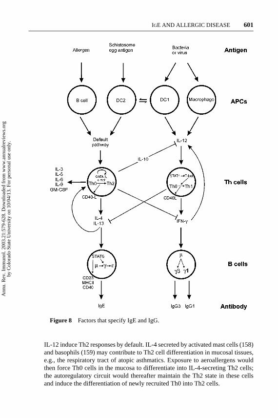
Th1 and Th2 cells differentiate from precursor cells when activated throughtheir T-cell receptor/CD3 complexes by processed antigen/class II MHC on APC(Figure 8). The signaling pathway for Th1 cell differentiation involves IL-12,interferon-γ (IFN-γ ), STAT4, STAT1, and the transcription factor, T-box, ex-pressed in T cells (T-bet). IL-12 is required for the activation of STAT4, which theninduces IFN-γ gene expression via T-bet. The resulting IFN-γ (through STAT1)forms part of an autoregulatory circuit with T-bet, whereby IFN-γ stimulates T-betgene expression and vice versa (146, 147). The IFN-γ secreted by the Th1 cellsdirects switching to IgGs but not to IgE in B cells. IFN-γ also antagonizes thedifferentiation of Th0 to Th2 cells, thus reinforcing the original lineage decision.
Th2 cell fate is governed by the transcription factors GATA-3 and c-maf; GATA-3 results in the production of the signature cytokines IL-4, IL-5, IL-9, and IL-13.GATA-3 autoregulates its own promoter and appears to control the accessibilityof the IL-4 gene for transcription. IL-4 stimulates class switching to IgG4 and IgEand also suppresses IFN-γ gene expression; consequently IFN-γ and IL-4 reasserttheir own lineage decision by positive and negative feedback mechanisms.
Th cell polarization is now understood in terms of cell division and IFN-γ andIL-4 chromatin remodeling. When Th0 cells are activated they begin to produceIL-2 immediately, but the synthesis of IFN-γ or IL-4 is linked to cell division (148–150). The frequency of IFN-γ gene expression in the cell population increasesthrough successive cell cycles, whereas IL-4 expression sets in only after threecell divisions. The concerted effects of cycling and cytokine signaling relieveepigenetic repression by demethylation and hyperacetylation of chromatin in theregion of the genes (149, 151–154).
APC, notably activated macrophages, and a subset of dendritic cells (DC1;see next section) specialize in the production of IL-12. The nature of the APC istherefore important because, in the absence of IL-12, activated Th cells generateIL-4, which downregulates IFN-γ and hence IL-12. This may explain why B cells,acting as APC (155–157) (see Figure 5), and dendritic cells that fail to produce
Ann
u. R
ev. I
mm
unol
. 200
3.21
:579
-628
. Dow
nloa
ded
from
ww
w.a
nnua
lrev
iew
s.or
gby
Col
orad
o St
ate
Uni
vers
ity o
n 10
/04/
13. F
or p
erso
nal u
se o
nly.
11 Feb 2003 17:8 AR AR180-IY21-18.tex AR180-IY21-18.sgm LaTeX2e(2002/01/18)P1: IKH
IGE AND ALLERGIC DISEASE 601
Figure 8 Factors that specify IgE and IgG.
IL-12 induce Th2 responses by default. IL-4 secreted by activated mast cells (158)and basophils (159) may contribute to Th2 cell differentiation in mucosal tissues,e.g., the respiratory tract of atopic asthmatics. Exposure to aeroallergens wouldthen force Th0 cells in the mucosa to differentiate into IL-4-secreting Th2 cells;the autoregulatory circuit would thereafter maintain the Th2 state in these cellsand induce the differentiation of newly recruited Th0 into Th2 cells.
Ann
u. R
ev. I
mm
unol
. 200
3.21
:579
-628
. Dow
nloa
ded
from
ww
w.a
nnua
lrev
iew
s.or
gby
Col
orad
o St
ate
Uni
vers
ity o
n 10
/04/
13. F
or p
erso
nal u
se o
nly.

11 Feb 2003 17:8 AR AR180-IY21-18.tex AR180-IY21-18.sgm LaTeX2e(2002/01/18)P1: IKH
602 GOULD ET AL.
The Role of Dendritic Cells
Whether dendritic cells direct Th cells into the Th1 or the Th2 pathway may be pre-determined by signals received from the antigen and the microenvironment (160).Bacterial constituents, such as lipopolysaccaride and oligonucleotides with CpGmotifs, induce the production of IL-12 and IL-18 in mouse DC1 and macrophages.Certain microbes,Bordetella pertussistoxin and poly(rI:C), the double-strandedviral RNA analogue, induce DC1 (corresponding to Th1) polarization, whereasextracellularVibrio choleraetoxin and extracts from schistosomal egg antigensinduce DC2 polarization (161). A glycolipid (LDN-DF) was recently identifiedas a schistosomal egg constituent, which may act at the host-parasite interface,perhaps triggering this innate immune response (162). The relevant receptors andsignaling pathways remain to be elucidated.
The locally acting agents responsible for DC2 differentiation may be prost-aglandin E2 (PGE2) and IL-10 (163, 164), which are known to suppress IL-12production in dendritic cells (164a). Other factors, such as the antigen (type, dose),the host (genetic background, age, prior infection history), and the environment(natural adjuvants) may all influence whether a dendritic cell becomes DC1 orDC2 (165–168).
Rat respiratory dendritic cells and murine Peyer’s patch dendritic cells elicit Th2responses (164a), whereas splenic dendritic cells engender Th1 responses (170).Viral antigens can induce Th2-committed (CD11c−) pre–dendritic cell matura-tion into dendritic cells that elicit IFN-γ -producing T cells (171). This shows theplasticity of dendritic cell function determined by antigens.
The Role of Allergens
There is no simple answer to the question, “What makes an antigen an allergen?”As noted above, there may be structural components recognized by the innateimmune system. For some antigens, the route of entry (the microenvironment) orparticle size, which in the case of aeroallergens affects the extent of penetrationinto the respiratory tract, may be overriding factors. Both the glycolipid structureand location in the lung mucosa may account for the propensity of the schis-tosomal egg antigen (LDN-DF) to induce an IgE response, although not all ofthe IgE is directed against the parasite. In addition, adjuvants in the environment(e.g., pollutants) or in vaccination protocols may influence the direction of classswitching.
Many common allergens, not least Der p I, the fecal antigen of the house dustmite, are proteases. Der p I attacks two known substrates at specific sites, namelyIL-2R (CD25) and CD23, which may affect the immunological outcome (Th1versus Th2). Inactivation of Der p I protease activity increases IgE synthesis invivo (172) and in B cells incubated with T cells and IL-4 in vitro (173). Treatmentof T cells with Der p I enhances their ability to stimulate IgE upon reconstitutionwith B cells in vitro (174). Accordingly, Der p I conditions T cells to producemore IL-4 and less IFN-γ , which could clearly contribute to dust mite stimulation
Ann
u. R
ev. I
mm
unol
. 200
3.21
:579
-628
. Dow
nloa
ded
from
ww
w.a
nnua
lrev
iew
s.or
gby
Col
orad
o St
ate
Uni
vers
ity o
n 10
/04/
13. F
or p
erso
nal u
se o
nly.

11 Feb 2003 17:8 AR AR180-IY21-18.tex AR180-IY21-18.sgm LaTeX2e(2002/01/18)P1: IKH
IGE AND ALLERGIC DISEASE 603
of IgE responses. Der p I also cleaves membrane-bound CD23 to release a 16-kDafragment (116a, 176), which may upregulate IgE synthesis by eliminating feedbackcontrol (see Multiple Functions of CD23, above).
Genes and the Environment
No description of the mechanisms of Th cell polarization would be completewithout mention of the more global effects of genetic polymorphisms and envi-ronmental risk factors. No less than half of the susceptibility to asthma is deter-mined by genetic predisposition (177), but attempts to identify the responsiblegenes have often led to conflicting answers owing to their multiplicity (178–180). Not surprisingly various “candidate” genes, e.g., the IL-4, IL-4Rα-chain,IL-9, IL-13, STAT6, and CD14 genes, determine the effective Th1/Th2 cell bal-ance. CD14 is the myeloid pattern-recognition receptor for lipopolysaccaride inAPC, which leads to Th1 cell polarization (181, 182). Other candidate genes, e.g.,the FcεRIβ and mast cell chymase genes, are related to effector mechanisms(178).
The fetus is exposed to a Th2 environment in the uterus, and the immunesystem is therefore Th2-polarized at birth. It is believed that immune deviationnormally occurs by the induction of Th1 responses and the resulting suppres-sion of Th2 responses following bacterial infections in the neonate. This may beaverted, especially in genetically predisposed individuals, by the high standardsof hygiene in industrialized countries (the “hygiene hypothesis”) and overexpo-sure to allergenic substances such as the fecal antigens of the house dust miteor fungal spores in the atmosphere. These conditions are thought to have con-tributed to the dramatic rise in the prevalence of allergic disease in recent decades(143, 178).
Mechanism of Class Switching to IgE
Class switching proceeds directly or sequentially to downstream CH genes fromCµ to Cγ , Cα, or Cε. Each class switching event occurs in three distinct stages:germline gene transcription, class switch recombination (CSR), and B cell differ-entiation into Ig-secreting plasma cells (183). As an example we describe directclass switching from IgM to IgE (Figure 7).
Theµ andε germline genes are transcribed from their intervening (I) exonpromoters. RNA polymerase traverses the I exon, switch (S) region, and CH exonsand stops at the normal (mRNA) polyA addition site of theµ chain andε chaingermline gene, and Iµ is spliced to Cµ, and Iε to Cε. The I exons contain stopcodons in all three frames and hence these transcripts are said to be “sterile.” Thechromosomal DNA is cleaved in the two switch regions (Sµ and Sε), and the endsof the chromosome and those of the excised intervening DNA fragment are joinedto make the rearranged chromosome and resultant “switch circle” DNA. The re-arrangedε gene can then be transcribed into theε chain mRNA precursor fromthe V region promoter, stimulated by the intronic enhancer (Eµ), and the initial
Ann
u. R
ev. I
mm
unol
. 200
3.21
:579
-628
. Dow
nloa
ded
from
ww
w.a
nnua
lrev
iew
s.or
gby
Col
orad
o St
ate
Uni
vers
ity o
n 10
/04/
13. F
or p
erso
nal u
se o
nly.

11 Feb 2003 17:8 AR AR180-IY21-18.tex AR180-IY21-18.sgm LaTeX2e(2002/01/18)P1: IKH
604 GOULD ET AL.
transcript is spliced to yield mRNA encoding either the membrane or secretedform of theε chain. The mRNAs are then translated into protein, assembled fromthe H- and L-chains and either transported to the cell membrane (mIgE) or, afterB cell differentiation, secreted (IgE).
Class switching is coupled to cell division and is therefore a slow process: Classswitching to IgE can be induced by incubation of Th cells (or a combination of IL-4and anti-CD40 or soluble CD40-L) with B cells in vitro. In vitro germline genetranscription is evident after a few hours, and CSR and mIgE expression at aroundday 6, followed by B cell differentiation and IgE secretion after day 10 (184). Thisis comparable to the time required for antibody generation in the primary immuneresponse in vivo.
ε Germline Gene Transcription
IL-4 or IL-13 bind to the commonα-chain of the IL-4 (IL-4R) and IL-13 (IL-13R)receptors to activateε germline gene transcription in B cells (185). The signaltransduction pathway is well understood (186). First the receptor is phospho-rylated by the associated JAK1 and JAK3 kinases. The phosphorylated receptorrecruits STAT6 from the cytoplasm, and the activated JAKS phosphorylate STAT6.Phosphorylated STAT6 molecules migrate to the cell nucleus and bind to responseelements in a number of genes, including one in the germline gene promoter(Figure 9).
Figure 9 (A) Transcription factor binding sites on the humanε-germline gene promo-tor. (B) Structure of an R-loop formed during germline gene transcription.
Ann
u. R
ev. I
mm
unol
. 200
3.21
:579
-628
. Dow
nloa
ded
from
ww
w.a
nnua
lrev
iew
s.or
gby
Col
orad
o St
ate
Uni
vers
ity o
n 10
/04/
13. F
or p
erso
nal u
se o
nly.

11 Feb 2003 17:8 AR AR180-IY21-18.tex AR180-IY21-18.sgm LaTeX2e(2002/01/18)P1: IKH
IGE AND ALLERGIC DISEASE 605
Figure 9A shows the more complete constellation of transcription factors con-trolling transcription of the humanε germline gene (187, 188). They includeNF-κB, PU-1, BSAP, and C/EBP and are counteracted by negatively acting factors,Myb, and possibly BCL-6, which must be displaced before transcription can pro-ceed. Crosslinking of CD40 on B cells activates NF-κB and enhancesε germlinegene transcription (189). Unstimulated NF-κB exists in a complex with an in-hibitor (IκB), in the cytoplasm. When phosphorylated by IKK1 and IKK2, IκB isdegraded and a nuclear localization sequence in NF-κB is unmasked. This allowsthe transcription factor to migrate from the cytoplasm to the nucleus and bind tothe accessible response elements in many genes, in addition to theε germline gene.Many NF-κB target genes belong to a wider family of inflammatory response genes(190). PU-1 is associated with differentiation of a common lymphoid/myeloid cellprecursor into the lymphocyte lineage and BSAP with the differentiation of acommon T/B cell precursor into B cells (191).
Recent evidence suggests that Myb inhibitsε germline gene transcription, prob-ably by blocking access to the 3′ side of the STAT6 site (192) (see Figure 9). Sucha factor or combination of factors may determine the probability of initiation ofε germline gene transcription in a stochastic manner (see Specification of IgE,below).
Class Switch Recombination
The mechanism of CSR (the term confined to the stage of DNA recombinationin class switching) proceeds by four sequential steps: (a) selection of the target Sregion, (b) recognition of the DNA target sequence or structure, (c) DNA cleavage,and (d) repair and ligation (193, 194).
S regions are located between the I exons and CH1 exons of each CH gene(except Cδ) in the germline DNA (Figure 7). They contain multiple pentameric(GAGCT and GGGGT) repeats and longer repeats extending over a total lengthof ∼1–10 kb; Sε is relatively short, comprising only 1–2 kb. Recent evidencesuggests that the essential property of the S sequences for CSR is their capacityto form hairpin loops by palindromic S sequences when the DNA is unwoundfor transcription (195, 196). Hairpin formation in the noncoding strand may befacilitated by the failure of the nascent RNA to dissociate from the coding strand,thus generating an “R loop” (197) (see Figure 9B). DNA breaks may be introducedat the exposed junctions between single and double-stranded regions in the hairpinsand at the ends of the RNA-DNA hybrid.
If, as expected from the R-loop model, some of the products of cleavage havestaggered ends, with little complementarity to those of potential S partners in CSR,the sequences would have to be modified before DNA ligation. Microhomologieshave been detected at switch junctions, suggesting that base pairing may nucleatethe formation of the hybrid S junction and allow the entry of repair enzymes (198).Error-prone polymerase(s) are implicated by the occurrence of point mutations andinsertions in the S junctions, whereas deletions from the germline DNA sequenceare assumed to reflect the activities of exonuclease(s).
Ann
u. R
ev. I
mm
unol
. 200
3.21
:579
-628
. Dow
nloa
ded
from
ww
w.a
nnua
lrev
iew
s.or
gby
Col
orad
o St
ate
Uni
vers
ity o
n 10
/04/
13. F
or p
erso
nal u
se o
nly.

11 Feb 2003 17:8 AR AR180-IY21-18.tex AR180-IY21-18.sgm LaTeX2e(2002/01/18)P1: IKH
606 GOULD ET AL.
Elements of the MRN DNA repair complex, Mre11, Rad50 or 51, and Nbs1,may be involved in the CSR mechanism. Nbs-1 stimulates the unwinding of DNAduplexes and hairpin cleavage, whereas Rad50 is responsible for ATP-bindingand joining of the noncomplementary ends (199). This complex, together withphosphorylated histone,γ -H2AX, is involved in repairing lesions in DNA causedby X-rays (200). Two components of the MRN complex, Nbs1 andγ -H2AX,colocalize with the H-chain gene locus during CSR; others were not detected(201).
Ku70/Ku80 and DNA–protein kinase are required for CSR, thus implicating thenonhomologous end-joining pathway in the mechanism. The Ku70/Ku80 complexbinds to single- or double-stranded DNA ends and recruits DNA–protein kinaseto such sites. It is not clear whether additional mechanisms help the broken DNAends to locate each other. One suggestion is that alternative splicing, whereby Iexons are joined to a distal CH1 exon, brings the associated DNA S regions intocloser proximity (figure 1 in Reference 194).
In B cells class switching occurs after several rounds of cell division, and the H-chain gene locus is replicated from a single origin downstream of the 3′ enhancer(Eα in Figure 6) early in the S phase of the cell cycle in B cells (202). Classswitching occurs at the G1/S stage of the cell cycle (203). What happens when thereplication fork and RNA polymerase, travelling in opposite directions, meet onthe DNA? Does this clean the slate for the resumption of transcription at G1/S ofthe next cycle or bypass the transcription complexes? This is unknown, and thereis little information in general about such situations.
The enzyme activation–induced cytidine deaminase (AID) is essential for bothCSR and SHM (193, 204, 205). AID is homologous to APOBEC-1, the catalyticsubunit of an RNA-editing complex, which deaminates C to U, thereby introducinga stop codon in the mRNA coding for apolipoprotein B, resulting in a truncatedprotein. AID itself exhibits cytidine deaminase activity in vitro. AID expressionis B cell–specific and particularly highly expressed in GC and during the G1/Sstage of the cell cycle, in accordance with the spatial and temporal requirementsfor activity in CSR (203). Ectopic expression of AID is sufficient to activate CSRin hybridomas (206) and fibroblasts (207). This implies that all other participatingproteins are either constitutively expressed or induced by AID. AID is not necessaryfor germline gene transcription or DNA cleavage (208, 209). Candidate targets forAID activity therefore include the DNA itself or mRNA encoding enzymes thatact in DNA repair or B cell survival during CSR (210). The identification of theAID substrate(s) is now a matter of priority. (See Note Added in Proof.)
B Cell Differentiation
Immediately after CSR the rearranged gene is expressed and mIg is exported to thecell surface. The differentiation of memory B cells into Ig-secreting plasma cellsentails gross changes in cell phenotype and function. The cells lose their motilityand responsiveness to many external signals. The nucleus contracts in response
Ann
u. R
ev. I
mm
unol
. 200
3.21
:579
-628
. Dow
nloa
ded
from
ww
w.a
nnua
lrev
iew
s.or
gby
Col
orad
o St
ate
Uni
vers
ity o
n 10
/04/
13. F
or p
erso
nal u
se o
nly.

11 Feb 2003 17:8 AR AR180-IY21-18.tex AR180-IY21-18.sgm LaTeX2e(2002/01/18)P1: IKH
IGE AND ALLERGIC DISEASE 607
to the shutdown of previous functions and develops an extensive endoplasmicreticulum for Ig synthesis and secretion; a plasma cell secretes up to 0.1 ng Ig perday, up to 50% of total protein synthesis (211).
How antigen triggers differentiation of B cells into plasma cells is now under-stood in broad outline (212, 213): Signals transmitted through theα andβ signalingsubunits of the receptor stimulate phosphorylation by mitogen-activated proteinkinase, leading to the degradation of BCL-6 (214). The function of BCL-6 hereis to repress the gene for B lymphocyte–induced maturation protein 1 (Blimp-1)(215, 216); thus, the degradation of Bcl-6 allows Blimp-1 expression. Blimp-1,in association with hGroucho and class I histone deacetylases, inhibits transcrip-tion of genes coding for the mIgα-chain, Myc, BSAP, STAT6, AID, Ku70/80,DNA–protein kinase, and other proteins involved in the processes occurring in theplasma cell precursors. BSAP represses the expression of X-box binding protein(XBP-1), required for terminal differentiation, so the lack of BSAP removes afurther block to differentiation. Terminal differentiation is reinforced by the in-hibition of renewed BCL-6 by Blimp-1, which appears following degradation ofBCL-6.
Differentiation to memory cells occurs through IL-10, secreted by Th2 cells,which upregulates CD27 in GC, allowing interaction with CD70+ T cells and Bcells (217). In response to IL-4 and CD40 crosslinking, CD27+ B cells prolif-erate and may thus undergo class switching to IgE (218, 219). CD40 signallingdownregulates the Blimp-1 gene (220), restraining this differentiation, whereasCD23 signalling through CD21 upregulates BCL-2 to rescue the B cells fromapoptosis (129). Together, CD40 and CD23 signaling promote cell proliferationand B cell survival, thus increasing the probability of class switching to IgE. CD23may also act in the preferential selection of IgE-committed B cells (see MultipleFunctions of CD23, above).
Specification of IgE
How does IL-4 specify IgE? The prevailing view is that it acts by facilitating tran-scription of theε germline gene, coupled to local unfolding of chromatin, much ashas been convincingly demonstrated for the IFN-γ and IL-4 genes in specifyingthe Th1 and Th2 lineages (see The Role of T Helper Cells, above). Transcriptionof theε germline gene was said to be accompanied by silencing ofγ genes (e.g.,γ2b in mouse) (221). The “chromatin accessibility model” for the direction ofclass switching, equating transcription with chromatin accessibility (see Figure 2in Reference 194), was built on this supposition. However, the chromatin structureof the CH genes has never been investigated. Developmental stage–specific un-folding of the murineκ andλ L-chain genes, in fact, precedes gene transcription(222).
The established fact is that IL-4 and IL-13 are the only cytokines that stimulateε germline gene transcription and (with CD40 signaling) class switching to IgEin both murine and human B cells. At the same time, IL-4 and CD40-L stimulate
Ann
u. R
ev. I
mm
unol
. 200
3.21
:579
-628
. Dow
nloa
ded
from
ww
w.a
nnua
lrev
iew
s.or
gby
Col
orad
o St
ate
Uni
vers
ity o
n 10
/04/
13. F
or p
erso
nal u
se o
nly.

11 Feb 2003 17:8 AR AR180-IY21-18.tex AR180-IY21-18.sgm LaTeX2e(2002/01/18)P1: IKH
608 GOULD ET AL.
transcription and CSR of all the other germline genes in human B cells, leading toCSR and secretion of the corresponding antibody classes (223, 224). In the latterstudy by Cerutti and co-workers the chosen mixture of cytokines contained notonly IL-4 and anti-CD40 but also IL-10 and IL-6, which are known to stimulate Bcell differentiation (225, 226). Another group found that IL-10, together with anti-CD40, stimulated class switching to IgGs but not to IgE, whereas IL-4 stimulatedCSR to IgGs and IgE, though only IgE was secreted (227).
Recent work in our laboratory has shown that the majority of mature, naive(IgD+) B cells exhibit constitutive transcription of one or more germline genes,including γ and ε genes, and that IL-4 and anti-CD40 upregulate the level oftranscription (D. Fear et al., N. McCloskey, G. Felsenfeld, H.J. Gould, unpublishedresults). IL-4 and IL-13 are the only cytokines capable of upregulatingε germlinegene transcription to approximately the same level as that of theγ germline genes.It has been suggested that a threshold level of transcription may be necessary, ifnot sufficient, for CSR (228).
IL-4 acts by way of STAT6 sites, which are found in all theγ germline genes,as well as in theε germline gene. If (as the best evidence indicates) all the genesare equally accessible, then the original question can be reformulated: How cantheγ germline gene transcription be upregulated in the absence of IL-4?
Hodgkin and co-workers have demonstrated that class switching to several iso-types, including IgE, in murine B cells is tightly coupled to the number of celldivisions. Switching to IgG1 occurs after three cell divisions and to IgE after five(229–232). This can be rationalized, even if the mechanism cannot be defined,by postulating an isotype-specific probability of switching at each round of repli-cation. This could depend, for instance, on the density of nascent germline genetranscripts (seeε Germline Gene Transcription, above). The highest probabilityof switching to a given isotype varied with the IL-4 concentration, but the numberof cell cycles at the peak was constant. Some cells underwent apoptosis beforearriving at the required number of cell divisions for class switching to IgE, andmore than half of the remainder failed to make the switch.
Sequential switching to downstream isotypes is a feature of both murine(233–235) and human (236–242) B cells. Thus, the lag in class switching to IgEmay result in part from the sequential process. However, it was observed that theprobabilities of switching from IgM or Ig2b to IgE were identical, suggesting theremay be an inherent delay in switching to IgE, the mechanism of which is not yetunderstood.
During sequential switching the I exons corresponding to previously expressedgermline genes are excised from the genome and only transiently transcribedfrom the resulting switch circles (243) (see Figure 7). The possibility of de-tecting germline gene transcripts from the intermediate isotypes must decreasewith time. This may account for the observation thatγ2b transcripts declinewhile ε transcripts increase between 3 and 5 h inmurine B cells stimulated withlipopolysaccharide and IL-4 (221), the cornerstone of the chromatin accessibilitymodel.
Ann
u. R
ev. I
mm
unol
. 200
3.21
:579
-628
. Dow
nloa
ded
from
ww
w.a
nnua
lrev
iew
s.or
gby
Col
orad
o St
ate
Uni
vers
ity o
n 10
/04/
13. F
or p
erso
nal u
se o
nly.

11 Feb 2003 17:8 AR AR180-IY21-18.tex AR180-IY21-18.sgm LaTeX2e(2002/01/18)P1: IKH
IGE AND ALLERGIC DISEASE 609
SOMATIC HYPERMUTATION OF IgE VH REGIONS
Somatic hypermutation (SHM) is characterized by the accumulation of point mu-tations across rearranged V region DNA, particularly at “hotspots” such as RGYWand its reverse complement WRCY (where R= A or G, Y = C or T, W = A orT). These hotspots occur more frequently in complementarity-determining regions(CDR) than framework regions (FR), making the CDRs especially susceptible tomutation (244). A single mutation can engender a 10-fold increase in antibodyaffinity (245). The ratio of replacement (R) relative to silent (S) mutations mayreveal evidence of antigen selection. In the absence of antigen selection R/S ratiosare generally twice as high in CDR as FR, reflecting the relative frequency ofRGYW motifs and the imprint of antigen selection (246).
Unusual patterns of R/S ratios and VH usage have been found in the V regionsfrom IgE H-chain (VH-Cε) sequences from allergic individuals. VH5 is one ofthe smallest VH gene families containing two members out of the total of 52functional VH genes (247). In patients with allergic rhinitis (248), allergic asthma(249–251), and atopic dermatitis (252) VH5 is greatly overrepresented in IgE,particularly in the target organs compared with peripheral blood B cells. In thenasal mucosa of patients with allergic rhinitis about one fifth of VH-Cε sequenceswere VH5, compared with about half that number in peripheral blood B cells fromthe same group of individuals (248). The bias was even more pronounced in apatient with severe asthma, one third of whose VH-Cε sequences were VH5 (251).A similar proportion of VH5-Cε sequences were detected in IgE+ B cells from thespleen of asthmatic patients (249), whereas the overall abundance of VH5 in thesplenic B cells of the combined antibody classes was only 8%. Two explanationshave been suggested: Either common allergens or autoallergens, perhaps acting assuperantigens, recognize FR, particularly FR3 (249), or FR3 imposes a favorablestructure on the CDRs (253).
Somatic mutations in the VH-Cε sequences of the allergic subjects peak inthe CDRs and occur at approximately the expected frequency in hotspot motifs.Analysis of the R/S ratios, however, revealed certain anomalies. In the study ofpatients with atopic dermatitis the majority of VH5-Cε sequences exhibited R/Sratios well below 2.9 in CDR (252), and in the patients with allergic rhinitis R/Sratios in the mucosa were 1.5 in CDR and 2.7 in FR (248). These unusual R/S ratioscould arise from the local activity of allergens recurrently or chronically stimulatingthe immune system, or from a superallergen, leaving the imprint of SHM in the FR.
Analysis of VH-Cε sequences in allergic individuals suggests the presence ofclonally related but diverged families of B cells, as indicated by the presence ofshared and unique mutations. These could emerge if the cells circulated repeat-edly through germinal centers (GC), with consequent accumulation of somaticmutations and clonal selection, but several observations suggest that this is notthe case. The occurrence of clonal families in mucosal tissues suggests that clonalexpansion may occur locally (248, 251). The coexistence of unmutated (germline)and varying extents of somatically mutated VH-Cε sequences in asthmatic lung
Ann
u. R
ev. I
mm
unol
. 200
3.21
:579
-628
. Dow
nloa
ded
from
ww
w.a
nnua
lrev
iew
s.or
gby
Col
orad
o St
ate
Uni
vers
ity o
n 10
/04/
13. F
or p
erso
nal u
se o
nly.

11 Feb 2003 17:8 AR AR180-IY21-18.tex AR180-IY21-18.sgm LaTeX2e(2002/01/18)P1: IKH
610 GOULD ET AL.
mucosa points to local somatic mutation (251). The argument is based on the lowprobability of migration of an unmutated clone to a lymph node, there to undergosomatic mutation, before returning to the very same site. The presence of clonallyrelated families comprising more than a single isotype (IgM, IgG, IgA, and IgE)in both the lung and nasal mucosa (248, 251) suggests by the same reasoning thatboth class switching and SHM occurred locally. These results and the observationof AID expression in the nasal mucosa of a patient with allergic rhinitis (248)provide supporting evidence for local class switching (see next section).
LOCAL REGULATION OF IgE
Local IgE Synthesis
Local synthesis of IgE has long been inferred on the grounds that IgE antibodiesare found in the nasal secretions of subjects with allergic rhinitis and in bron-choalveolar lavage fluid from patients with allergic asthma. However, exudationfrom serum or active transport of IgE, as recently demonstrated for CD23 (127)(see Multiple Functions of CD23, above), could not be excluded. More persua-sive evidence comes from the observation of antibodies in the secretions whenthey could not be detected in the serum by RAST assay or by skin prick test(9, 254). Recent work demonstrating de novo IgE synthesis in the nasal mucosa ofsubjects with allergic rhinitis and the lung mucosa of asthmatics has resolved thisissue (255, 256). Local IgE synthesis significantly allows maintainence of the stateof immediate hypersensitivity in the mucosal mast cells (see Compartmentation,below).
The source of IgE has therefore been determined, though not of that of the Bcells. As class switching to IgE undoubtedly occurs in GC, the committed cellscould have migrated from local lymphoid tissues through the circulation to theinflamed areas at sites of allergen provocation, but this is not easily reconciled withthe presence of clonal families of different isotypes (see Somatic Hypermutationof IgE VH Regions, above). The notion of clonal expansion and selection withinthe mucosa is supported by the high proportion of CD19+ B cells containingεchain mRNA in the nasal mucosa (a third and a tenth of the total number of cellsin allergic rhinitics and healthy nonatopic control subjects, respectively) (257).The mRNA is evidently translated, for the proportions of B cells and plasma cellsexpressing mIgE in the nasal mucosa of allergic rhinitis patients are much the same(258). Only one in 104–103 of peripheral blood B cells are committed to IgE (6, 7),so there is a 100- to a 1000-fold enrichment of these cells in the mucosa.
Persistence of IgE Synthesis
The synthesis of IgE antibodies in the nasal mucosa of grass pollen–sensitivesubjects continues outside the grass pollen season (255). How is the life of theIgE-secreting cells or clones extended to allow for a constant source of IgE without
Ann
u. R
ev. I
mm
unol
. 200
3.21
:579
-628
. Dow
nloa
ded
from
ww
w.a
nnua
lrev
iew
s.or
gby
Col
orad
o St
ate
Uni
vers
ity o
n 10
/04/
13. F
or p
erso
nal u
se o
nly.

11 Feb 2003 17:8 AR AR180-IY21-18.tex AR180-IY21-18.sgm LaTeX2e(2002/01/18)P1: IKH
IGE AND ALLERGIC DISEASE 611
allergen stimulation? IgE-expressing B cells may be effectively selected forlongevity if they have undergone a greater number of cell divisions and escapedapoptosis prior to class switching (see H-Chain Class Switching to IgE, above). Alarge proportion of IgE-secreting plasma cells are exceptionally resistant to killingby X-irradiation (259, 260).
The survival of IgE-committed B cells may be promoted following the upregu-lation of CD23b by IL-4 and the release of sCD23; sCD23 can form a heterotrimerwith mIgE and CD21 (135), and coligation of mIg and CD21 by CD23 stimulatesB cell proliferation (13, 131) (see Multiple Functions of CD23, above). Long-livedIgG antibody–secreting plasma cells in bone marrow are nurtured by stromal cellfactors (see H-Chain Class Switching to IgE, above). Recent studies of IgA reg-ulation suggest that this antibody class may be generated in the gastrointestinalmucosa of mouse by Th cell–independent class switching (261) or even direct VDJrecombination with Cα (262). Thus, different microenvironments evidently favorcertain antibody classes over others.
Allergen Stimulation of Local Class Switching to IgE
Evidence of local class switching to IgE comes from observations of varioustransient markers for the dynamic process of class switching (see H-Chain ClassSwitching to IgE, above). Local allergen challenge stimulates the appearance ofε germline gene transcripts in B cells within 24 h (257). Local allergen challengealso stimulates the appearance of IL-4 mRNA in Th cells in the same interval,suggesting a causal relationship. IL-4 mRNA in Th cells andε germline genetranscripts in B cells were also identified in the nasal mucosa of grass pollen–sensitive individuals during the grass pollen season (263). Allergen stimulation ofIL-4 synthesis by Th cells, which in turn induceε germline gene transcription inB cells, is thus again implied. More surpisinglyε-germline gene transcripts wereconstitutively expressed in the lungs of nonatopic asthmatics, suggesting that theIgE antibodies may be directed against autoantigens (264, 265).
To eliminate the possibility that the Th cells and B cells migrate from the circu-lation after mast cell activation of the tissue by allergen in the allergen challengestudies, explants from the nasal mucosa were incubated with allergen ex vivo.Again it was observed that IL-4 mRNA in Th cells andε germline gene transcriptsappeared in B cells after 24 h (266).
Although germline gene transcripts represent the first step in class switching,this is not to say that the process proceeds through the subsequent steps: CSR,B cell differentiation, and secretion of IgE (see H-Chain Class Switching to IgE,above). Switch circle DNA, transient markers of CSR, have been observed in thenasal secretions of allergic rhinitics challenged with ragweed allergen mixed withdiesel exhaust particles (267), and in the nasal mucosa of allergic rhinitics exposedto allergen (P. Takhar, L. Smurthwaite, S.R. Durham, & H.J. Gould, unpublishedresults). These observations suggest that allergen stimulates CSR in the nasalmucosa of allergic rhinitics.
Ann
u. R
ev. I
mm
unol
. 200
3.21
:579
-628
. Dow
nloa
ded
from
ww
w.a
nnua
lrev
iew
s.or
gby
Col
orad
o St
ate
Uni
vers
ity o
n 10
/04/
13. F
or p
erso
nal u
se o
nly.

11 Feb 2003 17:8 AR AR180-IY21-18.tex AR180-IY21-18.sgm LaTeX2e(2002/01/18)P1: IKH
612 GOULD ET AL.
Analogy Between Autoimmune and Allergic Disease
Repeated or chronic stimulation of inflammatory responses in mammalian tis-sues may result in SHM and class switching in abnormal locations. This is welldocumented in rheumatoid arthritis (268) and insulin-dependent diabetes mellitus(269). Synovial B cells in rheumatoid arthritis are long-term memory cells andthus play a part in the chronic inflammatory reactions. The microenvironment ofthe synovium allows the activation of naive and memory B cells, diversificationof their V gene repertoire, affinity maturation, and clonal expansion (268, 270).Synoviocytes support the differentiation of the activated B cells in vitro (271).
Chvatchko and coworkers found that germinal centers were formed in the lungafter airway antigenic challenge in mouse and that this led to local IgE synthesis(272). Together with the cellular and molecular studies described above this providecompelling evidence of a similar development in allergic disease.
COMPARTMENTATION
Is the Respiratory Tract Mucosa Self-Sufficient in IgE?
The allergic state, characterized by immediate hypersensitivity, depends on thepermanent sensitization of mast cells in the target organ. We have observed thatIgE synthesis occurs between seasons in the nasal mucosa of grass pollen–sensitvepatients with allergic rhinitis (see Local Regulation of IgE, above). We ask herewhether this is sufficient to maintain the sensitization of mast cells or whetherthe main source of IgE is outside the tissue. Nasal B cells synthesize∼4×10⁹ molecules/day/mm3 biopsy [calculated from the data of Smurthwaite et al.(255)]. Immunohistochemical studies of sections of the tissue yield a count of∼5000 mast cells/mm3 nasal biopsy (S.R. Durham, personal communication).There are∼2× 10⁵ receptor molecules per cell (69), and the half-life of IgEin skin is∼14 days (29, 29a, 273). Uninterrupted saturation of these receptorstherefore requires the de novo synthesis and secretion of 4× 10 7 molecules ofIgE/day/mm3. We conclude that the rate of IgE secretion generously exceeds theminimum requirement for saturation.
The figures are notional, there are other IgE-binding cells in the tissue, andabout half of the IgE is in any event nonspecific antibody (255), but maximumsensitization requires the occupancy of less than 10% of the FcεRI moleculeson the mast cell (69). The excess unbound IgE presumably corresponds to thatmeasured in secretions and in the serum, where it is diluted with IgE antibodiesfrom other mucosal sources. This IgE may also suffice to maintain the perpetualsensitization of Langerhans cells in the epithelium. Upon exposure to allergensthese cells would migrate to the local lymphoid tissue and initiate a GC reaction.This in turn would lead to a transient response, resulting in repopulation of themucosa with memory Th cells following their release into the circulation andrecruitment into the tissue. Scharenberg and Kinet refer to the more persistent IgEsynthesis in tissues as “pump priming” (274).
Ann
u. R
ev. I
mm
unol
. 200
3.21
:579
-628
. Dow
nloa
ded
from
ww
w.a
nnua
lrev
iew
s.or
gby
Col
orad
o St
ate
Uni
vers
ity o
n 10
/04/
13. F
or p
erso
nal u
se o
nly.

11 Feb 2003 17:8 AR AR180-IY21-18.tex AR180-IY21-18.sgm LaTeX2e(2002/01/18)P1: IKH
IGE AND ALLERGIC DISEASE 613
The above calculation indicates that the duration of mast cell sensitization byIgE is a critical characteristic. IgG diffuses out of tissue much more quickly thanIgE (29, 255, 273), a function both of the number of IgG receptors and the higherintrinsic dissociation rate of IgG from the receptors. The stability of the IgE-FcεRIcomplex is now understood in structural terms (45, 275) (see Crystal and NMRStructures, above). IgE antibody synthesis is also a critical factor; indeed it is adiagnostic feature of allergic rhinitis (29, 255, 257).
Coordination of the Local Response
Mast cells and basophils, along with IgE and IgE receptors, constitute a potentweapon against parasites but also threaten the host. A number of mechanisms arecoordinated to maximize the former and minimize the latter activity by focusingthe response on the affected tissue. IL-4 and other mast cell mediators induceselective recruitment of inflammatory cells, cell activation, and cell proliferation atthe site of antigen provocation, resulting in escalation of the inflammatory responsein the tissue. Eosinophils, stimulated by IL-5, play a particularly ambiguous rolein host protection versus host damage (86).
The release of IL-4 and IL-13 and other Th2 cytokines by allergen-activatedmast cells enhances the IgE response by stimulating Th2 cell differentiation, whichresults in class switching to IgE in B cells and increased production of IgE; thisin turn protects and resensitizes FcεRI on the mast cells in the tissue in a positivefeedback mechanism (green and blue arrows in Figure 5). Antigen presentation toTh2 cells may occur by dendritic cells expressing FcεRI, by B cells expressingCD23a, or directly by mast cells (black arrows in Figure 5A, B, andC, respec-tively). In any case the afferent IgE response to the allergen or parasite occursno further away than the local lymphoid tissue. The local Th1/Th2, IgE/IgG, andFcεRI/FcγR cell fate decisions are all mutually antagonistic (Figure 8). STAT6plays an important role in the coordination of Th2 responses. The regulation of IgEand the affinity of IgE for FcεRI are combined to exclude IgG, which would notconfer immune protection of such long duration as IgE antibodies, from the tissue.Local Th2 cell–mediated immunity operates in a coordinated fashion in expellingparasites and initiating the cascade of early- and late-phase allergic reactions.
SUMMARY
The reaction of IgE with its receptors is central to the phenomenon of allergy.X-ray crystallography and NMR have recently yielded detailed information aboutthe structures of these molecules and their interactions. Together with the outcomeof work on the equilibrium and dynamic characteristics of the antibody-receptorinteractions, considerations of the spatial and temporal factors of IgE regulationand effector functions enable us to assemble a relatively complete picture of themechanisms involved in the allergic response. This will undoubtedly open newavenues for therapeutic intervention in allergic disease.
Ann
u. R
ev. I
mm
unol
. 200
3.21
:579
-628
. Dow
nloa
ded
from
ww
w.a
nnua
lrev
iew
s.or
gby
Col
orad
o St
ate
Uni
vers
ity o
n 10
/04/
13. F
or p
erso
nal u
se o
nly.

11 Feb 2003 17:8 AR AR180-IY21-18.tex AR180-IY21-18.sgm LaTeX2e(2002/01/18)P1: IKH
614 GOULD ET AL.
TheAnnual Review of Immunologyis online at http://immunol.annualreviews.org
LITERATURE CITED
1. Sutton BJ, Gould HJ. 1993. The humanIgE network.Nature366:421–28
2. Metcalfe DD, Baram D, Mekori YA.1997. Mast cells.Physiol. Rev.77:1033–79
3. Galli SJ, Lantz CS. 1999.Allergy. SeeRef. 280, pp. 1127–1174
4. Williams AF, Barclay AN. 1988. Theimmunoglobulin superfamily—domainsfor cell surface recognition.Annu. Rev.Immunol.6:381–405
5. Lio A, Waldmann TA, Strober W. 1978.Metabolic study of human IgE: evidencefor an extravascular catabolic pathway.J. Immunol.20:1696–701
6. Lanzavecchia A, Parodi B. 1984. In vitrostimulation of IgE production at a sin-gle precursor level by human alloreac-tive T helper clones.Clin. Exp. Immunol.55:197–203
7. King CL, Poindexter RW, RagunathanJ, Fleisher TA, Ottesen EA, NutmanTB. 1991. Frequency analysis of IgE-secreting B lymphocytes in persons withnormal or elevated serum IgE levels.J.Immunol.146:1478–83
8. Patterson R, Suszko IM, Hsu CC,Roberts M, Oh SH. 1975. In vitro pro-duction of IgE by lymphocytes from a pa-tient with hyperimmunoglobulinaemiaE, eosinophilia and increased lympho-cytes carrying surface IgE.Clin. Exp. Im-munol.20:265–72
9. Smurthwaite L, Durham SR. 2002. Lo-cal IgE production in allergic rhinitisand asthma.Curr. Allergy Asthma Rep.2:231–38
10. Gauld SB, Dal Porto JM, Cambier JC.2002. B cell antigen receptor signaling:roles in cell development and disease.Science296:1641–42
11. Ravetch JV, Kinet J-P. 1991. Fc re-ceptors. Annu. Rev. Immunol.9:457–92
12. Daeron M. 1997. Fc receptor biology.Annu. Rev. Immunol.15:203–34
13. Gould HJ, Beavil RL, Relji´c R, Shi J,Ma CW, et al. 1997. IgE homeostasis: IsCD23 the safety switch? See Ref. 281,pp. 37–59
14. Weis WI, Taylor ME, Drickamer K.1998. The C-type lectin superfamily inthe immune system.Immunol. Rev.163:19–34
15. Padlan EA, Helm BA. 1993. Modellingof the lectin-homology domains of thehuman and murine low-affinity Fcε re-ceptor (FcεRII/CD23).Receptor3:325–41
16. Bajorath J, Aruffo A. 1996. Structure-based modeling of the ligand binding do-main of the human cell surface receptorCD23 and comparison of two indepen-dently derived molecular models.Pro-tein Sci.5:240–47
17. Dierks SE, Bartlett WC, Edmeades RL,Gould HJ, Rao M, Conrad DH. 1993.The oligomeric nature of the murineFcγ εRII/CD23: implications for func-tion. J. Immunol.150:2372–82
18. Kilmon MA, Ghirlando R, Strub M-P,Beavil RL, Gould HJ, Conrad DH. 2001.Regulation of IgE production requiresoligomerization of CD23.J. Immunol.167:3139–45
19. Szakonyi G, Guthridge JM, Li D, YoungK, Holers VM, Chen XS. 2001. Struc-ture of complement receptor 2 in com-plex with C3d ligand.Science292:1725–28
20. Zheng Y, Shopes B, Holowka D, BairdB. 1991. Conformations of IgE bound toits receptor FcεRI and in solution.Bio-chemistry30:9125–32
21. Deisenhofer J. 1981. Crysallographic re-finement and atomic model of a hu-man Fc fragment and its complexwith fragment B of protein A from
Ann
u. R
ev. I
mm
unol
. 200
3.21
:579
-628
. Dow
nloa
ded
from
ww
w.a
nnua
lrev
iew
s.or
gby
Col
orad
o St
ate
Uni
vers
ity o
n 10
/04/
13. F
or p
erso
nal u
se o
nly.

11 Feb 2003 17:8 AR AR180-IY21-18.tex AR180-IY21-18.sgm LaTeX2e(2002/01/18)P1: IKH
IGE AND ALLERGIC DISEASE 615
Staphylococcus aureus at 2.9 and 2.8AresolutionBiochemistry20:2361–70
22. Beavil AJ, Young R, Sutton BJ, PerkinsSJ. 1995. Bent domain structure of re-combinant human IgE-Fc fragment byX-ray and neutron scattering in con-junction with an automated curve fit-ting procedure.Biochemistry34:14449–61
23. Zheng Y, Shopes B, Holowka D, BairdB. 1992. Dynamic conformations com-pared for IgE and IgG1 in solution andbound to receptors.Biochemistry31:7446–56
24. Feinstein A, Richardson N, TaussigMJ. 1986. Immunoglobulin flexibility incomplement activation.Immunol. Today7:169–74
25. Perkins SJ, Nealis AS, Sutton BJ, Fein-stein A. 1991. Solution structure of hu-man and mouse immunoglobulin M bysynchrotron X-ray scattering and molec-ular graphic modelling.J. Mol. Biol.221:1345–66
26. Keown MB, Ghirlando R, Mackay GA,Sutton BJ, Gould HJ. 1997. Basis ofthe 1:1 stoichiometry of the high affinityreceptor FcεRI-IgE complex.Eur. Bio-phys. J.25:471–76
27. Maenaka K, van der Merwe PA, StuartDL, Jones EY, Sonderman PJ. 2001. Thehuman low affinity Fcγ receptors IIa,IIb, and III bind IgG with fast kineticsand distinct thermodynamic properties.J. Biol. Chem.276:44898–904
28. McDonnell JM, Calvert R, Beavil RL,Beavil AJ, Henry AJ, et al. 2001. Thestructure of the IgE Cε2 domain andits role in stabilizing the complex withits high-affinity receptor FcεRIα. Nat.Struct. Biol.8:437–41
29. Ishizaka K, Ishizaka T. 1971. Im-munoglobulin E and homocytotropicproperties. In:Progress in ImmunologyI, ed. B. Amos, pp. 859–74. New York:Academic
29a. Geha RS, Helm B, Gould H. 1985. In-hibition of the Prausnitz-K¨ustner reac-
tion by an immunoglobulin e-chain syn-thesized in E. coli.Nature 315:577–78
30. Shi J, Ghirlando R, Beavil RL, BeavilAL, Keown MB, et al. 1997. Interactionof the low affinity receptor CD23/FcεRIIlectin domain (sCD23) with the Fcε3-4fragment of IgE.Biochemistry36:2112–22
31. Wurzburg BA, Garman SC, JardetzkyTS. 2000. Structure of the human Cε3-Cε4 reveals conformational flexibility inthe antibody effector domains.Immunity13:375–85
32. Padlan EA, Davies DR. 1986. A modelof the Fc of immunoglobulin E.Mol. Im-munol.23:1063–75
33. Helm BA, Ling Y, Teale C, Padlan EA,Bruggemann M. 1991. The nature andimportance of the inter-epsilon chaindisulfide bonds in human IgE.Eur. J. Im-munol.21:1543–48
34. Garman SC, Kinet J-P, Jardetzky TS.1999. The crystal structure of the hu-man high-affinity IgE receptor (FcεRIα).Annu. Rev. Immunol.17:973–76
35. Garman SC, Kinet J-P, Jardetzky TS.1998. Crystal structure of the humanhigh-affinity IgE receptor.Cell 95:951–61
36. Garman SC, Sechi S, Kinet J-P, Jardet-zky TS. 2001. The analysis of the hu-man high affinity IgE receptor FcεRIαfrom multiple crystal forms.J. Mol. Biol.311:1049–62
37. Garman SC, Wurzburg BA, Tarchev-skaya SS, Kinet J-P, Jardetzky TS. 2000.Structure of the Fc fragment of humanIgE bound to its high-affinity receptorFcεRIα. Nature406:259–66
38. Maxwell KF, Powell MS, Hulett MD,Barton PA, McKenzie IF, et al. 1999.Crystal structure of the human leukocyteFc receptor, FcγRIIα. Nat. Struct. Biol.6:437–42
39. Sondermann P, Huber R, Jacob U. 1999.Crystal structure of the soluble form ofthe human FcγRIIβ: a new member of
Ann
u. R
ev. I
mm
unol
. 200
3.21
:579
-628
. Dow
nloa
ded
from
ww
w.a
nnua
lrev
iew
s.or
gby
Col
orad
o St
ate
Uni
vers
ity o
n 10
/04/
13. F
or p
erso
nal u
se o
nly.

11 Feb 2003 17:8 AR AR180-IY21-18.tex AR180-IY21-18.sgm LaTeX2e(2002/01/18)P1: IKH
616 GOULD ET AL.
the immunoglobulin superfamily at 1.7Aresolution.EMBO J.18:1095–103
40. Zhang Y, Boesen CC, Radaev S, BrooksAG, Fridman WH, et al. 2000. Crys-tal structure of the extracellular domainof a human FcγRIII. Immunity13:387–95
41. Sondermann P, Huber R, Oosthuizen V,Jacob U. 2000. The 3.2A-crystal struc-ture of the human IgG1 Fc fragment-FcγRIII complex.Nature406:267–73
42. Radaev S, Sun PD. 2001. Recognitionof IgG by Fcγ receptor. The role of Fcglycosylation and the binding of peptideinhibitors.J. Biol. Chem.276:16478–83
43. Radaev S, Motyka S, Fridman W-H,Sautes-Fridman C, Sun PD. 2001. Thestructure of a human type III Fcγ recep-tor in complex with Fc.J. Biol. Chem.276:16469–77
44. Wurzburg BA, Jardetzky TS. 2002.Structural insights into the interactionsbetween human IgE and its high affinityreceptor FcεRI.Mol. Immunol.38:1063–72
45. Wan T, Beavil RL, Fabiane SM, BeavilAJ, Sohi MK, et al. 2002. Crystal struc-ture of IgE Fc reveals an asymmetricallybent conformation.Nat. Immunol. 3:681–85
46. Sayers I, Cain SA, Swan JRM, Pick-ett MA, Watt PJ, et al. 1998. Aminoacid residues that influence FcεRI-mediated effector functions of humanimmunoglobulin E. Biochemistry 37:16152–64
47. Henry AJ, Cook JPD, McDonnell JM,Mackay GA, Shi J, et al. 1997. Partici-pation of the N-terminal region of Cε3in the binding of the human IgE to itshigh-affinity receptor FcεRI. Biochem-istry 361:5568–78
48. Cook JPD, Henry AJ, McDonnell JM,Owens RJ, Sutton BJ, Gould HJ. 1997.Identification of contact residues in theIgE binding site of human FcεRIα. Bio-chemistry36:15579–88
49. Sutton B, Corper A, Bonagura V, Taus-
sig M. 2000. The structure and originof rheumatoid factors.Immunol. Today21:177–83
50. Pawankar R, Okuda M, Yssel H, Oku-mura K, Ra C. 1997. Nasal mast cellsin perennial allergic rhinitics exhibit in-creased expression of the FcεRI, CD40L,IL-4, and IL-13, and can induce IgE syn-thesis in B cells.J. Clin. Invest.99:1492–99
51. Wedemeyer J, Galli SJ. 2000. Mast cellsand basophils in acquired immunity.Br.Med. Bull.56:936–55
52. Wedermeyer J, Tsai M, Galli SJ. 2000.Roles of mast cells and basophils in in-nate and acquired immunity.Curr. Opin.Immunol.12:624–31
53. Kay AB. 1997. Allergy and Allergic Dis-eases. Oxford: Blackwell Sci.
54. Holgate ST, Church MK, LichtensteinLM. 2001. Allergy. St. Louis, MO:Mosby
55. Gauchat J-F, Henchoz S, Mazzei G,Aubry J-P, Brunner T, et al. 1993. In-duction of human IgE synthesis in Bcells by mast cells and basophils.Nature365:340–43
56. Humbert M, Grant JA, Taborda-Barata L,Durham SR, Pfister R, et al. 1996. High-affinity IgE receptor (FcεRI)-bearingcells in bronchial biopsies from atopicand nonatopic asthma.Am. J. Respir.Crit. Care Med.153:1931–37
57. Hsu C, McGlashan DW Jr. 1996. IgEantibody up-regulates high affinity IgEbinding on murine bone marrow-derivedmast cells.Immunol. Lett.52:129–34
58. Toru H, Ra C, Nonoyama S, Suzuki K,Yata J-I, Nakahata T. 1996. Induction ofthe high-affinity receptor (FcεRI) on hu-man mast cells by IL-4.Int. Immunol.8:1367–73
59. Yano K, Yamaguchi M, de Mora F,Lantz CS, Butterfield JH, et al. 1997.Production of macrophage inflammatoryprotein-1α by human mast cells: in-creased anti-IgE-dependent secretion af-ter IgE-dependent enhancement of mast
Ann
u. R
ev. I
mm
unol
. 200
3.21
:579
-628
. Dow
nloa
ded
from
ww
w.a
nnua
lrev
iew
s.or
gby
Col
orad
o St
ate
Uni
vers
ity o
n 10
/04/
13. F
or p
erso
nal u
se o
nly.

11 Feb 2003 17:8 AR AR180-IY21-18.tex AR180-IY21-18.sgm LaTeX2e(2002/01/18)P1: IKH
IGE AND ALLERGIC DISEASE 617
cell IgE-binding ability. Lab. Invest.77:185–93
60. MacGlashan DW Jr, Bochner BS, Adel-man DC, Jardieu PM, Toglas A, et al.1997. Down-regulation of FcεRI expres-sion on human basophils during in vivotreatment of atopic patients with anti-IgE. J. Immunol.158:1438–45
61. MacGlashan DW Jr, Bochner BS, Adel-man DC, Jardieu PM, Toglas A, Licht-enstein LM. 1997. Serum IgE leveldrives basophil and mast cell IgE recep-tor display.Int. Arch. Allergy Immunol.113:45–47
62. Lantz CS, Yamaguchi M, Oettgen HC,Katona IM, Miyajama I, et al. 1997. IgEregulates mouse basophil FcεRI expres-sion in vivo.J. Immunol.158:2517–21
63. Yamaguchi M, Lantz CS, Oettgen HC,Katona IM, Fleming T, et al. 1997. IgEenhances mouse mast cell FcεRI expres-sion in vitro and in vivo: evidence fora novel amplification mechanism in IgEreactions.J. Exp. Med.185:663–72
64. Sihra BS, Kon OM, Grant JA, Kay AB.1997. Expression of high-affinity IgE re-ceptors (FcεRI) on peripheral blood ba-sophils, monocytes, and eosinophils inatopic and nonatopic subjects: relation-ship to total serum IgE concentrations.J.Allergy Clin. Immunol.99:699–706
65. MacGlashan DW Jr, McKenzie-WhiteJ, Chichester K, Bochner BS, Davis FM,et al. 1998. In vitro regulation of FcεRIαexpression on human basophils by IgEantibody.Blood91:1633–43
66. MacGlashan DW Jr, Lichtenstein LM,MacKenzie-White J, Chichester K,Henry AJ, et al. 1999. Upregulation ofFcεRI on human basophils by IgE anti-body is mediated by the interaction ofIgE with FcεRI. J. Allergy Clin. Im-munol.104:492–508
67. Yamaguchi M, Sayama K, Yano K, LantzCS, Noben-Trauth N, et al. 1999. IgEenhances Fcε receptor I expression andIgE-dependent release of histamine andlipid mediators from human umbilical
cord blood-derived mast cells: synergis-tic effect of IL-4 and IgE on human mastcell Fcε receptor I expression and medi-ator release.J. Immunol.162:5455–65
68. Saini SS, Klion AD, Holland SM, Hamil-ton RG, Bochner BS, MacGlashan DWJr. 2000. The relationship between serumIgE and surface levels of FcεRI onhuman leukocytes in various diseases:correlation of expression with FcεRIon basophils but not on monocytes oreosinophils.J. Allergy Clin. Immunol.2000:106:514–20
69. MacGlashan D Jr, Schroeder JT. 2000.Functional consequences of FcεRI up-regulation by IgE in human basophils.J.Leukoc. Biol.68:479–86
70. Borkowski TA, Jouvain M-H, Lin S-Y,Kinet J-P. 2001. Minimal requirementsfor IgE-mediated regulation of surfaceFcεRI. J. Immunol.167:1290–96
71. Katoh N, Kraft S, Wessendorf JHM,Bieber T. 2000. The high-affinity IgE re-ceptor (FcεRI) blocks apoptosis in nor-mal human monoytes.J. Clin. Invest.105:183–90
72. Asai K, Kitaura J, Kawakami Y, Yama-gata N, Tsai M, et al. 2001. Regulationof mast cell survival by IgE.Immunity14:791–800
73. Kalesnikoff J, Huber M, Lam V, DamenJE, Zhang J, et al. 2001. Monomeric IgEstimulates signalling pathways in mastcells that lead to cytokine production andcell survival.Immunity14:801–11
73a. Kawakami T, Galli SJ. 2002. Regula-tion of mast cell and basophil functionand survival by IgE.Nat. Rev. Immunol.2:773–86
74. Dombrowicz D, Flamand V, MiyajimaI, Ravetch JV, Galli SJ, Kinet J-P. 1997.Absence of FcεRIα chain results in up-regulation of FcγRIII -dependent mastcell degranulation and anaphylaxis: evi-dence of competition between FcεRI andFcγRIII for limiting amounts of FcRβand g chains.J. Clin. Invest.99:915–25
75. Kinet J-P. 1999. The high-affinity IgE
Ann
u. R
ev. I
mm
unol
. 200
3.21
:579
-628
. Dow
nloa
ded
from
ww
w.a
nnua
lrev
iew
s.or
gby
Col
orad
o St
ate
Uni
vers
ity o
n 10
/04/
13. F
or p
erso
nal u
se o
nly.

11 Feb 2003 17:8 AR AR180-IY21-18.tex AR180-IY21-18.sgm LaTeX2e(2002/01/18)P1: IKH
618 GOULD ET AL.
receptor (FcεRI): from physiology topathology.Annu. Rev. Immunol.17:931–72
76. Miyajima I, Dombrowicz D, Martin TR,Ravetch JV, Kinet J-P, Galli SJ. 1997.Systemic anaphylaxis in the mouse canbe mediated largely through IgG1 andFcγRIII. J. Clin. Invest.99:901–14
77. Strait RT, Morris SC, Yang M, Qu X-W,Finkelman FD. 2002. Pathways of ana-phylaxis in the mouse.J. Allergy Clin.Immunol.109:658–68
78. Bartlett WC, Graber P, Bonnefoy J-Y,Ahearn JM, Conrad DH. 1993. Evi-dence for lack of species specificity forthe CD23-CD21 interaction.J. Immunol.150:A223 (Abstr.)
79. Lack G, Renz H, Saloga J, Brandley KL,Loader J, et al. 1994. Nebulized but notparenteral IFN-γ decreases IgE produ-tion and normalizes airway function in amurine model of allergen sensitizationJ.Immunol.152:2546–54
80. Renz H, Enssle K, Lauffer L, Kur-rle R, Gelfand EW. 1995. Inhibition ofallergen-induced IgE and IgG1 produc-tion by soluble IL-4 receptor.Int. Arch.Allergy Immunol.106:46–54
81. Hamelmann E, Oshiba A, Loader J,Larsen GL, Gleich G, et al. 1997.Interleukin-5 antibody prevents airwayhyperresponsiveness in a murine modelof airway sensitisation.Am. J. Respir.Crit. Care Med.155:819–25
82. Dombrowicz D, Brini AT, Flamnd V,Hicks E, Snouwaert JKN, et al. 1996.Anaphylaxis mediated through a human-ized high affinity IgE receptor.J. Im-munol.157:1645–51
83. Fung-Leung W-P, De Sousa-Hitzler J,Ishaque A, Zhou L, Pang J, et al. 1996.Transgenic mice expressing the humanhigh-affinity immunoglobulin (Ig) E re-ceptor a-chain respond to human IgE inmast cell degranulation and in allergicreactions.J. Exp. Med.183:49–56
84. Dombrowicz D, Nutten S, DesreumauxP, Neut C, Torpier G, et al. 2001. Role of
the high affinity immunoglobulin E re-ceptor in bacterial translocation and in-testinal inflammation.J. Exp. Med.193:25–34
85. Dombrowicz D, Quatannens B, PapinJ-P, Capron A, Capron M. 2000. Ex-pression of a functional FcεRI on rateosinophils and macrophages.J. Im-munol.165:1266–71
86. Wardlaw AJ, Brightling C, GreenR, Woltmann G, Pavord I. 2000.Eosinophils in asthma and other allergicdiseases.Br. Med. Bull.56:985–1003
87. Kita H, Kaneko M, Bartemes KR, WeilerDA, Schimming AW, et al. 1999. DoesIgE bind to and activate eosinophilsfrom patients with allergy?J. Immunol.162:6901–11
88. Seminario M-C, Saini SS, MacGlashanDW Jr, Bochner BS. 1999. Intracellularexpression and release of FcεRIα by hu-man eosinophils.J. Immunol.162:6893–900
89. Kayaba H, Dombrowicz D, Woerly G,Papin J-P, Loiseau S, Capron M. 2001.Human eosinophils and human highaffinity IgE receptor transgenic mouseeosinophils express low levels of highaffinity IgE receptor, but release IL-10upon receptor activation.J. Immunol.167:995–1003
90. Gounni AS, Lamkhloued B, Ochial K,Yanaka Y, Delaporte E, et al. 1994. High-affinity IgE receptor on eosinophils is in-volved in defence against parasites.Na-ture367:183–86
91. Joseph M, Gounni AS, Vorng H, Sar-fati M, Kinet J-P, et al. 1997. Expres-sion and functions of the high-affinityIgE receptor on human platelets andmegakaryocyte precursors.Eur. J. Im-munol.27:2212–18
92. Dombrowicz D, Capron M. 2001. Eosi-nophils, allergy and parasites.Curr.Opin. Immunol.13:716–20
93. Karagiannis S, Wang Q, East N, Burke F,Riffard S, et al. 2003. Activity of humanmonocytes and basophils in surveillance
Ann
u. R
ev. I
mm
unol
. 200
3.21
:579
-628
. Dow
nloa
ded
from
ww
w.a
nnua
lrev
iew
s.or
gby
Col
orad
o St
ate
Uni
vers
ity o
n 10
/04/
13. F
or p
erso
nal u
se o
nly.

11 Feb 2003 17:8 AR AR180-IY21-18.tex AR180-IY21-18.sgm LaTeX2e(2002/01/18)P1: IKH
IGE AND ALLERGIC DISEASE 619
and killing of ovarian tumor cells. Sub-mitted
94. Grant JA, Humbert M, Taborada-BarataL, Sihra BS, Kon OM, et al. 1997. High-affinity IgE receptor FcεRI expression inallergic reactions.Int. Arch. Allergy Im-munol.113:376–78
95. Maurer D, Fiebiger E, Ebner C,Reininger B, Fischer GF, et al. 1996.Peripheral blood dendritic cells expressFcεRI as a complex composed of FcεRIαand FcεRI γ -chains and can use this re-ceptor for IgE-mediated allergen presen-tation.J. Immunol.157:607–16
96. Maurer D, Fiebiger E, Reininger B,Wolff-Winiski B, Jouvin M-H, et al.1994. Expression of functional highaffinity immunoglobulin E receptors(FcεRI) on monocytes of atopic individ-uals.J. Exp. Med.179:745–50
97. Reischl IG, Dubois GR, Peiritsch S,Brown KS, Wheat L, et al. 2000. Reg-ulation of FcεRI expression on humanmonocytic cells by ligand and IL-4.Clin.Exp. Allergy30:1033–40
98. Geiger E, Magerstaedt R, WessendorfJHM, Kraft S, Hanau D, Bieber T. 2000.IL-4 induces the intracellular expressionof theα chain of the high-affinity recep-tor for IgE in in vitro-generated dendriticcells.J. Allergy Clin. Immunol.105:150–56
99. Bjerke T, Hoffmann HJ, Christensen EI,Poulsen LK, Skjold T, Dahl R. 1999.Regulation of FcεRI synthesis in humaneosinophils.Int. Arch. Allergy Immunol.118:440–42
100. Terada N, Konnon A, Terada Y, FukudaS, Yamashita T, et al. 1995. IL-4upregulates FcεRI α-chain messengerRNA in eosinophils.J. Allergy Clin. Im-munol.96:1161–69
101. Lin S, Cicala C, Scharenberg AM, KinetJ-P. 1996. The FcRIβ subunit functionsas an amplifier of FcεRIγ -mediated cellactivation signals.Cell 85:985–95
102. Donnadieu E, Jouvin M-H, Kinet J-P.2000. A second amplifier function for the
allergy-associated FcεRI-β subunit.Im-munity12:515–23
103. Capron M, Capron A. 1994. Im-munoglobulin E and effector cells inschistosomiasis.Science264:1876–77
104. Verwaerde C, Joseph M, Capron M,Pierce RJ, Damonneville M, et al. 1987.Functional properties of a rat monoclonalIgE antibody specific for Schistosomamansoni.J. Immunol.138:4441–46
105. Capron A, Dombrowicz D, Capron M.1999. Regulation of the immune re-sponse in experimental and human schis-tosomiasis: the limits of an attractiveparadigm.Microbes Infect.1:485–90
106. King CL, Xianli J, Malhotra I, Liu S,Mahmoud AAF, Oettgen HC. 1997.Mice with a targeted deletion of theIgE gene have increased worm bur-dens and reduced granulomatous in-flammation following primary infectionwith Schistosoma mansoni.J. Immunol.158:294–300
107. Jankovic D, Kullberg MC, DombrowiczD, Barbieri S, Caspar P, et al. 1997.FcεRI-deficient mice infected withSchistosoma mansoni mount normalTh2-type responses while displaying en-hanced liver pathology.J. Immunol.159:1868–75
108. Finkelman FD, Shea-Donohue T, Gold-hill J, Sullivan CA, Morris SC, et al.1997. Cytokine regulation of host de-fense against parasitic gastrointestinalnematodes: Lesson from studies with ro-dent models.Annu. Rev. Immunol.15:505–33
109. Urban JF Jr, Noben-Trauth N, DonalsonDD, Madden KB, Morris SC, et al. 1998.IL-13, IL-4α and Stat6 are reqired for theexpulsion of the gastrointestinal nema-tode parasite Nippostrongylus brasilien-sis.Immunity8:255–64
110. Frandji P, Tzaczyk C, Osk´eritzian C,Lapeyre J, Peronet R, et al. 1995. Presen-tation of soluble antigens by mast cells:upregulation by interleukin-4 and gran-ulocyte/macrophage colony-stimulating
Ann
u. R
ev. I
mm
unol
. 200
3.21
:579
-628
. Dow
nloa
ded
from
ww
w.a
nnua
lrev
iew
s.or
gby
Col
orad
o St
ate
Uni
vers
ity o
n 10
/04/
13. F
or p
erso
nal u
se o
nly.

11 Feb 2003 17:8 AR AR180-IY21-18.tex AR180-IY21-18.sgm LaTeX2e(2002/01/18)P1: IKH
620 GOULD ET AL.
factor and downregulation by interferonγ . Cell. Immunol.163:37–46
111. Shi HZ, Humbles A, Gerard C, Jin Z,Weller PF. 2000. Lymph node traffick-ing and antigen presentation by endo-bronchial eosinophils.J. Clin. Invest.105:945–53
112. Maurer D, Ebner C, Reininger B,Fiebiger E, Kraft D, et al. 1995. The high-affinity IgE receptor (FcεRI) mediatesIgE-dependent allergen presentation.J.Immunol.154:6285–90
113. Stingl G, Maurer D. 1997. IgE-mediatedallergen presentation via FcεRI onantigen-presenting cells.Int. Arch. Al-lergy Immunol.113:24–29
114. Bieber T. 1997. FcεRI-expressing anti-gen-presenting cells: new players in theatopic game.Immunol. Today18:311–13
115. Mudde GC, Bheekha R, Bruijnzeel-Koomen CA. 1995. Consequences ofIgE/CD23-mediated antigen presenta-tion in allergy.Immunol. Today16:380–83
116. Marolewski AE, Buckle DR, Christie G,Earnshaw DL, Flamberg PL, et al. 1998.CD23 (FcεRII) release from cell mem-branes is mediated by a membrane-bound metalloprotease.Biochem. J.333:573–79
116a. Schulz O, Sutton BJ, Beavil RL, ShiJ, Sewell HF, et al. 1997. Cleavage ofthe low-affinity receptor for human IgE(CD23) by a mite cysteine protease: na-ture of the cleaved fragment in relation tothe structure and function of CD23.Eur.J. Immunol.27:584-88
117. Bonnefoy J-Y, Gauchat J-F, Lecoanet-Henchoz S, Graber P, Aubry J-P. 1996.Regulation of human IgE synthesis.Ann.NY Acad. Sci.796:59–71
118. van der Heijden FL, van Neerven RJJ,van Katwijk M, Bos JD, KapsenbergML. 1993. Serum IgE-facilitated aller-gen presentation in atopic disease.J. Im-munol.150:3643–50
119. Santamaria LF, Bheekha R, van ReijsenEC, Perez Soler MT, Suter M, et al. 1993.
Antigen focusing by specific monomericimmunoglobulin E bound to CD23 onEpstein-Barr virus-transformed B cells.Hum. Immunol.37:23–30
120. Karagiannis SN, Warrack JK, JenningsKH, Murdock PR, Christie G, et al. 2001.Endocytosis and recycling of the com-plex between CD23 and HLA-DR in hu-man B cells.Immunology103:319–31
121. Gagro A, Rabatic S. 1994. Allergen-induced CD23 on CD4+ T lymphocytesand CD21 on B lymphocytes in patientswith allergic asthma: evidence and regu-lation.Eur. J. Immunol.24:1109–14
122. Grosjean I, Lachaux A, Bella C, AugryJ-P, Bonnefoy J-Y, Kaiserlaian D. 1994.CD23/CD21 interaction is required forpresentation of soluble protein antigenby lymphoblastoid B cell lines to specificCD4+ T cell clones.Eur. J. Immunol.24:2982–86
123. Lamers MC, Yu P. 1995. Regulation ofIgE synthesis: lessons from the study ofIgE transgenic and CD23-deficient mice.Immunol. Rev.148:71–95
124. Payet M, Conrad DH. 1999. IgE regu-lation of CD23 knockout and transgenicmice.Allergy54:1125–29
125. Daeron M, Malbec O, Latour S, ArockM, Fridman WH. 1995. Regulation ofhigh-affinity IgE receptor-mediated mastcell activation by murine low-affinityIgG receptors.J. Clin. Invest.95:577–85
126. Ujike A, Ishikawa Y, Ono M, Yuasa T,Yoshino T, et al. 1999. Modulation of im-munoglobulin (Ig)E-mediated systemicanaphylaxis by low-affiniy Fc receptorsfor IgG. J. Exp. Med.189:1573–77
127. Yang P-C, Berin M, Yu LCH, ConradDH, Perdue MH. 2000. Enhanced intesti-nal transepithelial antigen transport in al-lergic rats is mediated by IgE and CD23(FcεRII). J. Clin. Invest.106:879–86
128. Kaiserlian D, Lachaux A, Grosjean I,Graber P, Bonnefoy J-Y. 1993. Intestinalepithelial cells express the CD23/FcεRIImolecule: enhanced expression in en-teropathies.Immunology80:90–95
Ann
u. R
ev. I
mm
unol
. 200
3.21
:579
-628
. Dow
nloa
ded
from
ww
w.a
nnua
lrev
iew
s.or
gby
Col
orad
o St
ate
Uni
vers
ity o
n 10
/04/
13. F
or p
erso
nal u
se o
nly.

11 Feb 2003 17:8 AR AR180-IY21-18.tex AR180-IY21-18.sgm LaTeX2e(2002/01/18)P1: IKH
IGE AND ALLERGIC DISEASE 621
128a. Tew JG, Wu J, Fakhar M, Szakal AK, QinD. 2001. Follicular dendritic cells: be-yond the necessity of T-cell help.TrendsImmunol.22:361–67
129. Liu Y-J, Cairns JA, Holder MJ, AbbotSD, Jansen KU, et al. 1991. Recombinant25 kDa CD23 and interleukin-1 promotethe survival of germinal center B cells:evidence for bifurcation in the develop-ment of centrocytes rescued from apop-tosis.Eur. J. Immunol.21:1107–14
130. Bonnefoy J-Y, Henchoz S, Hardie D,Holder MJ, Gordon J. 1993. A subset ofanti-CD21 antibodies promote the rescueof germinal center B cells from apopto-sis.Eur. J. Immunol.23:969–72
131. Reljic R, Cosentino G, Gould JH. 1997.Function of CD23 in the response of hu-man B cells to antigen.Eur. J. Immunol.27:572–75
132. Deleted in proof133. Aubry JP, Pochon S, Graber P, Jansen K,
Bonnefoy J-Y. 1992. CD21 is a ligandfor CD23 and regulates IgE production.Nature358:505–7
134. Pene J, Rousset F, Bri`ere F, Chr´etien,Wideman J, et al. 1988. Interleukin 5enhances interleukin 4-induced IgE pro-duction by normal human B cells. Therole of soluble CD23 antigen.Eur. J. Im-munol.18:929–35
135. Ma C. 2003.Functional studies of CD23.PhD thesis. Univ. London
136. Max EE. 1999. Immunoglobulins: mole-cular genetics. See Ref. 280, pp. 11–182
137. Deleted in proof138. Deleted in proof139. Warr GW, Magor KE, Higgins DA. 1995.
IgY: clues to the origins of modern anti-bodies.Immunol. Today16:392–98
140. Paul WE. 1999. The immune system: anintroduction. See Ref. 280, pp. 1–35
141. Romagnani S. 1994. Lymphokine pro-duction by human T cells in diseasestates.Annu. Rev. Immunol.12:227–57
142. Abbas AK, Murphy KM, Sher A. 1996.Functional diversity of helper T lympho-cytes.Nature383:787–93
143. Romagnani S. 2000. T-cell subsets (Th1versus Th2).Ann. Allergy Asthma Im-munol.85:9–18
144. Romagnani S. 2001. T-cell responses inallergy and asthma.Curr. Opin. AllergyClin. Immunol.1:73–78
145. Liew FY. 2002. Th1 and Th2 cells: a his-torical perspective.Nat. Rev. Immunol.2:55–60
146. Neurath MF, Sinotto S, Glimcher LH.2002. The role of Th1/Th2 polarizationin mucosal immunity.Nat. Med.8:567–73
147. O’Shea JJ, Paul WE. 2002. Regulationof TH1 differentiation—controlling thecontrollers.Nat. Immunol.3:506–8
148. Gett AV, Hodgkin PD. 1998. Cell divi-sion regulates the T cell cytokine reper-toire, revealing the mechanism underly-ing immune class regulation.Proc. Natl.Acad. Sci. USA95:9488–93
149. Bird JJ, Brown DR, Mjullen AC,Moskowitz NH, Mahowald MA, et al.1998. Helper T cell differentiation is con-trolled by the cell cycle.Immunity9:229–37
150. Coffman RL, Reiner SL. 1999. Instruc-tion, selection, or tampering with theodds.Science284:1283–85
151. O’Garra A, Arai N. 2000. The molecularbasis of T helper 1 and T helper 2 cell dif-ferentiation.Trends Cell Biol.10:542–50
152. Avni O, Rao A. 2000. T cell differen-tiation: a mechanistic view.Curr. Opin.Immunol.12:654–59
153. Takemoto N, Kamogawa Y, Lee HJ, Ku-rata H, Arai KI, et al. 2000. Chromatinremodeling at the IL-4/IL-13 intergenicregulatory region for Th2-specific genecluster.J. Immunol.165:6687–91
154. Avni O, Lee D, Macian F, SzaboSJ, Glimcher LH, Rao A. 2002. T(H)cell differentiation is accompanied bydynamic changes in histone acetylationof cytokine genes.Nat. Immunol.3:643–51
155. Fitch FW, McKisick MD, Lancki DW,
Ann
u. R
ev. I
mm
unol
. 200
3.21
:579
-628
. Dow
nloa
ded
from
ww
w.a
nnua
lrev
iew
s.or
gby
Col
orad
o St
ate
Uni
vers
ity o
n 10
/04/
13. F
or p
erso
nal u
se o
nly.

11 Feb 2003 17:8 AR AR180-IY21-18.tex AR180-IY21-18.sgm LaTeX2e(2002/01/18)P1: IKH
622 GOULD ET AL.
Gajewski TF. 1993. Differential regula-tion of murine T lymphocyte subsets.Annu. Rev. Immunol.11:29–48
156. Mason D. 1996. The role of B cells in theprogramming of T cells for IL-4 synthe-sis.J. Exp. Med.183:717–19
157. Stockinger B, Zal T, Zal A, Gray D. 1996.B cells solicit their own help from T cells.J. Exp. Med.183:891–99
158. Paul WE, Seder RA, Plaut M. 1993.Lymphokine and cytokine production byFcεRI+ cells.Adv. Immunol.53:1–29
159. Seder RA, Paul WE, Dvorak AM,Sharkis SJ, Kagey-Sobotka A, et al.1991. Mouse splenic and bone mar-row cell populations that express high-affinity Fcε receptors and produce IL-4 are high enriched in basophils.Proc.Natl. Acad. Sci. USA88:2835–39
160. Lambrecht BN. 2001. The dendritic cellin allergic airway diseases: a new playerto the game.Clin. Exp. Allergy31:206–18
161. De Jong EC, Viera PL, Kalinski P,Schuitemaker JHN, Tanaka Y, et al.2002. Microbial compounds selectivelyinduce Th1 cell-promoting or Th2 cell-promoting dendritic cells in vitro withdiverse Th cell-polarizing signals.J. Im-munol.168:1704–9
162. Van der Kleij D, Van Remoortere A,Schuitemaker JHN, Kapsenberg ML,Deelder AM, et al. 2002. Triggering ofinnate immune responses by schisto-some egg glycolipids and their car-bohydrate epitope GalNAcβ1-4(Fucα1-3)GlcNAc. J. Infect. Dis. 185:531–39
163. DeSmedt T, Van Mechelen M, DeBeckerG, Urbain J, Leo O, Moser M. 1997. Ef-fect of interleukin-10 on dendritic cellmaturation and function.Eur. J. Im-munol.27:1229–35
164. Kalinski P, Hilkens CMU, Snijders A,Kapsenberg ML. 1997. IL-12 deficientdendritic cells, generated in the pres-ence of prostaglandin E2, promote Type2 cytokine production in maturing hu-
man na¨ıve T helper cells.J. Immunol.159:28–35
164a. Stumbles PA, Thomas JA, Pumm CL,Lee PT, Venoulle TJ, et al. 1998. Rest-ing respiratory tract dendritic cells pref-erentially stimulate T helper cell type 2(Th2) responses and require obligatorycytokine signals for induction of Th1 im-munity.J. Exp. Med.188:2019–2031
165. Constant SL, Bottomly K. 1997. In-duction of Th1 and Th2 CD4+ T cellresponse: the alternative approaches.Annu. Rev. Immunol.15:297–322
166. Rogers PR, Croft M. 1999. Peptide dose,affinity, and time of differentiation cancontribute to the Th1/Th2 balance.J. Im-munol.163:1205–13
167. Kalinski P, Hilkens CMU, Wierenga EA,Kapsenberg ML. 1999. T-cell primingby type-1 and type-2 polarized dendriticcells: the concept of a third signal.Im-munol. Today20:561–67
168. Comoy E, Pestel J, Stewart GA, Finkel-man F, Capron A, Thyphronitis G. 1999.Do allergens induce type-2 immune re-sponses?Int. Arch. Allergy Immunol.119:399–402
169. Deleted in proof170. Iwasaki A, Kelsall BL. 1999. Freshly iso-
lated Peyer’s patch but not spleen, den-dritic cells produce interleukin 10 and in-duce the differentiation of T helper type2 cells.J. Exp. Med.190:229–39
171. Siegal FP, Kadowaki N, Shodell M,Fitzgerald-Bocarsly PA, Shah K, et al.1999. The nature of the principal typeI interferon-producing cells in humanblood.Science284:1835–37
172. Gough L, Schulz O, Sewell HF, ShakibF. 1999. The cysteine protease activityof the major dust mite allergen Der p1 selectively enhances the immunoglob-ulin E antibody response.J. Exp. Med.190:1897–901
173. Ghaemmaghami AM, Robins RA,Gough L, Sewell HF, Shakib F. 2001.Human T cell subset commitmentdetermined by the intrinsic property of
Ann
u. R
ev. I
mm
unol
. 200
3.21
:579
-628
. Dow
nloa
ded
from
ww
w.a
nnua
lrev
iew
s.or
gby
Col
orad
o St
ate
Uni
vers
ity o
n 10
/04/
13. F
or p
erso
nal u
se o
nly.

11 Feb 2003 17:8 AR AR180-IY21-18.tex AR180-IY21-18.sgm LaTeX2e(2002/01/18)P1: IKH
IGE AND ALLERGIC DISEASE 623
antigen: The proteolytic activity of themajor mite allergen Der p conditionsT cells to produce more IL-4 and lessIFN-g. Eur. J. Immunol.31:1211–16
174. Ghaemmaghami AM, Shakib F. 2002.Human T cells that have been condi-tioned by the proteolytic activity of themajor dust mite allergen Der p 1 triggerenhanced immunoglobulin E synthesisin B cells.Clin. Exp. Allergy32:728–32
175. Deleted in proof176. Shakib F, Schulz O, Sewell H. 1998. A
mite subversive: cleavage of CD23 andCD25 by Der p 1 enhances allergenicity.Immunol. Today7:313–16
177. Duffy DL, Martin NG, Battistutta D,Hopper JL, Mathews JD. 1990. Genet-ics of asthma and hay fever in Australiantwins. Am. Rev. Respir. Dis.142:1351–58
178. Ono SJ. 2000. Molecular genetics of al-lergic disease.Annu. Rev. Immunol.18:347–66
179. Patino CM, Martinez FD. 2001. Inter-actions between genes and environmentin the development of asthma.Allergy56:279–86
180. Rosenwasser LJ. 1997. Genetics of atopyand asthma: promoter-based candidategene studies for IL-4.Int. Arch. AllergyImmunol.113:61–64
181. Vercelli D, Baldini M, Martinez F.2001. The monocyte/IgE connection;may polymorphisms in the CD14 geneteach us about IgE regulation?Int. Arch.Allergy Immunol.124:20–24
182. Vercelli D, Baldini M, Stern D, LohmanIC, Halonen M, Martinez F. 2001. CD14:a bridge between innate immunity andadaptive IgE responses.J. EndotoxinRes.7:45–48
183. Gould HJ, Beavil RL, Vercelli D. 2000.IgE isotype determination: e-germlinegene transcription, DNA recombinationand B-cell differenation.Br. Med. Bull.56:908–24
184. Gauchat J-F, Lebman DA, Coffman RL,Gascan H, de Vries JE. 1990. Structure
and expression of germlineε transcriptsin human B cells induced by interleukin 4to switch to IgE production.J. Exp. Med.172:463–73
185. Nelms K, Keegan AD, Zamarono J, RyanJJ, Paul WE. 1999. The IL-4 receptor:signalling mechanisms and biologicalfunction.Annu. Rev. Immunol.17:701–38
186. Jiang H, Harris MB, Rothman P. 2000.IL-4/IL-13 signaling beyond JAK/STAT.J. Allergy Clin. Immunol.105:1063–70
187. Monticelli S, Vercelli D. 2001. Molecu-lar regulation of class switch recombina-tion to IgE throughε germline transcrip-tion. Allergy56:270–78
188. Oettgen H. 2000. Regulation of theIgE isotype switch: new insights on cy-tokine signals and the functions ofεgermline transcripts.Curr. Opin. Im-munol.12:618–23
189. Van Kooten C, Banchereau J. 1996.CD40-CD40 ligand: a multifunctionalreceptor.Adv. Immunol.61:1–77
190. Karin M, Lin A. 2002. NF-κB at thecrossroads of life and death.Nat. Im-munol.3:221–27
191. Schebesta M, Heavey B, BusslingerM. 2002. Transcriptional control of B-cell development.Curr. Opin. Immunol.14:216–23
192. Monticelli S, Ghittoni R, Kabesch M,Vercelli D. 2001. Myb proteins represshuman Igε germline transcription by in-hibiting STAT6-dependent promoter ac-tivation.Mol. Immunol.38:1129–38
193. Honjo T, Kinoshita K, Muramatsu M.2002. Molecular mechanism of classswitch recombination.Annu. Rev. Im-munol.20:165–96
194. Kinoshita K, Honjo T. 2000. Uniqueand unprecedented recombination mech-anisms in class switching.Curr. Opin.Immunol.12:195–98
195. Tashiro J, Kinoshita K, Honjo T. 2001.Palindromic but not G-rich sequencesare targets of class switch recombination.Int. Immunol.13:495–505
Ann
u. R
ev. I
mm
unol
. 200
3.21
:579
-628
. Dow
nloa
ded
from
ww
w.a
nnua
lrev
iew
s.or
gby
Col
orad
o St
ate
Uni
vers
ity o
n 10
/04/
13. F
or p
erso
nal u
se o
nly.

11 Feb 2003 17:8 AR AR180-IY21-18.tex AR180-IY21-18.sgm LaTeX2e(2002/01/18)P1: IKH
624 GOULD ET AL.
196. Mussmann R, Courtet M, Schwager J,Du Pasquier L. 1997. Microsites forimmunoglobulin switch recombinationbreakpoints fromXenopusto mammals.Eur. J. Immunol.27:2610–19
197. Tian M, Alt FW. 2000. Transcrip-tion induced cleavage of immunoglob-ulin switch.J. Biol. Chem.275:24163–72
198. Ehrenstein MR, Rada C, Jones A-M,Milstein C, Neuberger MS. 2001. Switchjunction sequences in PMS2-deficientmice reveal a microhomology-mediatedmechanism of Ig class switch recombi-nation.Proc. Natl. Acad. Sci. USA98:14553–58
199. Hopfner K-P, Craig L, Moncalian G,Zinkel RA, Usui T, et al. 2002. TheRad50 zinc-hook is a structure joiningMre11 complexes in DNA recombina-tion and repair.Nature418:562–66
200. Paull TT, Rogaku EP, Yamazaki V,Kirschgessner CU, Gellert M, BonnerWM. 2000. A critical role for H2AXin recruitment of repair factors to nu-clear foci after DNA damage.Curr. Biol.10:886–95
201. Petersen S, Casellas R, Reina-San-Martin B, Chen HT, Difilippantonio MJ,et al. 2001. AID is required to initiateNbs1/γ -H2AX focus formation and mu-tations at sites of class switching.Nature414:660–65
202. Zhou J, Ermakova OV, Riblet R, Bir-shtein BK, Schildkraut CL. 2002. Repli-cation and subnuclear location dynamicsof the immunoglobulin heavy-chain lo-cus in B-lineage cells.Mol. Cell. Biol.22:4876–89
203. Lundgren M, Str¨om L, Bergquist L-O, Skog S, Heiden T, et al. 1995.Cell cycle regulation of immunoglobu-lin class switch recombination and germ-line gene transcription: potential role ofEts family members.Eur. J. Immunol.25:2042–51
204. Muramatsu M, Kinoshita K, FagarasanS, Yamada S, Shinkai Y, Honjo T. 2000.
Class switch recombination and hyper-muation require activation-induced cyti-dine deaminase (AID), a potential RNAediting enzyme.Cell 102:553–63
205. Kinoshita K, Honjo T. 2001. Linkingclass-switch recombination with somatichypermutation. Nat. Rev. Mol. Biol.2:493–503
206. Martin A, Bardwell PD, Woo CJ, FanM, Shulman MJ, Scharff MD. 2002.Activation-induced cytidine deaminaseturns on somatic hypermutation in hy-bridomas.Nature415:802–6
207. Okazaki I, Kinoshita K, Muramatsu M,Yoshikawa K, Honjo T. 2002. The AIDenzyme induces class switch recombina-tion in fibroblasts.Nature416:340–45
208. Chua KE, Alt FW, Manis JP. 2002. Thefunction of AID in somatic mutation andclass switch recombination: upstream ordownstream of DNA breaks.J. Exp.Med.195:F37–41
209. Wuerffel RA, Du J, Thompson RJ, Ken-ter AL. 1997. Ig Sγ3 DNA-specific dou-ble strand breaks are induced in mitogen-activated B cells and are implicatedin switch recombination.J. Immunol.159:4139–44
210. Petersen-Mahrt SK, Harris RS, Neu-berger MS. 2002. AID mutates E. colisuggesting a DNA deamination mecha-nism for antibody diversification.Nature418:99–103
211. Hibi T, Dosch HM. 1986. Limiting dilu-tion analysis of the B cell compartmentin human bone marrow.Eur. J. Immunol.16:139–45
212. Calame KL. 2001. Plasma cells: findingnew light at the end of B cell develop-ment.Nat. Immunol.2:1103–8
213. Calame K. 2003. Regulatory mecha-nisms that determine the developmentand function of plasma cells.Annu. Rev.Immunol.21:205–230
214. Niu H, Ye BH, Dalla-Favera R.1998. Antigen receptor signalling in-duces MAP kinase-mediated phospho-rylation and degradation of the BCL-6
Ann
u. R
ev. I
mm
unol
. 200
3.21
:579
-628
. Dow
nloa
ded
from
ww
w.a
nnua
lrev
iew
s.or
gby
Col
orad
o St
ate
Uni
vers
ity o
n 10
/04/
13. F
or p
erso
nal u
se o
nly.

11 Feb 2003 17:8 AR AR180-IY21-18.tex AR180-IY21-18.sgm LaTeX2e(2002/01/18)P1: IKH
IGE AND ALLERGIC DISEASE 625
transcription factor.Genes Dev.12:1953–61
215. Reljic R, Wagner SD, Peakman JL,Fearon DT. 2000. Suppression of signaltransducer and activator of transcription-dependent B lymphocyte terminal differ-entiation by BCL-6.J. Exp. Med.192:1841–48
216. Shaffer AL, Yu X, He Y, Boldrick J, ChanEP, Staudt LM. 2000. BCL-6 repressesgenes that function in lymphocyte dif-ferentiation, inflammation and cell cyclecontrol.Immunity13:199–212
217. Jung J, Choe J, Li L, Choi YS. 2000. Reg-ulation of CD27 expression in the courseof germinal center B cell differentiation:the pivotal role of IL-10.Eur. J. Immunol.30:2437–43
218. Agematsu K, Hokibara S, Nagumo H,Komiyama A. 2000. CD27: a memoryB-cell marker.Trends Immunol.21:204–6
219. Nagumo H, Agematsu K, ShinozakiK, Hokibara S, Ito S, et al. 1998.CD27/CD70 interaction augments IgEsecretion by promoting the differentia-tion of memory B cells into plasma cells.J. Immunol.161:6496–502
220. Randall TD, Heath AW, Santos-Argumedo L, Howard MC, WeissmanIL, Lund FE. 1998. Arrest of B lym-phocyte terminal differentiation byCD40 signalling mechanism for lackof antibody-secreting cells in germinalcenters.Immunity8:733–42
221. Lutzker S, Rothman P, Pollock R, Coff-man R, Alt FW. 1988. Mitogen- and IL-4-regulated expression of germ-line Igγ2b transcripts: evidence for directedheavy chain class switching.Cell 53:177–84
222. Rose SM, Garrard WT. 1984. Diffe-rentiation-dependent chromatin alter-ations precede and accompany tran-scription of immunoglobulin light chaingenes.J. Biol. Chem.259:8534–44
223. Fujieda S, Zhang K, Saxon A. 1995. IL-4plus CD40 monoclonal antibody induces
human B cellsγ subclass-specific iso-type switch: switching toγ1,γ3, andγ4,but notγ2. J. Immunol.155:2318–28
224. Cerutti A, Zan H, Schaffer A, BergsagelL, Harindranath N, et al. 1998. CD40ligand and appropriate cytokines induceswitching to IgG, IgA and IgE and coor-dinated germinal center and plasmacy-toid phenotypic differentiation in a hu-man monoclonal IgM+, IgD+B cell line.J. Immunol.160:2145–57
225. Morse L, Chen D, Franklin D, Xiong Y,Chen-Kiang S. 1997. Induction of cellcycle arrest and B cell terminal differen-tiation by CDK inhibitor p18INK4c andIL-6. Immunity6:47–56
226. Jego G, Bataille R, Pellat-DeceunynckC. 2001. Interleukin-6 is a growth factorfor non-malignant human plasmablasts.Blood97:1817–22
227. Malisan F, Bri`ere F, Bridon J-M,Harindranath N, Mills FC, et al. 1996.Interleukin-10 induced immunoglobulinG isotype switch recombination in hu-man CD40-activated na¨ıve B lympho-cytes.J. Exp. Med.183:937–47
228. Snapper CM, Marcu KB, Zelazowski P.1997. The immunoglobulin class switch:beyond “accessibility.”Immunity6:217–23
229. Hasbold J, Lyons AB, Kehry MR,Hodgkin PD. 1998. Cell division numberregulates IgG1 and IgE switching of Bcells following stimulation by CD40 lig-and and IL-4.Eur. J. Immunol.28:1040–51
230. Deenick E, Hasbold J, Hodgkin PD.1999. Switching to IgG3, IgG2b, andIgA is division linked and independent,revealing a stochastic framework fordescribing differentiation.J. Immunol.163:4707–14
231. McCall MN, Hodgkin PD. 1999. Switchrecombination and germ-line transcrip-tion are division-regulated events in Blymphocytes.Biochim. Biophys. Acta1447:43–50
232. Rush JS, Hasbold J, Hodgkin PD. 2002.
Ann
u. R
ev. I
mm
unol
. 200
3.21
:579
-628
. Dow
nloa
ded
from
ww
w.a
nnua
lrev
iew
s.or
gby
Col
orad
o St
ate
Uni
vers
ity o
n 10
/04/
13. F
or p
erso
nal u
se o
nly.

11 Feb 2003 17:8 AR AR180-IY21-18.tex AR180-IY21-18.sgm LaTeX2e(2002/01/18)P1: IKH
626 GOULD ET AL.
Cross-linking surface Ig delays CD40ligand- and IL-4-induced class switch-ing and reveals evidence for independentregulation of B cell proliferation and dif-ferentiation.J. Immunol.168:2676–82
233. Yoshida K, Matsuoka M, Usuda S, MoriA, Ishizaki K, Sakno H. 1990. Im-munoglobulin switch circular DNA inthe mouse infected with Nippostrongy-lus brasiliensis: evidence for successiveclass switching fromµ toε viaγ1.Proc.Natl. Acad. Sci. USA87:7829–33
234. Siebenkotten G, Esser C, Wabl M, Rad-bruch A. 1992. The murine IgG1/IgEclass switch program.Eur. J. Immunol.22:1827–34
235. Mandler R, Finkelman FD, Levine AD,Snapper CM. 1993. IL-4 induction of IgEclass switching by lipopolysaccharide-activated murine B cells occurspredominantly through sequentialswitching.J. Immunol.150:407–18
236. Mills FC, Thyphronitis G, FinkelmanFD, Max EE. 1992. Igµ-ε isotype switchin IL-4-treated human B lymphoblastoidcells: evidence for a sequential switch.J.Immunol.149:1075–85
237. Gascan H, Gauchat J-F, Roncarolo M-G, Yssel H, Spits H, de Vries JE. 1991.Human B cell clones can be inducedto proliferate and to switch to IgE andIgG4 synthesis by interleukin 4 and asignal provided by activated CD4+T cellclones.J. Exp. Med.173:747–50
238. Zhang K, Mills FC, Saxon A. 1994.Switch circles from IL-4-directedε classswitching from human B lymphocytes:evidence for direct, sequential, and mul-tiple step sequential switch formµ toε Ig heavy chain gene.J. Immunol.152:3427–35
239. Mills FC, Mitchell MP, Hrindranath N,Max EE. 1995. Human Ig Sγ regions andtheir participation in sequential switch-ing to IgE.J. Immunol.155:3021–36
240. Zhang K, Cheah H-K, Saxon A. 1995.Secondary deletional recombination ofrearranged switch region in Ig isotype-
switched B cells: a mechanism forisotype stabilization.J. Immunol.154:2237–47
241. Baskin B, Islam KB, Evengard B, Emtes-tam L, Smith CIE. 1997. Direct and se-quential switching fromµ to ε in pa-tients with Schistosoma mansoni infec-tion and atopic dermatitis.Eur. J. Im-munol.27:130–35
242. Jabara HH, Loh R, Rameseser N, VercelliD, Geha RS. 1993. Sequential switch-ing fromµ to ε in human B cells stim-ulated with IL-4 and hydrocortisone.J.Immunol.151:4528–33
243. Kinoshita K, Hariga M, Fagarsan S, Mu-ramatsu M, Honjo T. 2001. Hallmark ofactive class switch recombination: tran-scripts directed by I exon promoters onlooped-out circular DNAs.Proc. Natl.Acad. Sci. USA98:12620–23
244. Neuberger MS, Ehrenstein MR, Klix N,Jolly CJ, Ye Lelamos J, et al. 1998.Monitoring and interpreting the intrinsicfeatures of somatic hypermutation.Im-munol. Rev.162:107–16
245. Berek C, Milstein C. 1987. Mutationaldrift and repertoire shift in the matura-tion of the immune response.Immunol.Rev.96:23–41
246. Shlomchik MJ, Nemazee DA, van SnickJ, Weigert M. 1987. Structure and func-tion of anti-DNA autoantibody-derivedfrom a single autoimmune mouse.Proc.Natl. Acad. Sci. USA84:9150–54
247. Cook GP, Tomlinson IM. 1995. Thehuman VH repertoire.Immunol. Today16:237–42
248. Coker H, Durham SR, Gould HJ.2003. Local somatic hypermutation,class switch recombination and VH5 biasof immunoglobulin epsilon genes in al-lergic rhinitis. Submitted
249. Snow RE, Chapman CJ, Frew AJ,Holgate ST, Stevenson FK. 1995. Anal-ysis of Ig VH region genes encodingIgE antibodies in splenic B lymphocytesof a patient with asthma.J. Immunol.154:5576–81
Ann
u. R
ev. I
mm
unol
. 200
3.21
:579
-628
. Dow
nloa
ded
from
ww
w.a
nnua
lrev
iew
s.or
gby
Col
orad
o St
ate
Uni
vers
ity o
n 10
/04/
13. F
or p
erso
nal u
se o
nly.

11 Feb 2003 17:8 AR AR180-IY21-18.tex AR180-IY21-18.sgm LaTeX2e(2002/01/18)P1: IKH
IGE AND ALLERGIC DISEASE 627
250. Snow RE, Chapman CJ, Frew AJ, Hol-gate ST, Stevenson FK. 1997. Pattern ofusage and somatic hypermutation in theVH5 gene segments of a patient withasthma: implications for IgE.Eur. J. Im-munol.27:162–70
251. Snow RE, Djukanovic R, Stevenson FK.1999. Analysis of immunoglobulin EVH transcripts in a bronchial biopsy ofan asthmatic patient confirms bias to-wards VH5, and indicates local clonalexpansion, somatic mutation and isotypeswitch events.Immunology98:646–51
252. Van der Stoep N, van der Linden J,Logtenberg T. 1993. Molecular evolu-tion of the human immunoglobulin Eresponse: high incidence of shared mu-tations and clonal relatedness amongεVH5 transcripts from three unrelatedpatients with atopic dermatitis.J. Exp.Med.177:99–107
253. Tomlinson IM, Walter G, Jones PT, DearPH, Sonnhammer ELL, Winter G. 1996.The imprint of somatic hypermutationon the rerpertoire of human germline Vgenes.J. Mol. Biol.256:813–17
254. Durham SR, Gould HJ, Hamid QA.1997. IgE regulation in tissues. See Ref.281, pp. 21–36
255. Smurthwaite L, Walker SN, Wilson DR,Birch DS, Merrett TG, et al. 2001. Per-sistent IgE synthesis in the nasal mucosaof hayfever patients.Eur. J. Immunol.31:3422–31
256. Wilson DR, Merrett TG, Varga EM,Smurthwaite L, Gould HJ, et al. 2002. In-creases in allergen-specific IgE in BALafter segmental allergen challenge inatopic asthmatics.Am. J. Respir. Crit.Care Med.165:22–26
257. Durham SR, Gould HJ, Thienes CP, Ja-cobson MR, Masuyama K, et al. 1997.Expression ofε germline gene tran-scripts and mRNA for theε heavy-chainof IgE in nasal B cells and the effects oftopical corticosteroid.Eur. J. Immunol.27:2899–906
258. KleinJan A, Vinke JG, Severinjnen
LWFM, Fokkens WJ. 2000. Local pro-duction and detection of (specific) IgE innasal B-cells and plasma cells of allergicrhinitis patients.Eur. Respir. J.15:491–97
259. Okudaira H, Ohta K, Ogita T, MiyamatoT. 1981. Human IgE antibody-formingcells: radio-resistant and radio-sensitivesubpopulations.Int. Arch. Allergy Appl.Immunol.65:162–67
260. Yanagihara Y, Yui Y, Shida T, OkudairaH, Miyamoto T. 1981. Anti-mite IgE an-tibody production in vitro by peripheralblood lymphocytes from mite-sensitiveatopic patients.Int. Arch. Allergy Appl.Immunol.66:251–58
261. Fagarasan S, Kinoshita K, MuramatsuM, Ikuta K, Honjo T. 2001. In situ classswitching and differentiation to IgA-producing cells in the gut lamina propria.Nature413:639–43
262. Macpherson AJ, Lamarre A, McCoy K,Harriman GR, Odermatt B, et al. 2001.IgA production withoutµ or δ chain ex-pression in developing B cells.Nat. Im-munol.2:625–31
263. Cameron LA, Durham SR, JacobsonMR, Masuyama K, Juliusson S, et al.1998. Expression of IL-4, C epsilon RNAand I epsilon RNA in the nasal mucosaof patients with seasonal rhinitis: effectof topical corticosteroids.J. Allergy Clin.Immunol.101:330–36
264. Ying S, Humbert M, Meng Q, Pfister R,Menz G, et al. 2001. Local expression ofε germline gene transcripts and RNA fortheε heavy chain of IgE in the bronchialmucosa in atopic and nonatopic asthma.J. Allergy Clin. Immunol. 107:686–92
265. Humbert M, Menz G, Ying S, CorriganCJ, Robinson DS, et al. 1999. The im-munopathology of extrinsic (atopic) andintrinsic (non-atopic) asthma: more simi-larities than differences.Immunol. Today20:528–33
266. Cameron L, Hamid Q, Wright E, Naka-mura Y, Christodolopoulos P, et al. 2000.
Ann
u. R
ev. I
mm
unol
. 200
3.21
:579
-628
. Dow
nloa
ded
from
ww
w.a
nnua
lrev
iew
s.or
gby
Col
orad
o St
ate
Uni
vers
ity o
n 10
/04/
13. F
or p
erso
nal u
se o
nly.

11 Feb 2003 17:8 AR AR180-IY21-18.tex AR180-IY21-18.sgm LaTeX2e(2002/01/18)P1: IKH
628 GOULD ET AL.
Local synthesis ofε germline gene tran-scripts, IL-4 and IL-13 in allergic nasalmucosa after ex vivo allergen exposures.J. Allergy Clin. Immunol.106:46–52
267. Fujieda S, Diaz-Sanchez D, Saxon A.1998. Combined nasal challenge withdiesel exhaust particles and allergen in-duces in vivo IgE switching.Am. J.Respir. Cell Mol. Biol.19:507–12
268. Berek C, Kim H-J. 1997. B-cell activa-tion and development within chronicallyinflamed synovium in rheumatoid and re-active arthritis.Semin. Immunol.9:261–68
269. Ludewig B, Odermat B, Landmann S,Hengartner H, Zinkernagel RM. 1998.Dendritic cells induce autoimmune dia-betes and maintain disease via de novoformation of local lymphoid tissue.J.Exp. Med.188:1493–501
270. Randen I, Brown D, Thompson KM,Hughes-Jones N, Pascual V, et al. 1992.Clonally related IgM rheumatoid fac-tors undergo affinity maturation in therheumatoid synovial tissue.J. Immunol.148:3296–301
271. Dechanet J, Merville P, Durand I, Banch-ereau J, Miossec P. 1995. The ability ofsynoviocytes to support terminal differ-entiation of activated cells may explainplasma cell accumulation in rheumatoidsynovium.J. Clin. Invest.95: 456–63
272. Chvatchko Y, Kosco-Vilbois MH, Her-ren S, Lefort J, Bonnefoy J-Y. 1996.Germinal center formation and local im-munoglobulin E (IgE) production in thelung after an airway antigenic challenge.J. Exp. Med.184:2353–60
273. Tada T, Okumura K, Platteau B, Beck-ers A, Bazin H. 1975. Half-lives of two
types of rat homocytotropic antibodiesin circulation and in the skin.Int. Arch.Allergy Appl. Immunol.48:116–31
274. Scharenberg AM, Kinet J-P. 1994. Is lo-calized immunoglobulin E synthesis theproblem?Curr. Biol. 4:140–42
275. Novak N, Bieber T. 2002. To bend or notto bend.Nat. Immunol.3:607–8
276. Saphire EO, Parren PWHI, PantophletR, Zwick MB, Morris GM, et al. 2001.Crystal structure of a neutralizing humanIgG against HIV-1: a template for vac-cine design.Science293:1155–59
277. Harris LJ, McPherson A. 1998. Crys-tallographic structure of an intact IgG1monoclonal antibody.J. Mol. Biol.277:861–72
278. Harris LJ, Larson SB, Hasel KW,McPherson A. 1997. Refined structureof an intact IgG2a monoclonal antibody.Biochemistry36:1581–97
279. Sondermann P, Kaiser J, Jacob U. 2001.Molecular basis for immune complexrecognition: a comparison of Fc-receptorstructure.J. Mol. Biol.309:737–49
280. Paul WE, ed. 1999.FundamentalImmunology.Philadelphia: Lippincott-Raven. 4th ed.
281. Vercelli D, ed. 1997.IgE Regulation.Colchester, UK: Wiley
282. Nola J, Neuberger MS. 2002. Alteringthe pathway of immunoglobulin hyper-mutation by inhibiting uracil-DNA gly-colase.Nature418:99–103
283. Rada C, Williams GT, Nilsen H, BarnesDE, Lindahl T, Neuberger MS. 2002. Im-munoglobulin isotype switching in in-hibited and somatic hypermutation per-turbed in UNG-deficient mice.Curr.Biol. 12:1746–55
NOTE ADDED IN PROOF
Recent studies on the effects of UNG uracil glycolase deficiencies in chicken Bcells and ung−/−mice (282, 283) provide further evidence that SHM and CSR in-volve dC→ dU deamination by AID and suggest that abasic sites may be targetsfor an apyrimidic nuclease and generation of adjacent mutations by an error-proneDNA polymerase.
Ann
u. R
ev. I
mm
unol
. 200
3.21
:579
-628
. Dow
nloa
ded
from
ww
w.a
nnua
lrev
iew
s.or
gby
Col
orad
o St
ate
Uni
vers
ity o
n 10
/04/
13. F
or p
erso
nal u
se o
nly.

19 Feb 2003 19:56 AR AR180-18-COLOR.tex AR180-18-COLOR.SGM LaTeX2e(2002/01/18)P1: GDL
Figure 2 The structure of IgE and its interaction with FcεRIα. Ribbon representationsof the polypeptide chain folds within the domains of (A) Cε3–4 (31), (B) Cε3–4complexed with FcεRIα (37), and (C) Fcε (45) in two orthogonal views, color-codedaccording to the schematic diagrams in (D).
Ann
u. R
ev. I
mm
unol
. 200
3.21
:579
-628
. Dow
nloa
ded
from
ww
w.a
nnua
lrev
iew
s.or
gby
Col
orad
o St
ate
Uni
vers
ity o
n 10
/04/
13. F
or p
erso
nal u
se o
nly.

19 Feb 2003 19:56 AR AR180-18-COLOR.tex AR180-18-COLOR.SGM LaTeX2e(2002/01/18)P1: GDL
Figure 3 Structural details of the interaction between Fcε and FcεRIα. (A) Conser-vation of amino acid residues and contacts of the Cε3–4:FcεRIα interfaces in IgG andIgG receptors (adapted from Ref. 37). (B) Variation in quaternary structure of the Cε3and Cε4 domains between Fcε (blue) (45) and Cε3–4 complexed (green) (37) anduncomplexed (red) (31), with the structures superimposed using their Cε4 domains.
Ann
u. R
ev. I
mm
unol
. 200
3.21
:579
-628
. Dow
nloa
ded
from
ww
w.a
nnua
lrev
iew
s.or
gby
Col
orad
o St
ate
Uni
vers
ity o
n 10
/04/
13. F
or p
erso
nal u
se o
nly.

19 Feb 2003 19:56 AR AR180-18-COLOR.tex AR180-18-COLOR.SGM LaTeX2e(2002/01/18)P1: GDL
Figure 4 (A) Model of the structure of the CD23 lectin head showing the Der p1cleavage site space filled in white (116a). (B) Modeling of the initial complex formedbetween IgE Fc and FcεRIα implies a conformational change on receptor binding.(i) Stereoscopic ribbon representation of FcεRI (green) docked onto Fcε (colored asin Figure 2) (based on the contacts formed between Cε3–4 and FcεRIα in Reference37). The residues on Cε2 identified by NMR to contact FcεRIα are shown green.Their location implies a substantial movement of Cε2 upon receptor binding, shownschematically in (ii ).
Ann
u. R
ev. I
mm
unol
. 200
3.21
:579
-628
. Dow
nloa
ded
from
ww
w.a
nnua
lrev
iew
s.or
gby
Col
orad
o St
ate
Uni
vers
ity o
n 10
/04/
13. F
or p
erso
nal u
se o
nly.

19 Feb 2003 19:56 AR AR180-18-COLOR.tex AR180-18-COLOR.SGM LaTeX2e(2002/01/18)P1: GDL
Figure 5 Positive feedback loops involving IgE synthesis, mast cell degranulation,and antigen presentation. The antigen-presenting cell may be a dendritic cell (A), a Bcell (B) or a mast cell (C).
Ann
u. R
ev. I
mm
unol
. 200
3.21
:579
-628
. Dow
nloa
ded
from
ww
w.a
nnua
lrev
iew
s.or
gby
Col
orad
o St
ate
Uni
vers
ity o
n 10
/04/
13. F
or p
erso
nal u
se o
nly.

P1: FRK
January 24, 2003 20:2 Annual Reviews AR180-FM
Annual Review of ImmunologyVolume 21, 2003
CONTENTS
FRONTISPIECE, Thomas A. Waldmann x
THE MEANDERING 45-YEAR ODYSSEY OF A CLINICAL IMMUNOLOGIST,Thomas A. Waldmann 1
CD8 T CELL RESPONSES TO INFECTIOUS PATHOGENS, Phillip Wongand Eric G. Pamer 29
CONTROL OF APOPTOSIS IN THE IMMUNE SYSTEM: BCL-2,BH3-ONLY PROTEINS AND MORE, Vanessa S. Marsdenand Andreas Strasser 71
CD45: A CRITICAL REGULATOR OF SIGNALING THRESHOLDS INIMMUNE CELLS, Michelle L. Hermiston, Zheng Xu, and Arthur Weiss 107
POSITIVE AND NEGATIVE SELECTION OF T CELLS, Timothy K. Starr,Stephen C. Jameson, and Kristin A. Hogquist 139
IGA FC RECEPTORS, Renato C. Monteiro and Jan G.J. van de Winkel 177
REGULATORY MECHANISMS THAT DETERMINE THE DEVELOPMENTAND FUNCTION OF PLASMA CELLS, Kathryn L. Calame, Kuo-I Lin,and Chainarong Tunyaplin 205
BAFF AND APRIL: A TUTORIAL ON B CELL SURVIVAL,Fabienne Mackay, Pascal Schneider, Paul Rennert, and Jeffrey Browning 231
T CELL DYNAMICS IN HIV-1 INFECTION, Daniel C. Douek,Louis J. Picker, and Richard A. Koup 265
T CELL ANERGY, Ronald H. Schwartz 305
TOLL-LIKE RECEPTORS, Kiyoshi Takeda, Tsuneyasu Kaisho,and Shizuo Akira 335
POXVIRUSES AND IMMUNE EVASION, Bruce T. Seet, J.B. Johnston,Craig R. Brunetti, John W. Barrett, Helen Everett, Cheryl Cameron,Joanna Sypula, Steven H. Nazarian, Alexandra Lucas,and Grant McFadden 377
IL-13 EFFECTOR FUNCTIONS, Thomas A. Wynn 425
LOCATION IS EVERYTHING: LIPID RAFTS AND IMMUNE CELLSIGNALING, Michelle Dykstra, Anu Cherukuri, Hae Won Sohn,Shiang-Jong Tzeng, and Susan K. Pierce 457
v
Ann
u. R
ev. I
mm
unol
. 200
3.21
:579
-628
. Dow
nloa
ded
from
ww
w.a
nnua
lrev
iew
s.or
gby
Col
orad
o St
ate
Uni
vers
ity o
n 10
/04/
13. F
or p
erso
nal u
se o
nly.
P1: FRK
January 24, 2003 20:2 Annual Reviews AR180-FM
vi CONTENTS
THE REGULATORY ROLE OF Vα14 NKT CELLS IN INNATE ANDACQUIRED IMMUNE RESPONSE, Masaru Taniguchi, Michishige Harada,Satoshi Kojo, Toshinori Nakayama, and Hiroshi Wakao 483
ON NATURAL AND ARTIFICIAL VACCINATIONS, Rolf M. Zinkernagel 515
COLLECTINS AND FICOLINS: HUMORAL LECTINS OF THE INNATEIMMUNE DEFENSE, Uffe Holmskov, Steffen Thiel, and Jens C. Jensenius 547
THE BIOLOGY OF IGE AND THE BASIS OF ALLERGIC DISEASE,Hannah J. Gould, Brian J. Sutton, Andrew J. Beavil, Rebecca L. Beavil,Natalie McCloskey, Heather A. Coker, David Fear, and Lyn Smurthwaite 579
GENOMIC ORGANIZATION OF THE MAMMALIAN MHC,Attila Kumanovics, Toyoyuki Takada, and Kirsten Fischer Lindahl 629
MOLECULAR INTERACTIONS MEDIATING T CELL ANTIGENRECOGNITION, P. Anton van der Merwe and Simon J. Davis 659
TOLEROGENIC DENDRITIC CELLS, Ralph M. Steinman, Daniel Hawiger,and Michel C. Nussenzweig 685
MOLECULAR MECHANISMS REGULATING TH1 IMMUNE RESPONSES,Susanne J. Szabo, Brandon M. Sullivan, Stanford L. Peng, andLaurie H. Glimcher 713
BIOLOGY OF HEMATOPOIETIC STEM CELLS AND PROGENITORS:IMPLICATIONS FOR CLINICAL APPLICATION, Motonari Kondo,Amy J. Wagers, Markus G. Manz, Susan S. Prohaska, David C. Scherer,Georg F. Beilhack, Judith A. Shizuru, and Irving L. Weissman 759
DOES THE IMMUNE SYSTEM SEE TUMORS AS FOREIGN OR SELF?Drew Pardoll 807
B CELL CHRONIC LYMPHOCYTIC LEUKEMIA: LESSONS LEARNEDFROM STUDIES OF THE B CELL ANTIGEN RECEPTOR,Nicholas Chiorazzi and Manlio Ferrarini 841
INDEXESSubject Index 895Cumulative Index of Contributing Authors, Volumes 11–21 925Cumulative Index of Chapter Titles, Volumes 11–21 932
ERRATAAn online log of corrections to Annual Review of Immunology chaptersmay be found at http://immunol.annualreviews.org/errata.shtml
Ann
u. R
ev. I
mm
unol
. 200
3.21
:579
-628
. Dow
nloa
ded
from
ww
w.a
nnua
lrev
iew
s.or
gby
Col
orad
o St
ate
Uni
vers
ity o
n 10
/04/
13. F
or p
erso
nal u
se o
nly.