synthesis and characterization of sio2 nanoparticles by ... · magnetically 50 min at temperature...
TRANSCRIPT

_____________________________________________________________________________________________________ *Corresponding author: Email: [email protected];
American Chemical Science Journal 5(1): 1-10, 2015, Article no.ACSj.2015.001
ISSN: 2249-0205
SCIENCEDOMAIN international
www.sciencedomain.org
Synthesis and Characterization of SiO2 Nanoparticles by Sol-Gel Process and Its
Degradation of Methylene Blue
Ruchi Nandanwar1, Purnima Singh1 and Fozia Z. Haque2*
1Department of Physics, Sarojini Naidu Govt Girls P.G. (Autonomous) College, Barkatullah University,
Bhopal 462016, Madhya Pradesh, India. 2Optical Nanomaterials Lab, Department of Physics, Maulana Azad National Institute of Technology,
Bhopal, Madhya Pradesh, India.
Authors’ contributions
This work was carried out in collaboration between all authors. Author RN designed the study, performed the statistical analysis, wrote the protocol, and wrote the first draft of the manuscript.
Authors PS and F ZH managed the analyses of the study. Author FZH managed the literature searches. All authors read and approved the final manuscript.
Article Information
DOI: 10.9734/ACSj/2014/10875
Editor(s): (1) Sang Hak LEE, Professor, Department of Chemistry, Kyungpook National University Daegu, 702-701, KOREA.
Reviewers: (1) Anupreet kaur, Department of chemistry, sri guru granth sahib world university, fatehgarh sahib, India.
(2) Anonymous, North Orissa University, India. Complete Peer review History: http://www.sciencedomain.org/review-history.php?iid=649&id=16&aid=5955
Received 14th
April 2014 Accepted 26
th May 2014
Published 5th
September 2014
ABSTRACT
Nanomaterials are used for the miniaturization of particular electronic device. But new era of technology demands a cheaper and more commercial method to produce excellent material especially silicon dioxide. The present work deals with the sol-gel synthesis of SiO2 material and also provides a basic understanding of the effect of calcination temperature on the growth of SiO2 by hydrolysis of TEOS with ethanol, deionized water and catalyst mixture. The properties of resulting materials were characterized by scanning electron microscopy (SEM), X-ray diffraction (XRD), AFM and optical properties through UV-visible spectroscopy and Photo Luminescence (PL). The XRD study of pure SiO2 with calcination temperature at 300°C shows well crystalline characteristics and having hexagonal crystal structure. SEM results show obtained silica particles were having spherical morphology. The proper temperature the PL intensity was reduced and the shape of the emission spectrum slightly split into sharp peaks. UV-visible absorbance spectra of the silica samples having wide band gap showing absorbance in the ultra violet region. The
Original Research Article

Nandanwar et al.; ACSj, 5(1): 1-10, 2015; Article no. ACSj.2015.001
2
degradation of methylene blue was selected as a test reaction to confer the photocatalytic activity of as obtained SiO2 samples. The results showed a strong correlation between the structure evolutions, crystallite size and photo degradation performance of SiO2. Although the photo degradation of silica calcined sample was found faster rate of MB breakdown than that of dried sample under visible irradiation.
Keywords: SiO2; sol-gel method; nanoparticles; morphology; optical properties; photo degradation.
1. INTRODUCTION
Metal oxides play a very important role in many areas of chemistry, physics and materials science. In technological applications, oxides are used in the fabrication of microelectronic circuits, sensors, piezoelectric devices, fuel cells, coatings for the passivation of surfaces against corrosion, and as catalysts [1]. Oxide nanoparticles can exhibit unique physical and chemical properties due to their limited size and a high density of corner or edge surface sites. Hence transition metal oxides (TMO) are the most widely used in the emerging field of magneto-electronics, catalysts, and photo catalysis, solar-cells, and gas- sensors applications [2]. SiO2 has been a subject of intensive research due to its outstanding physical and chemical characteristics. It exists in many crystalline forms, the better known being quartz, cristobalite, tridymite, stishovite, and coesite, but its best- known form is amorphous silicon dioxide. Therefore, amorphous silica continues as the focus of much fundamental research to understand its electronic structure, bonding characterization, defects, and optical properties. The inter-band optical properties having larger transition strengths and index of refraction of crystalline quartz and amorphous SiO2 vacuum in the ultraviolet UV region have been investigated using combined spectroscopic ellipsometry and UV spectroscopy [3,4,5]. Crystalline SiO2 has more sharp features in the inter-band transition strength spectrum than amorphous SiO2; the energy of the absorption edge for crystalline SiO2 is about 1eV higher than that for amorphous SiO2 [6,7]. Silica powder has demonstrated various properties according to its purity, shape, size and distribution and widely applied in the mechanical industry and as precise systems for chemical catalysts, ceramics, and photo-electricity elements etc. Applied fields for the silica corpuscular powder are expanding gradually because of the physicochemical merits of it. The silicon dioxide has been synthesized by various techniques. There is significant interest in the synthesis of crystalline and uniform
material, for the applications in microelectronics, optical, electrical and such various fields. The sol-gel process has been widely shown to be a very flexible route wet chemical techniques for the fabrication of a large variety of photonic materials in various configurations, such as monoliths, coatings, fibers and films for optical device applications [8]. The metal salt undergo hydrolysis and poly-condensation reactions to form a gel-like colloidal suspension consisting of both a liquid solvent and a solid metal oxide phase whose morphologies range from discrete particles to continuous polymer networks [9]. The sol-gel method has the merit of producing silica, by controlling the size and distribution at low temperatures. The nano- sized silica powders with very high specific surface area and low density were obtained using a low price water and HCl solution as a silica source [10]. It is well known that the structure of nano-oxides formed by sol-gel method depend on the preparation condition, the nature of the precursors and the ion source. The SiO2 sample having high purity to find potential applications in many fields such as controlled release application, sensor devices and catalysis and dielectric materials [11]. The sensitized mesoporous silica with acriflavin dye could find applications in nanosensors and in nanolasers [12]. The photocatalytic activity of oxide materials and polymers can be improved by the addition of SiO2 which increases the available surface area of the catalyst due to its large band gap [13,14]. It has been shown that the silica spherical nanostructure to obtain strong enhancement of the absorption rate or increase of the fluorescence emission depends upon the size of nanoparticles [15,16] in such a system. Kim et al. [17] proposed two models for titanium dioxide band gap energy was changed into higher form of energy in the introduction of silicon dioxide. The present paper is to investigate the influence of modifiers of organic and inorganic nature on the optical properties of SiO2 particles obtained by the sol-gel method. Multiple analytic techniques were used to monitor the prepared
Nandanwar et al.; ACSj, 5(1): 1-10, 2015; Article no. ACSj.2015.001
3
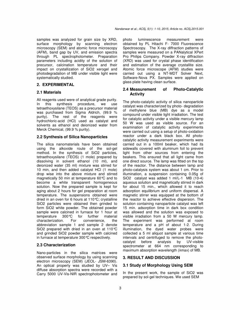
samples was analyzed for grain size by XRD, surface morphology by scanning electron microscopy (SEM) and atomic force microscopy (AFM), band gap by UV, and emission spectra through PL spectrophotometer. Preparation parameters including acidity of the solution of precursor, calcination temperature and their impact on crystallization of SiO2 xerogel and photodegradation of MB under visible light were systematically studied.
2. EXPERIMENTAL
2.1 Materials
All reagents used were of analytical grade purity. In this synthesis procedure, we use tetraethoxysilane (TEOS) as a precursor material was purchased from Sigma Aldrich, (99.9 % purity). The rest of the reagents were hydrochloric-acid (HCl) used as catalyst and solvents as ethanol and deionized water from Merck Chemical, (99.9 % purity).
2.2 Synthesis of Silica Nanoparticles
The silica nanomaterials have been obtained using the alkoxide route of the sol-gel method. In the synthesis of SiO2 particles, tetraethoxysilane (TEOS) (1 mole) prepared by dissolving in solvent ethanol (10 ml), and deionized water (35 ml) mixture was stirred for 10 min, and then added catalyst HCl (1 mole) drop wise into the above mixture and stirred magnetically 50 min at temperature 60°C and to become a white transparent homogeneous solution. Now the prepared sample is kept for aging about 2 hours for gel preparation at room temperature. The suspensions obtained were dried in an oven for 6 hours at 110°C; crystalline SiO2 particles were obtained then grinded to form SiO2 white powder. The obtained powder sample were calcined in furnace for 1 hour at temperature 300°C for further material characterization. For convenience, the abbreviation sample 1 and sample 2 denote SiO2 prepared with dried in an oven at 110°C and grinded SiO2 powder sample with calcined in furnace at temperature 300°C respectively.
2.3 Characterization
Nano-particles in the silica matrices were observed surface morphology by using scanning electron microscopy (SEM) (JEOL- JSM-6390). An optical property was studied by UV– Vis diffuse absorption spectra were recorded with a Carry 5000 UV-Vis-NIR spectrophotometer and
photo luminescence measurement were obtained by PL Hitachi F- 7000 Fluorescence Spectroscopy. The X-ray diffraction patterns of samples were measured on a PANalytical XPert Pro Philips Company. Powder X-ray diffraction (XRD) was used for crystal phase identification and estimation of the average crystallite size. Atomic force microscope (AFM) studies were carried out using a NT-MDT Solver Next, Software-Nova PX. Samples were applied on glass plate having clean surface.
2.4 Measurement of Photo-Catalytic Activity
The photo-catalytic activity of silica nanoparticle catalyst was characterized by photo- degradation of methylene blue (MB) dye as a model compound under visible light irradiation. The test for catalytic activity under a visible mercury lamp 50 W was used as visible source. For an examination of catalytic activity experiments were carried out using a setup of photo-oxidation reactor under a dark black box. All photo-catalytic activity measurement experiments were carried out in a 100ml beaker, which had its sidewalls covered with aluminum foil to prevent light from other sources from entering the beakers. This ensured that all light came from one direct source. The lamp was fitted on the top of the reactor. The distance between lamp and photo-catalysis system was about 1 cm. Prior to illumination, a suspension containing 0.05g of SiO2 catalyst was added 1 ml/L-1 MB (10-4) aqueous solution and magnetically stirred in dark for about 15 min., which allowed it to reach adsorption equilibrium and uniform dispersal. A magnetic stirrer was equipped at the bottom of the reactor to achieve effective dispersion. The solution containing nanoparticle catalyst was left 15 min. adsorption time in dark box condition was allowed and the solution was exposed to visible irradiation from a 50 W mercury lamp. The experiment was performed at room temperature and a pH of about 1-2. During illumination, the dyed water probes were collected a 5 ml aliquot sample at various time intervals and centrifuged to remove the photo-catalyst before analysis by UV-visible spectrometer at 664 nm corresponding to maximum absorption wavelength (λmax) of MB.
3. RESULT AND DISCUSSION
3.1 Study of Morphology Using SEM
In the present work, the sample of SiO2 was prepared by sol-gel techniques. We used SEM

to study the morphology of the sample prepared by using catalyst. It was found that the factors that affected the overall sol satiability include considerable emphasis of hydrochloric-acid and TEOS, and the steps mentioned for sol preparation. On sol stability on deleting the reaction that modifying HCl was an immediate precipitation and inhomogenety. A strong initial exothermic reaction and fast gelatine resulted from prereacting the TEOS with the hydrochloricFig. 1 shows the SEM micrographs ofparticles prepared at 110ºC dried temperature and calcinations temperature. As shown in Fig.
Fig. 1. SEM images of SiO2 samples: (a) dried at 110 3.2 Study of XRD of Sio2 Structures X-Ray Diffraction (XRD) analysis was carried out on powder form and typical diffraction patterns are shown in Fig. 2. From these graphs we can find the size of the sub-micrometer particles or crystallites by using Debye-Scherer equation as illustrated below: D = K λ/ β cosθ where, D is the average crystallite size, K is a constant depend on the crystallite shape (0.9), is the X-ray wavelength (in this case, Å for Cu-Kα radiation), β is the full width at half maxima (FWHM), and θ is the Bragg’s angle. Using the above equation we can determine the size of the particles. On comparing our obtained XRD spectrum from the JCPDS Card #850335 for SiO2, we can assure that the material formed is SiO2 (Quartz) nanoparticles, as the peaks reveal the formation of particles havidimensions in nm range and the reflection from (100), (110), (102), (111), (200) and (201)
Nandanwar et al.; ACSj, 5(1): 1-10, 2015; Article no.
4
to study the morphology of the sample prepared
It was found that the factors that affected the overall sol satiability include considerable
acid and TEOS, and the steps mentioned for sol preparation. On sol stability on deleting the reaction that modifying
precipitation and inhomogenety. A strong initial exothermic reaction and fast gelatine resulted from pre-reacting the TEOS with the hydrochloric-acid. Fig. 1 shows the SEM micrographs of SiO2
C dried temperature shown in Fig.
1(a), the as-prepared powder consists of spherical particles with poor agglomeration, and the aggregation takes place during the particle growth process at higher temperatures (Fig. 1b). Silica samples reveal that the dried sa110oC temperature, the SiO2 particles agglomerate and the boundaries diffuse to form discrete structures. When treated at proper temperature (calcined at 300oC temperature) conditions, the material could diffuse to form a bulk having well-defined boundaries. It is observed that, silica sample calcined at the 300oC having the spherical morphology and in regular in shape.
SEM images of SiO2 samples: (a) dried at 110ºC, (b) calcined at 300
of Sio2 Structures
Ray Diffraction (XRD) analysis was carried out on powder form and typical diffraction patterns are shown in Fig. 2. From these graphs we can
micrometer particles or Scherer equation as
(1)
where, D is the average crystallite size, K is a constant depend on the crystallite shape (0.9), λ
ray wavelength (in this case, λ = 1.5418 is the full width at half is the Bragg’s angle.
ove equation we can determine the size of the particles. On comparing our obtained XRD spectrum from the JCPDS Card #850335 for SiO2, we can assure that the material formed is SiO2 (Quartz) nanoparticles, as the peaks reveal the formation of particles having dimensions in nm range and the reflection from (100), (110), (102), (111), (200) and (201)
planes, at 2θ values 20.86139.470°, 40.296°, 42.457°, and 45.800the sample synthesized via sol-gel route. From the XRD pattern, it is clear that the material formed is having hexagonal crystal structure and is primitive lattice with lattice parameters a=b=4.913 Å and c=5.405 Å. Figs. 2(a) and (b) is instances of the Xdiffraction patterns taken from sol residues dried at 110°C and calcinated at 300pattern of the sample dried at 110characteristic of amorphous in nature; it shows that peptization can accelerate the crystallization and having crystallite size 70-80 nm. The XRD peaks become sharper as the calcinations300°C temperature and its crystallite size increases to 50-55 nm. As a result it can be concluded that the increase in heat treatment temperature to 300°C made a gradual crystallization of the material. The crystallite of powder samples obtained from solultrafine in dimensions, which means that the bonding between Si and O is spontaneous, the
; Article no. ACSj.2015.001
prepared powder consists of spherical particles with poor agglomeration, and the aggregation takes place during the particle growth process at higher temperatures (Fig. 1b).
Silica samples reveal that the dried sample at 110oC temperature, the SiO2 particles agglomerate and the boundaries diffuse to form discrete structures. When treated at proper temperature (calcined at 300oC temperature) conditions, the material could diffuse to form a
oundaries. It is observed that, silica sample calcined at the 300oC having the spherical morphology and in
C, (b) calcined at 300ºC
values 20.861°, 36.550°, , and 45.800° for
gel route. From s clear that the material
formed is having hexagonal crystal structure and is primitive lattice with lattice parameters
Figs. 2(a) and (b) is instances of the X-ray diffraction patterns taken from sol residues dried
calcinated at 300°C. The XRD pattern of the sample dried at 110°C is characteristic of amorphous in nature; it shows that peptization can accelerate the crystallization
80 nm. The XRD peaks become sharper as the calcinations at
C temperature and its crystallite size 55 nm. As a result it can be
concluded that the increase in heat treatment C made a gradual
crystallization of the material. The crystallite of ol-gel method is
ultrafine in dimensions, which means that the bonding between Si and O is spontaneous, the

Nandanwar et al.; ACSj, 5(1): 1-10, 2015; Article no. ACSj.2015.001
5
crystalline particles having random structure. However, with calcination temperature at 300°C there is an increase in the diffraction peaks intensity indicating an improvement in the crystallinity of the material. But in the presence of HCl enhances the acidic reaction in the solution [10]. This confirms the amorphous structure of the material which can be used for catalytic purposes and adsorption processes [18].
Fig. 2. XRD analysis of SiO2 samples: (a) dried at 110ºC, (b) calcined at 300ºC
3.3 Study of UV-Visible Spectroscopy of Sio2 Structures
Fig. 3 illustrates the UV-Visible absorption spectra of SiO2 synthesized via sol-gel route dried at 110°C and calcined at 300°C. The band gap energies (Eg) of samples were determined by the equation (2).
Eg = h c/λ (2)
Where, Eg is the band gap (eV), h is the Planck’s constant (4.135 x 10⁻⁶ eV), c is the velocity of light (3 x 10⁸ ms-1), and λ (nm) is the
wavelength of the absorption edge in the spectrum. The absorption edges around 350 nm were observed for the dried at 110°C (sample 1) and around 351 nm for calcinations at 300°C (sample 2). Compared SiO2 with dried temperature, the band gap was changed from 3.54429 eV to 3.53419 eV of calcination temperature. This change demonstrates a strategy for shaping photo-catalysis by an atomic-level doping for nano-catalyst. In addition, the absorption spectra of the sample 2 also showed a stronger absorption than sample 1 under UV irradiation, indicating that sample 2 could be a promising approach for increasing the catalytic activity. UV-visabsorbance spectra show wide band gap value of the prepared sample which makes it suitable for its application in electronic industries, substrates of computer chips and solar cells [19,20]. UV-visible spectroscopy results showing synthesized materials absorbance in the UV region and very low absorbance is shown in the visible region. There are some other peaks which may be due to impurities or presence of bi-products formed during synthesis.
3.4 PL Spectroscopy Study of Sio2 Structures Fig. 4 shows the PL emission spectrum of SiO2 nanoparticles. PL is done for justifying UV-visible results, to observe whether material is excited or not at wavelength showing maximum absorbance by UV. By this emission of full spectrum (200 nm to 900 nm) excited by particular wavelength gives some peaks of maximum wavelength. The bands are present in the UV regions and are attributed due to the presence of various types of defects in the as-synthesized SiO2. The probable defects can be interstitial Si or O2 (Sii or O2i), silica vacancy (Vsi) or the oxygen antisite (Osi). Luminescence features also depend on other crucial factor which includes solvent, atmosphere, temperature, etc. and is attributed to different processing conditions during synthesis process. Fig. 4 (a) shows that, a wide emission band was observed in the silica sample which is dried at 110°C. The result shows that a wide excitation in the ultraviolet range [21] similar to Nile Blue [22]. In Fig. 4 (b) the PL intensity was slightly reduced and the shape of the PL spectrum obviously split into sharp peaks, shows the formation of resolved manifold lines implies that positive ions were located on the well-defined lattices site in the SiO2 structures. A PL

Nandanwar et al.; ACSj, 5(1): 1-10, 2015; Article no. ACSj.2015.001
6
spectrum shows a slightly sharp emission band with a maximum intensity at 381.8 nm and some side peaks at 435.2 nm and 554.5 nm. 3.5 Atomic Force Microscopy Atomic force microscopy (AFM) was employed to characterize the surface morphology of the prepared samples. For AFM characterization the powder samples were dissolved in acetone and then sonicated using for 30 minutes. The mixture was then spincoated with Holmarc’s Spin Coater, Model no: HO-TH-05 at 6000rpm on thin glass substrate. This film was inserted under AFM tip for taking images. In Fig. 5 (a) and (b) the 2D topographic image shows that the SiO2 sample with dried temperature exhibits a flat and ill-defined surface, where as SiO2 sample with calcined temperature exhibits a rougher but still moderately flat texture and well-structured granular nano-surface with clear interstices between the particles/aggregates. The results shows that, the silica particle calcined at 300°C makes it possible to obtain the material with more highly developed surface. It creates a
higher degree of porosity, which should have a beneficial influence on the photo degradation of the materials dosed with a template provided that the pores are accessible for an organic pollutant. 3.6 Photo Degradation In order to study the photo degradation activity of the both prepared samples, we have used them to photodecomposition of methylene (MB) was investigated in aqueous heterogeneous suspensions, respectively. The maximal absorption of MB solutions is at 332 nm and 664 nm under our experimental conditions. Fig. 6 shows the normalized graph of photo-degradation performances of the SiO2 samples. Before irradiation of the photo- catalyst with visible light, an hour was allowed in order to account for any changes in concentration due to adsorption of organics on the photo-catalyst surface. The results show that the adsorption of MB dye without catalyst gets saturated after 60 min. lightirradiations; the concentration of MB does not increase any more with prolonging of light irradiation time.
Fig. 3. UV-Vis spectra of SiO2 samples: (a) dried at 110°C, (b) calcined at 300°C

Nandanwar et al.; ACSj, 5(1): 1-10, 2015; Article no. ACSj.2015.001
7
Fig. 4. PL spectra of SiO2 samples: (a) dried at 110ºC, (b) calcined at 300°C
In the photo-degradation of methylene blue by SiO2 at dried sample and calcined sample under visible light irradiation. Silica sample calcined at 300°C show increase in crystallite size, due to their large surface area the photo-degradation activity enhanced. The SiO2 sample dried at 110°C temperature the photo-
degradation of MB preceded at a much slower rate. In this case, sample 2 has higher degradation than sample 1. There are a difference in the performance of the photo-catalyst of SiO2 sample 1 and 2 under visible irradiation light source (indicated in Fig. 6).

Nandanwar et al.; ACSj, 5(1): 1-10, 2015; Article no. ACSj.2015.001
8
The photocatalysis activity are responsible for the two factors include surface phenomenon that is being very sensitive to the amount of surface OH groups which may act as the principal reactive oxidant in the photoreactions and increase in crystallinity [23]. The photocatalytic activity goes on increasing continued irradiation for both samples and no saturation or increase in photo degradation activity is observed indicating that these catalysts are stable under experimental conditions employed in this study. As one can see, the catalyst sample 2 absorbs the solute better than the catalyst of sample 1 under in visible irradiation, and there was marked improvement in the rate of photo-catalytic breakdown of MB.
Fig. 5. The AFM images (2D surface analysis)
of SiO2 samples: (a) dried at 110°C, (b) calcined at 300°C
Fig. 6 shows that the catalyst sample 2 of silica particle had faster rate of MB breakdown than sample 1 under visible light source. The visible region is a much wider spectrum, so this increases the chances of light of the correct
wavelength hitting the surface of the photocatalyst to active it. After few minute of exposure to visible light, about 50% of the methylene blue had been broken down. Compared with the photo degradation performance of two samples, the appropriate size of SiO2 obtained by appropriate annealing temperature will bring out the best result.
Fig. 6. Photo-degradation performances of
SiO2 samples: (a) without catalyst, (b) dried at 110°C and (c) calcined at 300°C
under visible light irradiation 4. CONCLUSION The ultrafine SiO2 particles were synthesized by sol-gel method and used as photocatalysts. The calcination temperature has played significant role in the dimensional as well as optical properties of material. From the XRD pattern, it is clear that the material formed is having hexagonal crystal structure. As the calcination temperature at 300°C, the crystallite size increases. The SEM images reveal that material formed has a discrete shape, varying in shape and structure. Both XRD and SEM studies confirms that material SiO2 so formed is amorphous in nature, which could be converted into crystalline material under appropriate temperature and suitable chemical environment. Powder morphology in these criteria is almost spherical, which is due to acidic condition that prevents agglomeration. The well-structured morphology displaced in the AFM image during the thermal treatment exhibit well-structured granular nano-surface. UV and PL spectra both reveal that both SiO2 samples show absorbance spectra in UV region.

Nandanwar et al.; ACSj, 5(1): 1-10, 2015; Article no. ACSj.2015.001
9
The silica samples having wide band gap showing absorbance in the ultra violet region which makes it suitable for its application in electronic industries. PL shows wide emission band in the silica sample which is dried at 110°C. SiO2 calcined at 300°C the PL intensity was reduced and the shape of the emission spectrum obviously split into sharp peaks which shows the well-defined lattices site in the silica structures. Detailed analysis of the results prove that SiO2 powder annealed at 300°C exhibited superior photoactivities over the other catalyst, indicating that the appropriate annealing temperature could improve their photoactivities performance. It can be due to its greater size and crystallinity of the solid sample. The photocatalytic activity of silica calcined sample performed best in photo degradation of MB breakdown than that of dried sample under visible light source. ACKNOWLEDGEMENT Author RN would like to thank the research scholars of Optical Nanomaterials Lab, Department of Physics, MANIT, Bhopal (M.P) for helpful discussions.
COMPETING INTERESTS Authors have declared that no competing interests exist. REFERENCES 1. Gleiter H. Nanostructured materials: state
of the art and perspectives, Journal of Nanostructured Materials. 1995;6:3-14.
2. Kung HH. Transition Metal Oxides: Surface Chemistry and Catalysis, Elsevier: Amsterdam; 1989.
3. Suzuki E. High-resolution scanning electron microscopy of immune gold-labeled cells by the use of thin plasma coating of osmium, Journal of Microscopy. 2002;208(3):153–157.
4. Lenza RFS, Vasconcelos WL. Preparation of Silica by Sol-Gel Method Using Formamide, Journal of Material Research. 2001;4:189-194,.
5. Henrich VE, Cox PA. The Surface Chemistry of Metal Oxides; Cambridge University Press: Cambridge, UK; 1994.
6. Valden M, Lai X, Goodman DW. Onset of Catalytic Activity of Gold Clusters on Titania with the Appearance of Nonmetallic Properties, Journal of
Science. 1998;281:1647. 7. Millot N, Aymes D, Bernard F, Niepc JC,
Traverse A, Bouree F, Cheng BL, Perriat PJ. Hydrothermal synthesis of nanostructured inorganic powders by a continuous process under supercritical conditions, Journal of Phys. Chem. 2003;B107:5740.
8. Brinker CJ, Scherer GW, Sol-Gel Science: The Physics and Chemistry of Sol- Gel Processing, Academic Press, inc. San Diego, CA. 1990;907.
9. Rahman I, Padavettan V. Synthesis of Silica Nanoparticles by Sol-Gel: Size- Dependent Properties, Surface Modification and Applications in Silica-Polymer Nanocomposite; A Review, Journal of Nanomaterials. 2012;10:1155.
10. Sang-Wook U, Seung-Jae L, Sang-Hoon L, Sung-Churl C, Control of the size and morphology of nano-size silica particles using a sodium silicate solution, Journal of Ceramic Processing Research. 2009;10(4):553-558.
11. Yelil Arasi A, Hema M, Tamilselvi P, Anbarasan R. Synthesis and characterization of SiO2 nanoparticles by sol-gel process, Indian Journal of Science. 2012;1(1):6-10.
12. Kumari S, Sahare PD, Gupta M, Sensitization of mesoporous silica nanoparticles by laser grade dye acriflavin, Journal of Advanced Materials Letters, VBRI Press. 2012:172. DOI: 10.5185 amlett.2012.icnano.
13. Ali M, Mohammad Ali B, Naser M, Characterization and photocatalytic activity of SiO2-TiO2 mixed oxide nanoparticles prepared by sol-gel method, Indian Journal of Chemistry. 2010;49:1593-1600.
14. Elias S, Panagiotis L, Chritophoros K. Dye sensitized photo-electrochemical cell using a nanocomposite SiO2/Poly (Ethylene Glycol) thin film as electrolyte support characterization by time-resolved luminescence and conductivit measurements, Journal of Phys. Chem. 2001;105:3486-3492.
15. Bujak L, N. Czechowski, D. Piatkowski, R. Litvin, S. Mackowski, THP Brotosudarmo, Cogdell RJ, Pichler S, Heiss W. Fluorescence enhancement of lightharvesting complex2 from purple bacteria coupled to spherical gold nanoparticles, Journal of Appl. Phys. Lett. 2011;99:173701.

Nandanwar et al.; ACSj, 5(1): 1-10, 2015; Article no. ACSj.2015.001
10
16. Bartosz K, Magdalena GR, Dawid P, Piotr N, Bartłomiej J, Eckhard H, Sebastian M. Silica nanoparticles as a tool for fluorescence collection efficiency enhancement, Journal of Nanoscale Research Letters. 2013;8:146.
17. Kim SW, Kang M, Choung SJ. Preparation of a TiO2 Film Using a TEOS Binder and its Application to the Photodegradation of Benzene, Journal of Ind. Eng. Chem. 2005;11(3)416-424.
18. Waseem M, Mustafa S, Naeem A, Shah KH, Irfan Shah, Ihsan-ul-Haque. Synthesis and characterization of silica by sol-gel method, Journal of Pak Mater Society. 2009;3(1).
19. Dahl A, Gharibi A, Swietlicki E, Gudmundsson A, Bohgard M, Ljungman A. Traffic-generated emission of ultrafine particles from pavement-tire interface, Journal of Atmospheric Environment. 2006;40:1314-1323.
20. Gustafsson M, Blomqvist G, Gudmundsson A, Dahl A, Swietlicki E, Lindbom J. Properties and toxicological effects of particles from the interaction between tyres, road pavement and winter traction material, Journal of Science of the Total Environment. 2008;393:226-240.
21. Vaccaro L, Vaccaro G, Agnello S, Buscarino G, Cannas M. Wide range excitation of visible luminescence in nano silica, Journal of Solid State Communications. 2010;50:2278-2280.
22. Fozia Z. Haque, Vazid Ali, M.Husain, Synthesis and Spectroscopic Characterization of Nile Blue Dye Doped Silica Gel Rods, OPTIK. 2013;124:4287-4291.
23. Hoffmann MR, Martin ST, Choi W, Bahnemann DW. Environmental applications of semiconductor photocatalysis, Journal of Chemical Reviews. 1995;95(1)69-96.
_________________________________________________________________________________ © 2015 Nandanwar et al.; This is an Open Access article distributed under the terms of the Creative Commons Attribution License (http://creativecommons.org/licenses/by/4.0), which permits unrestricted use, distribution, and reproduction in any medium, provided the original work is properly cited.
Peer-review history: The peer review history for this paper can be accessed here:
http://www.sciencedomain.org/review-history.php?iid=649&id=16 &aid=5955