switching from a unicellular to multicellular …switching from a unicellular to multicellular...
TRANSCRIPT

Switching from a Unicellular to Multicellular Organization in anAspergillus niger Hypha
Robert-Jan Bleichrodt,a,c,d Marc Hulsman,b,c,e Han A. B. Wösten,a,c Marcel J. T. Reindersb,c
Microbiology, Utrecht University, Utrecht, The Netherlandsa; Delft Bioinformatics Lab, Intelligent Systems, Department of Electrical Engineering, Mathematics andComputer Science, Delft University of Technology, Delft, The Netherlandsb; Kluyver Centre for Genomics of Industrial Fermentation, Delft, The Netherlandsc; ManchesterFungal Infection Group, Institute of Inflammation and Repair, Faculty of Medical and Human Sciences, University of Manchester, Manchester, United Kingdomd;Department of Clinical Genetics, VU University Medical Center, Amsterdam, The Netherlandse
R.-J.B. and M.H. contributed equally to this article.
ABSTRACT Pores in fungal septa enable cytoplasmic streaming between hyphae and their compartments. Consequently, the my-celium can be considered unicellular. However, we show here that Woronin bodies close ~50% of the three most apical septa ofgrowing hyphae of Aspergillus niger. The incidence of closure of the 9th and 10th septa was even >94%. Intercompartmentalstreaming of photoactivatable green fluorescent protein (PA-GFP) was not observed when the septa were closed, but open septaacted as a barrier, reducing the mobility rate of PA-GFP ~500 times. This mobility rate decreased with increasing septal age andunder stress conditions, likely reflecting a regulatory mechanism affecting septal pore diameter. Modeling revealed that suchregulation offers effective control of compound concentration between compartments. Modeling also showed that the incidenceof septal closure in A. niger had an even stronger impact on cytoplasmic continuity. Cytoplasm of hyphal compartments wasshown not to be in physical contact when separated by more than 4 septa. Together, data show that apical compartments ofgrowing hyphae behave unicellularly, while older compartments have a multicellular organization.
IMPORTANCE The hyphae of higher fungi are compartmentalized by porous septa that enable cytosolic streaming. Therefore, itis believed that the mycelium shares cytoplasm. However, it is shown here that the septa of Aspergillus niger are always closed inthe oldest part of the hyphae, and therefore, these compartments are physically isolated from each other. In contrast, only part ofthe septa is closed in the youngest part of the hyphae. Still, compartments in this hyphal part are physically isolated when sepa-rated by more than 4 septa. Even open septa act as a barrier for cytoplasmic mixing. The mobility rate through such septa re-duces with increasing septal age and under stress conditions. Modeling shows that the septal pore width is set such that its regu-lation offers maximal control of compound concentration levels within the compartments. Together, we show for the first timethat Aspergillus hyphae switch from a unicellular to multicellular organization.
Received 20 January 2015 Accepted 28 January 2015 Published 3 March 2015
Citation Bleichrodt R, Hulsman M, Wösten HAB, Reinders MJT. 2015. Switching from a unicellular to multicellular organization in an Aspergillus niger hypha. mBio 6(2):e00111-15. doi:10.1128/mBio.00111-15.
Invited Editor Reinhard Fischer, Karlsruhe Institute of Technology (KIT), Institute for Applied Biosciences Editor B. Gillian Turgeon, Cornell University
Copyright © 2015 Bleichrodt et al. This is an open-access article distributed under the terms of the Creative Commons Attribution-Noncommercial-ShareAlike 3.0 Unportedlicense, which permits unrestricted noncommercial use, distribution, and reproduction in any medium, provided the original author and source are credited.
Address correspondence to Han A. B. Wösten, [email protected], or Marcel J. T. Reinders, [email protected].
Animals have a multicellular organization. Intercellular cyto-solic continuity is restricted by gap junctions that allow
streaming of small molecules (�1,000 Da) such as ions, secondmessengers, and small metabolites (1). Plasmodesmata mediateintercellular cytosolic continuity in plants. They enable transloca-tion of water, metabolites, proteins, RNA, and viral genomes (2).Plants have been proposed to have a supracellular organizationbecause of their intercellular transport properties (3). Notably,intercellular transport in plants is controlled by changing the porediameter of plasmodesmata (4–6).
The mycelium of the lower fungi (like the Zygomycota) con-sists of hyphae that are nonseptate. As a consequence, the myce-lium can be considered to be unicellular. The hyphae of higherfungi (those belonging to the Ascomycota and Basidiomycotaphyla) are compartmentalized by septa. These septa have a centralpore of 50 to 500 nm that allows translocation of water, metabo-lites, proteins, RNA, and even organelles (7–10). Septa are closed
upon hyphal damage to prevent excessive loss of cytoplasm (7,11–16). They can also be closed in intact growing hyphae (17–19).The septal pore cap mediates septal plugging in the basidiomyceteSchizophyllum commune. It is a specialized form of the endoplas-mic reticulum (ER) that has been proposed to release pluggingmaterial into the septal pore (20). In ascomycetes, Woronin bod-ies plug septal pores. The lumen of these peroxisome-derived or-ganelles (13, 21–23) is filled with hexagonal rods of the HexAprotein (13, 24). Deletion of the gene encoding HexA in Neuro-spora crassa, Magnaporthe grisea, and Aspergillus oryzae results inthe absence of Woronin bodies (13–16). As a consequence, hyphalinjury results in severe cytoplasmic bleeding (13–16), while heter-ogeneity in cytosolic composition between neighboring hyphae ofA. oryzae is severely reduced (19).
Hyphae at the periphery of Aspergillus colonies are heteroge-neous with respect to cytosolic composition and transcriptionaland translational activity (25–32). For instance, only part of the
RESEARCH ARTICLE crossmark
March/April 2015 Volume 6 Issue 2 e00111-15 ® mbio.asm.org 1
on April 5, 2020 by guest
http://mbio.asm
.org/D
ownloaded from
hyphae highly expresses genes that are involved in substrate deg-radation (27, 28, 32). Heterogeneity is maintained by septal clo-sure that prevents intercompartmental cytosolic mixing withinand between hyphae (19). It was shown that 40% of the apicalsepta of hyphae at the periphery of A. oryzae colonies are closed byWoronin bodies. Closure of the septal pores was reversible andnot related to environmental conditions or to the plugging state ofneighboring septa. This suggests that septal plugging in A. oryzaeby Woronin bodies is a stochastic process.
Here, we studied the impact of both open and closed septa oncytosolic continuity in Aspergillus niger. The number of septa perhyphal unit, incidence and dynamics of septal closure, and rate ofstreaming of molecules within the hyphal compartments andthrough the septal pores was determined and used as input for amodel of cytosolic continuity. Our hypothesis was that cytosoliccontinuity is limited by septal plugging and by the diameter of theopen septal pore. It is shown that the mobility of moleculesthrough open septal pores is reduced over time and under stressconditions. Moreover, the incidence of plugging increases as thesepta become older. Modeling reveals that in this way young com-partments of A. niger hyphae switch, over time, from a unicellularto a multicellular organization.
RESULTSSeptal plugging in A. niger increases in older compartments.The wild-type Aspergillus niger strain N402 was grown on CDmedium (19) plus Met for 2 days. The first (apical) compartmentof hyphae at the colony periphery or one of the following 9 com-partments was dissected using a UV laser. Streaming of cytoplasmfrom the subapical, neighboring compartment to the damagedcompartment was used to score whether septa were open or closed(19) (see Text S1 in the supplemental material). It was found that57%, 57%, and 52% of the first, second, and third septa were open,respectively (95% confidence interval based on 40 scored septaeach, 42% to 72%, 42% to 72%, and 37% to 67%, respectively). Incontrast, only 5% (1% to 15%), 8% (2% to 19%), 10% (3% to22%), 5% (1% to 15%), and 13% (5% to 25%) of the 4th to 8thsepta were open, respectively, while the 9th and 10th septum werealmost always closed (0% to 6% open septa). Apical septa were
almost always open in the �hexA strain that has no Woronin bod-ies (33), showing that Woronin bodies are involved in plugging ofthe apical compartment. Notably, 15% (7% to 28%) of the 9th and10th septa were still found to be closed in this strain, indicating theexistence of another plugging system(s) in these subapical septa.
To determine the dynamics of septal plugging, A. niger strainRB#14.5 expressing photoactivatable green fluorescent protein(PA-GFP) from the gpdA promoter was grown for 2 days on MMagar with 25 mM maltose (25). PA-GFP was activated by 405-nmlaser light within the entire second compartment of hyphae at theperiphery of the colony. Hyphae that did not show streaming ofPA-GFP to neighboring compartments were selected. Time-lapsemovies showed that in 20 out of 52 compartments, PA-GFPstarted to stream to neighboring compartments within a 16-h pe-riod. This showed that plugging of septa in these 20 compartmentswas released. On average, septa reopened after 4.2 h � 5.0 h. Thus,septal plugging is reversible in A. niger.
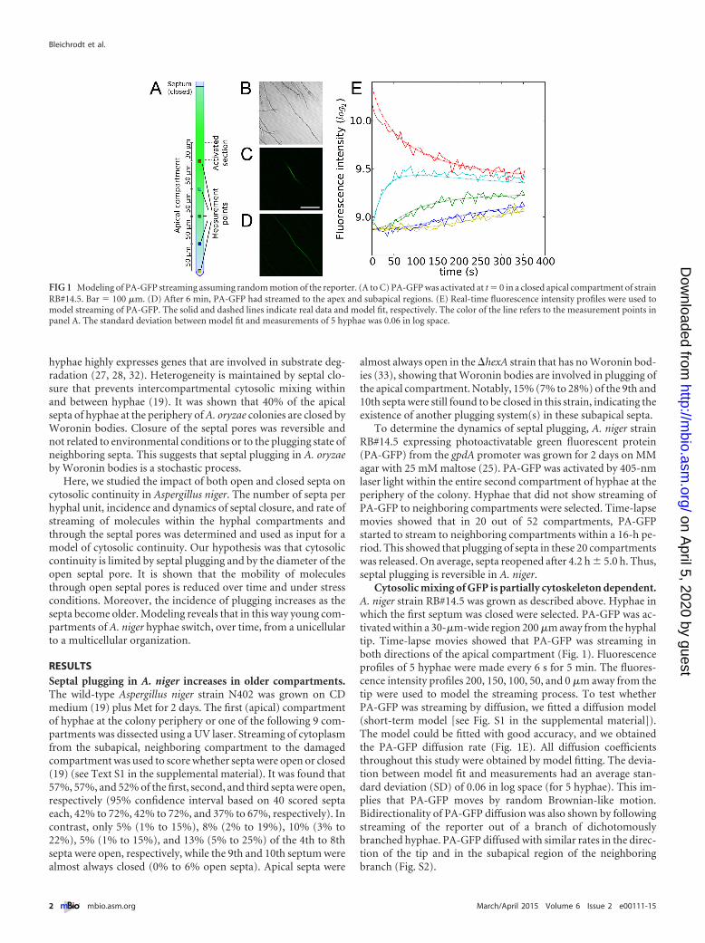
Cytosolic mixing of GFP is partially cytoskeleton dependent.A. niger strain RB#14.5 was grown as described above. Hyphae inwhich the first septum was closed were selected. PA-GFP was ac-tivated within a 30-�m-wide region 200 �m away from the hyphaltip. Time-lapse movies showed that PA-GFP was streaming inboth directions of the apical compartment (Fig. 1). Fluorescenceprofiles of 5 hyphae were made every 6 s for 5 min. The fluores-cence intensity profiles 200, 150, 100, 50, and 0 �m away from thetip were used to model the streaming process. To test whetherPA-GFP was streaming by diffusion, we fitted a diffusion model(short-term model [see Fig. S1 in the supplemental material]).The model could be fitted with good accuracy, and we obtainedthe PA-GFP diffusion rate (Fig. 1E). All diffusion coefficientsthroughout this study were obtained by model fitting. The devia-tion between model fit and measurements had an average stan-dard deviation (SD) of 0.06 in log space (for 5 hyphae). This im-plies that PA-GFP moves by random Brownian-like motion.Bidirectionality of PA-GFP diffusion was also shown by followingstreaming of the reporter out of a branch of dichotomouslybranched hyphae. PA-GFP diffused with similar rates in the direc-tion of the tip and in the subapical region of the neighboringbranch (Fig. S2).
FIG 1 Modeling of PA-GFP streaming assuming random motion of the reporter. (A to C) PA-GFP was activated at t � 0 in a closed apical compartment of strainRB#14.5. Bar � 100 �m. (D) After 6 min, PA-GFP had streamed to the apex and subapical regions. (E) Real-time fluorescence intensity profiles were used tomodel streaming of PA-GFP. The solid and dashed lines indicate real data and model fit, respectively. The color of the line refers to the measurement points inpanel A. The standard deviation between model fit and measurements of 5 hyphae was 0.06 in log space.
Bleichrodt et al.
2 ® mbio.asm.org March/April 2015 Volume 6 Issue 2 e00111-15
on April 5, 2020 by guest
http://mbio.asm
.org/D
ownloaded from

The diffusion rate of PA-GFP cannot be accurately expressed asa speed, as the fluorescence data are threshold dependent. How-ever, an approximation based on Fig. 2F revealed mobility ratesexceeding 5 �m s�1. In contrast, bulk flow of cytosolic particles (ofunknown identity) moved with a speed of 0.026 �m s�1 � 0.012�m s�1 toward the tips of hyphae. This matched the growth speedof the hypha (0.023 � 0.006 �m s�1) and is too slow to impact theshort-term (diffusion) model fit. To assess the role of the cytoskel-eton in cytosolic mixing of PA-GFP, activation of the reporter waseither preceded or not preceded with a 90-min incubation with1% dimethyl sulfoxide (DMSO), 100 �g ml�1 nocodazole (NOC)in 1% DMSO, 80 �g ml�1 cytochalasin A (CA) in 1% DMSO, or acocktail of cytochalasin A and nocodazole (CA-NOC) in 1%DMSO (see Fig. S3A in the supplemental material). NOC and CAcause degradation of the tubulin and actin cytoskeleton, respec-tively. The short-term model was fitted to measured fluorescenceprofiles (Fig. S3B to S3F). Only measurements with a high-qualitymodel fit (SD of �0.09) were considered (23 out of 30 measure-ments). Diffusion coefficients dropped significantly by 22% and28% when the cultures had been incubated with NOC or CA,respectively, but not when incubated with 1% DMSO alone(Fig. S3G). When the cultures had been incubated with a cocktailof NOC and CA, the diffusion coefficients dropped by 24%. Thisshows that cytosolic streaming is facilitated by both the actin andtubulin cytoskeleton but also by other unknown forces.
Next, we determined whether diffusion rates could be affectedby the plugging state of nearby septa. Therefore, the diffusioncoefficients of hyphae with a closed apical septum (Fig. 1) werecompared to those in which the two most apical septa were open(as determined by activating PA-GFP in the second compartment[Fig. 2A]). Indeed, intracellular diffusion coefficients of hyphae
with open septa were found to be significantly higher (increasingon average from 16 �m2 s�1 to 33 �m2 s�1 [P value of 0.013]).
Mobility of PA-GFP through the septal pore. PA-GFP doesnot stream to a neighboring compartment when the pore is closed(19) (Fig. 2D, top septum). Here it was assessed to which extentopen pores affect intercompartmental streaming. To this end,A. niger strain RB#14.5 was grown as described above. PA-GFPwas activated in the second compartment of leading hyphae(Fig. 2A to D), and 2-min time-lapse movies were made. Theshort-term model was extended with a septal diffusion coefficient(see Fig. S1B in the supplemental material). This enabled us tostudy the impact of open pores on cytosolic streaming. Hyphae inwhich the two most apical septa were open were selected. Mea-surement points for fluorescence intensity of PA-GFP were setdirectly before and after the first and second septa and 50 �m fromthe septal pore in the apical compartment (Fig. 2A). A mediandiffusion coefficient of 33.3 �m2 s�1 was found within the hyphae(1 SD, 21.6 to 51.2 �m2 s�1) (Fig. 2F). In contrast, the diffusioncoefficient for the first septum was only 0.07 �m2 s�1 (1 SD, 0.04to 0.11 �m2 s�1), representing almost a 500-fold difference. Thislow septal diffusion coefficient can be explained by the diameter ofthe septal pore. The calculated diameter of septal pores of A. nigeris on average 335 nm (Fig. 2H), which agrees with the pore sizesthat have been experimentally determined in filamentous fungi(7–10).
A simulation was performed to assess the impact of the lowseptal diffusion coefficient on intercompartmental streaming. Inthe simulation, a compound was produced in the 2nd compart-ment at a constant rate for 2 min. A 2-fold difference in concen-tration of the compound was found in this compartment after2 min of production in the presence of septa compared to their
FIG 2 Assessment of the diffusion coefficient of PA-GFP within hyphal compartments and through septal pores. (A to C) PA-GFP was activated in the secondcompartment of a leading hypha of strain RB#14.5. (D) After 2 min, PA-GFP had streamed to the apical compartment via the first septum. The second septumis closed. Arrowheads point to the septa in panels B to D. Bar � 100 �m. (E) Fluorescence intensity in time at the measurement points indicated in panel A. Thedashed lines indicate the model fit based on this data. (F) Diffusion coefficients within the compartment or through the septal pores. (G) Diffusion coefficientsof the septal pores in more detail. (H) Septal pore diameters, determined from hyphal diameter and septal and cytosolic diffusion coefficients. (I) Calculated porediameters of the 1st (red) and 2nd (green) septa under standard conditions (N 1 to N 5) and low-pH conditions (pH2 1 to pH2 5), assuming that the diameteris linearly related to the septal diffusion rate. The uncertainty in these parameter estimates is shown by plotting the distributions for the 95% confidence intervals(F to I).
Cytosolic Continuity in Aspergillus
March/April 2015 Volume 6 Issue 2 e00111-15 ® mbio.asm.org 3
on April 5, 2020 by guest
http://mbio.asm
.org/D
ownloaded from

absence (Fig. 3). This difference in concentration, which did notdepend on the production rate, had disappeared 8 min after pro-duction had stopped. Together, open septa form a barrier for in-tercompartmental streaming of compounds on a time scale ofminutes.
Relatively large differences were observed between septal dif-fusion coefficients (ranging from 0.03 to 0.11 �m2 s�1 [Fig. 2G]).These differences could be explained either by differences in thecytosolic diffusion rate (also affecting the diffusion rate within thepore) or by differences in the septal pore diameter. Only a limitedcorrelation between the cytosolic diffusion parameters and theseptal diffusion parameters was observed (Fig. 2F), suggesting thatthe septal pore diameter plays an important role. The differencesin septal pore diameter (with an average of 335 nm � 62 nm)
exceeded estimation uncertainty (Fig. 2H), indicating that septalpores indeed vary in width. Subsequently, we elaborated how theseptal pore diameter would affect the concentration of a com-pound in the producing compartment assuming that the rate ofdiffusion through the septal pore is linearly dependent on itscross-sectional area (note that diffusion rates will be lower close toseptal pore walls). Simulations revealed that changing the diame-ter of the septal pore has relatively large effects (Fig. 3D). Forinstance, almost no differences in concentration in the 2nd com-partment were observed after 10 min between a simulation withand without septa with a pore of 335 nm assuming a production ofa compound for 2 min in the 2nd compartment (ratio of 1.1 for alog2 ratio of 0 in Fig. 3D). However, reducing the septal porediameter to 25% of its size (from 335 to 84 nm; log2 ratio of �2 in
FIG 3 Simulation of PA-GFP diffusion rate in a hypha. (A) PA-GFP was activated at t � 0 in the region 200 to 230 �m away from the tip. The apical septumis closed. Diffusion coefficients were obtained by fitting experimental data (Fig. 2F). (B) Influence of open septa on concentration of compounds. A compoundis produced for 2 min at a constant rate in the 2nd compartment and is allowed to diffuse. (C) The concentration of the compound can reach levels up to 2-foldhigher in the first compartment than those in the second compartment due to the presence of an open septum compared to a situation without a septum. (D)Simulation of the effect of changing the septal pore diameter. The concentration ratio observed between an activated compartment (with open septa, as in panelB) and an activated region (without septa, as in panel C) after producing a certain compound for 2 min at a constant rate is plotted. Septal pores with widths asobserved in Fig. 2H (on average, 335 nm) were used as the reference (log2 ratio � 0; indicated by the vertical dashed line).
Bleichrodt et al.
4 ® mbio.asm.org March/April 2015 Volume 6 Issue 2 e00111-15
on April 5, 2020 by guest
http://mbio.asm
.org/D
ownloaded from

Fig. 3D) increased this ratio to 4.8. The 50- to 500-nm septal porediameter range reported (7–10) conforms to a log2 range of �2.7to 0.6, which covers the range that offers the largest effect onconcentrations (Fig. 3D, the log2 range of �2.7 to 0.6 on the x axiscorresponds to the steepest sections of the concentration ratiocurves). In comparison, the 5 hyphae (Fig. 2H) that were mea-sured under the same conditions were observed to cover a log2
diameter range of �0.5 to 0.3 (corresponding to diameters of 232to 411 nm).
Septal pore plugging is not affected by low pH (33). Here weassessed whether this environmental condition impacts the diffu-sion rate within the septal pore. The diffusion rates of the first andsecond septa were determined for hyphae grown under standardgrowth conditions (see above) and for hyphae that had been ex-posed to pH 2 for 1.5 h. Under both growth conditions, the diffu-sion rate was higher in the first septum than in the second septum.Data suggest that the diameter of the second pore is reduced 22%(which equals a reduction of 68 nm in diameter) compared to thatof the first septum. Data also showed that the septal pore diffusioncoefficients (�m2 s�1) of the first and second septa were signifi-cantly lower under low-pH conditions (on average, 0.061 versus0.034 and 0.040 versus 0.018, respectively [n � 5] [SD of mean,0.012, 0.002, 0.010, and 0.006, respectively]). These data suggestthat low pH induces the septal pore to narrow by 21% (equals areduction of 64 nm in diameter) (Fig. 2I).
Next, whether diffusion rates were similar through apical septathat had never been closed and septa that had reopened was ex-amined. To this end, PA-GFP was activated in the second hyphalcompartment. Time-lapse movies of 16 h were produced with animage taken every 15 min. Compartments that showed septaopening within this period were selected. The model was fitted toobtain the diffusion rates of PA-GFP. The pore widths of 2 out of3 reopened septa were smaller (143 and 207 nm) (see Fig. S4F inthe supplemental material) than those of septa that had never beenclosed (232 to 411 nm) (Fig. 2H). Notably, the longer the septalpore had been closed, the lower the calculated pore width was(Fig. S4E and S4F). This indicates that septa do not completelyreopen after septal plugging. It was also observed that reopenedsepta could plug again as indicated by the disequilibrium of hypha2 and 3 (Fig. S4C and S4D). This further substantiates the conceptof reversible septal plugging.
Factors that influence hyphal heterogeneity. The long-termsimulation model was used to determine which factors influenceheterogeneity between the apical sections of a dichotomouslybranched hypha at longer time scales (multiple hours) (Fig. 4A).To this end, the short-term model was extended with processesthat affect cytosolic streaming at these time scales (hyphal exten-sion, protein production/degradation, formation of septa, andplugging dynamics; see Fig. S5A in the supplemental material).Parameterization was based on a hyphal growth rate of 137 �13 �m h�1, an apical compartment length of 279 � 114 �m, andan average subapical compartment length of 45 � 19 �m. Diffu-sion coefficients were sampled from the experiment shown inFig. 2F. The compound degradation half-life and septal pluggingrate were varied to determine the extent of their effect. The con-centration of a compound was set at 1.0 in one of the two apicalcompartments at a given distance between the tips of the hyphae.The concentration profile of the compound at the tip of the otherhypha was determined over time through simulation (Fig. 4B).Diffusion effectively mixed the cytoplasm of the two hyphae in the
absence of septa (Fig. 4G). The effectiveness of mixing decreasedonly gradually when the distance between the tips increased due togrowth. Introducing the presence of open septa in the simulationreduced cytosolic mixing efficiency (as indicated by the maximumconcentration reached) by 2- to 3-fold (Fig. 4F). Mixing becameineffective when there were more than 4 septa that switched be-tween the open and closed states (Fig. 4E). As was found for shorttime scales, a reduction in diameter of the septal pore had a sig-nificant impact on diffusion, even when only 1 septum separatedthe apical compartments (Fig. 4H). Reducing the diameter of theseptal pore 2-fold (from 335 to 168 nm) reduced the maximumconcentration reached in the nonactivated apical compartment byapproximately 2.
The average septal plugging switch time [(�open � �closed)/2]and degradation time were both set at 1 h for the experimentsdescribed in the previous sections. Changing these values had lim-ited effect on the concentration peaks at the apical tip (Fig. 4D).For example, changing the septal plugging switch time from 1 to2 h only slightly reduced the average maximum concentrationfrom 0.25 to 0.22. The limited impact of the degradation rate canbe explained by the dilution taking place due to hyphal extension.At a 137 �m h�1 growth rate (see above), concentrations in closedapical compartments already have a half-life of approximately 2 hwithout any additional degradation occurring.
Three scenarios for emergence of heterogeneity between twoneighboring hyphae were tested. In the first scenario, a regulator isswitched on in one hypha at the moment that branching betweenthe two hyphae has occurred. In the second and third scenario, theregulator is either activated or inhibited in one of the hyphae aftera total of 2 septa have separated the two neighboring apices. Pro-duction of the regulator when activated was simulated to takeplace at a rate of the base production rate multiplied by (1.0 �current concentration min�1) using a degradation half-life of 1 hand a base production rate of 0.1. It was found that activation of agene after separation by two septa was the most effective way toobtain the largest concentration differences between two apicalneighboring tips (Fig. 4C). This conclusion also holds for a pro-duction rate that is 10 times slower or a degradation half-life of10 min, although in this case delaying the activation of the regu-lator provides only a small benefit.
The model showed that (reversible) septal plugging has a majorimpact on intercompartmental cytosolic streaming. Therefore, itwas tested to which extent septal plugging prevents cytosolicstreaming in vivo. Transformants RB#200.4, RB#200.6, andRB#200.7 expressing H2B-GFP (H2B stands for histone 2B) (nu-clear localization) and dTomato (freely diffusible) under controlof the inducible glaA promoter were grown on MM agar mediumwith 200 mM xylose (glaA repressing) or 25 mM maltose (glaAinducing). After 5 days, the colonies were transferred to 25 mMmaltose for 6 h. The fluorescence intensity of hyphae at the outerpart of the colony was measured (see Fig. S6M to S6P in the sup-plemental material). All transformants showed heterogeneousfluorescence intensities of H2B-GFP and dTomato after 6-h in-duction (Fig. S6A to S6L and Table S1). This indicates that cyto-solic streaming is limited. After 5-day induction, 2 out of 3 trans-formants still showed heterogeneity. This shows that cytosolicstreaming still has limited effect on reducing heterogeneity onlonger time scales. To test the extent of cytosolic streaming, Spear-man correlation coefficients were calculated between nuclearH2B-GFP and cytosolic dTomato fluorescence intensity within
Cytosolic Continuity in Aspergillus
March/April 2015 Volume 6 Issue 2 e00111-15 ® mbio.asm.org 5
on April 5, 2020 by guest
http://mbio.asm
.org/D
ownloaded from

individual hyphae. The correlation was 0.71 � 0.12 in the case ofthe 6-h induction, while it was significantly lower (0.46 � 0.08) inthe case of 5-day induction. This shows that reversible septal plug-ging maintains heterogeneity, but also allows cytosolic streamingbetween neighboring hyphae on long time scales (5 days) whenthey are separated by less than 4 septa.
DISCUSSION
A fungal mycelium consists of hyphae that grow at their tips andthat branch subapically. Zones within a mycelium show heteroge-neity in gene expression, growth, and secretion (25, 26, 30, 31,34–39). For instance, 9% of the active genes in colonies of A. nigerare expressed in only one out of five concentric zones, while 25%
of the active genes show at least a 2-fold difference in expressionbetween the outer and innermost zone of the colony (35). Evenhyphae within a zone of a mycelium are heterogeneous with re-spect to cytosolic composition (19, 25–32, 40). For instance, onlypart of the hyphae at the periphery of a mycelium of A. niger highlyexpresses the glucoamylase gene glaA (27). Hyphal heterogeneityin the mycelium is in conflict with the concept of cytosolic conti-nuity that would result from cytosolic mixing due to intercom-partmental streaming within and between hyphae enabled by thepresence of septal pores. However, we recently showed that part ofthe apical septa of growing vegetative hyphae of A. oryzae is closed(19). Plugging of apical septa was no longer observed in a �hexAstrain that does not contain Woronin bodies. As a consequence,
FIG 4 Long-term simulation of compound concentration in a branched hypha. (A) The compound concentration was measured in hyphal tips and branchpoints. (B) The compound concentration in apical compartment 1 is set at 1 at t � 0. In hyphal tip 2, the maximum concentration level reached by diffusion isnot maintained, but decreases quickly due to growth, degradation, and septum formation. (C) Scenarios of the emergence of heterogeneity. A switch in theregulatory program at the branch (one branch turns a gene on) compared to a delayed switch, either turning the gene off or on in one branch. The differences incompound concentrations between the tips is shown. (D) Effects of half-life and septal plugging switch time on the maximum concentration reached in apicaltip 2. (E) Cytosolic continuity between branches at different growth stages. Production in hyphal tip 1 after a certain branch length/number of septa have beenformed. (F and G) Same experiment as in panel E but with open septa or without septa, respectively. (H) Effect of septal pore diameter on cytosolic continuity.Apical compartment 1 is activated. The maximum compound concentration in hyphal tip 2 is shown. In panels B to H, the lines and shaded areas indicate themedian values and the ranges of the values (95% confidence interval), respectively. Expression changes/starts after 2 septa (B to D) or 1 septum (H) have formed.
Bleichrodt et al.
6 ® mbio.asm.org March/April 2015 Volume 6 Issue 2 e00111-15
on April 5, 2020 by guest
http://mbio.asm
.org/D
ownloaded from
hyphal heterogeneity was strongly reduced. Here, we elaboratedon this study assessing the impact of both open and closed septaon intercompartmental cytosolic streaming. It is shown that theyoungest part of a growing hypha can be considered unicellular,while the older part has a multicellular organization. A unicellularorganization may support growth of rapidly growing leading hy-phae. On the other hand, a multicellular organization may reduceinvasion of noncompatible partners and pathogens such as vi-ruses. In addition, multicellularity maintains heterogeneity be-tween hyphae and compartments, and this may increase survivalin a dynamic environment.
Cytosolic streaming can be described as a diffusion processwith a speed that exceeds 5 �m s�1. Particles in the hyphae (ofunknown identity) moved with approximately 200-fold-lowerspeed. This bulk flow was similar to the growth speed of the hy-phae and should therefore be sufficient to support apical growth.However, the bulk flow was too slow to impact the short-term(diffusion) model fit. In contrast, bulk flow does impact intercel-lular transport in N. crassa. The driving force behind this bulk flowis the osmotically regulated emergence of pressure gradients (41).The bulk flow is directed to the tip and can reach velocities as highas 60 �m s�1 (10). The cytoskeleton also has a role in transport inN. crassa by for instance providing retrograde and accelerated an-terograde movement of nuclei (42). The addition of cytochalasinA and nocodazole was used to assess the roles of actin and tubulin,respectively, on cytosolic streaming in A. niger. Both drugs or theircombination reduced cytosolic streaming rates by about 25%. Theimpact of the cytoskeleton on cytosolic streaming can be ex-plained by turbulence created by bidirectional movement of mo-lecular motors and their cargo along the cytoskeleton (43). Thefact that this turbulence would account for 25% of the streamingimplies the roles of other mechanisms such as turgor pressure(10).
About 60% of the three most apical septa of growing vegetativehyphae of A. oryzae are open (19). This is similar to the closureincidence of ~50% in the case of A. niger. The presence of a branchnear the septum did not correlate with increased plugging (datanot shown). Notably, only about 10% of the septa of the 4th to 8thcompartment were open in A. niger, while those of the 9th and10th compartment were almost always closed. The closure of septais reversible in the case of the apical septa, but in the case of the 9thand 10th septa, this is not expected to be the case. Since 40 septawere measured, there is still a small chance (up to 6%) that septa ofthe 10th compartment occasionally reopen. This is due to the factthat septa showed a reopening time of about 4.2 h. In this time, the2nd compartment will become at least a 10th compartment due tohyphal growth.
Modeling showed that an open septum is a short-term barrierfor intercompartmental mixing of the cytoplasm. The diffusionthrough septal pores was shown to be 500 times lower than thatwithin the hypha. This difference can be explained by the ratio ofthe cross-sectional area of the septal pore compared to that of thehypha. Based on electron microscopy images (16, 44; our data), itcan be calculated that the difference in cross-sectional area of aseptal pore compared to that of the hypha matches the differencein the fold change in diffusion. The lower diffusion coefficientwithin the septal pore can maintain heterogeneity on a time scaleof minutes. Interestingly, the diffusion coefficient was differentbetween septa, with consistent changes being observed betweenapical and nonapical septa (22%), with septa under low-pH con-
ditions (21%), and septa that had reopened after a period of clo-sure. It may well be that these differences in diffusion coefficientsare due to differences in the diameter of the septal pore. Septalpore sizes of 50 to 500 nm have been reported in filamentous fungi(7–10). This closely matches the results of our model that impliedan average diameter of 335 nm � 62 nm. Interestingly, this diam-eter is in the range where changes have the largest effects on com-partment concentrations. It is tempting to speculate that dynamicadjustment of the septal pore diameter is a way to regulate inter-compartmental mixing of the cytoplasm to reduce or increaseheterogeneity between hyphal compartments. The extent andfunctional identity of hyphal compartments are not yet knownand will be studied in the near future with single-compartmenttranscriptomics (30).
The variation in diameter of the septal pore may be explainedby septal pore-associated proteins (SPAs). These proteins accom-modate variation in septal pore diameter in N. crassa by formingaggregates (45). SPA-like proteins have also been found in Asper-gillus nidulans (46). Aggregates of SPA-like proteins may be in-volved in permanent closure of the septa in the 9th and 10th com-partments. These aggregates may do so by interacting withWoronin bodies or simply by forming a plug by themselves. In-teraction with the Woronin body is suggested by the finding that alonger period of plugging correlated with a lower predicted porewidth. A mechanism independent of Woronin bodies is suggestedfrom the finding that 15% of 9th and 10th septa were still closed ina �hexA strain of A. niger that does not form Woronin bodies. Itshould be noted that organelles like vacuoles regularly are foundnear the septal pore. These vacuoles can temporarily block theseptal pores and therefore are an additional factor impactingstreaming through the septa.
Modeling showed that the incidence of septal plugging (57%,57%, 52%, and 5% for the first 4 septa) impacted heterogeneity ona time scale of hours. In fact, the incidence of septal closure in A.niger makes that cytosolic continuity is restricted to a maximum offour apical compartments. Septal plugging thus profoundly im-pacts cytosolic continuity of the A. niger mycelium. It implies thatbulk flow of cytoplasm from the center to the periphery of thecolony (or vice versa) is nonexistent in the case of A. niger. Thisexplains why zones can be heterogeneous in protein and RNAcomposition (35, 38). Modeling showed that increasing half-lifeof RNA and/or proteins had limited effect on this cytosolic conti-nuity, as dilution due to hyphal extension was just as effective inreducing the concentrations. On the other hand, simulationsshowed that hyphal heterogeneity is built up most effectively byactivating a gene (rather than gene inhibition) and after brancheshad formed septa (rather than gene activation in the absence ofsepta).
The 500-fold difference in diffusion coefficients between septalpores and hyphal compartments is likely explained by the differ-ence in their cross-sectional areas. The diffusion rates within a gapjunction are on average 0.005 �m2 s�1 (47), which is more than6,600 times lower than that of septal pores of A. niger. This mayalso be explained, at least in part, by the cross-sectional area, be-cause pores in gap junctions are considerably smaller than those offungal septa (diameters of 2 to 3 nm [48] versus 50 to 500 nm[7–10]). In this respect, plasmodesmata with a diameter of 20 to50 nm (49) would have an intermediate diffusion coefficient. No-tably, intercellular transport in plants is controlled by changingthe pore diameter of plasmodesmata (3–5). Our data indicate that
Cytosolic Continuity in Aspergillus
March/April 2015 Volume 6 Issue 2 e00111-15 ® mbio.asm.org 7
on April 5, 2020 by guest
http://mbio.asm
.org/D
ownloaded from

fungi also regulate intercellular connectivity by changing the sep-tal pore diameter. This provides a way to reduce or increase het-erogeneity, the latter being important for developmental pro-cesses and possibly also to survive changing environmentalconditions.
MATERIALS AND METHODSStrains, growth conditions, and analysis of plugging. Strains used in thisstudy, growth conditions, and analysis of plugging are described inText S1 in the supplemental material.
Reporter studies and analysis of cytosolic bulk flow and hyphal ex-tension. Photoactivatable green fluorescent protein (PA-GFP) activation,fluorescence microscopy, correlation between fluorescence intensity ofreporters, and analyses of cytosolic bulk flow and hyphal extension aredescribed in Text S1 in the supplemental material.
Modeling. (i) Modeling cytosolic streaming. PA-GFP was used tomonitor streaming of cytosol. Streaming experiments (Fig. 1A) were re-created in silico assuming that the observed streaming is the result ofcytosolic mixing. The mixing process was modeled as a diffusion process(see Fig. S1B in the supplemental material). Hypha represent a quasi-one-dimensional (quasi-1D) environment, as the width of hypha is negligiblecompared to their length. Transverse transport due to diffusion is there-fore very fast compared to longitudinal transport. Based on this, a con-stant gradient along the transverse axis can be assumed, allowing a sim-plification of the model to 1D (along the hyphal length axis), with the
diffusion represented by the equation�f
�t� D
�2f
�x2 � 0. Here, f represents
fluorescence, t is time, x is the position in the hypha, and D is thediffusion coefficient. Hyphae were divided into a grid with a resolutionof �x � 2 �m. Note that the diameter of the hypha does not affect theestimate of the cytoplasmic diffusion coefficient D. Septa were mod-eled with grid points that were �xsepta � 0.25 �m apart, with thediffusion coefficient between these grid points set to the value Dsepta.Closed septa were assigned a value of Dsepta � 0. Each grid point in thesimulation had 2 neighboring grid points, except for the border (i.e., atthe tip) and branching points, which had 1 and 3 neighboring points,
respectively. The simulation time step �t was set to 0.5�x2
2D.
Septal locations and compartment sizes were determined experi-mentally or by an indirect fit based on fluorescence diffusion observa-tions (see parameter estimation below). Simulated fluorescence signalprofiles at locations between grid points were obtained through linearinterpolation.
(ii) Short-term model. The short-term model was based on parame-ters (see Data Set S1 in the supplemental material) describing the diffusioncoefficients D and Dsepta, the hyphal compartment lengths, septal plug-ging state, the initial fluorescence pulse intensity, and the backgroundfluorescence intensity. Unmeasured parameters (e.g., compartment sizewhen apical compartments were longer than the field of view) were esti-mated using experimental data, by encapsulating the short-term model ina Bayesian model (Fig. S1C). This allowed us to determine the posteriordistributions of the diffusion coefficients. Fluorescence signals were as-sumed to be normally distributed with the variance being distributedaccording to an inverse gamma prior (the conjugate prior of a normaldistribution with known �) with parameters � 1 and � 1. The septalstate was modeled using a Bernoulli distribution with probability P � 5.Compartment sizes were modeled using a uniform distribution when theyhad not been experimentally determined (e.g., due to being outside theimage frame).
Diffusion coefficients D and Dsepta were modeled using a log uniformdistribution, with limits of 20 to 28 for the cytoplasm and 2�10 to 26 forsepta. These limits prevented the Bayesian sampling process from sam-pling unreasonably high diffusion rates that would lead to infeasibly largesimulation times due to the associated reduction in the time step �t. Theinitial pulse intensity could not be directly measured due to limitations of
the microscope that induced a measurement delay of ~12 s after the initialpulse. The pulse intensity was therefore modeled using a log uniformdistribution with as the initial starting estimate a value obtained by ex-trapolating from the first 5 time points measured in the activated com-partment. The background fluorescence intensity was modeled similarly,with initial estimates obtained from measurement points that were not yetaffected by the activated PA-GFP.
To estimate the posterior distributions of the parameters, Markovchain Monte Carlo (MCMC) sampling was used by taking at least 250,000samples with a burn-in of 100,000 samples and a thinning factor of 10.
(iii) Estimation of septal diameter. On the basis of the observationthat the differences in the septal diffusion coefficient Dsepta and thecytosolic diffusion coefficient D could be explained by the differencebetween hyphal and septal diameter, we estimated the septal pore di-ameter from our measurements using the following equation:
septalporediameter��Dsepta *hyphaldiameter2
D.
This was done within the Bayesian framework to account for uncer-tainty in the diffusion coefficients.
(iv) Long-term model. The long-term model was based on the short-term model extended with simulation procedures for hyphal extension,compound production, and degradation, as well as septum formation anddynamic septal plugging, in order to model long-term behavior of thehypha across multiple hours (see Fig. S5 in the supplemental material).This is described in more detail in Text S1.
SUPPLEMENTAL MATERIALSupplemental material for this article may be found at http://mbio.asm.org/lookup/suppl/doi:10.1128/mBio.00111-15/-/DCSupplemental.
Text S1, DOCX file, 0.02 MB.Data set S1, PDF file, 0.1 MB.Figure S1, TIF file, 2.5 MB.Figure S2, TIF file, 1.4 MB.Figure S3, TIF file, 2.4 MB.Figure S4, TIF file, 0.7 MB.Figure S5, TIF file, 2.4 MB.Figure S6, TIF file, 1.8 MB.Table S1, PDF file, 0.1 MB.
ACKNOWLEDGMENT
This work was supported by The Netherlands Genomics Initiativethrough a grant of The Netherlands Consortium for Systems Biology.
REFERENCES1. Mese G, Richard G, White TW. 2007. Gap junctions: basic structure and
function. J Invest Dermatol 127:2516 –2524. http://dx.doi.org/10.1038/sj.jid.5700770.
2. Heinlein M, Epel BL. 2004. Macromolecular transport and signalingthrough plasmodesmata. Int Rev Cytol 235:93–164. http://dx.doi.org/10.1016/S0074-7696(04)35003-5.
3. Lucas WJ, Lee JY. 2004. Plasmodesmata as a supracellular control net-work in plants. Nat Rev Mol Cell Biol 5:712–726. http://dx.doi.org/10.1038/nrm1470.
4. Xu XM, Jackson D. 2010. Lights at the end of the tunnel: new views ofplasmodesmal structure and function. Curr Opin Plant Biol 13:684 – 692.http://dx.doi.org/10.1016/j.pbi.2010.09.003.
5. Lee JY, Lu H. 2011. Plasmodesmata: the battleground against intruders.Trends P lant Sc i 1 6 : 201–210 . h t tp : / /dx .do i .org /10 .1016/j.tplants.2011.01.004.
6. Maule AJ, Benitez-Alfonso Y, Faulkner C. 2011. Plasmodesmata �membrane tunnels with attitude. Curr Opin Plant Biol 14:683– 690.http://dx.doi.org/10.1016/j.pbi.2011.07.007.
7. Shatkin AJ, Tatum EL. 1959. Electron microscopy of Neurospora crassamycelia. J Biophys Biochem Cytol 6:423– 426. http://dx.doi.org/10.1083/jcb.6.3.423.
8. Moore RT, McAlear JH. 1962. Fine structures of mycota. Observations onsepta of ascomycetes and basidiomycetes. Am J Bot 49:86 –94. http://dx.doi.org/10.2307/2439393.
Bleichrodt et al.
8 ® mbio.asm.org March/April 2015 Volume 6 Issue 2 e00111-15
on April 5, 2020 by guest
http://mbio.asm
.org/D
ownloaded from
9. Gull K. 1978. Form and function of septa in filamentous fungi. The fila-mentous fungi III, p 7886 –93. In Smith JE, Berry DR (ed), Developmentalmycology. Wiley, New York, NY.
10. Lew RR. 2005. Mass flow and pressure-driven hyphal extension in Neu-rospora crassa. Microbiology 151:2685–2692. http://dx.doi.org/10.1099/mic.0.27947-0.
11. Trinci AP, Collinge AJ. 1974. Occlusion of the septal pores of damagedhyphae of Neurospora crassa by hexagonal crystals. Protoplasma 80:57– 67.http://dx.doi.org/10.1007/BF01666351.
12. Collinge AJ, Markham P. 1985. Woronin bodies rapidly plug septal poresof severed Penicillium chrysogenum hyphae. Exp Mycol 9:80 – 85. http://dx.doi.org/10.1016/0147-5975(85)90051-9.
13. Jedd G, Chua NH. 2000. A new self-assembled peroxisomal vesicle re-quired for efficient resealing of the plasma membrane. Nat Cell Biol2:226 –231. http://dx.doi.org/10.1038/35008652.
14. Tenney K, Hunt I, Sweigard J, Pounder JI, McClain C, Bowman EJ,Bowman BJ. 2000. hex-1, a gene unique to filamentous fungi, encodes themajor protein of the Woronin body and functions as a plug for septalpores. Fungal Genet Biol 31:205–217. http://dx.doi.org/10.1006/fgbi.2000.1230.
15. Soundararajan S, Jedd G, Li X, Ramos-Pamploña M, Chua NH, NaqviNI. 2004. Woronin body function in Magnaporthe grisea is essential forefficient pathogenesis and for survival during nitrogen starvation stress.Plant Cell 16:1564 –1574. http://dx.doi.org/10.1105/tpc.020677.
16. Maruyama J, Juvvadi PR, Ishi K, Kitamoto K. 2005. Three-dimensionalimage analysis of plugging at the septal pore by Woronin body duringhypotonic shock inducing hyphal tip bursting in the filamentous fungusAspergillus oryzae. Biochem Biophys Res Commun 331:1081–1088. http://dx.doi.org/10.1016/j.bbrc.2005.03.233.
17. van Peer AF, Müller WH, Boekhout T, Lugones LG, Wösten HAB.2009. Cytoplasmic continuity revisited: closure of septa of the filamentousfungus Schizophyllum commune in response to environmental conditions.PLoS One 4:e5977. http://dx.doi.org/10.1371/journal.pone.0005977.
18. van Peer AF, Wang F, van Driel KG, de Jong JF, van Donselaar EG,Müller WH, Boekhout T, Lugones LG, Wösten HAB. 2010. The septalpore cap is an organelle that functions in vegetative growth and mush-room formation of the wood-rot fungus Schizophyllum commune. Envi-ron Microbiol 12:833– 844. http://dx.doi.org/10.1111/j.1462-2920.2009.02122.x.
19. Bleichrodt RJ, van Veluw GJ, Recter B, Maruyama J, Kitamoto K,Wösten HA. 2012. Hyphal heterogeneity in Aspergillus oryzae is the resultof dynamic closure of septa by Woronin bodies. Mol Microbiol 86:1334 –1344. http://dx.doi.org/10.1111/mmi.12077.
20. van Driel KG, van Peer AF, Grijpstra J, Wösten HA, Verkleij AJ, MüllerWH, Boekhout T. 2008. Septal pore cap protein SPC18, isolated from thebasidiomycetous fungus Rhizoctonia solani, also resides in pore plugs. Eu-karyot Cell 7:1865–1873. http://dx.doi.org/10.1128/EC.00125-08.
21. Liu F, Ng SK, Lu Y, Low W, Lai J, Jedd G. 2008. Making two organellesfrom one: Woronin body biogenesis by peroxisomal protein sorting. J CellBiol 180:325–339. http://dx.doi.org/10.1083/jcb.200705049.
22. Escaño CS, Juvvadi PR, Jin FJ, Takahashi T, Koyama Y, Yamashita S,Maruyama J, Kitamoto K. 2009. Disruption of the Aopex11-1 gene in-volved in peroxisome proliferation leads to impaired Woronin body for-mation in Aspergillus oryzae. Eukaryot Cell 8:296 –305. http://dx.doi.org/10.1128/EC.00197-08.
23. Managadze D, Würtz C, Sichting M, Niehaus G, Veenhuis M, Rotten-steiner H. 2007. The peroxin PEX14 of Neurospora crassa is essential forthe biogenesis of both glyoxysomes and Woronin bodies. Traffic8:687–701. http://dx.doi.org/10.1111/j.1600-0854.2007.00560.x.
24. Managadze D, Würtz C, Wiese S, Meyer HE, Niehaus G, Erdmann R,Warscheid B, Rottensteiner H. 2010. A proteomic approach towards theidentification of the matrix protein content of the two types of microbod-ies in Neurospora crassa. Proteomics 10:3222–3234. http://dx.doi.org/10.1002/pmic.201000095.
25. Wösten HAB, Moukha SM, Sietsma JH, Wessels JG. 1991. Localizationof growth and secretion of proteins in Aspergillus niger. J Gen Microbiol137:2017–2023. http://dx.doi.org/10.1099/00221287-137-8-2017.
26. Wösten HA, van Veluw GJ, de Bekker C, Krijgsheld P. 2013. Hetero-geneity in the mycelium: implications for the use of fungi as cell factories.Biotechnol Lett 35:1155–1164. http://dx.doi.org/10.1007/s10529-013-1210-x.
27. Vinck A, Terlou M, Pestman WR, Martens EP, Ram AF, van denHondel CAMJJ, Wösten HAB. 2005. Hyphal differentiation in the ex-
ploring mycelium of Aspergillus niger. Mol Microbiol 58:693– 699. http://dx.doi.org/10.1111/j.1365-2958.2005.04869.x.
28. Vinck A, de Bekker C, Ossin A, Ohm RA, de Vries RP, Wösten HAB.2011. Heterogenic expression of genes encoding secreted proteins at theperiphery of Aspergillus niger colonies. Environ Microbiol 13:216 –225.http://dx.doi.org/10.1111/j.1462-2920.2010.02322.x.
29. Etxebeste O, Herrero-García E, Araújo-Bazán L, Rodríguez-Urra AB,Garzia A, Ugalde U, Espeso EA. 2009. The bZIP-type transcription factorFlbB regulates distinct morphogenetic stages of colony formation inAspergillus nidulans. Mol Microbiol 73:775–789. http://dx.doi.org/10.1111/j.1365-2958.2009.06804.x.
30. de Bekker C, Bruning O, Jonker MJ, Breit TM, Wösten HA. 2011. Singlecell transcriptomics of neighboring hyphae of Aspergillus niger. GenomeBiol 12:R71. http://dx.doi.org/10.1186/gb-2011-12-8-r71.
31. de Bekker C, van Veluw GJ, Vinck A, Wiebenga LA, Wösten HA. 2011.Heterogeneity of Aspergillus niger microcolonies in liquid shaken cultures.Appl Environ Microbiol 77:1263–1267. http://dx.doi.org/10.1128/AEM.02134-10.
32. van Veluw GJ, Teertstra WR, de Bekker C, Vinck A, van Beek N, MullerWH, Arentshorst M, van der Mei HC, Ram AF, Dijksterhuis J, WöstenHAB. 2013. Heterogeneity in liquid shaken cultures of Aspergillus nigerinoculated with melanised conidia or conidia of pigmentation mutants.Stud Mycol 74:47–57. http://dx.doi.org/10.3114/sim0008.
33. Bleichrodt R. 2012. Intercompartmental streaming in Aspergillus. Ph.D.thesis. University of Utrecht, Utrecht, The Netherlands.
34. Masai K, Maruyama J, Sakamoto K, Nakajima H, Akita O, Kitamoto K.2006. Square-plate culture method allows detection of differential geneexpression and screening of novel, region-specific genes in Aspergillusoryzae. Appl Microbiol Biotechnol 71:881– 891. http://dx.doi.org/10.1007/s00253-006-0429-z.
35. Levin AM, de Vries RP, Conesa A, de Bekker C, Talon M, Menke HH,van Peij NN, Wösten HAB. 2007. Spatial differentiation in the vegetativemycelium of Aspergillus niger. Eukaryot Cell 6:2311–2322. http://dx.doi.org/10.1128/EC.00244-07.
36. Levin AM, de Vries RP, Wösten HAB. 2007. Localization of proteinsecretion in fungal colonies using a novel culturing technique; the ring-plate system. J Microbiol Methods 69:399 – 401. http://dx.doi.org/10.1016/j.mimet.2007.01.003.
37. Kasuga T, Glass NL. 2008. Dissecting colony development of Neurosporacrassa using mRNA profiling and comparative genomics approaches. Eu-karyot Cell 7:1549 –1564. http://dx.doi.org/10.1128/EC.00195-08.
38. Krijgsheld P, Altelaar AF, Post H, Ringrose JH, Müller WH, Heck AJ,Wösten HAB. 2012. Spatially resolving the secretome within the myce-lium of the cell factory Aspergillus niger. J Proteome Res 11:2807–2818.http://dx.doi.org/10.1021/pr201157b.
39. Krijgsheld P, Nitsche BM, Post H, Levin AM, Müller WH, Heck AJ,Ram AF, Altelaar AF, Wösten HA. 2013. Deletion of flbA results inincreased secretome complexity and reduced secretion heterogeneity incolonies of Aspergillus niger. J Proteome Res 12:1808 –1819. http://dx.doi.org/10.1021/pr301154w.
40. Teertstra WR, Lugones LG, Wösten HAB. 2004. In situ hybridisation infilamentous fungi using peptide nucleic acid probes. Fungal Genet Biol41:1099 –1103. http://dx.doi.org/10.1016/j.fgb.2004.08.010.
41. Abadeh A, Lew RR. 2013. Mass flow and velocity profiles in Neurosporahyphae: partial plug flow dominates intra-hyphal transport. Microbiology159:2386 –2394. http://dx.doi.org/10.1099/mic.0.071191-0.
42. Ramos-García SL, Roberson RW, Freitag M, Bartnicki-García S,Mouriño-Pérez RR. 2009. Cytoplasmic bulk flow propels nuclei in maturehyphae of Neurospora crassa. Eukaryot Cell 8:1880 –1890. http://dx.doi.org/10.1128/EC.00062-09.
43. Steinberg G. 2007. Hyphal growth: a tale of motors, lipids, and the Spit-zenkörper. Eukaryot Cell 6:351–360. http://dx.doi.org/10.1128/EC.00381-06.
44. Momany M, Richardson EA, Van Sickle C, Jedd G. 2002. MappingWoronin body position in Aspergillus nidulans. Mycologia 94:260 –266.http://dx.doi.org/10.2307/3761802.
45. Lai J, Koh CH, Tjota M, Pieuchot L, Raman V, Chandrababu KB, YangD, Wong L, Jedd G. 2012. Intrinsically disordered proteins aggregate atfungal cell-to-cell channels and regulate intercellular connectivity. ProcNatl Acad Sci U S A 109:15781–15786. http://dx.doi.org/10.1073/pnas.1207467109.
46. Shen KF, Osmani AH, Govindaraghavan M, Osmani SA. 2014. Mitotic
Cytosolic Continuity in Aspergillus
March/April 2015 Volume 6 Issue 2 e00111-15 ® mbio.asm.org 9
on April 5, 2020 by guest
http://mbio.asm
.org/D
ownloaded from

regulation of fungal cell-to-cell connectivity through septal pores involvesthe NIMA kinase. Mol Biol Cell 25:763–775. http://dx.doi.org/10.1091/mbc.E13-12-0718.
47. Safranyos RG, Caveney S. 1985. Rates of diffusion of fluorescent mole-cules via cell-to-cell membrane channels in a developing tissue. J Cell Biol100:736 –747. http://dx.doi.org/10.1083/jcb.100.3.736.
48. Contreras JE, Sánchez HA, Véliz LP, Bukauskas FF, Bennett MV, SáezJC. 2004. Role of connexin-based gap junction channels and hemichan-nels in ischemia-induced cell death in nervous tissue. Brain Res Brain ResRev 47:290 –303. http://dx.doi.org/10.1016/j.brainresrev.2004.08.002.
49. Ehlers K, Kollmann R. 2001. Primary and secondary plasmodesmata:structure, origin, and functioning. Protoplasma 216:–1–30.
Bleichrodt et al.
10 ® mbio.asm.org March/April 2015 Volume 6 Issue 2 e00111-15
on April 5, 2020 by guest
http://mbio.asm
.org/D
ownloaded from