altered interactions between unicellular and multicellular ... · altered interactions between...
TRANSCRIPT
Altered interactions between unicellular andmulticellular genes drive hallmarks of transformationin a diverse range of solid tumorsAnna S. Trigosa,b, Richard B. Pearsonb,c,d, Anthony T. Papenfussa,b,e, and David L. Goodea,b,1
aComputational Cancer Biology Program, Peter MacCallum Cancer Centre, Melbourne, VIC 3000, Australia; bSir Peter MacCallum Department of Oncology,The University of Melbourne, Parkville, VIC 3010, Australia; cDepartment of Biochemistry and Molecular Biology, The University of Melbourne, Parkville, VIC3010, Australia; dDepartment of Biochemistry and Molecular Biology, Monash University, Clayton, VIC 3168, Australia; and eBioinformatics Division, TheWalter & Eliza Hall Institute of Medical Research, Parkville, VIC 3052, Australia
Edited by Robert H. Austin, Princeton University, Princeton, NJ, and approved April 17, 2017 (received for review November 18, 2016)
Tumors of distinct tissues of origin and genetic makeup displaycommon hallmark cellular phenotypes, including sustained pro-liferation, suppression of cell death, and altered metabolism. Thesephenotypic commonalities have been proposed to stem fromdisruption of conserved regulatory mechanisms evolved duringthe transition to multicellularity to control fundamental cellularprocesses such as growth and replication. Dating the evolutionaryemergence of human genes through phylostratigraphy uncoveredclose association between gene age and expression level in RNAsequencing data from The Cancer Genome Atlas for seven solidcancers. Genes conserved with unicellular organisms were stronglyup-regulated, whereas genes of metazoan origin were primarilyinactivated. These patterns were most consistent for processesknown to be important in cancer, implicating both selection andactive regulation duringmalignant transformation. The coordinatedexpression of strongly interacting multicellularity and unicellularityprocesses was lost in tumors. This separation of unicellular andmulticellular functions appeared to be mediated by 12 highlyconnected genes, marking them as important general drivers oftumorigenesis. Our findings suggest common principles closely tiedto the evolutionary history of genes underlie convergent changes atthe cellular process level across a range of solid cancers. We proposealtered activity of genes at the interfaces between multicellular andunicellular regions of human gene regulatory networks activateprimitive transcriptional programs, driving common hallmark fea-tures of cancer. Manipulation of cross-talk between biologicalprocesses of different evolutionary origins may thus presentpowerful and broadly applicable treatment strategies for cancer.
cancer | evolution | transcriptomics | systems biology | atavism
Progression to cancer involves repeated selection for commoncellular phenotypes, including sustained proliferation; altered
energy metabolism; and abnormal responses to signals controllingcell growth, adhesion, and differentiation. These hallmark fea-tures of cancer (1) demonstrate broad alteration of basic cellularprocesses is a consistent characteristic of tumors regardless oftissue of origin and genetic background. However, the overallprinciples guiding the convergence to shared molecular proper-ties in cancer remain unclear.Cancer has been suggested to result from an atavistic process,
whereby the activation of primitive, highly conserved programs(2, 3) leads to molecular phenotypes and population dynamics(4) similar to those of unicellular organisms (5). Genes com-monly involved in cancer associate with two major evolutionaryevents: the emergence of self-replicating cellular life and theappearance of simple multicellular organisms (6, 7). The dis-ruption of genes and processes that appeared in early metazoanlife to enhance intercellular cooperation is expected to be a re-current driver of carcinogenesis, as implicated by the widespreadoccurrence of cancer across the tree of multicellular life (8, 9)
and the common dysregulation of pathways that evolved tosustain multicellularity, such as Wnt and integrins (10, 11).The evidence presented to date to support atavistic trans-
formation in tumors has been primarily observational, with limitedcomprehensive molecular evidence. It has been reported thatdisruption of genes tied to multicellularity confers advantages tomalignant tumor clones (12), the expression of highly conservedgenes is a feature of drug resistance in tumor cells (7), and there isglobal convergent activation in tumors of transcriptional programsassociated with dedifferentiation (12, 13). These findings suggestdeeper understanding of the differences in the expression andregulation of ancient unicellular and more recently evolved mul-ticellular gene sets during malignant transformation will be crucialfor uncovering the molecular basis of common tumor phenotypesand will provide new targets and strategies for cancer therapy.To investigate atavistic transformation as a core element of
tumorigenesis, we combined phylogenetic and interaction datawith RNA-sequencing data from The Cancer Genome Atlas(TCGA) for seven solid tumor types to determine how changesin gene expression patterns in tumors are tied to evolutionaryhistories of the genes and processes involved.
ResultsGenes Originating in Unicellular and Multicellular Ancestors of HumansShow Divergent Expression Patterns in Tumors. We determined thepoint of emergence in evolutionary history of 17,318 human genes
Significance
Cancer represents a breakdown of molecular mechanisms evolvedby multicellular life to impose constraints on cell growth, resultingin more “primitive” proliferative cellular phenotypes. This suggestsinterpreting the activity of genes in cancer according to theirevolutionary origins may provide insights into common mecha-nisms driving tumorigenesis. We incorporated phylogenetic andinteraction data into expression analysis of seven solid tumors,revealing universal strong preferential expression of genes sharedwith unicellular species in tumors, alongsidewidespread disruptionof links between unicellular and multicellular components of generegulatory networks. Considering how the constraints imposed onthese networks by evolution were altered in tumors identifiedmolecular processes that could be manipulated for therapeuticbenefit in cancer and uncovered several promising drug targets.
Author contributions: A.S.T., R.B.P., A.T.P., and D.L.G. designed research; A.S.T. and D.L.G.performed research; A.S.T. analyzed data; and A.S.T., R.B.P., A.T.P., and D.L.G. wrote the paper.
The authors declare no conflict of interest.
This article is a PNAS Direct Submission.
See Commentary on page 6160.1To whom correspondence should be addressed. Email: [email protected].
This article contains supporting information online at www.pnas.org/lookup/suppl/doi:10.1073/pnas.1617743114/-/DCSupplemental.
6406–6411 | PNAS | June 13, 2017 | vol. 114 | no. 24 www.pnas.org/cgi/doi/10.1073/pnas.1617743114
by phylostratigraphy (14). Human genes were classified into 16clades (phylostrata) that represent the major evolutionary innova-tions, based on the most distant species with a clear ortholog (SIAppendix, Figs. S1 and S2, and Dataset S1). Phylostrata assign-ments were corroborated by functional enrichment analysis (SIAppendix, Fig. S3), with genes assigned to more primitive phylos-trata enriched for basal cellular processes, as opposed to thoseinvolved in complex cellular functions assigned to later phylostrata,demonstrating our assignments are not unduly affected by potentialbiases (15). Human genes assigned to phylostrata 1–3 date back tounicellular ancestors (UC genes), whereas genes assigned to laterphylostrata emerged in multicellular ancestors (MC genes).To investigate how the expression of genes in tumors is related
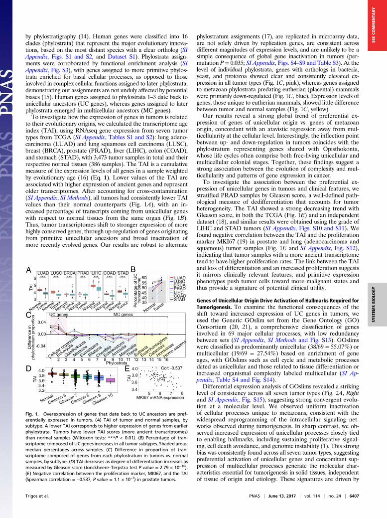
to their evolutionary origins, we calculated the transcriptome ageindex (TAI), using RNAseq gene expression from seven tumortypes from TCGA (SI Appendix, Tables S1 and S2): lung adeno-carcinoma (LUAD) and lung squamous cell carcinoma (LUSC),breast (BRCA), prostate (PRAD), liver (LIHC), colon (COAD),and stomach (STAD), with 3,473 tumor samples in total and theirrespective normal tissues (386 samples). The TAI is a cumulativemeasure of the expression levels of all genes in a sample weightedby evolutionary age (16) (Eq. 1). Lower values of the TAI areassociated with higher expression of ancient genes and representolder transcriptomes. After accounting for cross-contamination(SI Appendix, SI Methods), all tumors had consistently lower TAIvalues than their normal counterparts (Fig. 1A), with an in-creased percentage of transcripts coming from unicellular geneswith respect to normal tissues from the same organ (Fig. 1B).Thus, tumor transcriptomes shift to stronger expression of morehighly conserved genes, through up-regulation of genes originatingfrom primitive unicellular ancestors and broad inactivation ofmore recently evolved genes. Our results are robust to alternate
phylostratum assignments (17), are replicated in microarray data,are not solely driven by replication genes, are consistent acrossdifferent magnitudes of expression levels, and are unlikely to be asimple consequence of global gene inactivation in tumors (per-mutation P = 0.035; SI Appendix, Figs. S4–S9 and Table S3). At thelevel of individual phylostrata, genes with orthologs in bacteria,yeast, and protozoa showed clear and consistently elevated ex-pression in all tumor types (Fig. 1C, pink), whereas genes assignedto metazoan phylostrata predating eutherian (placental) mammalswere primarily down-regulated (Fig. 1C, blue). Expression levels ofgenes, those unique to eutherian mammals, showed little differencebetween tumor and normal samples (Fig. 1C, yellow).Our results reveal a strong global trend of preferential ex-
pression of genes of unicellular origin vs. genes of metazoanorigin, concordant with an atavistic regression away from mul-ticellularity at the cellular level. Interestingly, the inflection pointbetween up- and down-regulation in tumors coincides with thephylostratum representing genes shared with Opisthokonta,whose life cycles often comprise both free-living unicellular andmulticellular colonial stages. Together, these findings suggest astrong association between the evolution of complexity and mul-ticellularity and patterns of gene expression in cancer.To investigate the association between the preferential ex-
pression of unicellular genes in tumors and clinical features, westratified PRAD samples by Gleason score, a well-defined path-ological measure of dedifferentiation that accounts for tumorheterogeneity. The TAI showed a strong decreasing trend withGleason score, in both the TCGA (Fig. 1E) and an independentdataset (18), and similar results were obtained using the grade ofLIHC and STAD tumors (SI Appendix, Figs. S10 and S11). Wefound negative correlation between the TAI and the proliferationmarker MKI67 (19) in prostate and lung (adenocarcinoma andsquamous) tumor samples (Fig. 1E and SI Appendix, Fig. S12),indicating that tumor samples with a more ancient transcriptometend to have higher proliferation rates. The link between the TAIand loss of differentiation and an increased proliferation suggestsit mirrors clinically relevant features, and primitive expressionphenotypes push tumor cells toward more malignant states andthus provide a signature of potential clinical utility.
Genes of Unicellular Origin Drive Activation of Hallmarks Required forTumorigenesis. To examine the functional consequences of theshift toward increased expression of UC genes in tumors, weused the Generic GOslim set from the Gene Ontology (GO)Consortium (20, 21), a comprehensive classification of genesinvolved in 69 major cellular processes, with low redundancybetween sets (SI Appendix, SI Methods and Fig. S13). GOslimswere classified as predominantly unicellular (38/69 = 55.07%) ormulticellular (19/69 = 27.54%) based on enrichment of geneages, with GOslims such as cell cycle and metabolic processesdated as unicellular and those related to tissue differentiation orincreased organismal complexity labeled multicellular (SI Ap-pendix, Table S4 and Fig. S14).Differential expression analysis of GOslims revealed a striking
level of consistency across all seven tumor types (Fig. 2A, Rightand SI Appendix, Fig. S15), suggesting strong convergent evolu-tion at a molecular level. We observed uniform inactivationof cellular processes unique to metazoans, consistent with thewidespread reprogramming of the intracellular signaling net-works observed during tumorigenesis. In sharp contrast, we ob-served increased expression of unicellular processes closely tiedto enabling hallmarks, including sustaining proliferative signal-ing, cell death avoidance, and genomic instability (1). This strongbias was consistently found across all seven tumor types, suggestingpreferential activation of unicellular genes and concomitant sup-pression of multicellular processes generate the molecular char-acteristics essential for tumorigenesis in solid tissues, independentof tissue of origin and etiology. These signatures are driven by
C
D E
5
0
5
0
5
0
B
Cor: −0.537
LUAD LUSC BRCA PRAD LIHC COAD STAD
*** *** *** *** *** *** ***
3
4
5
6
A LUAD LUSC BRCA PRAD LIHC COAD STAD
TAI
34
5
6
Normal Tumor
Normal Tumor
Normal Tumor
Normal Tumor
Normal Tumor
Normal Tumor
Normal Tumor
Normal Tumor
LUAD LUSC BRCAPRADLIHC COADSTAD
Per
cent
age
of U
C
trans
crip
tom
e
35 40 45 50 55 60
Normal
Gleason 6
Gleason 7
Gleason 8
Gleason 9
Gleason 10 3.2 3.4 3.6 3.8 4.0
TAI
TAI
3.4
3.6 3.8 4.0
MKI67 mRNA expression 5 6 7 8
Cor: -0.537 Phylostrata
Diff
eren
ce in
ph
ylos
tratu
m p
ropo
rtion
-0.04
0.00
0.04
UC genes MC genes
Cellular organism
s
Eukaryota
Opisthokonta
Metazoa
Eumetazoa
Bilateria
Chordata
Euteleostomi
Ammiota
Mammalia
TheriaEutheria
Euarchontoglire
s
Catarrhini
Homininae
Homo sapiens
1 2 3 4 5 6 7 8 9 10 11 12 13 14 15 16
Fig. 1. Overexpression of genes that date back to UC ancestors are pref-erentially expressed in tumors. (A) TAI of tumor and normal samples, bysubtype. A lower TAI corresponds to higher expression of genes from earlierphylostrata. Tumors have lower TAI scores (more ancient transcriptomes)than normal samples (Wilcoxon tests: ***P < 0.01). (B) Percentage of tran-scriptome composed of UC genes increases in all tumor subtypes. Shaded areas:median percentages across samples. (C) Difference in proportion of tran-scriptome composed of genes from each phylostratum in tumors vs. normalsamples, by subtype. (D) TAI decreases as degree of differentiation increases asmeasured by Gleason score (Jonckheere–Terpstra test P value = 2.79 × 10⁻¹⁶).(E) Negative correlation between the proliferation marker, MKI67, and the TAI(Spearman correlation = −0.537, P value = 1.1 × 10⁻⁷) in prostate tumors.
Trigos et al. PNAS | June 13, 2017 | vol. 114 | no. 24 | 6407
SYST
EMSBIOLO
GY
SEECO
MMEN
TARY
processes beyond dedifferentiation, as they are distinct fromthose observed in stem cells (SI Appendix, SI Note 1 and Figs.S16 and S17).However, whether a unicellular process was activated or not was
dependent on its functional role, as we observed consistent down-regulation of many unicellular processes, particularly metabolicprocesses involving complex molecules, reflecting the metabolicreprogramming commonly seen in tumors. These patterns sug-gest induction of primitive processes in tumors is not merely aside effect of progressive stochastic loss of metazoan gene reg-ulatory mechanisms, but rather the result of coordinated andselective processes targeting specific pathways. Thus, a unifyingtheme behind the emergence of many of the common hallmarksof cancer could be a controlled transition to more primitivecellular phenotypes.We hypothesized the transcriptional states of individual cel-
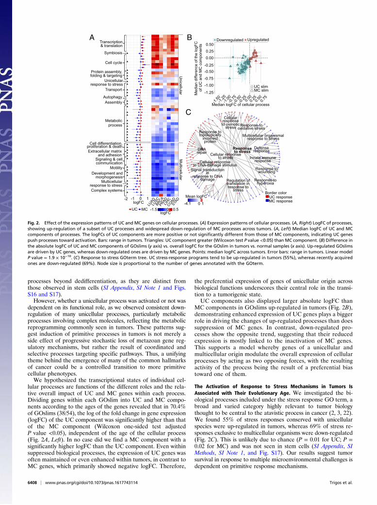
lular processes are functions of the different roles and the rela-tive overall impact of UC and MC genes within each process.Dividing genes within each GOslim into UC and MC compo-nents according to the ages of the genes revealed that in 70.4%of GOslims (38/54), the log of the fold change in gene expression(logFC) of the UC component was significantly higher than thatof the MC component (Wilcoxon one-sided test adjustedP value <0.05), independent of the age of the cellular process(Fig. 2A, Left). In no case did we find a MC component with asignificantly higher logFC than the UC component. Even withinsuppressed biological processes, the expression of UC genes wasoften maintained or even enhanced within tumors, in contrast toMC genes, which primarily showed negative logFC. Therefore,
the preferential expression of genes of unicellular origin acrossbiological functions underscores their central role in the transi-tion to a tumorigenic state.UC components also displayed larger absolute logFC than
MC components in GOslims up-regulated in tumors (Fig. 2B),demonstrating enhanced expression of UC genes plays a biggerrole in driving the changes of up-regulated processes than doessuppression of MC genes. In contrast, down-regulated pro-cesses show the opposite trend, suggesting that their reducedexpression is mostly linked to the inactivation of MC genes.This supports a model whereby genes of a unicellular andmulticellular origin modulate the overall expression of cellularprocesses by acting as two opposing forces, with the resultingactivity of the process being the result of a preferential biastoward one of them.
The Activation of Response to Stress Mechanisms in Tumors IsAssociated with Their Evolutionary Age. We investigated the bi-ological processes included under the stress response GO term, abroad and varied category highly relevant to tumor biologythought to be central to the atavistic process in cancer (2, 3, 22).We found 55% of stress responses conserved with unicellularspecies were up-regulated in tumors, whereas 69% of stress re-sponses exclusive to multicellular organisms were down-regulated(Fig. 2C). This is unlikely due to chance (P = 0.01 for UC; P =0.02 for MC) and was not seen in stem cells (SI Appendix, SIMethods, SI Note 1, and Fig. S17). Our results suggest tumorsurvival in response to multiple microenvironmental challenges isdependent on primitive response mechanisms.
UC
MC
y
yyy
y
y
r
y
Transcription & translation
Symbiosis
Cell cycle
Protein assembly, folding & targeting
AutophagyAssembly
Transport
Unicellularresponse to stress
Metabolicprocess
Multicellular response to stress Complex systems
Cell differentiation,proliferation & death Extracellular matrix
and adhesion Signaling & cell communication
Motility Development and
morphogenesis
Unicellular
Multicellular
-2 -1 0 1 logFC
UC MC
LUAD
LUSC
BRCAPRADLIH
C COADSTA
D
-1.5 0 0.5 logFC
Med
ian
diffe
renc
e of
the
logF
C o
f UC
and
MC
com
pone
nts
-1.25
-1.00
-0.75
-0.50 -0.25
0.00
0.25
0.50
Median logFC of cellular process -1.
25
-1.00
-0.
75
-0.50
-0.
25 0.0
0 0.2
5 0.5
0 -1.
50
0.75
Downregulated
UC slimMC slim
Cellular response to stress
Response to stress
Response tooxidative stress
Multicellular organismal response to stress
Defense response
Innate immune response
Response towounding
Response tohyperoxia
Regulation of translation in response to
stress
Cellularresponse to osmotic
stress Response totopologically
incorrectprotein
Cellular response to DNA damage stimulus
DNArepair
Signal transduction in
response to DNAdamage
Border colorUC response MC response10
Mean logFC
C
-4.1
BA Upregulated
Fig. 2. Effect of the expression patterns of UC and MC genes on cellular processes. (A) Expression patterns of cellular processes. (A, Right) LogFC of processes,showing up-regulation of a subset of UC processes and widespread down-regulation of MC processes across tumors. (A, Left) Median logFC of UC and MCcomponents of processes. The logFCs of UC components are more positive or not significantly different from those of MC components, indicating UC genespush processes toward activation. Bars: range in tumors. Triangles: UC component greater (Wilcoxon test P value <0.05) than MC component. (B) Difference inthe absolute logFC of UC and MC components of GOslims (y axis) vs. overall logFC for the GOslim in tumors vs. normal samples (x axis). Up-regulated GOslimsare driven by UC genes, whereas down-regulated ones are driven by MC genes. Points: median logFC across tumors. Error bars: range in tumors. Linear modelP value = 1.9 × 10⁻¹⁰. (C) Response to stress GOterm tree. UC stress-response programs tend to be up-regulated in tumors (55%), whereas recently acquiredones are down-regulated (69%). Node size is proportional to the number of genes annotated with the GOterm.
6408 | www.pnas.org/cgi/doi/10.1073/pnas.1617743114 Trigos et al.
This increased expression of processes originating in unicel-lular ancestors of modern life, such as DNA damage stimulus, isconsistent with mechanisms needed to withstand the geneticinstability typical of tumors. In contrast, advanced DNA repairprocesses exploited by multicellular organisms, such as pyrimi-dine dimer repair and specific double-strand break repair, weredown-regulated. An exception was the up-regulation of signaltransduction in response to DNA damage, which likely evolvedas a support system for the more primitive responses to DNAdamage, resulting in strong coregulation.The transition to a UC state in tumors is supported by pref-
erential activation of stress response mechanisms developed towithstand stresses encountered by unicellular ancestors, many ofwhich (hypoxia, nutrient deprivation, and DNA damage) wouldbe similar to environmental pressures encountered by rapidlyexpanding tumors. Conversely, the damping down of multicel-lular functions in tumors appears to extend to stress responseprocesses as well. The resulting phenotypic alterations couldsignificantly impact tumor evolution and response to treatment.
Disruption of the Coexpression Between Unicellular and MulticellularProcesses in Tumors Enhances Hallmark Phenotypes. We hypothe-sized convergent patterns of expression of cellular processes withrespect to evolutionary age were supported by coregulationmechanisms between processes. We first calculated the activityof cellular processes, using single-sample gene set enrichmentanalysis (ssGSEA) (23), which calculates the degree of coordi-nated up- and down-regulation of genes in individual samplesand can correctly classify samples according to subtype (SI Ap-pendix, Fig. S18). The Spearman correlation between pairs ofprocesses was calculated using ssGSEA scores, constructing anetwork of correlation of expression between cellular processes.To distinguish highly coexpressed pairs of processes with high
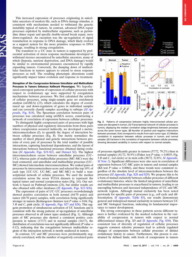
numbers of physical and regulatory interactions from those pairswhere coexpression occurred indirectly, we developed a metric,interconnectedness (I), to quantify the degree of interaction be-tween cellular processes (Eq. 3). Our metric normalizes thenumber of direct physical and genetic interactions between pro-teins of every pair of processes by the total number of possibleinteractions, capturing functional dependencies, and the layout ofinteractions between functional processes obtained during evolu-tion (SI Appendix, Figs. S19–S21, and Dataset S2). The highestinterconnectedness occurs between unicellular processes (UC–UC), whereas pairs of multicellular processes (MC–MC) were theleast connected, and unicellular and multicellular processes (UC–MC) showed intermediate interconnectedness. We ranked pairs ofprocesses by interconnectedness score and selected the top 10% ofeach type (UC–UC, UC–MC, and MC–MC) to build a tran-scriptional network of cellular processes. We used the mediancorrelation across the seven TCGA datasets to represent thetypical tumor and normal coexpression states (Fig. 3A). Our net-work is based on PathwayCommons (24), but similar results arealso obtained with other databases (SI Appendix, Figs. S22–S26).The expression of pairs of UC–UC processes overwhelmingly
showed positive correlation in both normal (64/70 pairs) andtumor (66/70 pairs) samples, with correlation being significantlystronger in tumors (Kolmogorov–Smirnov test P value = 0.04; Fig.3 B and C, pink circle; SI Appendix, Figs. S27 and S28). This sug-gests promotion of simultaneous activation of UC functions and isconsistent with the strong patterns of up-regulation of unicellularprocesses observed in all tumor types analyzed (Fig. 1). Althoughpairs of MC processes also showed a consistent positive coex-pression in tumors (17/17) and in normal samples (16/17), thedistribution of correlations was not significantly different (P value =0.12), indicating that the coregulation between multicellular re-gions of the interaction network is mostly unaltered in tumors.In contrast, UC and MC processes were predominantly neg-
atively correlated, with the number of negatively correlated pairs
of processes significantly greater in tumors (57/72, 79.2%) than innormal samples (41/72, 56.9%) (Fisher test P value = 0.0035; Fig.3 B and C, red circle) or in stem cells (38/72, 52.8%; SI Appendix,SI Note 1). Significant differences were also seen in correlation ofexpression between UC–MC pairs in tumors and normal samples(KS test P value = 0.0066), and these trends were consistent re-gardless of the absolute level of interconnectedness between theprocesses (SI Appendix, Figs. S28 and S29). We propose this to bea form of mutual exclusivity between cellular processes of differentevolutionary histories, where the limited integration of unicellularand multicellular processes is exacerbated in tumors, leading to anuncoupling between and increased independence of UC and MCnetwork regions. Although mutual exclusivity has been notedpreviously for specific pairs of processes (e.g., cell cycle and dif-ferentiation; SI Appendix, Table S5), our results demonstrategeneral and widespread mutual exclusivity in tumors between UCand MC biological functions, indicating its fundamental impor-tance to tumor development.The strong convergence in these patterns across different tu-
mors is further evidenced by the marked reduction in the vari-ability of coexpression in tumors with respect to normaldifferentiated tissues (Fig. 3D and SI Appendix, Fig. S30). Thisconsistent loss of diversity of regulation of cellular processessuggests common selective pressures lead to actively regulatedchanges of coexpression between cellular processes of distinctevolutionary history in cancer. Furthermore, this process is con-strained by defined limits, with levels of coexpression in tumors
A
0
5
0
5
0
UC_UCUC_MCMC_MC
C
UC-UC UC-MCMC-MC
PositiveNegative
UpregulatedDownregulated
UC process MC process
Normal Tumour
C C C C C C
B
DCorrelation in normals
Cor
rela
tion
in tu
mor
s
Med
ian
coun
t
75
50
25
0 UC-U
C UC-M
CMC-M
CUC-U
C UC-M
CMC-M
C
-1.0 -0.5 0.0 0.5 1.0
1.0
0.5
0.0
-0.5
-1.0
UC-UC UC-MCMC-MC
Normal
Tumor
Nor
mal Tu
mor
Normal
Tumor
Varia
nce
of e
dges
ac
ross
tiss
ues
0.00
0.05
0.10
0.15
Normal Tumor
PositiveNegative
Interactions: Correlation:
MC-MCUC-MCUC-UC
Fig. 3. Patterns of coexpression between highly interconnected cellular pro-cesses are disrupted in tumors. (A) Transcriptional network of cellular processes intumors, displaying the median correlation (edge brightness) between processesacross the seven tumor types. (B) Number of positive and negative interactionsbetween processes. Dots correspond to results from each tumor type. (C) Mediancorrelation in expression between cellular processes in tumors and normalsamples. (D) Variance in the correlation of expression between cellular processes,showing decreased variability in tumors with respect to normal samples.
Trigos et al. PNAS | June 13, 2017 | vol. 114 | no. 24 | 6409
SYST
EMSBIOLO
GY
SEECO
MMEN
TARY
consistently within the ranges of those of normal samples (SI Ap-pendix, Fig. S31), suggesting that system-level constraints limit theviable paths taken by tumor cells.We found 20 UC–MC interacting pairs switched from posi-
tive to negative correlation, whereas only 4 pairs switched inthe opposite direction (Fig. 3C, orange square; SI Appendix,Table S6). Nearly half of them involved cell death (11/24;45.83%), consistent with the view that cell death is tied to manyof the major regulatory and signaling changes that occur intumors. The strong selection for mutually exclusive associa-tions between these specific gene sets across multiple tumortypes indicates disruption of the links between them maydrive tumor development.
Identifying Genes Modulating the Altered Interactions BetweenNetwork Regions of Different Evolutionary Age. We hypothesizedthe disruption in coexpression of UC and MC processes was dueto altered interactions between genes linking these two parts of thehuman gene network, forming key vulnerabilities. We focused ona pair of cellular processes whose coexpression was among themost strongly disrupted in tumors, cellular junction organizationand chromosome organization, which displayed a pronounced shiftfrom positive correlation in normal tissues (median: 0.21) to strongnegative correlation in tumors (median: −0.36). This selection for adrastic change in coexpression suggests mutual exclusivity betweenthese processes is advantageous for tumorigenesis.We reasoned the key genes modulating this change would
be highly connected across the two gene sets and have alteredcoexpression with many of their interaction partners. We devel-oped a “hubness” metric for each gene with annotated interac-tions between cellular junction organization and chromosomeorganization, calculated as the sum of the absolute difference ofchange in expression correlation between its connecting partnergenes (Eq. 4). This metric is highly correlated with the number ofinteractions of a gene (SI Appendix, Fig. S32), a property asso-ciated with relative importance in modulating functional re-sponses (25). Ranking by this metric uncovered 12 genes (RCC2,TLN1, VASP, ACTG1, PLEC, CTTN, DSP, ILK, PKN2, CTNNA1,CTNND1, and PKP3) whose hubness was consistently in the top10% across all seven tumor types, identifying a set of genes thatcommonly mediate changes in coexpression between cellular junc-tion organization and chromosome organization.All 12 genes interact with genes belonging to a signature of
chromosomal instability associated with poor clinical outcome andmetastasis (26) (SI Appendix, Fig. S33), suggesting these genes haveroles in regulatory networks linking genomic instability and me-tastasis during tumor progression. Many are involved in pathwayshighly relevant to cancer (SI Appendix, Table S8), including4 within the Rap1 signaling pathway, which has yet to be largelystudied in the context of cancer, and experimental evidence sug-gests they can modulate malignant characteristics in specific tumortypes (SI Appendix, Table S7). However, here we associate thesegenes with seven tumor types, uncovering a wider role as general,pan-cancer modulators of tumor development. CRISPR screendata for these genes and 91 additional genes involved in other pairsof UC–MC processes negatively correlated across tumors revealedknockdown of many of the identified UC genes hinders growth ofmultiple cancer cell lines (SI Appendix, SI Note 2, Fig. S34, andTables S9 and S10), indicating their potential as viable drug targets.The association of key genes promoting mutual exclusivity in
tumors with main features of tumorigenesis supports the viewthat that mutual exclusivity in the coregulation of unicellular andmulticellular processes is under positive selection during tumorformation and progression across multiple tumor types. Drugsthat target these fundamental points of vulnerability or thatabolish this mutual exclusivity would have great potential as ef-fective broad-spectrum treatment strategies.
DiscussionDetailed transcriptome analysis of 3,473 tumor and 386 normalsamples from TCGA demonstrates gene expression changes in tu-mors are closely tied to the evolutionary ages of the genes involved.Our findings suggest convergence of tumors to similar molecularphenotypes is tied to common principles guiding patterns of coex-pression of cellular processes according to their evolutionary his-tories. This is the most comprehensive molecular evidence to datethat a widespread shift to preferential expression of genes conservedin primitive, single-celled species is a common feature of tumors.This is concordant with the atavism hypothesis, which states cancerresults from a transition to a more “selfish” unicellular mode of life,not merely through a passive stochastic occurrence but as an active,directed process driven by selection (2, 3, 27). Our findings dem-onstrate the up-regulation of unicellular GO terms is limited tocertain processes and pathways, indicative of selection, and loss ofcoordinated expression between multicellular and unicellular pro-cesses across multiple tumor types, implicating altered regulation.Although convergent evolution in tumors has been described
in the context of gene expression (13), here we show convergentevolution is also apparent at the level of coexpression of cellularprocesses, according to their point of evolutionary emergence.Tumorigenesis reinforces the interdependence between unicel-lular genes while enhancing segregation between the unicellularand multicellular components of the gene regulatory network.Such mutual exclusivity would promote loss of multicellular fea-tures in tumors as activation of unicellular genes occurs in re-sponse to selective pressures favoring increased replication oractivation of basal cellular processes, leading to an increasinglyatavistic malignant phenotype with increased selective advantage.Gene expression in cancer with respect to evolutionary age is
not a simple dichotomy, as multicellular biological processessuch as hormone receptors drive several tumor types. However,the highly reproducible nature of our observations and the signsof regulatory control behind them suggests treatment strategiesthat manipulate the fundamental systems-level rewiring at theinterface between more primitive and more advanced compo-nents of gene regulatory networks in cancer could have broadtherapeutic application and high specificity for tumor cells.Several compounds already in clinical use target primitive funda-
mental biological functions, e.g., ref. 28. Our results showing manyprimitive functions up-regulated together in tumors raise the possi-bility of going a step beyond, to simultaneously target multiple in-dependent unicellular processes. Another approach involves stressingmulticellular systems that are inactive or diminished in cancer, a“target the weakness” approach (29) by altering the intra- or extra-cellular environment of tumor cells to put cells that have lost orinactivated a particular multicellular pathway at a selective disadvan-tage. Our analysis shows stress response pathways composed primarilyof multicellular genes could also be manipulated for clinical benefit.Given strong anticorrelation in expression between multicel-
lular and unicellular genes is apparent in many tumors, rees-tablishing the balance between the activities of unicellular andmulticellular processes could push tumors back to a more normalstate and/or achieve a form of synthetic lethality. Empiricalsupport for this approach comes from studies showing inhibitionof glycolysis increased sensitivity of cancer cells to pharmaco-logically induced apoptosis (30). Cell death displayed consis-tently altered correlations with many core metabolic and celldivision processes, suggesting manipulation of other biologicalfunctions could further prime tumor cells for apoptosis.Our approach uncovered previously unappreciated association be-
tween biological processes exploited by tumors. We could narrowdown 12 key genes bridging the cell junction organization and chro-mosome organization processes, which are biomarkers or regulatorsof malignancy in vitro for at least one cancer, validating the approach,but our analysis also implicates them as potential common drivers of a
6410 | www.pnas.org/cgi/doi/10.1073/pnas.1617743114 Trigos et al.
number of cancers. To our knowledge, no studies have yet pub-lished potential therapeutic agents for these genes, making themattractive targets for prioritization in future drug screens.Our study applies a detailed molecular framework to view
cancer as a failure of the systems supporting increased organis-mal complexity (31, 32). This is an important step toward un-derstanding how macroevolutionary processes left vulnerabilitiesthat lead to cancer and how they may be exploited in practicalterms to improve treatment outcomes, facilitating the applica-tion of “Darwinian medicine” to oncology (33).
MethodsPhylostratigraphy of Human Genes. A total of 17,318 human genes weremapped to a phylogenetic tree (Dataset S1), consisting of 16 clades (phylos-trata), ranging from including all cellular organisms (phylostratum 1) to Homosapiens (phylostratum 16) (SI Appendix, Fig. S1). The most ancient phylos-tratum represented in a group of orthologs from the OrthoMCL databaseversion 5 (34) was considered as the point of emergence of the human protein.
Transcriptomic Analysis Incorporating Gene Ages. RNAseq expression datawere downloaded from TCGA (https://portal.gdc.cancer.gov/) (SI Appendix,Table S2). The transcriptome ages of tumor and normal samples were cal-culated with the TAI method (16),
TAI=Pn
i=1psi × eiPni=1ei
, [1]
where psi is the phylostratum of each gene i and ei is the gene expressionvalue of gene i.
The proportion of the total library size represented by each phylostratumin each sample was calculated using Eq. 2,
Ppsi =
Pmij=1eijPn
i=1
Pmij=1eij
, [2]
where Ppsi is the proportion of expression abundance corresponding togenes of phylostratum i, eij is the expression value of gene j in the phylos-tratum i, mi is the total number of genes in phylostratum i, and n is the totalnumber of phylostrata. The Ppsi values were averaged across all samples foreach normal and tumor type, and the tumor vs. normal difference in pro-portions was determined by subtraction.
Functional Analyses. GOslims were obtained from Gene Ontology (gen-eontology.org/), and their ages were calculated by permutation of the agesof the genes annotated with each GOslim (SI Appendix, Table S4). Tumor vs.normal differential expression analysis of GOslims and UC and MC compo-nents was conducted using QuSAGE (35). For the response to stress tree, theaverage logFC obtained by QuSAGE for each term was calculated acrosstumors, regardless of the significance of individual false discovery rate.Statistical significance of trends was assessed using permutation tests (SIAppendix, SI Methods).
Construction of Transcriptional Coexpression Networks. The degree of in-teraction I (Dataset S2) was calculated for each pair of GOslims (i and j),
I=Eij −Unique
�Ei∩ij + Ej∩ij
��Gi −Gi∩j
�*�Gj −Gi∩j
� =Number of edges between GOslimsTotal number of possible edges
, [3]
with Eij being the number of edges joining genes of GOslim i withGOslim j, and Ei∩ij and Ej∩ij are the number of edges in GOslim i or j thatare also found in Eij. Gi and Gj are the numbers of genes of pathways iand j, respectively, and Gi∩j is the number of genes shared by GOslims iand j.
Gene Hubness Score. Edges connecting genes of cell junction organization andchromosome organization processes were weighted by the Spearman cor-relation of expression in each tumor and normal type. We defined
Hubness of genei =XN
j=1
��Correlation tumorj −Correlation normalj��, [4]
with i being a gene in the bipartite graph and j an edge linking genes inseparate processes.
Code is available at https://github.com/cancer-evolution/Evolutionary-analysis-of-cancer-transcriptomes.
ACKNOWLEDGMENTS. We thank David Bowtell, Patrick Humbert, DavidThomas, Arcadi Cipponi, Ismael Vergara, and Jason Li for helpful comments.This work was supported by a Melbourne International Engagement Awardand a Melbourne International Fee Remission Scholarship (to A.S.T.), a Na-tional Health & Medical Research Council of Australia (NHMRC) Peter Doh-erty Early Career Fellowship (to D.L.G.) (APP1052904), and NHMRC ProgramGrant 1053792 (to R.B.P.), as well as by NHMRC Senior Research Fellowships(to R.B.P. and A.T.P.).
1. Hanahan D, Weinberg RA (2011) Hallmarks of cancer: The next generation. Cell 144:646–674.
2. Davies PC, Lineweaver CH (2011) Cancer tumors as Metazoa 1.0: Tapping genes ofancient ancestors. Phys Biol 8:015001.
3. Vincent M (2012) Cancer: A de-repression of a default survival program common to allcells?: A life-history perspective on the nature of cancer. BioEssays 34:72–82.
4. Merlo LM, Pepper JW, Reid BJ, Maley CC (2006) Cancer as an evolutionary and eco-logical process. Nat Rev Cancer 6:924–935.
5. Lambert G, et al. (2011) An analogy between the evolution of drug resistance inbacterial communities and malignant tissues. Nat Rev Cancer 11:375–382.
6. Domazet-Loso T, Tautz D (2010) Phylostratigraphic tracking of cancer genes suggestsa link to the emergence of multicellularity in metazoa. BMC Biol 8:66.
7. Wu A, et al. (2015) Ancient hot and cold genes and chemotherapy resistance emer-gence. Proc Natl Acad Sci USA 112:10467–10472.
8. Aktipis CA, et al. (2015) Cancer across the tree of life: Cooperation and cheating inmulticellularity. Philos Trans R Soc Lond B Biol Sci 370:20140219.
9. Domazet-Lošo T, et al. (2014) Naturally occurring tumours in the basal metazoanHydra. Nat Commun 5:4222.
10. Ruiz-Trillo I, Nedelcu A (2015) Evolutionary transitions to multicellular life. Principlesand Mechanisms, eds Ruiz-Trillo I, Nedelcu A (Springer, Dordrecht, The Netherlands),pp 47–78.
11. Engler AJ, Humbert PO, Wehrle-Haller B, Weaver VM (2009) Multiscale modeling ofform and function. Science 324:208–212.
12. Chen H, Lin F, Xing K, He X (2015) The reverse evolution from multicellularity tounicellularity during carcinogenesis. Nat Commun 6:6367.
13. Chen H, He X (2016) The convergent cancer evolution toward a single cellular des-tination. Mol Biol Evol 33:4–12.
14. Domazet-Loso T, Brajkovi�c J, Tautz D (2007) A phylostratigraphy approach to uncover thegenomic history of major adaptations in metazoan lineages. Trends Genet 23:533–539.
15. Moyers BA, Zhang J (2015) Phylostratigraphic bias creates spurious patterns of ge-nome evolution. Mol Biol Evol 32:258–267.
16. Domazet-Lošo T, Tautz D (2010) A phylogenetically based transcriptome age indexmirrors ontogenetic divergence patterns. Nature 468:815–818.
17. Domazet-Loso T, Tautz D (2008) An ancient evolutionary origin of genes associatedwith human genetic diseases. Mol Biol Evol 25:2699–2707.
18. Erho N, et al. (2013) Discovery and validation of a prostate cancer genomic classifierthat predicts early metastasis following radical prostatectomy. PLoS One 8:e66855.
19. Andor N, et al. (2016) Pan-cancer analysis of the extent and consequences of intra-tumor heterogeneity. Nat Med 22:105–113.
20. Ashburner M, et al.; The Gene Ontology Consortium (2000) Gene ontology: Tool forthe unification of biology. Nat Genet 25:25–29.
21. Gene Ontology Consortium (2015) Gene Ontology Consortium: Going forward.Nucleic Acids Res 43:D1049–D1056.
22. Cipponi A, Thomas DM (2014) Stress-induced cellular adaptive strategies: Ancient evolu-tionarily conserved programs as new anticancer therapeutic targets. BioEssays 36:552–560.
23. Barbie DA, et al. (2009) Systematic RNA interference reveals that oncogenic KRAS-driven cancers require TBK1. Nature 462:108–112.
24. Cerami EG, et al. (2011) Pathway Commons, a web resource for biological pathwaydata. Nucleic Acids Res 39:D685–D690.
25. Jeong H, Mason SP, Barabási AL, Oltvai ZN (2001) Lethality and centrality in proteinnetworks. Nature 411:41–42.
26. Carter SL, Eklund AC, Kohane IS, Harris LN, Szallasi Z (2006) A signature of chromo-somal instability inferred from gene expression profiles predicts clinical outcome inmultiple human cancers. Nat Genet 38:1043–1048.
27. Greaves M (2015) Evolutionary determinants of cancer. Cancer Discov 5:806–820.28. Devlin JR, et al. (2016) Combination therapy targeting ribosome biogenesis and mRNA
translation synergistically extends survival in MYC-driven lymphoma. Cancer Discov 6:59–70.29. Lineweaver CH, Davies PC, Vincent MD (2014) Targeting cancer’s weaknesses (not its
strengths): Therapeutic strategies suggested by the atavistic model. BioEssays 36:827–835.30. Meynet O, et al. (2012) Glycolysis inhibition targets Mcl-1 to restore sensitivity of
lymphoma cells to ABT-737-induced apoptosis. Leukemia 26:1145–1147.31. Aktipis CA, Nesse RM (2013) Evolutionary foundations for cancer biology. Evol Appl 6:
144–159.32. Aktipis CA, Boddy AM, Gatenby RA, Brown JS, Maley CC (2013) Life history trade-offs
in cancer evolution. Nat Rev Cancer 13:883–892.33. Greaves M (2007) Darwinian medicine: A case for cancer. Nat Rev Cancer 7:213–221.34. Li L, Stoeckert CJ, Jr, Roos DS (2003) OrthoMCL: Identification of ortholog groups for
eukaryotic genomes. Genome Res 13:2178–2189.35. Yaari G, Bolen CR, Thakar J, Kleinstein SH (2013) Quantitative set analysis for gene
expression: A method to quantify gene set differential expression including gene-gene correlations. Nucleic Acids Res 41:e170.
Trigos et al. PNAS | June 13, 2017 | vol. 114 | no. 24 | 6411
SYST
EMSBIOLO
GY
SEECO
MMEN
TARY