swift: a novel method to track the neural correlates of recognition
TRANSCRIPT

NeuroImage 81 (2013) 273–282
Contents lists available at SciVerse ScienceDirect
NeuroImage
j ourna l homepage: www.e lsev ie r .com/ locate /yn img
SWIFT: A novel method to track the neural correlates of recognition
Roger Koenig-Robert ⁎, Rufin VanRullenCentre de Recherche Cerveau et Cognition, Université Paul Sabatier, Université de Toulouse, Toulouse, FranceCNRS, CerCo, Toulouse, France
⁎ Corresponding author at: School of Psychology and PNursing and Health Sciences, Monash University, Clayto
E-mail address: [email protected] (R. Koenig-Ro
1053-8119/$ – see front matter © 2013 Elsevier Inc. Allhttp://dx.doi.org/10.1016/j.neuroimage.2013.04.116
a b s t r a c t
a r t i c l e i n f oArticle history:Accepted 28 April 2013Available online 9 May 2013
Keywords:Conscious recognitionObject representationHigh-level visionVisual dynamicsFrequency taggingConsciousness
Isolating the neural correlates of object recognition and studying their fine temporal dynamics have been agreat challenge in neuroscience. A major obstacle has been the difficulty to dissociate low-level feature ex-traction from the actual object recognition activity. Here we present a new technique called semanticwavelet-induced frequency-tagging (SWIFT), where cyclic wavelet-scrambling allowed us to isolate neuralcorrelates of object recognition from low-level feature extraction in humans using EEG. We show thatSWIFT is insensitive to unrecognized visual objects in natural images, which were presented up to 30 s,but is highly selective to the recognition of the same objects after their identity has been revealed. Theenhancement of object representations by top-down attention was particularly strong with SWIFT due toits selectivity for high-level representations. Finally, we determined the temporal dynamics of object repre-sentations tracked by SWIFT and found that SWIFT can follow a maximum of between 4 and 7 different objectrepresentations per second. This result is consistent with a reduction in temporal capacity processing fromlow to high-level brain areas.
© 2013 Elsevier Inc. All rights reserved.
Introduction
How visual objects are represented as meaningful items in ourbrains and become part of our conscious experience is one of themost fascinating questions in neuroscience. Current models, largelyinspired by invasive studies in monkeys, propose a view of the visualsystem where object representations emerge progressively from a hi-erarchical cascade of processing stages (Felleman and Van Essen,1991; Riesenhuber and Poggio, 1999). Early stages are devoted toextracting simple visual features such as luminance (Amthor et al.,2005), contrast (Sclar et al., 1990), contours (Hubel and Wiesel,1968) and intersecting lines (Hegdé and Van Essen, 2000). Down-stream in the ventral pathway, the integration of these simple fea-tures implies that neurons become selective to more and morecomplex forms, e.g. in area V4 (Gallant et al., 1993). At the highestpurely visual area in the ventral stream, the inferotemporal cortex(IT), neurons can be selective to single object categories (Kobatakeand Tanaka, 1994; Tanaka, 1996).
While neuronal selectivities in the ventral stream of the monkeyvisual system are well understood, their associated semantic valueis difficult to access. Where and when do meaningful object represen-tations emerge? Non-invasive techniques have been developed totrack visual stimulus representations in the human brain – forwhich perceptual meaning can be more readily assessed. Functional
sychiatry, Faculty of medicine,n, Australia.bert).
rights reserved.
magnetic resonance imaging (fMRI) has played a major role in under-standing the human brain areas engaged in object representations.For example, fMRI has revealed that some regions of the temporallobe are selective to faces in the FFA (Kanwisher et al., 1997), scenesin the PPA (Epstein et al., 1999) or body parts in sub-regions of theLOC (Downing et al., 2001), and there is good evidence that these re-gions respond more strongly when the corresponding stimuli areconsciously perceived by the subjects (Bar et al., 2001; Grill-Spectoret al., 2000; Hesselmann and Malach, 2011; Tong et al., 1998). How-ever, the temporal dynamics of object representations on the scaleof a few tenths of a second are unattainable to the slower temporalresolution of fMRI. Electroencephalography (EEG) has been exten-sively used to explore these temporal dynamics in humans. Moreparticularly, steady-state visual evoked potentials (SSVEP) can trackthe activity elicited by a given visual stimulus in near-real time. Thismethod, also known as frequency tagging, involves the modulationof a stimulus' intensity over time at a fixed temporal frequency f0; aneural response is evoked at the same frequency f0 (and usually itsharmonics), thus providing a frequency label (or tag) for the stimulusrepresentation in the brain (Appelbaum and Norcia, 2009; Regan,1977; Srinivasan et al., 2006). The frequency-tagged response hasbeen found to depend on attention (Ding et al., 2006; Kim et al.,2007; Morgan et al., 1996; Müller et al., 1998) and on the subject'sperceptual state (Kaspar et al., 2010; Srinivasan and Petrovic, 2006;Sutoyo and Srinivasan, 2009; Tononi et al., 1998). One limitation ofSSVEP is that they normally rely on themodulation of stimulus contrastor luminance; as a result, both semantic object-representations andlow-level feature extraction mechanisms are simultaneously tagged

274 R. Koenig-Robert, R. VanRullen / NeuroImage 81 (2013) 273–282
at the modulation frequency. As a result, previous studies reportednon-consistent effects of object recognition on SSVEP amplitude acrosstagging frequencies, with recognized images sometimes leading tohigher and sometimes to lower SSVEP amplitudes than unrecognizedones (Kaspar et al., 2010). In order to try to disentangle low-level fea-ture extraction processes from semantic object-representations, we de-veloped a novel technique called SWIFT (semantic wavelet-inducedfrequency tagging) in which we equalized low-level physical attributes(luminance, contrast and spatial frequency spectrum) across all framesof a sequence, while modulating, at a fixed frequency f0, the mid- andhigher-level image properties carried by the spatial configuration oflocal contours.
In order to validate the sensitivity of our technique to high-levelvisual representations, we reasoned that SWIFT should satisfy 3criteria that we tested in separate experiments. First, activity elicitedby explicitly recognized objects should be clearly differentiated fromactivity elicited by non-recognized objects: indeed, we found thatSWIFT is insensitive to unrecognized objects presented up to 30 s,but is highly selective to the recognition of the same objects oncetheir identity has been explicitly revealed. Second, as a consequenceof the top-down transmission of attention signals (Lauritzen et al.,2009; Saalmann et al., 2007), attentional modulation intensity shouldbe greater for high-level visual representations than for lower ones;indeed, we demonstrated that SWIFT responses are strongly modu-lated by top-down attention – considerably more so than classicSSVEP signals. Third, as a result of a reduction in temporal processingcapacity from early visual cortex to higher areas (Gauthier et al.,2012; Holcombe, 2009; McKeeff et al., 2007), high-level representa-tions should be limited in their temporal sensitivity: indeed, wefound that SWIFT responses reached a limit between 4 and 7 itemsper second.
Material and methods
SWIFT sequences creation
SWIFT sequences were created by cyclic wavelet scrambling in thewavelets 3D space. We chose wavelet image decomposition ratherthan other types of image transformation (such as the Fourier transform)because wavelet functions (contrary to Fourier functions) are localizedin space: this allowed us to scramble contours while conserving locallow-level attributes. The first step was to apply a wavelet transformbased on the discreteMeyer (dmey)wavelet and 6 decomposition levels,using the Wavelet toolbox under Matlab (MathWorks); in other words,the image was converted to a multi-scale pyramid of spatially organizedmaps. At each location and scale, the local contour is represented by a 3Dvector v1
→, with the 3 dimensions representing the strengths of horizon-
tal, vertical and diagonal orientations. The vector length v1→����
���� is ameasure
of local contour energy. In a second step, for each location and scale, two
random vectors (v2→
and v3→) were defined that shared the length of the
original vector ( v1→����
���� ¼ v2→����
���� ¼ v3→����
����), thus conserving local energy. By def-inition, the 3 vectors describe a unique circular path over an isoenergeticspherewhere all surface points share the same energy (i.e., the same Eu-clidian distance from the origin) but represent differently oriented ver-sions of the local image contour. The cyclic wavelet-scrambling wasthen performed by rotating each original vector (representing the actualimage contour), along the circular path defined above. Somewavelet el-ements (defined by a specific spatial location and decomposition scale)underwent this rotation once per cycle (i.e., at the fundamental frequencyf0) while others rotated multiple (integer) times per cycle (i.e., at har-monic frequencies of f0, from the 2nd up to the 5th harmonic). The intro-duction of harmonics was crucial to spread the temporal luminancemodulation over a broader frequency band, avoiding low-level evoked
activity at the tagging frequency f0. The 5 harmonic frequencies were dis-tributed equally and randomly among all the wavelet elements. Finally,the inverse wavelet transform was used to obtain the image sequencesin the pixel domain. By construction, the original unscrambled imageappeared once in each cycle, with a number of intervening wavelet-scrambled frames that depended on the monitor refresh rate and thetagging frequency f0. For each original image, several distinct wavelet-scrambling cycles were computed (5 cycles in experiment 1, 2 cycles inexperiment 2 and 4 cycles in experiment 3), with different randomlychosen values for thewavelet-scrambling trajectories and the harmonicrotation frequency at eachwavelet element. These different cycles werepresented in random alternation during the experimental sequences.Two final normalization steps were necessary in order to ensurethat the temporal luminance modulation for every pixel was constant(i.e. without any peaks at the individual harmonics frequencies) withinthe range of harmonic modulation frequencies, and also to ensure theconservation of the mean luminance across frames. First, we calculatedthe Fourier transform across frames for every pixel and normalizedtheir luminance modulation spectra. Second, mean frame luminancewas equalized over time. (NB: A Matlab script following this procedureto create a wavelet-scrambling sequence based on any given originalimage is available as Supplementary Material).
Subjects
All subjects gave informed consent to take part in these studiesthat were approved by the local ethics committee. A total of 49 ob-servers (26 women, aged 22 to 53) participated in the 3 experiments(19 in Experiment 1, 8 in Experiment 2 and 24 in Experiment 3).
Stimuli and procedure
For all 3 experiments, subjects were placed at 57 cm of a CRTscreen with a refresh rate of 170 Hz in a dark room.
In Experiment 1, 100 SWIFT sequences containing either grayscalenatural images (bodies with faces 29%, bodies with no visible faces16%, animals 21% and manmade objects 14%, downloaded from the In-ternet) or low-levelmatched textures synthesized using the texture syn-thesis algorithm developed by Portilla and Simoncelli (2000) wereshown. The number of images in each category was chosen in order tomaximize the number of non-canonical images and thus promote the oc-currence of ‘unrecognized’ images. The image contours were modulatedcyclically over time at f0 = 1.4953 Hz. The experiment was divided in 4blocks of 25 trials each. Each trial lasted 42 s (30 s of naïve period + 2 sof steady image presentation + 10 s of cognizant period). Sequences(10.5° × 10.5° visual angle) were presented at the center of the screenover a gray background. Subjects were asked to keep their fixationover a red cross at the center of the display during the trial. They gavetheir responses (presence of a non-abstract item) at any time duringthe first naïve period by pressing the left arrow of the computer key-board for the low confidence threshold (key 1: “I perceive an object-like item, but I am not sure of which object it is”) and the right arrowfor the high confidence threshold (key 2: “I see an object and I have iden-tified it confidently”). Trials were classified as ‘quickly recognized’whena natural imagewas presented and the subject recognized an object withhigh confidence within the first 10 s of the naïve period. Trials wereclassified as ‘tardily recognized’ when a natural image was presentedbut the subject did not recognize an object during the 30 s of the naïveperiod. Trials were classified as ‘no-object’ when abstract textures werepresented. Two of 19 subjects were not considered in the analysis be-cause they had less than 7 tardily recognized trials. For the 17 remainingsubjects, the mean number of quickly recognized trials was 22.2, tardilyrecognized was 22.4 and 20 no-object trials were presented systemati-cally (the remaining trials, corresponding to incomplete or erroneousrecognition, were not included in the analysis). Response time for key2 (“I see an object and I have identified it confidently”) in fast recognized

275R. Koenig-Robert, R. VanRullen / NeuroImage 81 (2013) 273–282
trials was 4.4 s (mean) and 3.5 s (median). More than half of key 2responses (51.56%) were delivered within 5 s of stimulus onset. Key 2responses followed a negative exponential distribution, with 24.74% ofresponses between 5 and 10 s, 12.76% of responses between 15 and20 s, 4.17% of responses between 15 and 20 s, 3.9% of responses between20 and 25 s and 2.86% responses between 25 and 30 s. This shows thatfor most quickly recognized trials, object recognition emerged after afew cycles. Quickly recognized trials were classified as such if key 2was pressed within the 10 first seconds of the naïve period. All naïve pe-riod cycles were considered in the analysis of quickly recognized trials.
In Experiment 2, 360 pairs of sequences containing human faces(n = 120, downloaded from the Internet) were presented. The 2 se-quences (8.5° × 8.5° visual angle) were presented over a gray back-ground, on either side of the screen, separated by 5.8° visual angle.In half of the trials both face pictures were modulated in the contrastdomain (by manipulating the contrast from 0 to 100% through a sinu-soidal envelope: “classic SSVEP”) and the other half in the wavelet do-main (SWIFT). In each trial, the two sequences were modulated attwo different, non-harmonically related frequencies = 1.4953 Hz or2.0253 Hz (counter balanced per presentation side across trials).The experiment was divided in 4 blocks of 90 trials each. Subjectswere instructed to attend the sequence indicated by the central cue(a red arrow of 0.8° visual angle) and ignore the other, while keepingfixation on the central arrow. The task consisted in detecting a deviantcycle (present in 25% of trials) which was only presented in the targetsequence. Deviant cycles under SWIFT modulation were produced byinserting a cycle in which the embedded image was itself a waveletscrambled version of the face showed in the sequence, with the resultthat the face did not reappear in this deviant cycle. For the classicSSVEP modulation, the original face was replaced by a wavelet scram-bled version in the deviant cycle. Deviant cycles were placed randomlyduring the trial, excluding the first and last cycle. Each trial lasted 10 sand the response (presence/absence of a deviant cycle) was givenat the end of the trial by pressing either the left (presence) or right(absence) arrow on a computer keyboard.
In experiment 3, SWIFT sequences (10.5 × 10.5° visual angle)containing natural images (41% animals, 16% faces, 14% bodies withvisible faces and 29% manmade, downloaded from the Internet), werepresented at the center of the screen over a gray background. Partici-pants were asked to keep fixation over a red cross at the center ofthe sequence during the trial. SWIFT modulation was performed ondifferent trials at 8 different, non-harmonically related frequencies(f0 = 1.4953, 2.0253, 2.6230, 3.4043, 4.3243, 6.9665, 8.4211 or12.3077 Hz). The experimental session contained 160 trials, eachlasting 10 s. The task consisted in detecting the deviant cycles (present20% of trials) as explained above. This low-level change detection taskwould be independent of object recognition and was deliberately cho-sen as such since wewanted to avoid subject's motivation and vigilancedropping (and the subsequent decrease in the amount of tagging)whenthe object in the sequence was hard to recognize (i.e., at high taggingfrequencies). The answer was given at the end of the trial by pressingthe left arrow key (present) or right arrow key (absent). Dprimes foreach of the eight tagging frequencies, from 1.4953 to 12.3077 Hz,were 1.31, 1.73, 1.90, 2.45, 2.60, 2.64, 2.92 and 2.79, respectively.
EEG acquisition and data processing
Continuous EEG was acquired with a 64-channel ActiveTwo system(Biosemi). Two additional electrodes [CMS (common mode sense) andDRL (driven right leg)] were used as reference and ground. Electrodeswere placed according to the international 10/10 system. The verticaland horizontal electrooculogram were recorded by attaching additionalelectrodes below the left eye and at the outer canthi of both eyes. An ac-tive electrode (CMS) and a passive electrode (DRL) were used to com-pose a feedback loop for amplifier reference. Details of this circuitrycan be found on the Biosemi website (www.biosemi.com/faq/cms&drl.
htm). All signals were digitized at 1024 Hz, 24-bit A/D conversion. Alldata were analyzed off-line under Matlab (MathWorks) using theEEGLAB toolbox (Delorme and Makeig, 2004). Average reference wascomputed, the data was downsampled at 128 Hz and band-pass filteredbetween 0.5 and 30 Hz. DC offset was removed from continuous EEGdata. The linear trend was removed from each trial, using AndreasWidman's function for EEGLAB. Independent component analysis (ICAplugin fromEEGLAB)was performed to remove eye blinks andmuscularartifacts. Artifactual components were removed manually (from 2 to 5out of 64 components per subject) according to their activity timecourse and topography (frontal for eye blinks and temporal formuscularartifacts).
Event related potentials (ERP) analysis
Data was first low-pass filtered at 3.5 Hz in order to concentrate theanalysis on the responses evoked at the tagging frequency (1.4952 Hz).ERP epochs were selected for the naïve and cognizant periods between-0.1 and 0.6688 s, locked to the onset of the embedded image in eachSWIFT cycle (44.86 cycles in the naïve period and 14.95 in the cognizantperiod; only full cycles were considered). A bootstrap procedure(resampling without replacement, n = 1000) was used to equalizethe number of trials entered in the computation of the ERP for all condi-tions, periods (naïve vs. cognizant) and subjects, resampling 80 epochsat each iteration. 4 central-parietal channels (Cz, CP1, CPz and CP2)were selected as a region of interest using the amplitude of differentialactivities between perceived/non-perceived conditions as the selectioncriterion. Onset latencies were evaluated using the statistical criteriaproposed by Rugg et al. (1995): at least 15 consecutive time pointswith p-values (two-tailed, paired t test) equal or below 0.05.
Phase-locking analysis
Time-locked responses generated at the tagging frequency f0 wereassessed by measuring the response phase delay conservation, relativeto the onsets of the semantic information (i.e. the onsets of the embed-ded image during the trial), at f0. Phase-locking analysis was selectedover other spectral measures (i.e. spectral power) because of its relativerobustness to the noise. By definition, noise is not time locked with thestimulus and can be readily discarded by phase conservation analysis,whereas power analysis is insensitive to the delay of the response andthusmore prone to be influenced by noise. The phase of the EEG signalswas calculated by means of the fast Fourier transform (FFT) algorithmunder Matlab (MathWorks). In Experiments 2 and 3, the FFT algorithmwas applied over the entire epoch (10 s, 1280 time-points, frequencyresolution = 0.1 Hz) and the phase was extracted at the tagging fre-quency (f0 = 1.4953 Hz for Experiment 1, f0 = 1.4953, 2.0253 Hz forExperiment 2 and f0 = 1.4953, 2.0253, 2.6230, 3.4043, 4.3243, 6.9665,8.4211 or 12.3077 Hz for Experiment 3). The FFT transform at f0 ofthe time-domain signal S for trial k is a complex number in which Arepresents the amplitude of the signal and φ its phase:
F Sk;f0� �
¼ Ak;f0eiφk;f0
Phase-locking factor (PLF, also called inter-trial coherence or phase-locking value) was calculated as follows:
PLFf0 ¼ 1n
Xkn¼1
ei φk;f02πð Þ�����
�����
The PLF measure takes values between 0 and 1. A value of 0 repre-sents absence of synchronization across trials between EEG data andthe time-locking events, and a value of 1 indicates perfect synchroni-zation. PLF is computed by normalizing the lengths of the complexvectors (representing amplitude and phase) to 1 for all trials and

1 The Fourier-based global spatial frequency spectrum can be somewhat distorted bySWIFT, because the rotation of each wavelet maintains the local energy and spatial fre-quency but disrupts the long-range orientation alignment that is the basis of a Fourierdecomposition.
276 R. Koenig-Robert, R. VanRullen / NeuroImage 81 (2013) 273–282
then computing their complex average. Thus, only the informationabout the phase of the spectral estimate of each trial is taken intoaccount.
In Experiment 2, the attentional effect Awas calculated as follows:
Af0 ¼ 100PLFTPLFD
−1� �
Were PLFT and PLFD are the phase-locking factors at the tagging fre-quency of the target and the distractor, respectively. The ratio was cal-culated frequency-wise in order to compare activities elicited at thesame frequencies. This was done by dividing the PLF of the target anddistractor at the same tagging frequency (either both at f0 = 1.4953or both at 2.0253 and thus representing activities elicited in differenttrials). Overall attentional effect Awas obtained by averaging the ratiosat the 2 tagging frequencies.
In Experiment 3, the SWIFT response T as a function of the differ-ent tagging frequencies (f0 = 1.4953, 2.0253, 2.6230, 3.4043, 4.3243,6.9665, 8.4211 or 12.3077 Hz) was obtained directly as the PLF at thegiven frequency:
Tf0 ¼ PLFf0
PLF values for each f0were averaged across subjects and the standarderror of the mean (s.e.m.) among them was calculated (shaded areaFig. 4b). Significance was assessed by comparing the PLF values at eachtagging frequency against the null hypothesis that all phaseswere distrib-uted randomly (the random PLF value over 16 trials = 0.2102, MonteCarlo simulation, 106 iterations) by two-tailed, one-sample t-test. Tocorrect for multiple comparisons, we analyzed the resulting distributionsof p values with the false discovery rate (FDR) procedure to compute ap threshold that set the expected rate of falsely rejected null hypothesesto 5%.
Inter-subject phase-locking factor in Experiment 1 was obtained byapplying the FFT transform to the ERP waveform of each subject(0.6641 s, 85 time-points, frequency resolution 1.5058 Hz). The same4 central-parietal channels of the ERP calculation were selected (Cz,CP1, CPz and CP2) and PLF at f0 = 1.4952 across subjects was calculat-ed as follows:
PLFf0 ¼ 1n
Xs
n¼1
ei φs;f02πð Þ�����
�����
Where s is the number of subjects (n = 17). Significance thresh-olds were estimated by Monte Carlo simulation. After 106 iterationsthe random PLF value over 17 subjects is 0.2037 (i.e. chance level).Based on the PLF values distribution of the Monte Carlo simulation,5% confidence interval is represented by a PLF value of 0.4172 and1% confidence interval by a PLF value = 0.5112.
Results
SWIFT sequences properties
By scrambling information in the wavelet domain (see Materialand methods and Fig. 1), we created image sequences in which thecontours of the image were disrupted cyclically at a fixed frequencyf0, while low-level physical properties (in particular global lumi-nance, local luminance modulation, contrast and the local distributionof spatial frequencies) were preserved at all time-points. As a result,visual processing mechanisms that are sensitive to these low-levelphysical features should be similarly engaged by all frames in the se-quence, and their response profile should thus be largely independentof f0. On the other hand, higher-level mechanisms responsible forextracting semantic information would only come into play onceduring each cycle: around the onset of the original, unscrambled
image. By analyzing the neural activity evoked at f0 we should thusbe able to isolate form-sensitive and higher-level mechanisms.
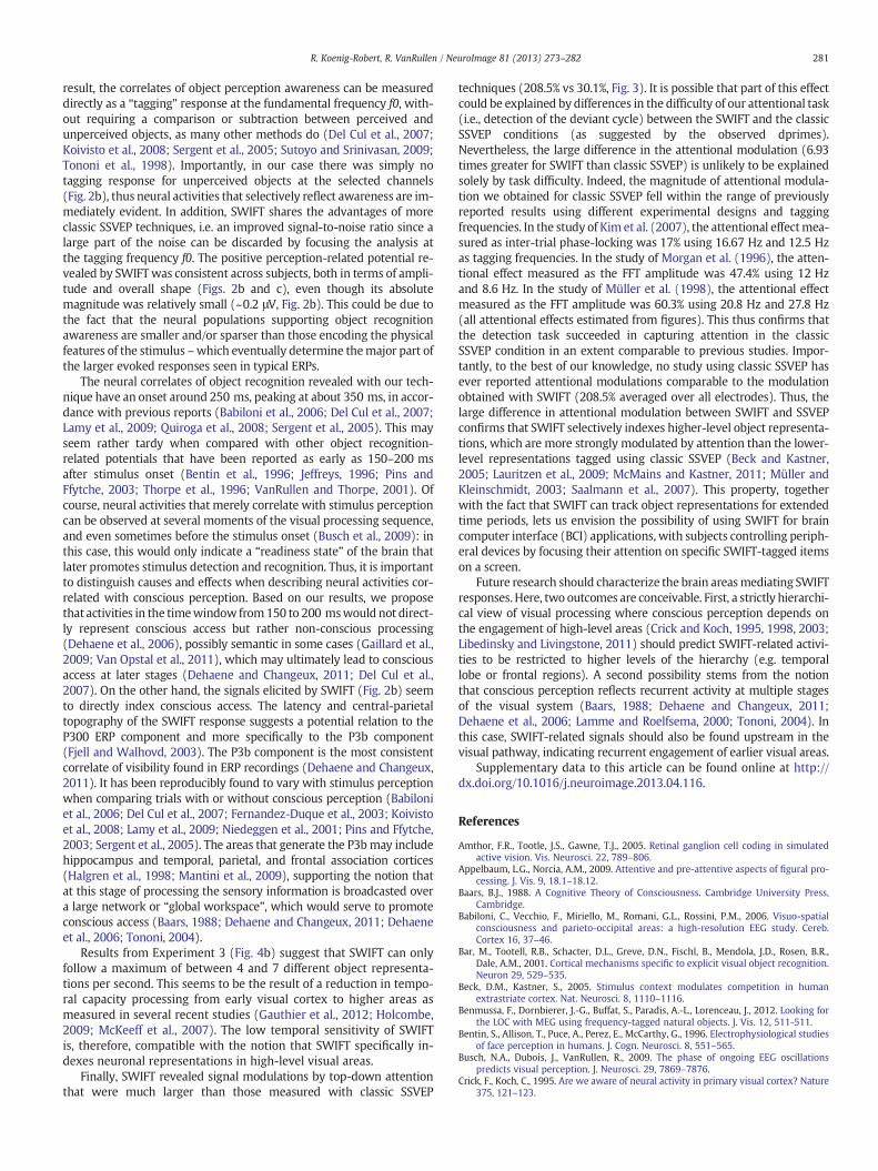
Our first step was to verify that our SWIFT procedure indeed pre-served low-level image features at both the local and global levels. Inour procedure (see Material and methods section), each wavelet wastemporally modulated at a different harmonic (i.e. multiple) frequencyof f0, such that individual contour elements returned to their originalorientation several times per cycle – but the entire image was restoredonly once per cycle. As a result, the luminance of individual pixels wasmodulated equally over a wide range of frequencies, and the averagetemporal frequency spectrum of pixel luminance was thus found to beconstant around the frequency of interest f0 (Fig. 1b, red curve); in con-trast, classic SSVEP methods using luminance or contrast modulationproduce a sharp peak at f0 in this temporal frequency spectrum(Fig. 1b, black curve). Similarly, global image contrastwas also preservedover time in SWIFT sequences (Fig. 1c, red curve), but not in classicSSVEP sequences (Fig. 1c, black curve). Finally, our wavelet scramblingprocedure also conserves the local distribution of spatial frequencies,since (by design) it only affects the orientation, but not the energy orspatial frequency of each wavelet.1
Phenomenologically, SWIFT sequences are perceived as a periodicflow of discernible information (i.e. the embedded image) recurringlyfading-out into an indistinguishable pattern – like an image reflectedin a water surface suddenly vanishing into chaotic ripples (see MovieS1). The recurrence of the embedded image is pulsatile, rather thansinusoidal. This is a consequence of the introduction of multiple har-monic frequencies in the wavelet scrambling: the original image isonly visible when the phases of the different harmonic modulationscome into alignment. With the five harmonics used here (seeMaterial and methods section) the embedded image was recogniz-able in only ~10% of the individual frames (see Fig. S1). This visibilitycan bemanipulated by varying the temporal frequency f0 and the numberof harmonic frequencies used in the wavelet scrambling. We exploitedthis property to create sequences where the embedded object was diffi-cult to identify. Crucially, after priming the content of the sequence by re-vealing the original image, the sameunrecognized objects became easy torecognize in a second presentation, enabling us to compare EEG activitywhen subjects were aware and unaware of the embedded object, as illus-trated in the next section.
Isolating object recognition awareness
To determine our method's sensitivity to semantic processing, wecompared the activity elicited by SWIFT sequences in three different con-ditions: (1) when the semantic content was present and perceived, (2)when it was present and not perceived and (3) when there was nosemantic content. To create these conditions, SWIFT sequences wereproduced containing natural images that could be either recognized(i.e. semantic information is available and perceived) or not recognized(i.e. semantic information is available but not perceived); we also pro-duced sequences containing abstract textures (i.e. no semantic informa-tion), which were used as a baseline. We showed each sequence twice,the second time after having explicitly revealed (i.e., primed) the embed-ded semantic content (when available), such that we could compare theactivity elicited by exactly the same sequence before and after the sub-ject had access to the semantic information.
More precisely, we presented SWIFT sequences (n = 100) containingeither grayscale natural imageswith one principal visual object (80%) orlow-level matched textures (20%). The image contoursweremodulated

2 The criterion by which subjects should ascertain proper recognition of an objectwas to have at least identified its basic categorization level (i.e., dog), but post-experiment interviews revealed that many objects were actually identified at the sub-ordinate level (i.e., German shepherd dog) during the naïve period.
Fig. 1. Construction and properties of SWIFT sequences. A. (1) The image is transformed into wavelet coefficients, representing local contours' energy and orientation. (2) For eachwavelet element represented by a 3D vector, two new vectors with the same length but random orientations are created: low level attributes (spatial frequency, contrast) are con-served but the local contour is disrupted. (3) The 3 vectors define a circular isoenergetic path. The original vector goes around this path at a multiple frequency of f0 (from 1 to 5).(4) Using the inverse wavelet transform, a cyclic sequence is generated showing the original image once per cycle at f0, and conserving low-level attributes at each frame. B. Pixelluminance modulation spectrum over time showing the spreading of the local luminance modulation over the harmonic frequencies of the cyclic phase-scrambling (red line). Thisensures a continuous modulation of visual stimulation in which the energy is not concentrated in any particular frequency, avoiding low-level evoked responses at f0. Note the peak(black line) at f0 obtained with classic SSVEP using sinusoidal contrast modulation. C. Global image contrast modulation spectrum over time. The contrast modulation at f0 used inclassic SSVEP (black line) evokes a strong neural response at the same frequency. In contrast, there is no peak of contrast modulation at f0 for the SWIFT technique (red line).
277R. Koenig-Robert, R. VanRullen / NeuroImage 81 (2013) 273–282
cyclically over time at f0 = 1.5 Hz, meaning that the original imagereappeared regularly with a period of ~0.67 s. Importantly, each se-quence contained only one embedded image (either a natural imageor an abstract texture), and was used only once in the experiment.Each trial consisted of two distinct periods (Fig. 2a): the first “naïve” pe-riod (30 s), followed by a steady presentation of the original imagecontained in the sequence (2 s), and finally the “cognizant” period(10 s). In the naïve period, subjects saw the SWIFT sequence for thefirst time; their task consisted in reporting the identification of a visualobject (i.e. a non-abstract item) by a button press as fast as possible.Two different response keys were provided corresponding to distinctconfidence levels: “I perceive an object-like item, but I am not sure ofwhich object it is” (key 1), and “I see an object and I have identified itconfidently” (key 2). It was possible to press the second key directly, orthe first key subsequently followed by the second one. When subjectsdid not perceive any object nokey presswas required. After the naïve pe-riod, the image embedded in the sequence was revealed and presentedsteadily for 2 s. Subjects were instructed to explore it freely and to seekthe features (if any) that had not been recognized during the naïve peri-od. Finally, in the cognizant period the SWIFT sequence was showedagain (10 s), and subjects were asked to try and identify the objectand/or the details they had missed during the naïve period. In case akey had been pressed during the naïve period, subjects had to report atthe end of the trial whether the object perceived in the naïve period ac-tually corresponded to the true object in the steady image (i.e. the‘prime’ presented in-between the naïve and the cognizant periods).
Only correctly identified objects were considered as recognized2. Thenatural images used to construct the sequences contained non-canonical views of bodies, faces, animals and manmade objects, whichmeans that several imageswere not recognized during the naïve period,but only during the cognizant period, i.e. after their identity had beenrevealed. This allowed us to compare the activity elicited by SWIFTwhen the same object was recognized and when it was not.
Trials were classified as ‘quickly recognized’when a natural imagewas presented and the subject recognized an object with high confi-dence within the first 10 s of the naïve period (22.2% of trials). Trialswere classified as ‘tardily recognized’ when a natural image waspresented but the subject did not recognize an object during the30 s of the naïve period (22.4% of trials). Trials were classified as‘no-object’ when abstract textures where presented (20%, regardlessof the subject's response: the false alarm rate at low confidence was2.86%, and at high confidence only 0.14%). Sequences presenting nat-ural images where the objects were not confidently detected (i.e. onlywith low confidence, or with high confidence but only after the first10 s of presentation) were not analyzed (35.4% of trials).
We analyzed EEG data (64 channels) of 17 participants. Eachwavelet-scrambling cycle (669 ms), starting at the moment when

Fig. 2. Isolating object perception awareness. A. Paradigm to compare activity elicited by SWIFT under recognition and nonrecognition of objects (see Results section). Two SWIFT epochs com-posed each trial: the naïve period (30 s,where the sequencewas presented for the first time) and the cognizant period (10 s,where the sequencewas presented again). In between, the originalimagewas presented steadily for 2 s, ensuring that the object (if any)would be later perceived in the cognizant period. Trials inwhich the objectwas detected in thefirst 10 s of thenaïve periodwere labeled as quickly recognized trials (QR). Trials inwhich objectswere recognized only in the cognizant period (no button press during the naïve period)were labeled as tardily recognizedtrials (TR). In no-object trials (NO), abstract textureswere shown and no objectwas ever recognized. B. ERP analysis over 4 central electrodes (marked by a dot in the topographies) taking eachwavelet-scrambling cycle (667 ms) as an epoch. Zero time indicates the frame where the original (unscrambled) imagewas presented. Shaded areas represent S.E.M. During the naïve period,only the activity elicited in QR, but not in TR trials differed (p b 0.05, gray box) from the baselinemeasured in the NO trials. In the cognizant period, however, both the QR and the TR conditionsdiffered from the NO condition (p b 0.05, gray box), and the waveforms elicited by QR and TR trials were very similar in this period. To summarize, a significant response only occurred whensubjects consciously recognized the objects in the sequences. Topographieswere calculated over the time points fallingwithin the gray box (marking statistical significance). Object recognitionactivity peaked over central electrodes, and was only observed when an object was recognized. C. Phase-locking analysis representing the similarity of ERP waveforms across subjects. EEGphase-locking across subjects at f0was significant (p b 0.05) when an object was recognized, but did not differ from chance level when no object was recognized.
278 R. Koenig-Robert, R. VanRullen / NeuroImage 81 (2013) 273–282

279R. Koenig-Robert, R. VanRullen / NeuroImage 81 (2013) 273–282
the original image was fully recognizable, was taken as an epoch tocompute the event-related potential (ERP). Based on the ERP topogra-phy (Fig. 2b), we selected 4 central electrodes where the ERP washighest, and we present the corresponding ERP in Fig. 2b. The ERPfor no-object trials can be used as a baseline (green line, Fig. 2b),since there was no semantic information present in these trials.The quickly recognized trials (where the semantic information waspresent and perceived, black line, Fig. 2b) showed a positive peakaround 300 ms, both during the naïve period (with a significant differ-ence to the ‘no-object’ baseline from 261 to 387 ms, two-tailed, pairedt-test, p b 0.05) and during the cognizant period (significant differ-ence from 269 to 411 ms). Tardily recognized trials in the naïve period(where the semantic information was present but not yet perceived,red line, Fig. 2b, left panel), on the other hand, were not significantlydifferent from no-object trials (two-tailed, paired t-test, p > 0.05),but only showed a significant difference during the cognizant period(where the semantic informationwas present and this time perceived,red line, Fig. 2b, right panel) from 269 to 388 ms (two-tailed, pairedt-test, p b 0.05). ERP topographies, computed over time points whenquickly-recognized ERPs differed from baseline for the naïve period(261–387 ms), and over time points when both quickly-recognizedERPs and tardily-recognized ERPs differed from baseline for the cogni-zant period (269–388 ms), show a positive central potential which isselective to object recognition: it is present whenever an object is rec-ognized (whether spontaneously, during the naïve period, or after ex-plicit priming, during the cognizant period), but it is absent otherwise.
Finally, the similarity of SWIFT responses across subjects was assessedby computing the phase-locking value of ERPs, band-pass filtered at f0,across the subjects' population (see Material and methods for details).Whenever an object was recognized (quickly recognized trials in bothnaïve and cognizant periods, and tardily recognized trials in cognizantperiod) a significant phase-locking emerged, indicative of a globallysimilar ERP waveform across subjects (Fig. 2c). Importantly, no signifi-cant phase locking was observed for unrecognized objects or no-object sequences. Thus, SWIFT responses appear to constitute a reliableindex of conscious recognition.
Measuring attention deployment
Our second criterion to validate the SWIFT technique involvedmeasuring top-down attentional modulations. These modulationsare known to be stronger in higher-level visual areas than in lower-level ones (Beck and Kastner, 2005; Lauritzen et al., 2009; McMainsand Kastner, 2011; Müller and Kleinschmidt, 2003; Saalmann et al.,2007). As a result, the conscious representation of an overtly recog-nized object (assuming that it is encoded in higher-level areas)must be enhanced more strongly by attention than the physical attri-butes of the same object. Hence, SWIFT responses should be morestrongly modulated by attention than the responses from more clas-sic frequency-tagging methods. To test this we quantified attentiondeployment to a face image using either (on randomly interleavedtrials) a simple contrast modulation (classic SSVEP) or our wavelet-scrambling method (SWIFT). In each trial, two faces were presentedon either side of fixation, each one oscillating at a different frequency(1.5 Hz or 2.03 Hz, positions counterbalanced across trials). A centralcue, pointing either to the left or right, indicated which face had to beattended (Fig. 3a). In order to engage the subjects' attention, the taskwas to report whether a “deviant” cycle (25% probability) had oc-curred in the attended sequence (i.e. a cycle in which the circularwavelet scrambling or the SSVEP contrast modulation did not returnto the original image, see Material and methods for details).
Dprimes for classic SSVEP and SWIFT conditions were 3.83 and 1.2respectively (Fig. 3b). The task was performed above chance level(d’ = 0) for both classic SSVEP (p = 3.5∙10−9, two-tailed, one-samplet-test) and SWIFT trials (p = 3.3∙10−5, two-tailed, one-sample t-test).This shows that the task was easier for SSVEP than for SWIFT trials,
whichmust be kept inmindwhen interpreting themeasured attention-al effect. The effect of attention was measured as the relative increase ofphase-locking values across trials at f0 over all channels, for the targetsequence compared to the distractor sequence (Fig. 3c; see Materialand methods for details). The attentional effect was predominant overcontralateral parietal electrodes for the classic SSVEP condition (Fig. 3c,upper panel, right) as previously documented (Kim et al., 2007;Morgan et al., 1996; Müller et al., 1998); whereas, for the SWIFT condi-tion, an additional central hot spot was present on the topographies(Fig. 3c, upper panel, left). The amplitude of the attentional effect re-vealed by the classic SSVEP technique was 30.1% (±6%, s.e.m. across 8subjects), consistent with existing findings (17%, 47.4% and 60.3% inKim et al., 2007; Morgan et al., 1996; Müller et al., 1998 studies, respec-tively). In SWIFT trials, the attentional effect reached 208.5% (±23%,s.e.m.). Even taking into account the difference in behavioral perfor-mance, this difference (p b 10−4, two-tailed, paired t-test) is consistentwith a selective tagging of high-level object representations by SWIFT,which is predicted to be more strongly modulated by top-down atten-tion than the lower-level feature extraction processes associated withmore classic SSVEP sequences.
Probing the temporal dynamics of object representations tracked by SWIFT
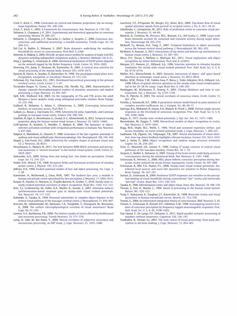
Finally, we measured the temporal dynamics of SWIFT responses todetermine the rate at which the high-level object representations thatare selectively tagged by SWIFT can be processed by the visual system,expecting this rate to be strongly limited due to the reduction in tem-poral capacity processing from early visual cortex to higher areas(Gauthier et al., 2012; Holcombe, 2009; McKeeff et al., 2007). To thisaim,we presented SWIFT sequences at differentmodulation frequenciesf0: the maximum frequency at which SWIFT can successfully tag objectrepresentations should indicate the fastest rate at which the visualsystem can extract and integrate visual information to lead to overtrecognition of the objects contained in the SWIFT sequences. SWIFT se-quences containing grayscale natural images (n = 20) were shown at 8frequencies in different trials (1.5, 2.03, 2.62, 3.4, 4.32, 6.96, 8.42 and12.31 Hz). As in the previous experiment, the subjects' task was todetect any deviant cycle (present in 20% of trials). We measured thetagging as the mean phase-locking factor across trials at f0 (Fig. 4; seeMaterial and methods). This tagging was significant from 1.5 to4.32 Hz (two-tailed, one sample t-test, fdr-corrected, p = 3.3∙10−5,2.6∙10−5, 8.5∙10−4, 3.3∙10−5, 2.9∙10−5). Thus, the high-level objectrepresentations that are selectively tagged by the SWIFTmethod appearto reach their limit between 4 and 7 object representations per second.
Discussion
Wehavedeveloped a new technique to explore theneural correlatesof object perception awareness which combines imagemanipulation inthewavelets domain and frequency-tagging. Other imagemanipulationtechniques have been proposed previously to modulate high-levelimage properties while conserving some physical attributes (Moca etal., 2011; Sadr and Sinha, 2004). A first particularity of SWIFT comparedwith previous techniques is that we were able to combine it withfrequency-tagging to track high-level image representationswhilemin-imizing neural activity evoked by changes in the physical properties ofthe stimulation. This was achieved in part by introducing several tem-poral harmonic frequencies (i.e., multiples of f0) into the cyclic waveletscrambling; as a result, the average luminance temporal frequencyspectrum was constant around the tagging frequency f0 (Fig. 1b, seeMaterial and methods for details). One advantage of SWIFT overFourier-based image manipulation approaches (i.e., Sadr and Sinha,2004) is that wavelet-based scrambling conserves local energy and spa-tial frequencies. In SWIFT, the orientation of local contours at each spa-tial frequency is disrupted but their energy and relative position in theimage is not altered. Conversely, phase-scrambling in the Fourier

280 R. Koenig-Robert, R. VanRullen / NeuroImage 81 (2013) 273–282
domain disperses energy and spatial frequency information across theentire image (but leaves the relative strength of each orientationunaltered). This implies that the large-scale pattern of neural activationin any retinotopic visual area will be severely distorted by such globalimage scrambling approaches (e.g., if a region of the original imagehas particularly strong energy at high spatial frequencies, this energywill be randomly redistributed among high-spatial frequencies in vari-ous other regions of the scrambled image, thus recruiting different
Fig. 4. Temporal dynamics of object recognition. SWIFT was applied to natural imagesat 8 different frequencies (from 1.4953 to 12.3077 Hz). The tagging response was cal-culated as the phase-locking factor across trials at f0 (after trials were aligned relativeto the first appearance of the original unscrambled image in the sequence). A signifi-cant tagging (p b 0.05) was obtained at frequencies from 1.4953 to 4.3243, but notat frequencies of 6.97 Hz and above. This suggests that the visual system can form amaximum of 4 to 7 different high-level representations per second.
retinotopic neuronal populations in the original and scrambled images).SWIFT, on the other hand, should preserve the large-scale retinotopicpattern of neuronal activation (e.g. if a part of the original image isrich in high spatial frequencies, the corresponding energy will beretained at this location and spatial frequency in the scrambled image;only the corresponding orientations will be altered).
Another class of image manipulation methods, the so-called gen-erative techniques (see for example Benmussa et al., 2012; Moca etal., 2011) take a very different approach: they generate images frombasic structural elements (such as dots, color patches, line segments,Gabors, etc.) in order to gain better control over image properties.However, by avoiding the pitfalls of natural image manipulation tech-niques (a.k.a. transformative techniques), this kind of method alsoprecludes the utilization of natural images as stimuli. Finally, it isworth pointing out that no image manipulation technique can everdisrupt an image's semantic contents without affecting its low-levelphysical properties in someway (indeed, two images that have strict-ly identical low-level physical properties are necessarily identicalcopies, and thus also have identical semantic contents). Each imagemanipulation technique must therefore choose to equate some phys-ical properties at the expense of others; we believe that our SWIFTtechnique represents a fair trade-off between local and global imagephysical properties conservation.
As a consequence of the conservation of low-level physical featureson each frame, SWIFT isolates high-level activities – i.e. the brain'stime-locked responses to the pulsatile flow of semantic informationcarried by the frames where the embedded images are visible. As a
Fig. 3. Measuring attention deployment. A. The effect of attention on frequency-tagging was calculated using SWIFT (wavelet scrambling modulation) or classicSSVEP (contrast modulation) in different trials. Two faces were presented on eitherside of the screen at different modulation frequencies (1.4953 Hz and 2.0253, random-ly counterbalanced). Top-down attention was manipulated by a central cue pointing tothe face to be attended, and a difficult detection task in the corresponding sequence.B. Dprimes for the detection task in the SWIFT and classic SSVEP conditions. C. Attentionmodulation of the frequency-tagged response. Upper panel: topographies showing theincrease (percentage) in phase-locking at f0 for the target compared to the distractorstimulus. Topographies represent activations elicited for target (T) presented on the leftside and distractor (D) on the right side of the fixation point (for targets presented onthe right, topographies were flipped around the central axis.) Lower panel: increase inphase-locking at f0 for the target compared to the distractor stimulus averaged over all64 electrodes.
281R. Koenig-Robert, R. VanRullen / NeuroImage 81 (2013) 273–282
result, the correlates of object perception awareness can be measureddirectly as a “tagging” response at the fundamental frequency f0, with-out requiring a comparison or subtraction between perceived andunperceived objects, as many other methods do (Del Cul et al., 2007;Koivisto et al., 2008; Sergent et al., 2005; Sutoyo and Srinivasan, 2009;Tononi et al., 1998). Importantly, in our case there was simply notagging response for unperceived objects at the selected channels(Fig. 2b), thus neural activities that selectively reflect awareness are im-mediately evident. In addition, SWIFT shares the advantages of moreclassic SSVEP techniques, i.e. an improved signal-to-noise ratio since alarge part of the noise can be discarded by focusing the analysis atthe tagging frequency f0. The positive perception-related potential re-vealed by SWIFTwas consistent across subjects, both in terms of ampli-tude and overall shape (Figs. 2b and c), even though its absolutemagnitude was relatively small (~0.2 μV, Fig. 2b). This could be due tothe fact that the neural populations supporting object recognitionawareness are smaller and/or sparser than those encoding the physicalfeatures of the stimulus –which eventually determine themajor part ofthe larger evoked responses seen in typical ERPs.
The neural correlates of object recognition revealed with our tech-nique have an onset around 250 ms, peaking at about 350 ms, in accor-dance with previous reports (Babiloni et al., 2006; Del Cul et al., 2007;Lamy et al., 2009; Quiroga et al., 2008; Sergent et al., 2005). This mayseem rather tardy when compared with other object recognition-related potentials that have been reported as early as 150–200 msafter stimulus onset (Bentin et al., 1996; Jeffreys, 1996; Pins andFfytche, 2003; Thorpe et al., 1996; VanRullen and Thorpe, 2001). Ofcourse, neural activities that merely correlate with stimulus perceptioncan be observed at several moments of the visual processing sequence,and even sometimes before the stimulus onset (Busch et al., 2009): inthis case, this would only indicate a “readiness state” of the brain thatlater promotes stimulus detection and recognition. Thus, it is importantto distinguish causes and effects when describing neural activities cor-related with conscious perception. Based on our results, we proposethat activities in the timewindow from150 to 200 mswould not direct-ly represent conscious access but rather non-conscious processing(Dehaene et al., 2006), possibly semantic in some cases (Gaillard et al.,2009; Van Opstal et al., 2011), which may ultimately lead to consciousaccess at later stages (Dehaene and Changeux, 2011; Del Cul et al.,2007). On the other hand, the signals elicited by SWIFT (Fig. 2b) seemto directly index conscious access. The latency and central-parietaltopography of the SWIFT response suggests a potential relation to theP300 ERP component and more specifically to the P3b component(Fjell and Walhovd, 2003). The P3b component is the most consistentcorrelate of visibility found in ERP recordings (Dehaene and Changeux,2011). It has been reproducibly found to vary with stimulus perceptionwhen comparing trials with or without conscious perception (Babiloniet al., 2006; Del Cul et al., 2007; Fernandez-Duque et al., 2003; Koivistoet al., 2008; Lamy et al., 2009; Niedeggen et al., 2001; Pins and Ffytche,2003; Sergent et al., 2005). The areas that generate the P3bmay includehippocampus and temporal, parietal, and frontal association cortices(Halgren et al., 1998; Mantini et al., 2009), supporting the notion thatat this stage of processing the sensory information is broadcasted overa large network or “global workspace”, which would serve to promoteconscious access (Baars, 1988; Dehaene and Changeux, 2011; Dehaeneet al., 2006; Tononi, 2004).
Results from Experiment 3 (Fig. 4b) suggest that SWIFT can onlyfollow a maximum of between 4 and 7 different object representa-tions per second. This seems to be the result of a reduction in tempo-ral capacity processing from early visual cortex to higher areas asmeasured in several recent studies (Gauthier et al., 2012; Holcombe,2009; McKeeff et al., 2007). The low temporal sensitivity of SWIFTis, therefore, compatible with the notion that SWIFT specifically in-dexes neuronal representations in high-level visual areas.
Finally, SWIFT revealed signal modulations by top-down attentionthat were much larger than those measured with classic SSVEP
techniques (208.5% vs 30.1%, Fig. 3). It is possible that part of this effectcould be explained by differences in the difficulty of our attentional task(i.e., detection of the deviant cycle) between the SWIFT and the classicSSVEP conditions (as suggested by the observed dprimes).Nevertheless, the large difference in the attentional modulation (6.93times greater for SWIFT than classic SSVEP) is unlikely to be explainedsolely by task difficulty. Indeed, the magnitude of attentional modula-tion we obtained for classic SSVEP fell within the range of previouslyreported results using different experimental designs and taggingfrequencies. In the study of Kim et al. (2007), the attentional effectmea-sured as inter-trial phase-locking was 17% using 16.67 Hz and 12.5 Hzas tagging frequencies. In the study of Morgan et al. (1996), the atten-tional effect measured as the FFT amplitude was 47.4% using 12 Hzand 8.6 Hz. In the study of Müller et al. (1998), the attentional effectmeasured as the FFT amplitude was 60.3% using 20.8 Hz and 27.8 Hz(all attentional effects estimated from figures). This thus confirms thatthe detection task succeeded in capturing attention in the classicSSVEP condition in an extent comparable to previous studies. Impor-tantly, to the best of our knowledge, no study using classic SSVEP hasever reported attentional modulations comparable to the modulationobtained with SWIFT (208.5% averaged over all electrodes). Thus, thelarge difference in attentional modulation between SWIFT and SSVEPconfirms that SWIFT selectively indexes higher-level object representa-tions, which are more strongly modulated by attention than the lower-level representations tagged using classic SSVEP (Beck and Kastner,2005; Lauritzen et al., 2009; McMains and Kastner, 2011; Müller andKleinschmidt, 2003; Saalmann et al., 2007). This property, togetherwith the fact that SWIFT can track object representations for extendedtime periods, lets us envision the possibility of using SWIFT for braincomputer interface (BCI) applications, with subjects controlling periph-eral devices by focusing their attention on specific SWIFT-tagged itemson a screen.
Future research should characterize the brain areasmediating SWIFTresponses. Here, two outcomes are conceivable. First, a strictly hierarchi-cal view of visual processing where conscious perception depends onthe engagement of high-level areas (Crick and Koch, 1995, 1998, 2003;Libedinsky and Livingstone, 2011) should predict SWIFT-related activi-ties to be restricted to higher levels of the hierarchy (e.g. temporallobe or frontal regions). A second possibility stems from the notionthat conscious perception reflects recurrent activity at multiple stagesof the visual system (Baars, 1988; Dehaene and Changeux, 2011;Dehaene et al., 2006; Lamme and Roelfsema, 2000; Tononi, 2004). Inthis case, SWIFT-related signals should also be found upstream in thevisual pathway, indicating recurrent engagement of earlier visual areas.
Supplementary data to this article can be found online at http://dx.doi.org/10.1016/j.neuroimage.2013.04.116.
References
Amthor, F.R., Tootle, J.S., Gawne, T.J., 2005. Retinal ganglion cell coding in simulatedactive vision. Vis. Neurosci. 22, 789–806.
Appelbaum, L.G., Norcia, A.M., 2009. Attentive and pre-attentive aspects of figural pro-cessing. J. Vis. 9, 18.1–18.12.
Baars, B.J., 1988. A Cognitive Theory of Consciousness. Cambridge University Press,Cambridge.
Babiloni, C., Vecchio, F., Miriello, M., Romani, G.L., Rossini, P.M., 2006. Visuo-spatialconsciousness and parieto-occipital areas: a high-resolution EEG study. Cereb.Cortex 16, 37–46.
Bar, M., Tootell, R.B., Schacter, D.L., Greve, D.N., Fischl, B., Mendola, J.D., Rosen, B.R.,Dale, A.M., 2001. Cortical mechanisms specific to explicit visual object recognition.Neuron 29, 529–535.
Beck, D.M., Kastner, S., 2005. Stimulus context modulates competition in humanextrastriate cortex. Nat. Neurosci. 8, 1110–1116.
Benmussa, F., Dornbierer, J.-G., Buffat, S., Paradis, A.-L., Lorenceau, J., 2012. Looking forthe LOC with MEG using frequency-tagged natural objects. J. Vis. 12, 511-511.
Bentin, S., Allison, T., Puce, A., Perez, E., McCarthy, G., 1996. Electrophysiological studiesof face perception in humans. J. Cogn. Neurosci. 8, 551–565.
Busch, N.A., Dubois, J., VanRullen, R., 2009. The phase of ongoing EEG oscillationspredicts visual perception. J. Neurosci. 29, 7869–7876.
Crick, F., Koch, C., 1995. Are we aware of neural activity in primary visual cortex? Nature375, 121–123.
282 R. Koenig-Robert, R. VanRullen / NeuroImage 81 (2013) 273–282
Crick, F., Koch, C., 1998. Constraints on cortical and thalamic projections: the no-strong-loops hypothesis. Nature 391, 245–250.
Crick, F., Koch, C., 2003. A framework for consciousness. Nat. Neurosci. 6, 119–126.Dehaene, S., Changeux, J.-P., 2011. Experimental and theoretical approaches to conscious
processing. Neuron 70, 200–227.Dehaene, S., Changeux, J.-P., Naccache, L., Sackur, J., Sergent, C., 2006. Conscious, pre-
conscious, and subliminal processing: a testable taxonomy. Trends Cogn. Sci. 10,204–211.
Del Cul, A., Baillet, S., Dehaene, S., 2007. Brain dynamics underlying the nonlinearthreshold for access to consciousness. PLoS Biol. 5, e260.
Delorme, A.,Makeig, S., 2004. EEGLAB: an open source toolbox for analysis of single-trial EEGdynamics including independent component analysis. J. Neurosci. Methods 134, 9–21.
Ding, J., Sperling, G., Srinivasan, R., 2006. Attentional modulation of SSVEP power dependson the network tagged by the flicker frequency. Cereb. Cortex 16, 1016–1029.
Downing, P.E., Jiang, Y., Shuman, M., Kanwisher, N., 2001. A cortical area selective forvisual processing of the human body. Science 293, 2470–2473.
Epstein, R., Harris, A., Stanley, D., Kanwisher, N., 1999. The parahippocampal place area:recognition, navigation, or encoding? Neuron 23, 115–125.
Felleman, D.J., Van Essen, D.C., 1991. Distributed hierarchical processing in the primatecerebral cortex. Cereb. Cortex 1, 1–47.
Fernandez-Duque, D., Grossi, G., Thornton, I.M., Neville, H.J., 2003. Representation ofchange: separate electrophysiological markers of attention, awareness, and implicitprocessing. J. Cogn. Neurosci. 15, 491–507.
Fjell, A.M., Walhovd, K.B., 2003. On the topography of P3a and P3b across the adultlifespan–a factor-analytic study using orthogonal procrustes rotation. Brain Topogr.15, 153–164.
Gaillard, R., Dehaene, S., Adam, C., Clémenceau, S., 2009. Converging intracranialmarkers of conscious access. PLoS Biol. 7.
Gallant, J.L., Braun, J., Van Essen, D.C., 1993. Selectivity for polar, hyperbolic, and Cartesiangratings in macaque visual cortex. Science 259, 100–103.
Gauthier, B., Eger, E., Hesselmann, G., Giraud, A.-L., Kleinschmidt, A., 2012. Temporal tuningproperties along the human ventral visual stream. J. Neurosci. 32, 14433–14441.
Grill-Spector, K., Kushnir, T., Hendler, T., Malach, R., 2000. The dynamics of object-selective activation correlate with recognition performance in humans. Nat. Neurosci.3, 837–843.
Halgren, E., Marinkovic, K., Chauvel, P., 1998. Generators of the late cognitive potentials inauditory and visual oddball tasks. Electroencephalogr. Clin. Neurophysiol. 106, 156–164.
Hegdé, J., Van Essen, D.C., 2000. Selectivity for complex shapes in primate visual areaV2. J. Neurosci. 20, RC61.
Hesselmann, G., Malach, R., 2011. The link between fMRI-BOLD activation and percep-tual awareness is “stream-invariant” in the human visual system. Cereb. Cortex 21,2829–2837.
Holcombe, A.O., 2009. Seeing slow and seeing fast: two limits on perception. TrendsCogn. Sci. 13, 216–221.
Hubel, D.H., Wiesel, T.N., 1968. Receptive fields and functional architecture of monkeystriate cortex. J. Physiol. 195, 215–243.
Jeffreys, D.A., 1996. Evoked potential studies of face and object processing. Vis. Cogn. 3,1–38.
Kanwisher, N., McDermott, J., Chun, M.M., 1997. The fusiform face area: a module inhuman extrastriate cortex specialized for face perception. J. Neurosci. 17, 4302–4311.
Kaspar, K., Hassler, U., Martens, U., Trujillo-Barreto, N., Gruber, T., 2010. Steady-state vi-sually evoked potential correlates of object recognition. Brain Res. 1343, 112–121.
Kim, Y.J., Grabowecky, M., Paller, K.A., Muthu, K., Suzuki, S., 2007. Attention inducessynchronization-based response gain in steady-state visual evoked potentials.Nat. Neurosci. 10, 117–125.
Kobatake, E., Tanaka, K., 1994. Neuronal selectivities to complex object features in theventral visual pathway of the macaque cerebral cortex. J. Neurophysiol. 71, 856–867.
Koivisto, M., Lähteenmäki, M., Sørensen, T.A., Vangkilde, S., Overgaard, M., Revonsuo,A., 2008. The earliest electrophysiological correlate of visual awareness? BrainCogn. 66, 91–103.
Lamme, V.A., Roelfsema, P.R., 2000. The distinct modes of vision offered by feedforwardand recurrent processing. Trends Neurosci. 23, 571–579.
Lamy, D., Salti, M., Bar-Haim, Y., 2009. Neural correlates of subjective awareness andunconscious processing: an ERP study. J. Cogn. Neurosci. 21, 1435–1446.
Lauritzen, T.Z., D'Esposito, M., Heeger, D.J., Silver, M.A., 2009. Top-down flow of visualspatial attention signals from parietal to occipital cortex. J. Vis. 9, 18.1–18.14.
Libedinsky, C., Livingstone, M., 2011. Role of prefrontal cortex in conscious visual per-ception. J. Neurosci. 31, 64–69.
Mantini, D., Corbetta, M., Perrucci, M.G., Romani, G.L., Del Gratta, C., 2009. Large-scalebrain networks account for sustained and transient activity during target detec-tion. Neuroimage 44, 265–274.
McKeeff, T.J., Remus, D.A., Tong, F., 2007. Temporal limitations in object processingacross the human ventral visual pathway. J. Neurophysiol. 98, 382–393.
McMains, S., Kastner, S., 2011. Interactions of top-down and bottom-up mechanisms inhuman visual cortex. J. Neurosci. 31, 587–597.
Moca, V.V., Ţincaş, I., Melloni, L., Mureşan, R.C., 2011. Visual exploration and objectrecognition by lattice deformation. PLoS One 6, e22831.
Morgan, S.T., Hansen, J.C., Hillyard, S.A., 1996. Selective attention to stimulus locationmodulates the steady-state visual evoked potential. Proc. Natl. Acad. Sci. U. S. A.93, 4770–4774.
Müller, N.G., Kleinschmidt, A., 2003. Dynamic interaction of object- and space-basedattention in retinotopic visual areas. J. Neurosci. 23, 9812–9816.
Müller, M.M., Picton, T.W., Valdes-Sosa, P., Riera, J., Teder-Sälejärvi, W.A., Hillyard, S.A.,1998. Effects of spatial selective attention on the steady-state visual evoked poten-tial in the 20–28 Hz range. Cogn. Brain Res. 6, 249–261.
Niedeggen, M., Wichmann, P., Stoerig, P., 2001. Change blindness and time to con-sciousness. Eur. J. Neurosci. 14, 1719–1726.
Pins, D., Ffytche, D., 2003. The neural correlates of conscious vision. Cereb. Cortex 13,461–474.
Portilla, J., Simoncelli, E.P., 2000. A parametric texture model based on joint statistics ofcomplex wavelet coefficients. Int. J. Comput. Vis. 40, 49–71.
Quiroga, R.Q., Mukamel, R., Isham, E.A., Malach, R., Fried, I., 2008. Human single-neuronresponses at the threshold of conscious recognition. Proc. Natl. Acad. Sci. U. S. A.105, 3599–3604.
Regan, D., 1977. Steady-state evoked potentials. J. Opt. Soc. Am. 67, 1475–1489.Riesenhuber, M., Poggio, T., 1999. Hierarchical models of object recognition in cortex.
Nat. Neurosci. 2, 1019–1025.Rugg, M.D., Doyle, M.C., Wells, T., 1995. Word and nonword repetition within- and
across-modality: an event-related potential study. J. Cogn. Neurosci. 7, 209–227.Saalmann, Y.B., Pigarev, I.N., Vidyasagar, T.R., 2007. Neural mechanisms of visual atten-
tion: how top-down feedback highlights relevant locations. Science 316, 1612–1615.Sadr, J., Sinha, P., 2004. Object recognition and random image structure evolution.
Cognit. Sci. 28, 259–287.Sclar, G., Maunsell, J.H., Lennie, P., 1990. Coding of image contrast in central visual
pathways of the macaque monkey. Vision Res. 30, 1–10.Sergent, C., Baillet, S., Dehaene, S., 2005. Timing of the brain events underlying access to
consciousness during the attentional blink. Nat. Neurosci. 8, 1391–1400.Srinivasan, R., Petrovic, S., 2006. MEG phase follows conscious perception during bin-
ocular rivalry induced by visual stream segregation. Cereb. Cortex 16, 597–608.Srinivasan, R., Bibi, F.A., Nunez, P.L., 2006. Steady-state visual evoked potentials: dis-
tributed local sources and wave-like dynamics are sensitive to flicker frequency.Brain Topogr. 18, 167–187.
Sutoyo, D., Srinivasan, R., 2009. Nonlinear SSVEP responses are sensitive to the percep-tual binding of visual hemifields during conventional “eye” rivalry and interocular“percept” rivalry. Brain Res. 1251, 245–255.
Tanaka, K., 1996. Inferotemporal cortex and object vision. Annu. Rev. Neurosci. 19, 109–139.Thorpe, S., Fize, D., Marlot, C., 1996. Speed of processing in the human visual system.
Nature 381, 520–522.Tong, F., Nakayama, K., Vaughan, J.T., Kanwisher, N., 1998. Binocular rivalry and visual
awareness in human extrastriate cortex. Neuron 21, 753–759.Tononi, G., 2004. An information integration theory of consciousness. BMC Neurosci. 5, 42.Tononi, G., Srinivasan, R., Russell, D.P., Edelman, G.M., 1998. Investigating neural corre-
lates of conscious perception by frequency-tagged neuromagnetic responses. Proc.Natl. Acad. Sci. U. S. A. 95, 3198–3203.
Van Opstal, F., De Lange, F.P., Dehaene, S., 2011. Rapid parallel semantic processing ofnumbers without awareness. Cognition 120, 136–147.
VanRullen, R., Thorpe, S.J., 2001. The time course of visual processing: from early per-ception to decision-making. J. Cogn. Neurosci. 13, 454–461.

Supplementary figure 1. The 107 frames of a SWIFT sequence modulated at f0 = 1.4953Hz. Each
frame was presented during 6.25 ms. The green outline (not shown in the experiment), indicates the
frame where the semantic information was fully available (unscrambled image), corresponding to
zero-time in the ERP analysis and to the reference time for the phase-locking calculation at f0.