interactive report neural correlates of memory retrieval...
TRANSCRIPT
Ž .Cognitive Brain Research 9 2000 209–222www.elsevier.comrlocaterbres
Interactive report
Neural correlates of memory retrieval and evaluation1
Charan Ranganath 2, Ken A. Paller )
Department of Psychology, Northwestern UniÕersity, 2029 Sheridan Road, EÕanston, IL 60208-2710, USA
Accepted 3 October 1999
Abstract
Results from recent neuroimaging studies have led to a controversy as to whether right or left prefrontal regions are relatively moreimportant for episodic retrieval. To address this issue, we recorded event-related brain potentials during two recognition tests withidentical stimuli but differing retrieval demands. In both tests, participants viewed a sequence of object drawings, half of which wereidentical to ones viewed earlier except for a change in size and half of which were new. Instructions were to discriminate between old and
Ž . Ž .new objects general test or to additionally decide whether old objects were larger or smaller at study specific test . Frontal brainpotentials that were more positive during the specific than during the general test for both old and new objects were interpreted as neuralcorrelates of the process by which specific attributes of test cues are compared with information retrieved from memory. Another ERPdifference between the specific and general tests, which was observed for old objects only, had a left posterior scalp topography and wasinterpreted to reflect the reactivation of memories for studied objects. Frontal and posterior potentials thus reflected two memoryprocesses important for accurate episodic retrieval. Furthermore, our findings suggest that both left and right prefrontal regions wereengaged when demands to retrieve and evaluate perceptual information increased. q 2000 Elsevier Science B.V. All rights reserved.
Keywords: Human memory; Event-related potentials; ERPs; Prefrontal cortex; Episodic memory; Source monitoring; Recollection
1. Introduction
A great deal of neuropsychological evidence has impli-cated prefrontal cortex in operations engaged during the
w xformation and retrieval of memories for events 29,52,57 .Although patients with prefrontal lesions are not amnesic,they show distinctive episodic memory impairments; theyare mildly impaired on tests of item recognition and cued
w xrecall 14,56,58,59,63 , they are disproportionately im-w xpaired at free recall 9,12,13,16,55 , and they are signifi-
cantly impaired on tests of memory for context informa-w xtion 6,15,24,26,28 . In addition, patients with prefrontal
damage may exhibit memory distortions such as falsew xrecognition 7,40,45,46,50,59 , and in the extreme, confab-
w xulation 1,21,22,30,54 .Interest in the role of prefrontal cortex in episodic
memory has intensified due to results from recent neu-roimaging studies showing robust activation of multiple
) Corresponding author. Tel.: q1-847-467-3370; Fax: q1-847-491-7859; E-mail: [email protected]; URL: http:rrwww.nwu.edurpeoplerkap
1 Published on the World Wide Web on 12 October 1999.2 Present address: Department of Neurology, University of Pennsylva-
nia, 3 West Gates, 3400 Spruce St., Philadelphia, PA 19104-4283, USA.
w xprefrontal regions 2,5,32,33,61,62 . Findings from thesestudies suggest not only that prefrontal cortex is importantfor memory processing but also that left and right pre-
w xfrontal regions make distinct contributions 32,33,61 .These laterality patterns were noted in the Hemispheric
Ž .Encoding Retrieval Asymmetry HERA model proposedw xby Tulving and colleagues 61 . According to this model,
left prefrontal cortex is more involved in episodic memoryencoding and in semantic memory retrieval, whereas rightprefrontal cortex is more involved in episodic retrieval.This view was supported by a meta-analysis conducted by
w xNyberg and colleagues 33 showing that of 16 positronŽ .emission tomography PET studies involving encoding
tasks, 13 reported significant prefrontal activations, all inthe left hemisphere. And of 31 PET studies of episodicretrieval, 29 reported significant prefrontal activation and26 of these reported larger prefrontal blood flow increasesin the right hemisphere. Subsequent studies using func-
Ž .tional magnetic resonance imaging fMRI have also re-ported right prefrontal activation during episodic retrievalw x3,4,38,49 .
Findings of hemispheric asymmetry during episodicretrieval were interpreted differently by Nolde, Johnson,
w xand Raye 32 . These researchers noted that findings of
0926-6410r00r$ - see front matter q 2000 Elsevier Science B.V. All rights reserved.Ž .PII: S0926-6410 99 00048-8
( )C. Ranganath, K.A. PallerrCognitiÕe Brain Research 9 2000 209–222210
right prefrontal activation were based on comparisons be-tween episodic retrieval tasks and non-memory baselinetasks. On the other hand, comparisons in some experi-ments were made between difficult and relatively lessdifficult episodic memory tests. In these contrasts, pre-frontal activations were more often left-lateralized or bilat-
w xeral. For example, Nolde, Johnson, and D’Esposito 31conducted an event-related fMRI study in which partici-pants were scanned during recognition testing with studiedwords, words corresponding to studied pictures, and un-studied words. In the oldrnew condition, participants at-tempted to discriminate between words that were or were
Ž .not studied either as a picture or as a word . In the sourcecondition, participants were asked to specify whether eachword was studied as a picture, studied as a word, orpreviously unstudied. Thus, the source condition placedgreater demands on retrieval and evaluation of specificperceptual attributes of memories than did the oldrnewcondition. Results showed that left prefrontal regions weremore active during the source condition than during theoldrnew condition, whereas right prefrontal regions wereactivated equally during the two test conditions.
w xTo explain these results, Nolde and colleagues 32suggested that right prefrontal regions implement ‘heuristicprocessing’ or evaluation of memories based on a singledimension, such as familiarity. In contrast, they argued thatleft prefrontal regions implement ‘systematic processing’or evaluation of multiple memory characteristics in orderto make a memory judgment. Thus, in contrast with theHERA model, these investigators proposed that left pre-frontal regions play an important role in episodic retrieval.
Whereas the neuroimaging studies reviewed above usedmethods that rely on hemodynamic correlates of neuralactivity, other studies have examined patterns of frontal
Ž .activity by recording event-related brain potentials ERPsw xduring tests of episodic retrieval 8,19,43,44,51,60,64–68 .
ERPs are scalp-recorded measures of electroencephalo-graphic activity synchronized to an external event. ERPsthus allow direct measurement of brain activity with tem-poral resolution on the order of milliseconds. Althoughpresent methods do not allow for precise localization ofintracranial sources of scalp-recorded ERPs, their hightemporal resolution can provide a useful complement to
w xneuroimaging methods 10,23,34,48 .In a previous experiment, we found that ERPs impli-
w xcated prefrontal regions in episodic memory retrieval 44 .Participants studied pictures of objects and were testedwith three types of stimuli: studied objects, studied objectswith altered aspect ratios, and previously unstudied ob-jects. Results from two memory tests were compared. Inthe general test, participants were instructed to disregardalterations in making oldrnew judgments, whereas in thespecific test they were instructed to judge an object ‘old’only if it was identical to a studied object and to judge allother objects ‘new.’ Thus, the specific test placed greaterdemands on the retrieval and evaluation of specific percep-
tual information than did the general test. We reasoned thatfrontal ERP differences between the specific and generaltests could provide insights into the role of prefrontalcortex in retrieval as follows. If prefrontal regions areselectively engaged following successful retrieval of spe-cific event information, then frontal ERP differences wouldbe prominent only for old pictures. But if prefrontal re-gions implement strategic processing during retrieval at-tempts, then frontal ERP differences would be observedfor both old and new pictures.
The central electrophysiological finding was that ERPswere enhanced during the specific test relative to thegeneral test. These effects were maximal over left frontalscalp regions. The finding that these ERP differences weresimilar for old and new pictures suggested that they didnot reflect successful recall of specific perceptual details ofstudied pictures. We concluded instead that we hadrecorded a neural correlate of a process wherein specificperceptual attributes of recognition cues were comparedwith the contents of memory. The left frontal scalp topog-raphy of this effect and the neuroimaging findings re-viewed above support the hypothesis that this evaluativeprocess is implemented by left prefrontal cortex.
Note that in the specific test employed in our previousw xstudy 44 , participants were required to judge whether or
not studied pictures had been changed, but they were notrequired to remember anything specific about the change.Conceivably, participants were able to evaluate the extentto which studied pictures were altered without engaging inthe more active process of recalling the precise way inwhich studied pictures were altered. Accordingly, it re-mains an open question whether frontal brain potentialsassociated with successful retrieval would be produced ifparticipants were given the more demanding requirementof recalling perceptual detail, rather than just evaluatingwhether or not there was a match between the recognitioncue and the contents of memory.
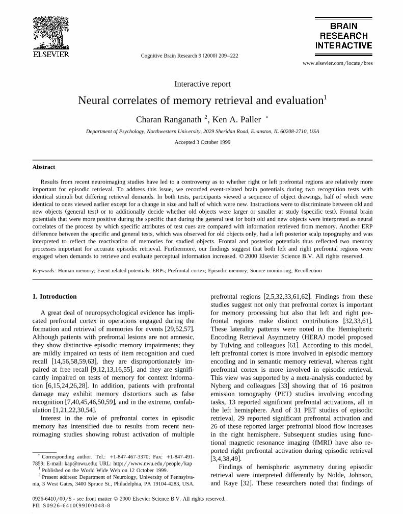
In the present study, we investigated whether frontalbrain potentials associated with successful retrieval wouldbe observed if participants were encouraged to recall per-ceptual detail. The design of the study is schematicallydepicted in Fig. 1. Participants studied line drawings of
Fig. 1. Sample stimuli and responses in each test condition.
( )C. Ranganath, K.A. PallerrCognitiÕe Brain Research 9 2000 209–222 211
objects and were tested with size-changed versions ofstudied objects and new objects. In the general test, theywere required to make old–new judgments, whereas in thespecific test they were additionally required to indicatewhether old objects were studied in a larger or smallersize. By comparing brain potentials recorded during thespecific and general test conditions, we were able toinvestigate frontal activity and its association with strategicretrieval and with successful recall of specific perceptualinformation.
2. Methods
2.1. Participants
Two men and ten women from the Northwestern Uni-versity community were paid for participating in the exper-iment. They were right-handed and ranged in age from 18to 22 years. Data from four additional participants were
Ždiscarded two because of technical difficulties and two.because of excessive eye and muscle artifacts .
2.2. Stimuli
Stimuli were derived from 300 pictures of objects usedw xin previous studies of picture memory 39,53 . Each pic-
Žture was manipulated to create a small version 75%. Ž .scaling and a large version 135% scaling so that a total
of 900 pictures were used. The average picture size wasapproximately 40 = 40 mm.
2.3. Procedure
ŽEach participant was fitted with an electrode cap see.below and seated in a sound-attenuating chamber. Partici-
pants were given task instructions and also instructed to tryto relax neck and facial muscles and to avoid blinking ormoving while performing the experimental tasks. Stimuliwere presented on a video monitor about 140 cm away.Responses were made with a joystick held in the righthand. Participants practiced mock trials of each task beforebeginning the experiment to assure that they fully under-stood the task requirements.
The experiment consisted of 20 study-test blocks. In theŽstudy phase of each block, ten pictures half small and half
.large versions were presented twice, each time in a differ-ent random order. The exposure duration for each pictureduring the study phase was 630 ms. A fixation crossappeared after each picture and the next trial began 2.65 safter the response. Participants were to push the joystick tothe right if the highest point on the right half of the picturewas higher than the highest point on the left half, and tothe left if the highest point on the left half was higher or ifboth points were equally high. This study task was used toensure that participants encoded each drawing in a percep-tually detailed manner. After the last picture, a fillerpicture was shown, followed by a rectangle, and the
participant was asked if the last picture was wider than therectangle. This question was asked to minimize recencyeffects. Participants were then given feedback on theirperformance during the study run and allowed to blink orstretch. The average delay between the last study pictureand the test phase was approximately 1 min.
At the beginning of the test phase, a cue was presentedindicating whether a general test or a specific test wouldbe given. Participants were instructed that three types ofpictures would be presented during the test phase:
Ž .reduced-size versions of studied pictures oldrsmaller ,Ž .enlarged-size versions of studied pictures oldrlarger , and
Ž .unstudied pictures new . In the general test, instructionswere to ignore size changes and push the joystick up forold pictures and down for new pictures. In the specific test,instructions were to push the joystick to the left foroldrsmaller pictures, to the right for oldrlarger pictures,
Žand down for new pictures. Fifteen unscaled pictures five.of each type were shown in a random order during the test
Žphase of each block the oldrsmaller pictures had beenstudied in enlarged format and the oldrlarger in reduced
.format . Each picture was presented for 300 ms and thenreplaced by a fixation cross. The next trial began 2.65 msafter the participant’s response.
The specific and general tests were given in a pseudo-random order, with the provision that there were no morethan two consecutive blocks of either test type. The map-
Ž .ping of pictures to tests specific vs. general and stimulusŽ .types oldrlarger vs. oldrsmaller vs. new was counterbal-
anced across participants. Behavioral and ERP data forstudied pictures were collapsed across the oldrsmaller andoldrlarger categories because no differences were ex-pected or observed between these two types of pictures.
2.4. ERP recording and analysis
Electroencephalographic recordings were made using anelastic cap with 21 tin electrodes at standard scalp loca-
Žtions Fpz, Fp1, Fp2, Fz, F3, F4, F7, F8, Cz, C3, C4, Pz,.P3, P4, T3, T4, T5, T6, Oz, O1, O2 . Scalp electrode
impedances were reduced below 5 K Ohms. Electrooculo-Ž .graphic EOG recordings were made using an electrode
Ž .below the right eye vertical EOG and electrodes lateral toŽ .each eye horizontal EOG . The band pass was .1–100 Hz.
Scalp and vertical EOG electrodes were referenced to aleft mastoid electrode during recording and the referencewas changed to the average of the left and right mastoidrecordings off-line. Trials containing artifacts due to eyemovements that occurred from 100 ms pre-stimulus to1200 ms post-stimulus were excluded prior to averagingŽ .Ms11.0%, SEs3.2% .
2.5. ERP analysis methods
Statistical analyses were restricted to artifact-free trialson which participants correctly identified an item as old ornew. We characterized the primary effects by focusing on
( )C. Ranganath, K.A. PallerrCognitiÕe Brain Research 9 2000 209–222212
ŽERPs from a selection of eight scalp locations F7, F8, T3,.T4, P3, P4, O1, O2 . Topographic maps were used to
display additional distributional information. The analysesmost relevant to the goals of this study concerned ERPdifferences between the specific and general tests, referredto as ‘test effects’. In light of findings from our previous
w xstudy 44 , we expected ERPs recorded during the specifictest to exhibit enhanced positivity relative to those recordedduring the general test, and we expected this effect to bemost pronounced at frontal scalp sites. Planned compar-isons were thus conducted on test effects observed atfrontal sites using an alpha level of .05 for each compari-son. In addition, we conducted analyses on ERP testeffects observed at other scalp locations, and aBonferroni-corrected alpha level was used for these analy-
Ž .ses .017 , based on the number of tests computed for eachŽ .epoch three . Interaction terms that were significant by
these criteria were followed up with t-tests to determinethe source of the effect. Analyses that revealed significantTest = Hemisphere interactions were repeated after rescal-ing mean amplitudes by the vector-length method to avoidconfounding amplitude differences with topography differ-
w xences 27 .
3. Results
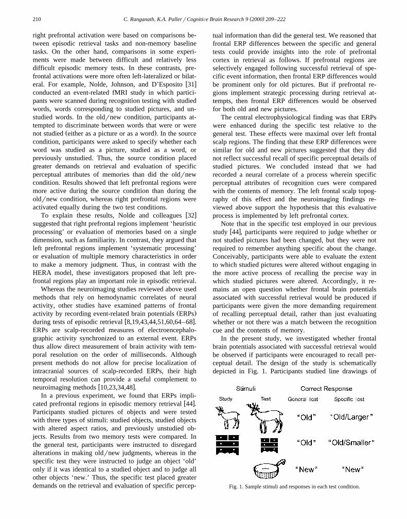
3.1. BehaÕioral results
Ž .Recognition accuracy results are shown in Fig. 2 A .Ž .An analysis of variance ANOVA revealed no significant
differences in item recognition accuracy between old andw Ž . xnew pictures F 1,11 -1 or between test conditions
w Ž . xF 1,11 s1.22, ps .294 and no significant Stimulusw Ž . xType = Condition interaction F 1,11 -1 . The mean
accuracy of judgments of size change in the specific testŽ .was 82.5% ranges72–92; SDs8.7 .
Ž .Reaction times RTs for correct responses are shown inŽ .Fig. 2 B . RTs were slower in the specific test than the
w Ž . xgeneral test F 1,11 s97.56, p- .001 and slower forw Ž .old pictures than for new pictures F 1,11 s74.67, p-
x.001 . However, a significant Stimulus Type = Conditionw Ž . xinteraction was also observed F 1,11 s42.91, p- .001 ,
and t-tests showed that RTs were significantly slower forw Ž .old than for new pictures in the specific test t 11 s7.74,
x w Ž .p- .001 but not in the general test t 11 s1.48, psx.167 .
3.2. ERP results
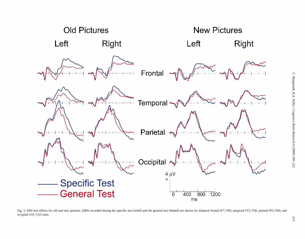
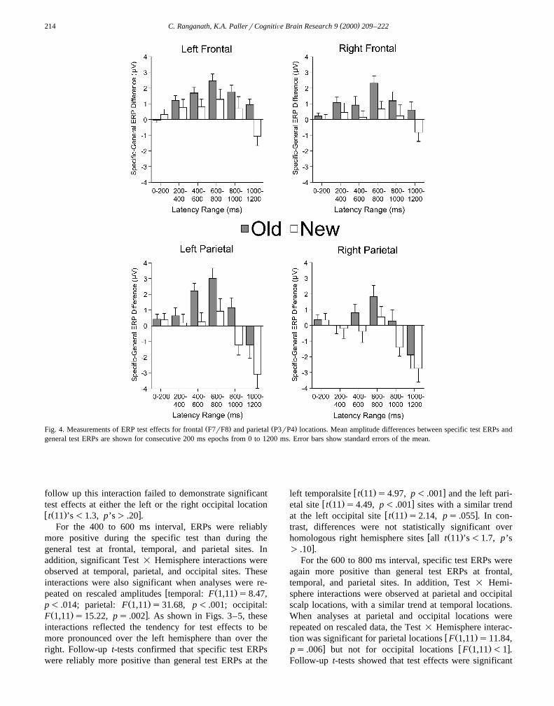
ERPs recorded during each test condition are shown forold and new pictures in Fig. 3. Mean amplitude measure-ments were made over successive 200 ms intervals tocalculate ERP differences between the specific and general
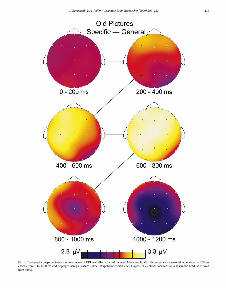
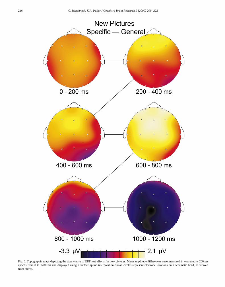
Ž .tests Fig. 4 . Topographic maps of these ERP test effectsare shown for old pictures in Fig. 5 and for new pictures in
Ž .Fig. 2. Behavioral results for each condition. A Mean recognitionŽ .accuracy. B Mean reaction time for correct responses.
Fig. 6. As described below in more detail, three differenttest effects were apparent. First, frontal potentials weremore positive during the specific test than during thegeneral test from approximately 200 to 1000 ms, and thisERP test effect was larger in magnitude for old than fornew pictures. Second, a test effect maximal over leftposterior scalp regions from approximately 400 to 900 mswas only seen for old pictures. Third, a test effect wasobserved after 900 ms at parietal and occipital scalplocations for both old and new pictures.
3.3. Test effects for old pictures
Mean ERP amplitudes to old pictures recorded frompairs of left and right frontal, temporal, parietal, and
Ž .occipital electrodes as shown in the left portion of Fig. 3were analyzed in successive 200 ms intervals from 200 to
Ž1200 ms. Results from corresponding Test specific vs.. Ž .general = Hemisphere left vs. right ANOVAs are shown
in Table 1. For the 200 to 400 ms interval, specific testERPs were significantly more positive than general testERPs at frontal locations with a similar trend at temporallocations. In addition, as shown in the table, a significantTest = Hemisphere interaction was observed at occipitallocations during this interval, with a similar marginalinteraction at parietal sites. The interaction at occipitalsites was also significant when the analysis was repeated
w Ž . xon rescaled mean amplitudes F 1,11 s20.73, ps .001 ,suggesting that the interaction reflected a true topographydifference between the two conditions. T-tests designed to
()
C.R
anganath,K.A
.Pallerr
CognitiÕe
Brain
Research
92000
209–
222213
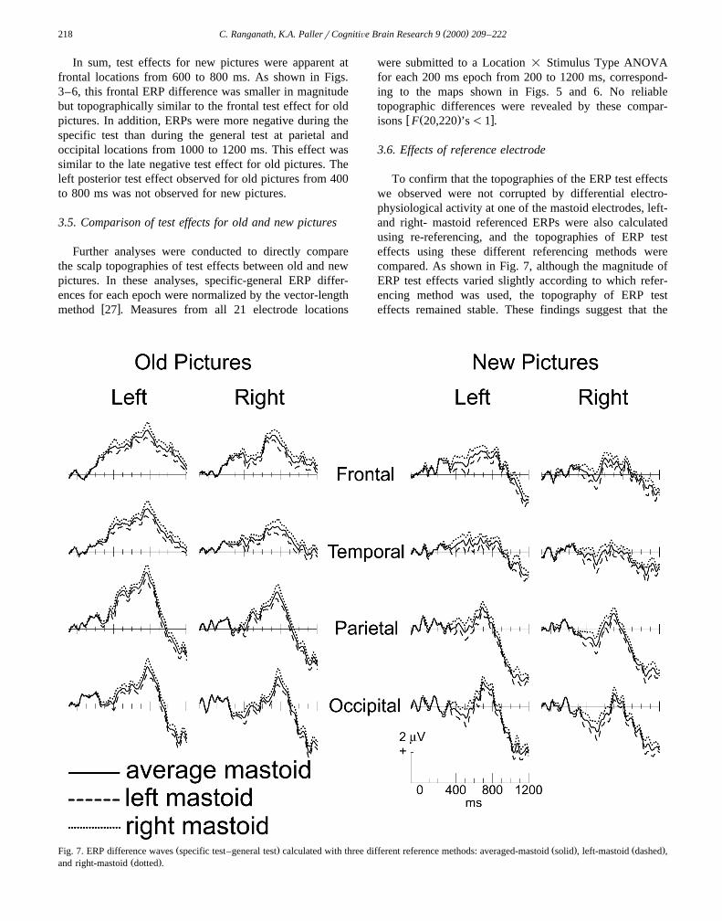
Ž . Ž . Ž . Ž . Ž .Fig. 3. ERP test effects for old and new pictures. ERPs recorded during the specific test solid and the general test dotted are shown for bilateral frontal F7rF8 , temporal T3rT4 , parietal P3rP4 , andŽ .occipital O1rO2 sites.
( )C. Ranganath, K.A. PallerrCognitiÕe Brain Research 9 2000 209–222214
Ž . Ž .Fig. 4. Measurements of ERP test effects for frontal F7rF8 and parietal P3rP4 locations. Mean amplitude differences between specific test ERPs andgeneral test ERPs are shown for consecutive 200 ms epochs from 0 to 1200 ms. Error bars show standard errors of the mean.
follow up this interaction failed to demonstrate significanttest effects at either the left or the right occipital locationw Ž . xt 11 ’s-1.3, p’s) .20 .
For the 400 to 600 ms interval, ERPs were reliablymore positive during the specific test than during thegeneral test at frontal, temporal, and parietal sites. Inaddition, significant Test = Hemisphere interactions wereobserved at temporal, parietal, and occipital sites. Theseinteractions were also significant when analyses were re-
w Ž .peated on rescaled amplitudes temporal: F 1,11 s8.47,Ž .p- .014; parietal: F 1,11 s31.68, p- .001; occipital:
Ž . xF 1,11 s15.22, ps .002 . As shown in Figs. 3–5, theseinteractions reflected the tendency for test effects to bemore pronounced over the left hemisphere than over theright. Follow-up t-tests confirmed that specific test ERPswere reliably more positive than general test ERPs at the
w Ž . xleft temporalsite t 11 s4.97, p- .001 and the left pari-w Ž . xetal site t 11 s4.49, p- .001 sites with a similar trend
w Ž . xat the left occipital site t 11 s2.14, ps .055 . In con-trast, differences were not statistically significant over
w Ž .homologous right hemisphere sites all t 11 ’s-1.7, p’sx) .10 .
For the 600 to 800 ms interval, specific test ERPs wereagain more positive than general test ERPs at frontal,temporal, and parietal sites. In addition, Test = Hemi-sphere interactions were observed at parietal and occipitalscalp locations, with a similar trend at temporal locations.When analyses at parietal and occipital locations wererepeated on rescaled data, the Test = Hemisphere interac-
w Ž .tion was significant for parietal locations F 1,11 s11.84,x w Ž . xps .006 but not for occipital locations F 1,11 -1 .
Follow-up t-tests showed that test effects were significant
( )C. Ranganath, K.A. PallerrCognitiÕe Brain Research 9 2000 209–222 215
Fig. 5. Topographic maps depicting the time course of ERP test effects for old pictures. Mean amplitude differences were measured in consecutive 200 msepochs from 0 to 1200 ms and displayed using a surface spline interpolation. Small circles represent electrode locations on a schematic head, as viewedfrom above.
( )C. Ranganath, K.A. PallerrCognitiÕe Brain Research 9 2000 209–222216
Fig. 6. Topographic maps depicting the time course of ERP test effects for new pictures. Mean amplitude differences were measured in consecutive 200 msepochs from 0 to 1200 ms and displayed using a surface spline interpolation. Small circles represent electrode locations on a schematic head, as viewedfrom above.
( )C. Ranganath, K.A. PallerrCognitiÕe Brain Research 9 2000 209–222 217
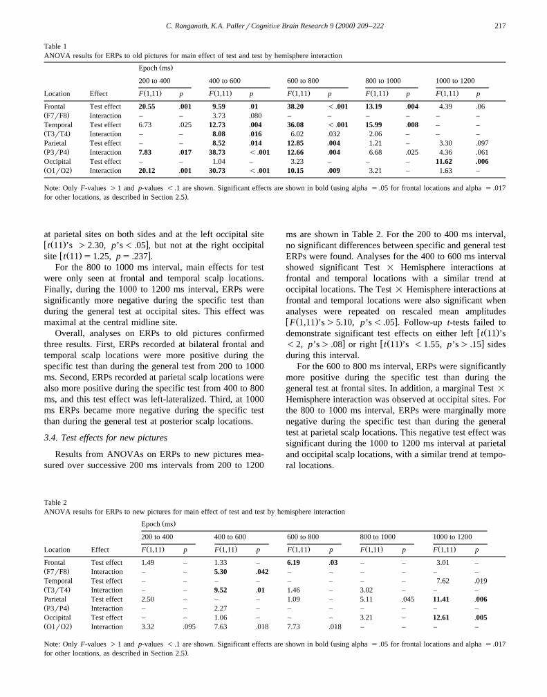
Table 1ANOVA results for ERPs to old pictures for main effect of test and test by hemisphere interaction
Ž .Epoch ms
200 to 400 400 to 600 600 to 800 800 to 1000 1000 to 1200
Ž . Ž . Ž . Ž . Ž .Location Effect F 1,11 p F 1,11 p F 1,11 p F 1,11 p F 1,11 p
Frontal Test effect 20.55 .001 9.59 .01 38.20 - .001 13.19 .004 4.39 .06Ž .F7rF8 Interaction – – 3.73 .080 – – – – – –Temporal Test effect 6.73 .025 12.73 .004 36.08 - .001 15.99 .008 – –Ž .T3rT4 Interaction – – 8.08 .016 6.02 .032 2.06 – – –Parietal Test effect – – 8.52 .014 12.85 .004 1.21 – 3.30 .097Ž .P3rP4 Interaction 7.83 .017 38.73 - .001 12.66 .004 6.68 .025 4.36 .061Occipital Test effect – – 1.04 – 3.23 – – – 11.62 .006Ž .O1rO2 Interaction 20.12 .001 30.73 - .001 10.15 .009 3.21 – 1.63 –
ŽNote: Only F-values )1 and p-values - .1 are shown. Significant effects are shown in bold using alpha s .05 for frontal locations and alpha s .017.for other locations, as described in Section 2.5 .
at parietal sites on both sides and at the left occipital sitew Ž . xt 11 ’s )2.30, p’s- .05 , but not at the right occipital
w Ž . xsite t 11 s1.25, ps .237 .For the 800 to 1000 ms interval, main effects for test
were only seen at frontal and temporal scalp locations.Finally, during the 1000 to 1200 ms interval, ERPs weresignificantly more negative during the specific test thanduring the general test at occipital sites. This effect wasmaximal at the central midline site.
Overall, analyses on ERPs to old pictures confirmedthree results. First, ERPs recorded at bilateral frontal andtemporal scalp locations were more positive during thespecific test than during the general test from 200 to 1000ms. Second, ERPs recorded at parietal scalp locations werealso more positive during the specific test from 400 to 800ms, and this test effect was left-lateralized. Third, at 1000ms ERPs became more negative during the specific testthan during the general test at posterior scalp locations.
3.4. Test effects for new pictures
Results from ANOVAs on ERPs to new pictures mea-sured over successive 200 ms intervals from 200 to 1200
ms are shown in Table 2. For the 200 to 400 ms interval,no significant differences between specific and general testERPs were found. Analyses for the 400 to 600 ms intervalshowed significant Test = Hemisphere interactions atfrontal and temporal locations with a similar trend atoccipital locations. The Test = Hemisphere interactions atfrontal and temporal locations were also significant whenanalyses were repeated on rescaled mean amplitudesw Ž . xF 1,11 ’s)5.10, p’s- .05 . Follow-up t-tests failed to
w Ž .demonstrate significant test effects on either left t 11 ’sx w Ž . x-2, p’s) .08 or right t 11 ’s -1.55, p’s) .15 sides
during this interval.For the 600 to 800 ms interval, ERPs were significantly
more positive during the specific test than during thegeneral test at frontal sites. In addition, a marginal Test =
Hemisphere interaction was observed at occipital sites. Forthe 800 to 1000 ms interval, ERPs were marginally morenegative during the specific test than during the generaltest at parietal scalp locations. This negative test effect wassignificant during the 1000 to 1200 ms interval at parietaland occipital scalp locations, with a similar trend at tempo-ral locations.
Table 2ANOVA results for ERPs to new pictures for main effect of test and test by hemisphere interaction
Ž .Epoch ms
200 to 400 400 to 600 600 to 800 800 to 1000 1000 to 1200
Ž . Ž . Ž . Ž . Ž .Location Effect F 1,11 p F 1,11 p F 1,11 p F 1,11 p F 1,11 p
Frontal Test effect 1.49 – 1.33 – 6.19 .03 – – 3.01 –Ž .F7rF8 Interaction – – 5.30 .042 – – – – – –Temporal Test effect – – – – – – – – 7.62 .019Ž .T3rT4 Interaction – – 9.52 .01 1.46 – 3.02 – – –Parietal Test effect 2.50 – – – 1.09 – 5.11 .045 11.41 .006Ž .P3rP4 Interaction – – 2.27 – – – – – – –Occipital Test effect – – 1.06 – – – 3.21 – 12.61 .005Ž .O1rO2 Interaction 3.32 .095 7.63 .018 7.73 .018 – – – –
ŽNote: Only F-values )1 and p-values - .1 are shown. Significant effects are shown in bold using alpha s .05 for frontal locations and alpha s .017.for other locations, as described in Section 2.5 .
( )C. Ranganath, K.A. PallerrCognitiÕe Brain Research 9 2000 209–222218
In sum, test effects for new pictures were apparent atfrontal locations from 600 to 800 ms. As shown in Figs.3–6, this frontal ERP difference was smaller in magnitudebut topographically similar to the frontal test effect for oldpictures. In addition, ERPs were more negative during thespecific test than during the general test at parietal andoccipital locations from 1000 to 1200 ms. This effect wassimilar to the late negative test effect for old pictures. Theleft posterior test effect observed for old pictures from 400to 800 ms was not observed for new pictures.
3.5. Comparison of test effects for old and new pictures
Further analyses were conducted to directly comparethe scalp topographies of test effects between old and newpictures. In these analyses, specific-general ERP differ-ences for each epoch were normalized by the vector-length
w xmethod 27 . Measures from all 21 electrode locations
were submitted to a Location = Stimulus Type ANOVAfor each 200 ms epoch from 200 to 1200 ms, correspond-ing to the maps shown in Figs. 5 and 6. No reliabletopographic differences were revealed by these compar-
w Ž . xisons F 20,220 ’s-1 .
3.6. Effects of reference electrode
To confirm that the topographies of the ERP test effectswe observed were not corrupted by differential electro-physiological activity at one of the mastoid electrodes, left-and right- mastoid referenced ERPs were also calculatedusing re-referencing, and the topographies of ERP testeffects using these different referencing methods werecompared. As shown in Fig. 7, although the magnitude ofERP test effects varied slightly according to which refer-encing method was used, the topography of ERP testeffects remained stable. These findings suggest that the
Ž . Ž . Ž .Fig. 7. ERP difference waves specific test–general test calculated with three different reference methods: averaged-mastoid solid , left-mastoid dashed ,Ž .and right-mastoid dotted .
( )C. Ranganath, K.A. PallerrCognitiÕe Brain Research 9 2000 209–222 219
pattern of results was not corrupted by using averagemastoid referenced ERPs for our statistical analyses.
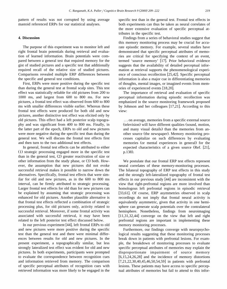
4. Discussion
The purpose of this experiment was to monitor left andright frontal brain potentials during retrieval and evalua-tion of learned information. Brain potentials were com-pared between a general test that required memory for thegist of studied pictures and a specific test that additionallyrequired recall of the relative size of studied pictures.Comparisons revealed multiple ERP differences betweenthe specific and general test conditions.
First, ERPs were more positive during the specific testthan during the general test at frontal scalp sites. This testeffect was statistically reliable for old pictures from 200 to1000 ms, and largest from 600 to 800 ms. For newpictures, a frontal test effect was observed from 600 to 800ms with smaller differences visible earlier. Whereas thesefrontal test effects were produced for both old and newpictures, another distinctive test effect was elicited only byold pictures. This effect had a left posterior scalp topogra-phy and was significant from 400 to 800 ms. Finally, inthe latter part of the epoch, ERPs to old and new pictureswere more negative during the specific test than during thegeneral test. We will discuss the frontal test effects firstand then turn to the two additional test effects.
In general, frontal test effects can be attributed to eitherŽ .1 strategic processing engaged more in the specific test
Ž .than in the general test, 2 greater reactivation of size orŽ .other information from the study phase, or 3 both. How-
ever, the assumption that new pictures did not evokesuccessful retrieval makes it possible to narrow down thealternatives. Specifically, frontal test effects that were sim-ilar for old and new pictures, as in the 600 to 800 msinterval, can be firmly attributed to strategic processing.Larger frontal test effects for old than for new pictures canbe explained by assuming that strategic processing wasenhanced for old pictures. Another plausible alternative isthat frontal test effects reflected a combination of strategicprocessing plus, for old pictures only, activity related tosuccessful retrieval. Moreover, if some frontal activity wasassociated with successful retrieval, it may have beenrelated to the left posterior test effect discussed below.
w xIn our previous experiment 44 , left frontal ERPs to oldand new pictures were more positive during the specifictest than the general test and there were minimal differ-ences between results for old and new pictures. In thepresent experiment, a topographically similar, but lessstrongly lateralized test effect was evident for old and newpictures. In both experiments, participants were promptedto evaluate the correspondence between recognition cuesand information retrieved from memory. The comparisonof specific perceptual attributes of recognition cues withretrieved information was more likely to be engaged in the
specific test than in the general test. Frontal test effects inboth experiments can thus be taken as neural correlates ofthe more extensive evaluation of specific perceptual at-tributes in the specific test.
Findings from a series of behavioral studies suggest thatthis memory monitoring process may be crucial for accu-rate episodic memory. For example, several studies havedemonstrated that specific perceptual attributes of memo-ries are critical for specifying the context of an event,
w xtermed ‘source memory’ 17 . Prior behavioral evidencesuggests that the availability of detailed perceptual infor-mation at retrieval supports the phenomenological experi-
w xence of conscious recollection 25,42 . Specific perceptualinformation is also a major cue in differentiating memoriesof thoughts, mental images, or imagined events from mem-
w xories of experienced events 18,20 .The importance of retrieval and evaluation of specific
perceptual information during episodic recollection wasemphasized in the source monitoring framework proposed
w xby Johnson and her colleagues 17,21 . According to thisview:
. . . on average, memories from a specific external sourceŽ . Žtelevision will have different qualities sound, motion,
.and many visual details than the memories from an-Ž .other source the newspaper . Memory monitoring pro-
cesses capitalize on such differences by evaluatingŽ .memories or mental experiences in general for the
Ž w xexpected characteristics of a given source Ref. 21 ,.p.138 .
We postulate that our frontal ERP test effects representneural correlates of these memory-monitoring processes.The bilateral topography of ERP test effects in this studyand the strongly left-lateralized topography of frontal test
w xeffects in our previous study 44 are inconsistent with theview that right-prefrontal regions are more involved thanhomologous left prefrontal regions in episodic retrievalw x33,61 . Of course, ERP asymmetries observed in scalprecordings do not imply that frontal neural activity isequivalently asymmetric, given that activity in one hemi-sphere can generate scalp potentials over the contralateralhemisphere. Nonetheless, findings from neuroimagingw x11,31,32,44 converge on the view that left and rightprefrontal regions are important in implementing thesememory monitoring processes.
Furthermore, our findings converge with neuropsycho-logical results suggesting that these monitoring processesbreak down in patients with prefrontal lesions. For exam-ple, the breakdown of monitoring processes to evaluatespecific perceptual attributes of memories may explain thedisproportionate impairment of source memoryw x6,15,24,26,28 and the incidence of memory distortionsw x7,21,22,30,40,45,46,50,54,59 in patients with prefrontallesions. These patients may have access to specific percep-tual attributes of memories but fail to attend to this infor-
( )C. Ranganath, K.A. PallerrCognitiÕe Brain Research 9 2000 209–222220
mation when attempting to recall the source of a memoryw x7 .
In addition to the frontal test effects described above, aleft parietal test effect was apparent for old but not for new
Ž .pictures Figs. 3–6 . This ERP test effect was topographi-cally similar to ERP modulations reported in previousstudies of source memory conducted by Wilding and col-
w xleagues 64–68 . In these studies, participants studied wordsspoken in a male or female voice. Next, they were given arecognition test with visual words and were asked tospecify the gender of the speaker for words judged old.Across these studies, ERPs to old items were more posi-tive-going than those to new items, with these differenceslargest over left posterior and right frontal scalp regions.Furthermore, these ‘old–new effects’ were larger in mag-nitude when the speaker’s voice was correctly recalled,suggesting that they were associated with recollection ofthe voice information associated with studied words. Wehave also linked posterior ERPs to recollective processingin prior studies that made use of memory dissociations
w xbetween implicit and explicit memory tests 35–37 .Although our primary intentions in the present experi-
ment were to focus on differences in brain potentialsbetween the two test conditions, it should be noted that theleft posterior test effect can also be conceptualized as atype of old–new effect. ERPs to old and new pictures ineach condition at the left parietal scalp site are shown inFig. 8. An old–new effect was observed in both testconditions from 200 to 500 ms, but old–new differencescontinued from 500 to 1200 ms during the specific test. Inlight of various findings associating similar ERP effects
Ž w x .with recollection see Ref. 47 for review , we proposethat the left posterior test effect seen in this study reflectsthe recollection of additional details of studied pictures inthe specific test. Our ERP results thus demonstrated adissociation between posterior activity reflecting the reacti-vation of stored information and anterior activity reflectingthe monitoring and evaluation of retrieved information.These findings parallel recent neuroimaging results associ-ating posterior cortical activity with reactivation of storedinformation and prefrontal activity with the monitoring and
w xmanipulation of that information 41 .The finding that ERPs after about 900 ms were more
negative during the specific test than during the generaltest was unexpected, given that no such effect was appar-
w xent in our previous study 44 . The posterior distribution ofthis effect may have been exaggerated due to overlap withanterior test effects of the opposite polarity from 200 to1000 ms. The finding that the late test effect appeared tobegin earlier and to have a broader topography for newthan for old pictures may have actually been related to thefact that the overlapping frontal test effect was smaller fornew pictures, rather than to differences in the late effectper se. In light of the late onset of the negative test effectrelative to reaction times, however, it is unlikely that itreflects processes that were critical for making memory
Fig. 8. Old–new ERP effects during each test condition for the leftparietal scalp location.
judgments in either test condition. Instead, the effect mayreflect a divergence in cognitive activities between the twotest conditions that occurred after participants made recog-nition decisions. For example, it is possible that partici-pants were more confident in recognition responses madeduring the general test than in responses made during thespecific test. It is also possible that continued processingand evaluation of relative size information continued in thespecific test but not in the general test, and that thispost-decision processing was associated with widespreadnegative potentials.
In summary, results from the present study have re-vealed several insights into the role of prefrontal cortex inepisodic memory retrieval. First, in contrast to the HERAmodel, our results support the view that left and rightprefrontal regions both make important contributions toepisodic retrieval. Second, our results demonstrate thatfrontal activity can be associated both with the reactivation
Ž .of stored information for old pictures and with evaluativeŽ .processing for old and new pictures . We also observed
( )C. Ranganath, K.A. PallerrCognitiÕe Brain Research 9 2000 209–222 221
left posterior activity related to the reactivation of storedinformation. Although it remains unclear exactly how leftand right prefrontal regions differ in their functions duringepisodic retrieval, our results indicate that these asymme-tries are more complex than suggested by models such asHERA.
Acknowledgements
This research was supported by grant NS34639 fromthe National Institute of Neurological Disorders and Stroke.We thank Brian Gonsalves, Marcia K. Johnson, WilliamRevelle, J. Peter Rosenfeld, and two anonymous reviewersfor their helpful comments and Ted Whalen for technicalsupport.
References
w x1 D.F. Benson, A. Djenderedjian, B.L. Miller, N.A. Pachana, L.Chang, L. Itti, I. Mena, Neural basis of confabulation, Neurology 46Ž .1996 1239–1243.
w x2 R.L. Buckner, Beyond HERA: Contributions of specific prefrontalbrain areas to long-term memory retrieval, Psychonom. Bull. Rev. 3Ž .1996 149–158.
w x3 R.L. Buckner, W. Koutstaal, D.L. Schacter, A.M. Dale, M. Rotte,B.R. Rosen, Functional-anatomic study of episodic retrieval. II.Selective averaging of event-related fMRI trials to test the retrieval
Ž .success hypothesis, Neuroimage 7 1998 163–175.w x4 R.L. Buckner, W. Koutstaal, D.L. Schacter, A.D. Wagner, B.R.
Rosen, Functional-anatomic study of episodic retrieval using fMRI.Ž .I. Retrieval effort versus retrieval success, Neuroimage 7 1998
151–162.w x5 R.L. Buckner, S.E. Petersen, What does neuroimaging tell us about
the role of prefrontal cortex in memory retrieval?, Semin. Neurosci.Ž .8 1996 47–55.
w x6 M.A. Butters, A.W. Kaszniak, E.L. Glisky, P.W. Eslinger, D.L.Schacter, Recency discrimination deficits in frontal patients, Neu-
Ž .ropsychology 8 1994 343–353.w x7 T. Curran, D.L. Schacter, K.A. Norman, L. Galluccio, False recogni-
tion after a right frontal lobe infarction: Memory for general andŽ .specific information, Neuropsychologia 35 1997 1035–1049.
w x8 E. Duzel, R. Cabeza, T.W. Picton, A.P. Yonelinas, H. Scheich, H.J.¨Heinze, E. Tulving, Task-related and item-related brain processes of
Ž .memory retrieval, Proc. Natl. Acad. Sci. USA 96 1999 1794–1799.w x9 F.B. Gershberg, A.P. Shimamura, Serial position effects in implicit
and explicit tests of memory, J. Exp. Psychol.: Learning, Memory,Ž .and Cognition 20 1994 1370–1378.
w x10 E. Halgren, PET may image the gates of awareness, not its center,Ž .Behav. Brain Sci. 18 1995 358–359.
w x11 R.N.A. Henson, T. Shallice, R.J. Dolan, Right prefrontal cortex andepisodic memory retrieval: a functional MRI test of the monitoring
Ž .hypothesis, Brain 122 1999 1367–1381.w x12 W. Hirst, B.T. Volpe, Memory strategies with brain damage, Brain
Ž .Cognit. 8 1988 379–408.w x13 A. Incisa della Rochetta, B. Milner, Strategic search and retrieval
Ž .initiation: the role of the frontal lobes, Neuropsychologia 31 1993503–524.
w x14 J.S. Janowsky, A.P. Shimamura, M. Kritchevsky, L.R. Squire, Cog-nitive impairment following frontal lobe damage and its relevance to
Ž .human amnesia, Behav. Neurosci. 103 1989 548–560.
w x15 J.S. Janowsky, A.P. Shimamura, L.R. Squire, Source memory im-pairment in patients with frontal lobe lesions, Neuropsychologia 27Ž .1989 1043–1056.
w x16 W. Jetter, U. Poser, R.B. Freeman, H.J. Markowitsch, A verballong-term memory deficit in frontal lobe damaged patients, Cortex
Ž .22 1986 229–242.w x17 M.K. Johnson, S. Hashtroudi, D. Lindsay, Source monitoring, Psy-
Ž .chol. Bull. 114 1993 3–28.w x18 M.K. Johnson, M.A. Foley, A.G. Suengas, C.L. Raye, Phenomenal
characteristics for perceived and imagined autobiographical events,Ž .J. Exp. Psychol.: General 117 1988 371–376.
w x19 M.K. Johnson, J. Kounios, S.F. Nolde, Electrophysiological brainŽ .activity and memory source monitoring, Neuroreport 8 1997
1317–1320.w x20 M.K. Johnson, C.L. Raye, Reality monitoring, Psychol. Rev. 88
Ž .1981 67–85.w x21 M.K. Johnson, C.L. Raye, False memories and confabulation, Trends
Ž .Cognit. Sci. 2 1998 137–145.w x22 R. Joseph, Confabulation and delusional denial: Frontal lobe and
Ž .lateralized influences, J. Clin. Psychol. 42 1986 507–520.w x23 M. Kutas, A. Dale, Electrical and magnetic readings of mental
Ž .functions, in: M.D. Rugg Ed. , Cognitive Neuroscience, MIT Press,Cambridge, MA, 1997, pp. 197–242.
w x24 J.A. Mangels, Strategic processing and memory for temporal orderŽ .in patients with frontal lobe lesions, Neuropsychology 11 1997
207–221.w x25 T. Mantyla, Recollections of faces: Remembering differences and¨ ¨
knowing similarities, J. Exp. Psychol.: Learning, Memory, andŽ .Cognition 23 1997 1203–1216.
w x26 M.P. McAndrews, B. Milner, The frontal cortex and memory forŽ .temporal order, Neuropsychologia 29 1991 849–859.
w x27 G. McCarthy, C.C. Wood, Scalp distributions of event-related poten-tials: An ambiguity associated with analysis of variance models,
Ž .Electroencephalogr. Clin. Neurophysiol. 62 1985 203–208.w x28 B. Milner, P. Corsi, G. Leonard, Frontal-lobe contribution to recency
Ž .judgements, Neuropsychologia 29 1991 601–618.w x29 B. Milner, M. Petrides, Behavioural effects of frontal-lobe lesions in
Ž .man, Trends Neurosci. 7 1984 403–407.w x30 M. Moscovitch, B. Melo, Strategic retrieval and the frontal lobes:
Evidence from confabulation and amnesia, Neuropsychologia 35Ž .1997 1017–1034.
w x31 S.F. Nolde, M.K. Johnson, M. D’Esposito, Left prefrontal activationduring episodic remembering: an event-related fMRI study, Neurore-
Ž .port 9 1998 3509–3514.w x32 S.F. Nolde, M.K. Johnson, C.L. Raye, The role of prefrontal cortex
Ž .during tests of episodic memory, Trends Cognit. Sci. 2 1998399–406.
w x33 L. Nyberg, R. Cabeza, E. Tulving, PET studies of encoding andŽ .retrieval: The HERA model, Psychonom. Bull. Rev. 3 1996 135–
148.w x34 K.A. Paller, If a Picture is Worth 1,000 Words, How Many Pictures
Ž .is a Word Worth?, Behav. Brain Sci. 18 1995 367–368.w x35 K.A. Paller, V.S. Bozic, C. Ranganath, M. Grabowecky, S. Yamada,
Brain waves following remembered faces index conscious recollec-Ž .tion, Cognit. Brain Res. 7 1999 519–531.
w x36 K.A. Paller, M. Kutas, Brain potentials during memory retrievalprovide neurophysiological support for the distinction between con-
Ž .scious recollection and priming, J. Cognit. Neurosci. 4 1992375–391.
w x37 K.A. Paller, M. Kutas, H.K. McIsaac, Monitoring conscious recol-Ž .lection via the electrical activity of the brain, Psychol. Sci. 6 1995
107–111.w x38 K.A. Paller, C. Ranganath, K.S. Labar, T.B. Parrish, D.R. Gitelman,
V.S. Bozic, M.M. Mesulam, Neural correlates of memory for faces:Differential frontal activity for retrieval success versus retrieval
w x Ž .effort abstract , NeuroImage 9 1999 S962.
( )C. Ranganath, K.A. PallerrCognitiÕe Brain Research 9 2000 209–222222
w x39 S. Park, A. Raine, T. Lencz, S. Bihrle, L. LaCasse, Structural andfunctional correlates of working memory and olfactory identification
w x Ž .in schizotypal subjects abstract , Schizophrenia Res. 24 1997 135.w x40 A.J. Parkin, C. Bindschaedler, L. Harsent, C. Metzler, Pathological
false alarm rates following damage to left frontal cortex, BrainŽ .Cognit. 32 1996 14–27.
w x41 B.R. Postle, J.S. Berger, M. D’Esposito, Functional neuroanatomicaldouble dissociation of mnemonic and executive control processescontributing to working memory performance, Proc. Natl. Acad. Sci.USA, in press.
w x42 S. Rajaram, Perceptual effects on remembering: Recollective pro-cesses in picture recognition memory, J. Exp. Psychol.: Learning,
Ž .Memory, and Cognition 22 1996 365–377.w x43 C. Ranganath, K.A. Paller, Frontal brain activity during episodic and
semantic retrieval: insights from event-related potentials, J. Cognit.Ž .Neurosci. 11 1999 598–609.
w x44 C. Ranganath, K.A. Paller, Frontal brain potentials during recogni-tion are modulated by requirements to retrieve perceptual detail,
Ž .Neuron 22 1999 605–613.w x45 S.Z. Rapcsak, A.W. Kaszniak, S.L. Reminger, M.L. Glisky, E.L.
Glisky, J.F. Comer, Dissociation between verbal and autonomicmeasures of memory following frontal lobe damage, Neurology 50Ž .1998 1259–1265.
w x46 S.Z. Rapcsak, M.R. Polster, M.L. Glisky, J.F. Comer, False recogni-tion of unfamiliar faces following right hemisphere damage: neu-
Ž .ropsychological and anatomical observations, Cortex 32 1996593–611.
w x47 M.D. Rugg, ERP studies of memory, in: M.D. Rugg, M.G.H. ColesŽ .Eds. , Electrophysiology of Mind, Oxford University Press, NewYork, 1995, pp. 132–170.
w x48 M.D. Rugg, Convergent approaches to electrophysiological andhemodynamic investigations of memory, Human Brain Mapp. 6Ž .1998 394–398.
w x49 D.L. Schacter, R.L. Buckner, W. Koutstaal, A.M. Dale, B.R. Rosen,Late onset of anterior prefrontal activity during true and false
Ž .recognition: An event-related fMRI study, Neuroimage 6 1997259–269.
w x50 D.L. Schacter, T. Curran, L. Galluccio, W.P. Milberg, J.F. Bates,False recognition and the right frontal lobe: A case study, Neuropsy-
Ž .chologia 34 1996 793–808.w x51 A.J. Senkfor, C. Van Petten, Who said what? An event-related
potential investigation of source and item memory, J. Exp. Psychol.:Ž .Learning, Memory, and Cognition 24 1998 1005–1025.
w x52 A.P. Shimamura, The role of prefrontal cortex in monitoring andŽ .controlling memory processes, in: L. Reder Ed. , Implicit Memory
and Metacognition, Erlbaum, Mahwah, NJ, 1996, pp. 259–274.
w x53 J. Snodgrass, M. Vanderwort, A standardized set of 260 pictures:norms for name agreement, image agreement, familiarity, and visualcomplexity, J. Exp. Psychol.: Learning, Memory, and Cognition 6Ž .1980 174–215.
w x54 D.T. Stuss, M.P. Alexander, A. Lieberman, H. Levine, An extraordi-Ž .nary form of confabulation, Neurology 28 1978 1166–1172.
w x55 D.T. Stuss, M.P. Alexander, C.L. Palumbo, L. Buckle et al., Organi-zational strategies with unilateral or bilateral frontal lobe injury in
Ž .word learning tasks, Neuropsychology 8 1994 355–373.w x56 D.T. Stuss, D.F. Benson, Neuropsychological studies of the frontal
Ž .lobes, Psychol. Bull. 95 1984 3–28.w x57 D.T. Stuss, D.F. Benson, The Frontal Lobes, Raven Press, New
York, 1986.w x58 D. Swick, R.T. Knight, Is prefrontal cortex involved in cued recall?
A neuropsychological test of PET findings, Neuropsychologica 34Ž .1996 1019–1028.
w x59 D. Swick, R.T. Knight, Contributions of prefrontal cortex to recogni-tion memory: electrophysiological and behavioral evidence, Neu-
Ž .ropsychology 13 1999 155–170.w x60 C.T. Trott, D. Friedman, W. Ritter, M. Fabiani, Item and source
memory: differential age effects revealed by event-related potentials,Ž .Neuroreport 8 1997 3373–3378.
w x61 E. Tulving, S. Kapur, F.I.M. Craik, M. Moscovitch, S. Houle,Hemispheric encodingrretrieval asymmetry in episodic memory —Positron Emission Tomography findings, Proc. Natl. Acad. Sci.
Ž .USA 91 1994 2016–2020.w x62 A.D. Wagner, Working memory contributions to human learning
Ž .and remembering, Neuron 22 1999 19–22.w x63 M.A. Wheeler, D.T. Stuss, E. Tulving, Frontal lobe damage pro-
duces episodic memory impairment, J. Int. Neuropsychol. Soc. 1Ž .1995 525–536.
w x64 E.L. Wilding, Separating retrieval strategies from retrieval success:An event- related potential study of source memory, Neuropsycholo-
Ž .gia 37 1999 441–454.w x65 E.L. Wilding, M.C. Doyle, M.D. Rugg, Recognition memory with
and without retrieval of context — an event-related potential study,Ž .Neuropsychologia 33 1995 743–767.
w x66 E.L. Wilding, M.D. Rugg, An event-related potential study ofrecognition memory with and without retrieval of source, Brain 119Ž .1996 889–905.
w x67 E.L. Wilding, M.D. Rugg, An event-related potential study ofmemory for words spoken aloud or heard, Neuropsychologia 35Ž .1997 1185–1195.
w x68 E.L. Wilding, M.D. Rugg, Event-related potentials and the recogni-Ž .tion memory exclusion task, Neuropsychologia 35 1997 119–128.