surgical management of neovascular glaucoma - endo … · surgical management of neovascular...
TRANSCRIPT
Surgical Management of Neovascular GlaucomaRobert F. Haverly, MD, FACS
Abstract: Neovascular glaucoma is a vision- and eye-threatening dis-ease that carries a poor prognosis. Understanding the cause of neovas-cular glaucoma with early diagnosis and treatment is important for apositive outcome. There are numerous surgical treatment options avail-able, but to have the best outcome, deciding which surgical technique touse is important. Some patients are better candidates than others for thevarious surgical techniques. An early- or late-stage clinical presentation,as well as the other ocular conditions associated with neovascular glau-coma, will dictate the appropriate surgical plan. This article provides astep-by-step approach to surgically managing neovascular glaucoma andexplains new surgical techniques to help with the best chance of a pos-itive patient outcome.
(Tech Ophthalmology 2010;8: 87Y93)
Neovascular glaucoma is a serious complication of systemicand retinal disease. The prognosis is poor, and early, ag-
gressive treatment is a must to achieve the best outcome. Manyeyes with neovascular glaucoma eventually need an enucleation,but the incidence of enucleation for neovascular glaucoma hasgradually been on the decline.1 Early detection and bettermedical and surgical techniques are most likely the reason forimproved outcomes. Unfortunately, it is all too common to firstsee a patient with a red, painful eye, with vision loss, and apressure in the 60- or 70-mm Hg range. Although, the prognosisis generally poor, some cases can have a surprisingly goodoutcome. To help in the diagnosis of neovascular glaucoma,early screening for patients at high risk is important. Examiningthe iris for rubeosis and the angle and the retina for neovascu-larization is important in the screening process. Understandingthe systemic diseases associated with glaucoma is a must tohave a heightened suspicion for neovascular glaucoma. Vasculardiseases such as diabetes, hypertension, heart disease, and pe-ripheral vascular disease are common underlying causes ofneovascular glaucoma. Ischemia to ocular tissue is theorized tobe the genesis of neovascular glaucoma.2 The most commoncauses of neovascular glaucoma related to the systemic diseasesinclude ischemic central retinal vein occlusion, proliferative di-abetic retinopathy, and carotid artery disease associated withocular ischemic syndrome. Other causes of neovascular glau-coma include hemiretinal and branch retinal vein occlusions,branch or central retinal artery occlusion, and giant cell arteritis.Treatment of the ocular ischemia associated with these condi-tions can reduce the incidence of neovascular glaucoma.2
Neovascular glaucoma develops from the induced hypoxiafrom ischemic retinal disease, which releases vascular endo-thelial growth factor (VEGF), a vasoproliferative substance thatacts upon healthy endothelial cells of viable capillaries to stim-ulate the formation of a fragile meshwork of vessels (neo-
vascularization). In extreme retinal hypoxia, there are few viableretinal capillaries. In this instance, VEGF is theorized to diffuseto the nearest area of viable capillaries, which in many cases isthe posterior iris. Neovascularization develops from these cap-illaries and progresses along the posterior iris, into the pupil,anterior iris, and also into the angle of the eye.3 Once in theangle, the neovascularization develops a fibrovascular supportmembrane that acts to both physically block the angle as well asbridge the angle that blocks the trabecular meshwork. Peripheralanterior synechiae with permanent angle closure can happenquickly. This results in secondary angle closure glaucoma with-out papillary block. Blood in the angle and a hyphema arecommon at this stage. The intraocular pressure can increase toextreme levels greater than 50 mm Hg. At this stage, the diag-nosis is readily apparent, but the clinical presentation may bemore subtle in other cases.4
CLINICAL PRESENTATION
Early StageEarly signs and symptoms of neovascular glaucoma may
only include rubeosis and/or mild vascularization of the iriswithout vision loss or pain. There may be minimal visual symp-toms associated with the presentation at this time, so other com-mon causes of neovascular glaucoma such as chronic carotidartery occlusive disease,5,6 sickle cell disease,7 neoplasias,8Y11
Fuchs heterochromic iridocyclitis,12 and pseudoexfoliation syn-drome13 must be ruled out. It is difficult to predict how long ittakes to progress from the early signs of neovascular glaucomato the advanced stage.
Advanced StageAdvanced stage of neovascular glaucoma can occur rap-
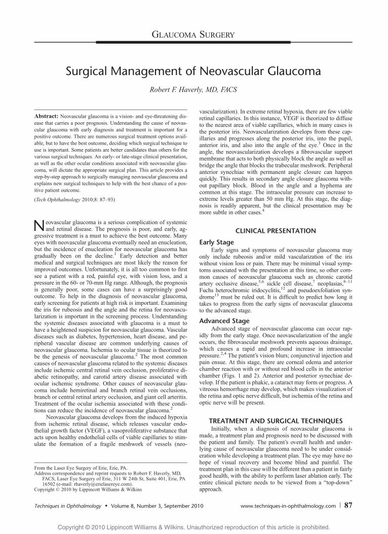
idly from the early stage. Once neovascularization of the angleoccurs, the fibrovascular meshwork prevents aqueous drainage,which causes a rapid and profound increase in intraocularpressure.2,4 The patient’s vision blurs; conjunctival injection andpain ensue. At this stage, there are corneal edema and anteriorchamber reaction with or without red blood cells in the anteriorchamber (Figs. 1 and 2). Anterior and posterior synechiae de-velop. If the patient is phakic, a cataract may form or progress. Avitreous hemorrhage may develop, which makes visualization ofthe retina and optic nerve difficult, but ischemia of the retina andoptic nerve will be present.
TREATMENT AND SURGICAL TECHNIQUESInitially, when a diagnosis of neovascular glaucoma is
made, a treatment plan and prognosis need to be discussed withthe patient and family. The patient’s overall health and under-lying cause of neovascular glaucoma need to be under consid-eration while developing a treatment plan. The eye may have nohope of visual recovery and become blind and painful. Thetreatment plan in this case will be different than a patient in fairlygood health, with the ability to perform laser ablation early. Theentire clinical picture needs to be viewed from a Btop-down[approach.
GLAUCOMA SURGERY
Techniques in Ophthalmology & Volume 8, Number 3, September 2010 www.techniques-in-ophthalmology.com 87
From the Laser Eye Surgery of Erie, Erie, PA.Address correspondence and reprint requests to Robert F. Haverly, MD,
FACS, Laser Eye Surgery of Erie, 311 W 24th St, Suite 401, Erie, PA16502 (e-mail: [email protected]).
Copyright * 2010 by Lippincott Williams & Wilkins
Copyright @ 20 Lippincott Williams & Wilkins. Unauthorized reproduction of this article is prohibited.10
Ophthalmologists and their patients are fortunate to havemany different treatment modalities at their disposal. In decidingan initial treatment, the first order is to determine if the retinacan be visualized, and if there is a clear view for pan retinalphotocoagulation. If the answer is yes, this should be done assoon as possible.14,15 If this cannot be performed, which hap-pens very often in the advanced stage, it is important to knowthe underlying cause of the neovascular glaucoma. If it is froman underlying tumor, treatment will be different. In cases of auveal melanoma, retinoblastoma, or metastatic disease, an enu-cleation may be the best choice, but radiation, chemotherapy,and/or a radioactive plaque may also be an option. If there isno visual potential and no tumor is present, transscleral diodelaser, transscleral cryotherapy, enucleation, and retrobulbar in-jection of alcohol are all possible options.
If the retina is visible, perform panretinal photocoagulation.I find it best to give a retrobulbar block and perform panretinalphotocoagulation 360 degrees from the arcades and as far intothe periphery as possible with a Rodenstock, goniolens, or otherwide-field lenses. Many times, the pupil will not dilate well, andthe peripheral retina is difficult to ablate, but do as much as pos-sible. I prefer to administer an intravitreal injection of an anti-
VEGF agent (bevacizumab [Avastin] or ranibizumab [Lucentis];Genentech) after the retinal ablation. The patient is given aA-blocker, acetazolamide, >-blockers, and prostaglandin drops.If possible, oral carbonic anhydrase inhibitor is started. In somecases, this may be all the treatment necessary to reduce the in-traocular pressure and treat the neovascular glaucoma.
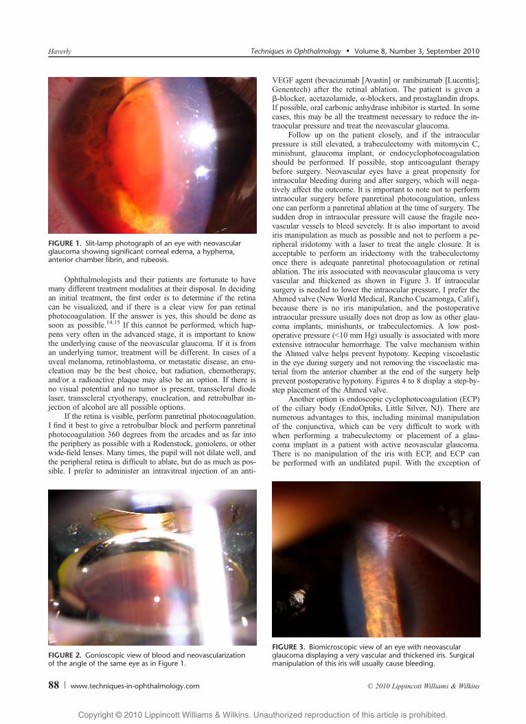
Follow up on the patient closely, and if the intraocularpressure is still elevated, a trabeculectomy with mitomycin C,minishunt, glaucoma implant, or endocyclophotocoagulationshould be performed. If possible, stop anticoagulant therapybefore surgery. Neovascular eyes have a great propensity forintraocular bleeding during and after surgery, which will nega-tively affect the outcome. It is important to note not to performintraocular surgery before panretinal photocoagulation, unlessone can perform a panretinal ablation at the time of surgery. Thesudden drop in intraocular pressure will cause the fragile neo-vascular vessels to bleed severely. It is also important to avoidiris manipulation as much as possible and not to perform a pe-ripheral iridotomy with a laser to treat the angle closure. It isacceptable to perform an iridectomy with the trabeculectomyonce there is adequate panretinal photocoagulation or retinalablation. The iris associated with neovascular glaucoma is veryvascular and thickened as shown in Figure 3. If intraocularsurgery is needed to lower the intraocular pressure, I prefer theAhmed valve (NewWorld Medical, Rancho Cucamonga, Calif ),because there is no iris manipulation, and the postoperativeintraocular pressure usually does not drop as low as other glau-coma implants, minishunts, or trabeculectomies. A low post-operative pressure (G10 mm Hg) usually is associated with moreextensive intraocular hemorrhage. The valve mechanism withinthe Ahmed valve helps prevent hypotony. Keeping viscoelasticin the eye during surgery and not removing the viscoelastic ma-terial from the anterior chamber at the end of the surgery helpprevent postoperative hypotony. Figures 4 to 8 display a step-by-step placement of the Ahmed valve.
Another option is endoscopic cyclophotocoagulation (ECP)of the ciliary body (EndoOptiks, Little Silver, NJ). There arenumerous advantages to this, including minimal manipulationof the conjunctiva, which can be very difficult to work withwhen performing a trabeculectomy or placement of a glau-coma implant in a patient with active neovascular glaucoma.There is no manipulation of the iris with ECP, and ECP canbe performed with an undilated pupil. With the exception of
FIGURE 1. Slit-lamp photograph of an eye with neovascularglaucoma showing significant corneal edema, a hyphema,anterior chamber fibrin, and rubeosis.
FIGURE 2. Gonioscopic view of blood and neovascularizationof the angle of the same eye as in Figure 1.
FIGURE 3. Biomicroscopic view of an eye with neovascularglaucoma displaying a very vascular and thickened iris. Surgicalmanipulation of this iris will usually cause bleeding.
Haverly Techniques in Ophthalmology & Volume 8, Number 3, September 2010
88 www.techniques-in-ophthalmology.com * 2010 Lippincott Williams & Wilkins
Copyright @ 20 Lippincott Williams & Wilkins. Unauthorized reproduction of this article is prohibited.10
making the incisions during surgery, there is no abrupt drop inintraocular pressure.
If panretinal photocoagulation is not possible because ofa limited view, there are several options. Transscleral cycloa-blation can be performed with or without a glaucoma implant.However, transscleral diode laser cyclophotocoagulation andtransscleral cryotherapy are very destructive to the eye and vi-sion.16,17 Usually, vision is lost after this type of treatment, andhypotony and phthisis can ensue. Another option is removingthe opacity that is preventing panretinal photocoagulation suchas cataract extraction and/or vitreous hemorrhage and then per-forming panretinal photocoagulation as well as placement of aglaucoma implant or a trabeculectomy. However, in many cases,the pupil will still need to be surgically dilated, and this leads toadditional intraocular hemorrhaging. If the cornea is clouded orscarred, this approach is even more difficult. It is not unusual in
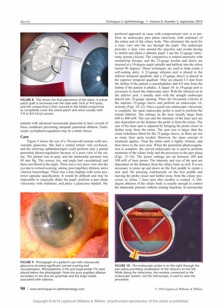
FIGURE 5. A preferred location for the seton is in the superior,temporal quadrant where a large peritomy is created using bluntWescott scissors. The conjunctival pocket is deepened withtenotomy scissors in a reverse cutting fashion.
FIGURE 6. The seton is placed into the conjunctival pocketand sutured with 8-0, 9-0, 10-0 Nylon between 8 and 10 mmfrom the limbus between the rectus muscles. A lateral relaxingincision of the conjunctiva helps to provide visualization forsuturing. It is very important to prime the implant with balancesalt solution or sterile water through the drainage tube with ablunt 26 to 30 gauge cannula before implantation of the seton.
FIGURE 7. The tube is cut to the appropriate length so that it doesnot block the visual axis but is far enough into the eye to remainin the anterior chamber. A scleral tunnel approximately 2.0 mmfrom the limbus with a 23-gauge needle creates the track for thetube. The tube can be anchored with 8-0 or 9-0 Nylon at its scleralinsertion. Viscoelastic placed in the anterior chamber through aparacentesis site helps with tube placement.
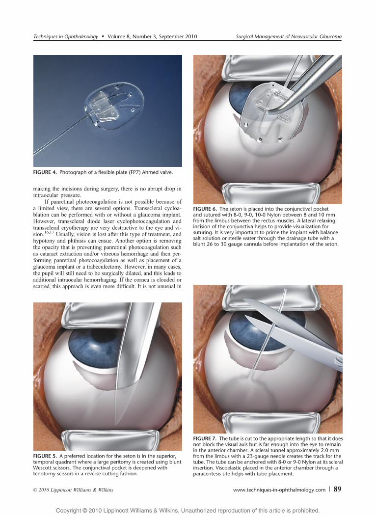
FIGURE 4. Photograph of a flexible plate (FP7) Ahmed valve.
Techniques in Ophthalmology & Volume 8, Number 3, September 2010 Surgical Management of Neovascular Glaucoma
* 2010 Lippincott Williams & Wilkins www.techniques-in-ophthalmology.com 89
Copyright @ 20 Lippincott Williams & Wilkins. Unauthorized reproduction of this article is prohibited.10
patients with advanced neovascular glaucoma to have several ofthese conditions preventing adequate panretinal ablation. Endo-scopic cyclophotocoagulation may be a better choice.
CaseFigure 9 shows the eye of a 70-year-old woman with neo-
vascular glaucoma. She had a central retinal vein occlusion,and the referring ophthalmologist could perform only a partialpanretinal photocoagulation because of a poor view of the ret-ina. The patient was in pain, and the intraocular pressure was68 mm Hg. The cornea, iris, and angle had vascularized, andthere was blood in the angle. There was a very poor view into theeye due to corneal scarring, edema, poor pupillary dilation, and avitreous hemorrhage. There was a lens implant with some pos-terior capsular opacification. It would be difficult and may beimpossible to surgically dilate the pupil, perform a pars planavitrectomy with endolaser, and place a glaucoma implant. My
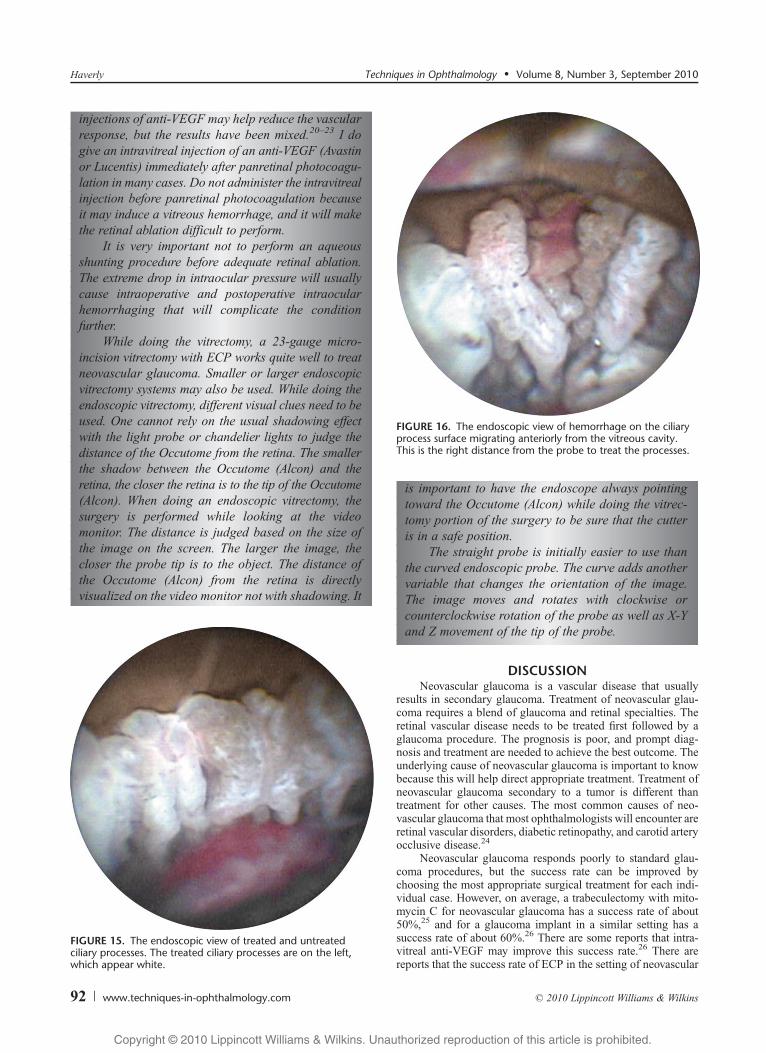
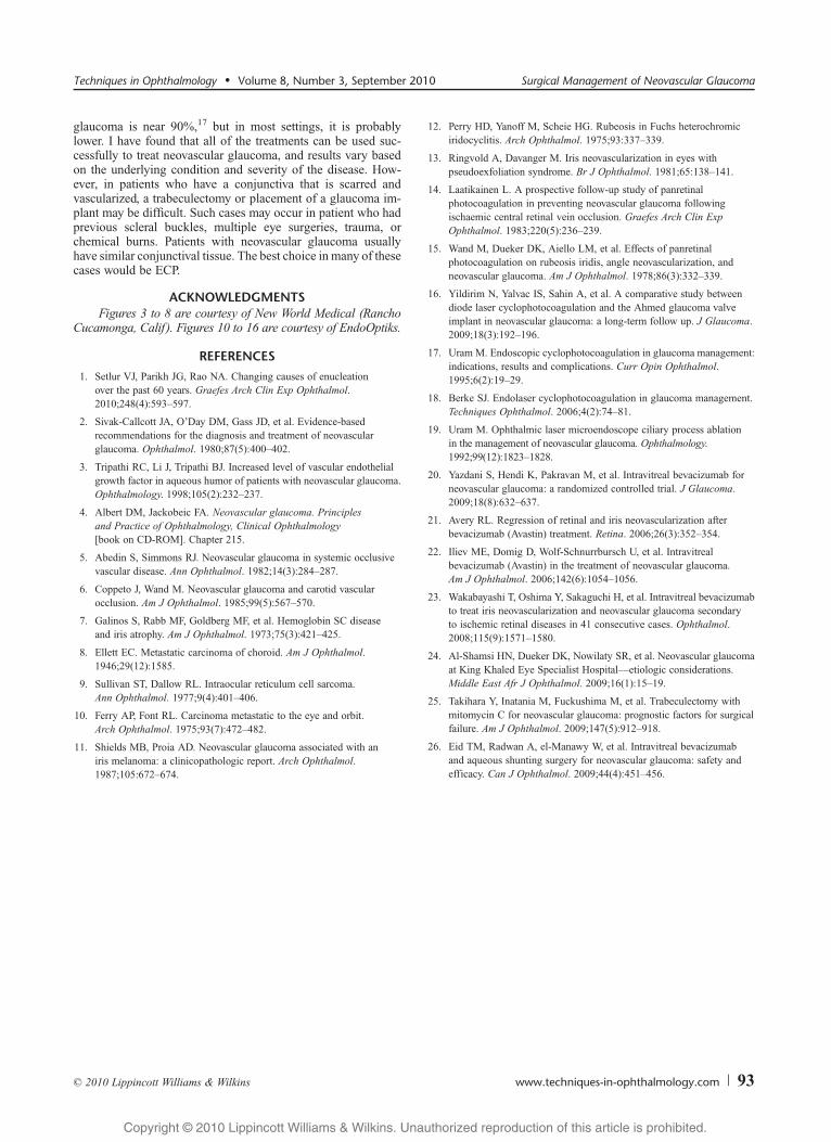
preferred approach in cases with compromised view is to per-form an endoscopic pars plana vitrectomy with endolaser ofthe retina and of the ciliary body. This eliminates the need fora clear view into the eye through the pupil. The endoscopeprovides a clear view around the opacities and avoids havingto stretch and dilate a rubeotic pupil. I use the 23-gauge vitrec-tomy system (Alcon). The conjunctiva is rotated anteriorly withserrated-tip forceps, and the 23-gauge trochar and sleeve areinserted at a 10-degree angle initially and halfway into the scleraturned 90 degrees. These techniques are used to help create aself-sealing entry. A 23-gauge infusion port is placed in theinferior temporal quadrant, and a 23-gauge sleeve is placed inthe superior temporal quadrant. They are placed 3.5 mm fromthe limbus if the patient is pseudophakic and 4.0 mm from thelimbus if the patient is phakic. A larger 18- to 19-gauge port isnecessary to insert the endoscopic laser. With the infusion on inthe inferior port, I usually start with the straight endoscopeplaced into 18-gauge opening. Place the Occutome (Alcon) inthe superior 23-gauge sleeve and perform an endoscopic vit-rectomy (Figs. 10Y12). Once a good core endoscopic vitrectomyis complete, the same endoscopic probe is used to perform theretinal ablation. The settings on the laser usually range from600 to 800 mW. The size and the intensity of the laser spot arealso dependent on the distance the probe is from the retina. Thesize of the laser spot is adjusted by bringing the probe closer orfarther away from the retina. The spot size is larger than theusual endolasers fitted for the 23-gauge sleeve, so there are notas many laser spots needed. However, the same concept oftreatment applies. Treat the retina until it lightly whitens, andthen move to the next area. When the panretinal photocoagula-tion is complete, the curved endoscopic tip is used to performtreatment of the ciliary body and the processes to the pars plana(Figs. 13Y16). The power settings are set between 200 and300 mW of laser power. The intensity and size of the spot aredependent on the distance from the ciliary body as well. It is notnecessary to press up and down on the foot peddle to create anew spot. By pressing continuously on the foot peddle andmoving the probe closer and farther away from the ciliary pro-cesses or retina, 1 laser spot after another is created. A 180-degree ablation of the ciliary body is usually enough to controlthe intraocular pressure without creating hypotony. In neovascular
FIGURE 9. Photograph of a patient’s eye with neovascularglaucoma showing significant corneal scarring andvascularization. Phenylephrine 2.5% and tropicamide 1% wereplaced before the photograph. Note the poor pupillary dilationsecondary to the lens-iris synechiae and the large vesselsassociated with rubeosis.
FIGURE 10. The endoscopic probe is on the right through thepars plana providing visualization of the vitrector on the left.While doing the vitrectomy, the monitor connected to theendoscopic system, not the microscope, is used to view theprocedure.
FIGURE 8. This shows the final placement of the seton. A scleralpatch graft is anchored over the tube with 10-0 or 9-0 nylon,and the conjunctiva is then sutured to the limbal conjunctivato completely cover the scleral patch and seton usually with7-0 or 8-0 Vicryl sutures.
Haverly Techniques in Ophthalmology & Volume 8, Number 3, September 2010
90 www.techniques-in-ophthalmology.com * 2010 Lippincott Williams & Wilkins
Copyright @ 20 Lippincott Williams & Wilkins. Unauthorized reproduction of this article is prohibited.10
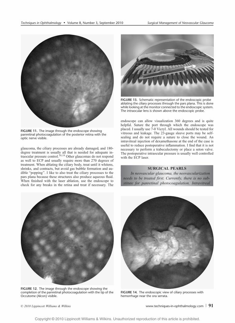
glaucoma, the ciliary processes are already damaged, and 180-degree treatment is usually all that is needed for adequate in-traocular pressure control.18,19 Other glaucomas do not respondas well to ECP and usually require more than 270 degrees oftreatment. When ablating the ciliary body, treat until it whitens,shrinks, and contracts, but avoid gas bubble formation and au-dible Bpopping[. I like to also treat the ciliary processes to thepars plana because these structures also produce aqueous fluid.When finished with the laser ablation, use the endoscope tocheck for any breaks in the retina and treat if necessary. The
endoscope can allow visualization 360 degrees and is quitehelpful. Suture the port through which the endoscope wasplaced. I usually use 7-0 Vicryl. All wounds should be tested forvitreous and leakage. The 23-gauge sleeve ports may be self-sealing and do not require a suture to close the wound. Anintravitreal injection of dexamethasone at the end of the case isuseful to reduce postoperative inflammation. I find that it is notnecessary to perform a trabeculectomy or place a seton valve.The postoperative intraocular pressure is usually well controlledwith the ECP laser.
SURGICAL PEARLSIn neovascular glaucoma, the neovascularization
needs to be treated first. Currently, there is no sub-stitute for panretinal photocoagulation. Intravitreal
FIGURE 13. Schematic representation of the endoscopic probeablating the ciliary processes through the pars plana. This is donewhile looking at the monitor connected to the endoscopic system.The intraocular lens is shown above the endoscopic probe.
FIGURE 12. The image through the endoscope showing thecompletion of the panretinal photocoagulation with the tip of theOccutome (Alcon) visible.
FIGURE 14. The endoscopic view of ciliary processes withhemorrhage near the ora serrata.
FIGURE 11. The image through the endoscope showingpanretinal photocoagulation of the posterior retina with theoptic nerve visible.
Techniques in Ophthalmology & Volume 8, Number 3, September 2010 Surgical Management of Neovascular Glaucoma
* 2010 Lippincott Williams & Wilkins www.techniques-in-ophthalmology.com 91
Copyright @ 20 Lippincott Williams & Wilkins. Unauthorized reproduction of this article is prohibited.10
injections of anti-VEGF may help reduce the vascularresponse, but the results have been mixed.20Y23 I dogive an intravitreal injection of an anti-VEGF (Avastinor Lucentis) immediately after panretinal photocoagu-lation in many cases. Do not administer the intravitrealinjection before panretinal photocoagulation becauseit may induce a vitreous hemorrhage, and it will makethe retinal ablation difficult to perform.
It is very important not to perform an aqueousshunting procedure before adequate retinal ablation.The extreme drop in intraocular pressure will usuallycause intraoperative and postoperative intraocularhemorrhaging that will complicate the conditionfurther.
While doing the vitrectomy, a 23-gauge micro-incision vitrectomy with ECP works quite well to treatneovascular glaucoma. Smaller or larger endoscopicvitrectomy systems may also be used. While doing theendoscopic vitrectomy, different visual clues need to beused. One cannot rely on the usual shadowing effectwith the light probe or chandelier lights to judge thedistance of the Occutome from the retina. The smallerthe shadow between the Occutome (Alcon) and theretina, the closer the retina is to the tip of the Occutome(Alcon). When doing an endoscopic vitrectomy, thesurgery is performed while looking at the videomonitor. The distance is judged based on the size ofthe image on the screen. The larger the image, thecloser the probe tip is to the object. The distance ofthe Occutome (Alcon) from the retina is directlyvisualized on the video monitor not with shadowing. It
is important to have the endoscope always pointingtoward the Occutome (Alcon) while doing the vitrec-tomy portion of the surgery to be sure that the cutteris in a safe position.
The straight probe is initially easier to use thanthe curved endoscopic probe. The curve adds anothervariable that changes the orientation of the image.The image moves and rotates with clockwise orcounterclockwise rotation of the probe as well as X-Yand Z movement of the tip of the probe.
DISCUSSIONNeovascular glaucoma is a vascular disease that usually
results in secondary glaucoma. Treatment of neovascular glau-coma requires a blend of glaucoma and retinal specialties. Theretinal vascular disease needs to be treated first followed by aglaucoma procedure. The prognosis is poor, and prompt diag-nosis and treatment are needed to achieve the best outcome. Theunderlying cause of neovascular glaucoma is important to knowbecause this will help direct appropriate treatment. Treatment ofneovascular glaucoma secondary to a tumor is different thantreatment for other causes. The most common causes of neo-vascular glaucoma that most ophthalmologists will encounter areretinal vascular disorders, diabetic retinopathy, and carotid arteryocclusive disease.24
Neovascular glaucoma responds poorly to standard glau-coma procedures, but the success rate can be improved bychoosing the most appropriate surgical treatment for each indi-vidual case. However, on average, a trabeculectomy with mito-mycin C for neovascular glaucoma has a success rate of about50%,25 and for a glaucoma implant in a similar setting has asuccess rate of about 60%.26 There are some reports that intra-vitreal anti-VEGF may improve this success rate.26 There arereports that the success rate of ECP in the setting of neovascular
FIGURE 16. The endoscopic view of hemorrhage on the ciliaryprocess surface migrating anteriorly from the vitreous cavity.This is the right distance from the probe to treat the processes.
FIGURE 15. The endoscopic view of treated and untreatedciliary processes. The treated ciliary processes are on the left,which appear white.
Haverly Techniques in Ophthalmology & Volume 8, Number 3, September 2010
92 www.techniques-in-ophthalmology.com * 2010 Lippincott Williams & Wilkins
Copyright @ 20 Lippincott Williams & Wilkins. Unauthorized reproduction of this article is prohibited.10
glaucoma is near 90%,17 but in most settings, it is probablylower. I have found that all of the treatments can be used suc-cessfully to treat neovascular glaucoma, and results vary basedon the underlying condition and severity of the disease. How-ever, in patients who have a conjunctiva that is scarred andvascularized, a trabeculectomy or placement of a glaucoma im-plant may be difficult. Such cases may occur in patient who hadprevious scleral buckles, multiple eye surgeries, trauma, orchemical burns. Patients with neovascular glaucoma usuallyhave similar conjunctival tissue. The best choice in many of thesecases would be ECP.
ACKNOWLEDGMENTSFigures 3 to 8 are courtesy of New World Medical (Rancho
Cucamonga, Calif ). Figures 10 to 16 are courtesy of EndoOptiks.
REFERENCES
1. Setlur VJ, Parikh JG, Rao NA. Changing causes of enucleationover the past 60 years. Graefes Arch Clin Exp Ophthalmol.2010;248(4):593Y597.
2. Sivak-Callcott JA, O’Day DM, Gass JD, et al. Evidence-basedrecommendations for the diagnosis and treatment of neovascularglaucoma. Ophthalmol. 1980;87(5):400Y402.
3. Tripathi RC, Li J, Tripathi BJ. Increased level of vascular endothelialgrowth factor in aqueous humor of patients with neovascular glaucoma.Ophthalmology. 1998;105(2):232Y237.
4. Albert DM, Jackobeic FA. Neovascular glaucoma. Principlesand Practice of Ophthalmology, Clinical Ophthalmology[book on CD-ROM]. Chapter 215.
5. Abedin S, Simmons RJ. Neovascular glaucoma in systemic occlusivevascular disease. Ann Ophthalmol. 1982;14(3):284Y287.
6. Coppeto J, Wand M. Neovascular glaucoma and carotid vascularocclusion. Am J Ophthalmol. 1985;99(5):567Y570.
7. Galinos S, Rabb MF, Goldberg MF, et al. Hemoglobin SC diseaseand iris atrophy. Am J Ophthalmol. 1973;75(3):421Y425.
8. Ellett EC. Metastatic carcinoma of choroid. Am J Ophthalmol.1946;29(12):1585.
9. Sullivan ST, Dallow RL. Intraocular reticulum cell sarcoma.Ann Ophthalmol. 1977;9(4):401Y406.
10. Ferry AP, Font RL. Carcinoma metastatic to the eye and orbit.Arch Ophthalmol. 1975;93(7):472Y482.
11. Shields MB, Proia AD. Neovascular glaucoma associated with aniris melanoma: a clinicopathologic report. Arch Ophthalmol.1987;105:672Y674.
12. Perry HD, Yanoff M, Scheie HG. Rubeosis in Fuchs heterochromiciridocyclitis. Arch Ophthalmol. 1975;93:337Y339.
13. Ringvold A, Davanger M. Iris neovascularization in eyes withpseudoexfoliation syndrome. Br J Ophthalmol. 1981;65:138Y141.
14. Laatikainen L. A prospective follow-up study of panretinalphotocoagulation in preventing neovascular glaucoma followingischaemic central retinal vein occlusion. Graefes Arch Clin ExpOphthalmol. 1983;220(5):236Y239.
15. Wand M, Dueker DK, Aiello LM, et al. Effects of panretinalphotocoagulation on rubeosis iridis, angle neovascularization, andneovascular glaucoma. Am J Ophthalmol. 1978;86(3):332Y339.
16. Yildirim N, Yalvac IS, Sahin A, et al. A comparative study betweendiode laser cyclophotocoagulation and the Ahmed glaucoma valveimplant in neovascular glaucoma: a long-term follow up. J Glaucoma.2009;18(3):192Y196.
17. Uram M. Endoscopic cyclophotocoagulation in glaucoma management:indications, results and complications. Curr Opin Ophthalmol.1995;6(2):19Y29.
18. Berke SJ. Endolaser cyclophotocoagulation in glaucoma management.Techniques Ophthalmol. 2006;4(2):74Y81.
19. Uram M. Ophthalmic laser microendoscope ciliary process ablationin the management of neovascular glaucoma. Ophthalmology.1992;99(12):1823Y1828.
20. Yazdani S, Hendi K, Pakravan M, et al. Intravitreal bevacizumab forneovascular glaucoma: a randomized controlled trial. J Glaucoma.2009;18(8):632Y637.
21. Avery RL. Regression of retinal and iris neovascularization afterbevacizumab (Avastin) treatment. Retina. 2006;26(3):352Y354.
22. Iliev ME, Domig D, Wolf-Schnurrbursch U, et al. Intravitrealbevacizumab (Avastin) in the treatment of neovascular glaucoma.Am J Ophthalmol. 2006;142(6):1054Y1056.
23. Wakabayashi T, Oshima Y, Sakaguchi H, et al. Intravitreal bevacizumabto treat iris neovascularization and neovascular glaucoma secondaryto ischemic retinal diseases in 41 consecutive cases. Ophthalmol.2008;115(9):1571Y1580.
24. Al-Shamsi HN, Dueker DK, Nowilaty SR, et al. Neovascular glaucomaat King Khaled Eye Specialist HospitalVetiologic considerations.Middle East Afr J Ophthalmol. 2009;16(1):15Y19.
25. Takihara Y, Inatania M, Fuckushima M, et al. Trabeculectomy withmitomycin C for neovascular glaucoma: prognostic factors for surgicalfailure. Am J Ophthalmol. 2009;147(5):912Y918.
26. Eid TM, Radwan A, el-Manawy W, et al. Intravitreal bevacizumaband aqueous shunting surgery for neovascular glaucoma: safety andefficacy. Can J Ophthalmol. 2009;44(4):451Y456.
Techniques in Ophthalmology & Volume 8, Number 3, September 2010 Surgical Management of Neovascular Glaucoma
* 2010 Lippincott Williams & Wilkins www.techniques-in-ophthalmology.com 93
Copyright @ 20 Lippincott Williams & Wilkins. Unauthorized reproduction of this article is prohibited.10