case report neovascular glaucoma following stereotactic ... · neovascular glaucoma following...
TRANSCRIPT
252
Korean J Ophthalmol 2010;24(4):252-255DOI: 10.3341/kjo.2010.24.4.252pISSN: 1011-8942 eISSN: 2092-9382
Case Report
Neovascular Glaucoma Following Stereotactic Radiosurgery for an Optic Nerve Glioma: A Case Report
Sohee Jeon, Na Young Lee, Chan Kee ParkDepartment of Ophthalmology and Visual Science, Seoul St. Mary’s Hospital,
The Catholic University of Korea School of Medicine, Seoul, Korea
A 13-year-old girl with a right intraorbital optic nerve glioma (ONG) was referred to our glaucoma clinic because of uncontrolled intraocular pressure (IOP) in her right eye. The IOP reached as high as 80 mmHg. Several months ear-lier, she had undergone stereotactic image-guided robotic radiosurgery using the CyberKnife for her ONG; the mass had become smaller after treatment. Her visual acuity was no light perception. Slit lamp examination revealed rubeosis iridis, a swollen pale optic disc, and vitreous hemorrhage. After medical treatment, the IOP decreased to 34 mmHg, and no pain was reported. Although the mass effect of an ONG can cause neovascular glaucoma (NVG), this case shows that stereotactic radiosurgery may also cause NVG, even after reducing the mass of the tumor. Patients who undergo radiosurgery targeting the periocular area should be followed carefully for complications.
Key Words: Neovascular glaucoma, Optic nerve glioma, Radiosurgery
ⓒ2010 The Korean Ophthalmological SocietyThis is an Open Access article distributed under the terms of the Creative Commons Attribution Non-Commercial License (http://creativecommons.org/licenses/by-nc/3.0/) which permits unrestricted non-commercial use, distribution, and reproduction in any medium, provided the original work is properly cited.
Received: May 1, 2009 Accepted: September 18, 2009
Reprint requests to Chan Kee Park. Department of Ophthalmology and Visual Science, Seoul St. Mary’s Hospital, The Catholic University of Korea School of Medicine, #505 Banpo-dong, Seocho-gu, Seoul 137-040,Korea. Tel: 82-2-590-1523, Fax: 82-2-590-1544, E-mail: [email protected]
Optic nerve gliomas (ONGs) are the most common tumors of the optic pathways in childhood. They are usually diag-nosed early in life, at a median age of 4.5 years, and rarely present after age 10. The prognosis is highly variable. ONGs are usually slow-growing tumors that sometimes regress spontaneously, but can occasionally progress rapidly [1]. The signs and symptoms that occur in patients with ONGs in-clude decreased visual function, proptosis, optic disc swel-ling or pallor, and strabismus. Chronic compression of the vessels may give rise to central retinal vein occlusion, venous stasis retinopathy, optociliary shunt vessels, and rarely, ru-beosis iridis with neovascular glaucoma (NVG) [2]. NVG is a rare complication of ONGs, two different cases have been reported. We report a case of NVG that developed in a patient who underwent stereotactic radiosurgery for an ONG.
Case Report
A 13-year-old girl was referred to our glaucoma clinic for uncontrolled intraocular pressure (IOP). She had a history of
bilateral lateral rectus and inferior oblique muscle resection at 3 years of age to treat exotropia, with bilateral inferior obli-que muscle overactivity detected at 3 months of age. Her vis-ual acuity had been 20/80 (OD), 20/50 (OS), and corrected to 20/25 (OU) when she was 3 years old.
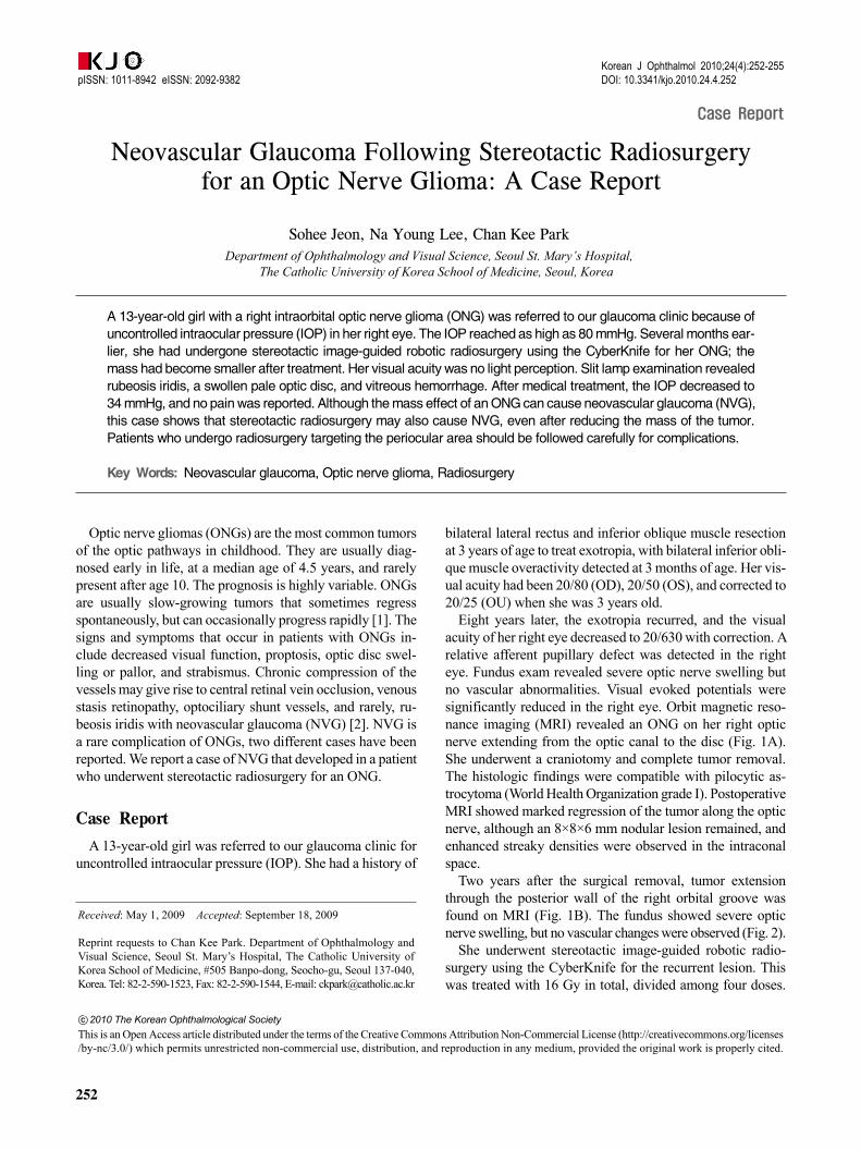
Eight years later, the exotropia recurred, and the visual acuity of her right eye decreased to 20/630 with correction. A relative afferent pupillary defect was detected in the right eye. Fundus exam revealed severe optic nerve swelling but no vascular abnormalities. Visual evoked potentials were significantly reduced in the right eye. Orbit magnetic reso-nance imaging (MRI) revealed an ONG on her right optic nerve extending from the optic canal to the disc (Fig. 1A). She underwent a craniotomy and complete tumor removal. The histologic findings were compatible with pilocytic as-trocytoma (World Health Organization grade I). Postoperative MRI showed marked regression of the tumor along the optic nerve, although an 8×8×6 mm nodular lesion remained, and enhanced streaky densities were observed in the intraconal space.
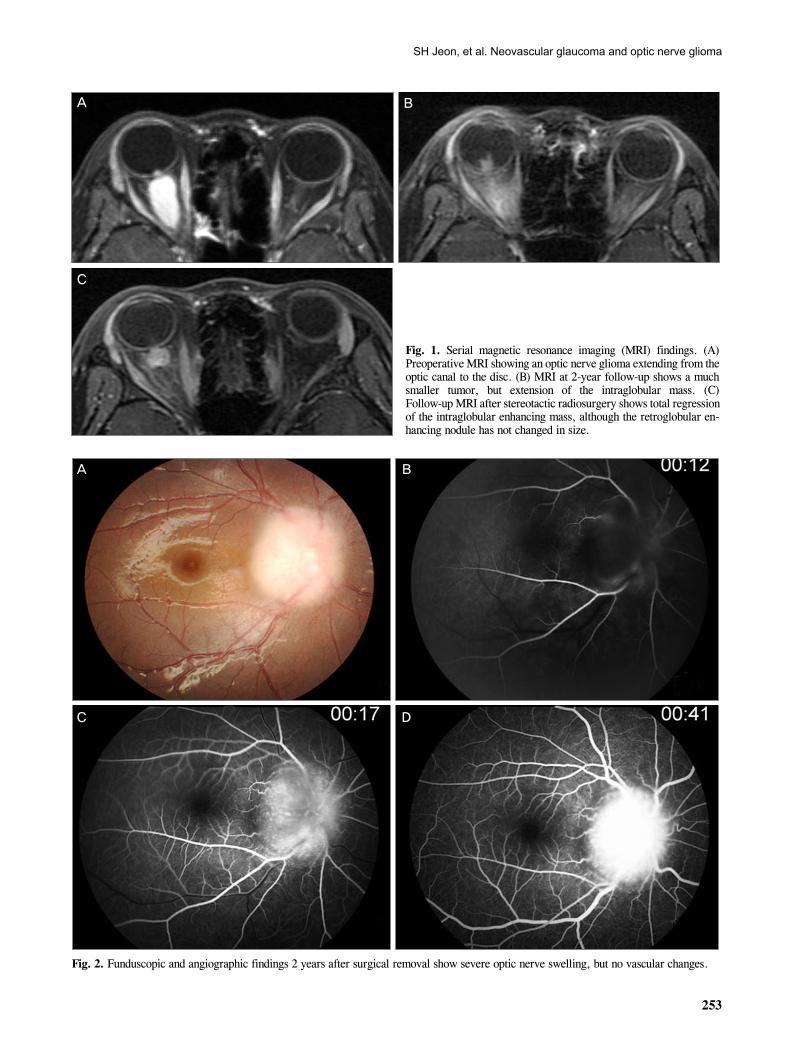
Two years after the surgical removal, tumor extension through the posterior wall of the right orbital groove was found on MRI (Fig. 1B). The fundus showed severe optic nerve swelling, but no vascular changes were observed (Fig. 2).
She underwent stereotactic image-guided robotic radio-surgery using the CyberKnife for the recurrent lesion. This was treated with 16 Gy in total, divided among four doses.
SH Jeon, et al. Neovascular glaucoma and optic nerve glioma
253
A B
C
Fig. 1. Serial magnetic resonance imaging (MRI) findings. (A) Preoperative MRI showing an optic nerve glioma extending from the optic canal to the disc. (B) MRI at 2-year follow-up shows a much smaller tumor, but extension of the intraglobular mass. (C) Follow-up MRI after stereotactic radiosurgery shows total regression of the intraglobular enhancing mass, although the retroglobular en-hancing nodule has not changed in size.
A B
C D
Fig. 2. Funduscopic and angiographic findings 2 years after surgical removal show severe optic nerve swelling, but no vascular changes.
Korean J Ophthalmol Vol.24, No.4, 2010
254
A B
C D
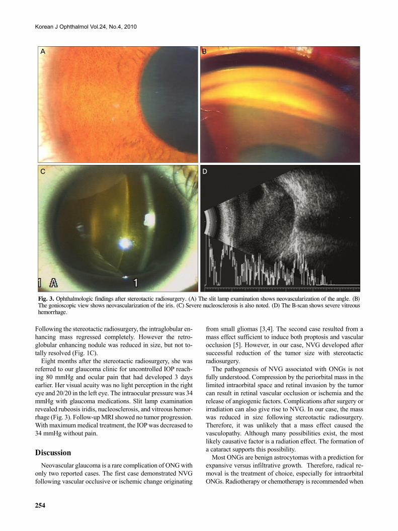
Fig. 3. Ophthalmologic findings after stereotactic radiosurgery. (A) The slit lamp examination shows neovascularization of the angle. (B) The gonioscopic view shows neovascularization of the iris. (C) Severe nucleosclerosis is also noted. (D) The B-scan shows severe vitreous hemorrhage.
Following the stereotactic radiosurgery, the intraglobular en-hancing mass regressed completely. However the retro-globular enhancing nodule was reduced in size, but not to-tally resolved (Fig. 1C).
Eight months after the stereotactic radiosurgery, she was referred to our glaucoma clinic for uncontrolled IOP reach-ing 80 mmHg and ocular pain that had developed 3 days earlier. Her visual acuity was no light perception in the right eye and 20/20 in the left eye. The intraocular pressure was 34 mmHg with glaucoma medications. Slit lamp examination revealed rubeosis iridis, nucleosclerosis, and vitreous hemor-rhage (Fig. 3). Follow-up MRI showed no tumor progression. With maximum medical treatment, the IOP was decreased to 34 mmHg without pain.
Discussion
Neovascular glaucoma is a rare complication of ONG with only two reported cases. The first case demonstrated NVG following vascular occlusive or ischemic change originating
from small gliomas [3,4]. The second case resulted from a mass effect sufficient to induce both proptosis and vascular occlusion [5]. However, in our case, NVG developed after successful reduction of the tumor size with stereotactic radiosurgery.
The pathogenesis of NVG associated with ONGs is not fully understood. Compression by the periorbital mass in the limited intraorbital space and retinal invasion by the tumor can result in retinal vascular occlusion or ischemia and the release of angiogenic factors. Complications after surgery or irradiation can also give rise to NVG. In our case, the mass was reduced in size following stereotactic radiosurgery. Therefore, it was unlikely that a mass effect caused the vasculopathy. Although many possibilities exist, the most likely causative factor is a radiation effect. The formation of a cataract supports this possibility.
Most ONGs are benign astrocytomas with a prediction for expansive versus infiltrative growth. Therefore, radical re-moval is the treatment of choice, especially for intraorbital ONGs. Radiotherapy or chemotherapy is recommended when
SH Jeon, et al. Neovascular glaucoma and optic nerve glioma
255
progression occurs after surgical removal [6]. Currently, however, stereotactic radiosurgery is used more frequently to avoid surgical complications. Although several reports have presented NVG as a complication after stereotactic radio-surgery for intraocular tumors [5], no report has described NVG after stereotactic radiosurgery for an ONG.
We suggest that ONGs be irradiated very selectively and followed carefully for unwanted complications such as NVG, even after a successful reduction in tumor size.
Conflict of Interest
No potential conflict of interest relevant to this article was reported.
References
1. Charles NC, Nelson L, Brookner AR, et al. Pilocytic astrocytoma of the optic nerve with hemorrhage and extreme cystic degeneration. Am J Ophthalmol 1981;92:691-5.
2. Henkind P, Benjamin JV. Vascular anomalies and neoplasms of the optic nerve head. Trans Ophthalmol Soc UK 1976;96:418-23.
3. Hovland KR, Ellis PP. Hemorrhagic glaucoma with optic nerve glioma. Arch Ophthalmol 1966;75:806-9.
4. Buchanan TA, Hoyt WF. Optic nerve glioma and neovascular glaucoma: report of a case. Br J Ophthalmol 1982;66:96-8.
5. Bergman L, Nilsson B, Lundell G, et al. Ruthenium brachy- therapy for uveal melanoma, 1979-2003: survival and functional outcomes in the Swedish population. Ophthalmology 2005;112: 834-40.
6. Demaerel P, de Ruyter N, Casteels I, et al. Visual pathway glioma in children treated with chemotherapy. Eur J Paediatr Neurol 2002;6:207-12.